1
This is a revised manuscript.
Original article can be found:
https://link.springer.com/article/10.1007%2Fs00726-018-2630-7 DOI: https://doi.org/10.1007/s00726-018-2630-7
Amino Acids (2018).
2 Éva Kiss1, Gergő Gyulai1, Edit Pári1, Kata Horváti2,3, Szilvia Bősze2
1 Laboratory of Interfaces and Nanostructures, Institute of Chemistry, Eötvös Loránd University, Budapest 112, P.O. Box 32, H-1518 Budapest, Hungary
2 MTA-ELTE Research Group of Peptide Chemistry, Budapest 112, P.O. Box 32, H-1518 Budapest, Hungary
3 Institute of Chemistry, Eötvös Loránd University, Budapest 112, P.O. Box 32, H-1518 Budapest, Hungary
Membrane affinity and fluorescent labelling: comparative study of monolayer interaction, cellular uptake and cytotoxicity profile of carboxyfluorescein conjugated cationic peptides
Running title: Effect of labelling of membrane active peptides
Abstract
Fluorescent labelling is a common approach to reveal the molecular details of cellular uptake, internalisation, transport, distribution processes in biological systems. The conjugation with a fluorescent moiety might affect relevant physicochemical and in vitro transport properties of the bioactive component. A representative set of seven cationic peptides – including cell penetrating peptides as well as antimicrobial peptides and synthetic derivatives – was selected for our comparative study. Membrane affinity of the peptides and their 5(6)-carboxyfluorescein (Cf) derivatives was determined quantitatively and compared applying Langmuir monolayer of zwitterionic (DPPC) and negatively charged (DPPC+DPPG) lipids as cell membrane models. The interaction with neutral lipid layer is mainly governed by the overall hydrophobicity of the molecule which is remarkably increased by Cf-conjugation for the most hydrophobic Magainin, Melittin and Transportan. A significantly enhanced membrane affinity was detected to negatively charged lipid model monolayer for all of the peptides since the combination of electrostatic and hydrophobic interaction is active in that case.
The Cf-conjugation improved the penetration ability of Penetratin and Dhvar4 suggesting that both the high charged character (Z/n) and the increased hydrophobicity by Cf-conjugation present important contribution to membrane interaction. This effect might also responsible for the observed high in vitro internalisation rate of Penetratin and Dhvar4, while according to in vitro studies they did not cause damage of cell membrane. From the experiments with the given seven cationic peptides it can be concluded, that the Cf-conjugation alters the degree of membrane interaction of such peptides which are moderately hydrophobic and highly charged.
Key words: fluorescent labelling, membrane affinity, cell penetrating peptides, lipid monolayer, penetration, cellular uptake
Author for correspondence: Éva Kiss e-mail: kisseva@caesar.elte.hu
Introduction
Detailed investigations of biological processes as well as the following of the transport of a given component generally require labelling with a fluorophore. Proteins, nucleic acids, lipids, or other molecules are labelled with fluorescent dyes which can be small organic molecules, proteins or quantum dots (Jensen 2012). Fluorescent proteins are used for monitoring gene activation, selective labelling and analysis of single proteins, cellular organelles or the whole cell. The colour palette of fluorescent proteins extends from far-red to blue in addition to the special ones with light modulated spectral properties. However, the photophysical properties of fluorescent proteins such as brightness or stability are not as good as organic dyes. Quantum dots introduced recently possess size-controlled fluorescence providing an outstanding alternative in labelling due to their unique optical properties. Their excellent photostability, long fluorescence lifetime, availability in many colours allow the complex imaging both in vitro and in vivo. On the other hand, the possible toxicity and relatively large size are the main issues limiting their wide spread application. Therefore, the organic fluorophores are currently the most popular and versatile agents used for bioimaging.
Any kind of chemical coupling raises the question whether the labelling influences the behaviour of the system investigated. There are signs in different experiments that the labelling can affect the processes, but it is also accepted that it is very much systems dependent. Kitchens and co-workers reported on the transport of various surface modified poly(amidoamine) dendrimers across Caco-2 monolayers (Kitchens et al. 2006). The comparison revealed that fluorescent labelling of the polymer enhanced its permeability comparing to the unlabelled one due to opening of tight junctions.
Phagocytosis of Bordetella pertussis labelled extrinsically with fluorescein isothiocyanate (FITC) or genetically with green fluorescent protein (GFP) was followed by Weingart and co-workers (Wiengart et al. 1999). The covalent coupling of FITC induced such alteration which led to increased phagocytosis and additionally at least one extracellular virulence
3 factor was also affected by FITC labelling. Considering the misleading results, the authors concluded that care should be taken when using labelled bacteria in phagocytosis assays.
FITC was also used for specifically targeting of human cervical cancer cells (Liu et al. 2018). For this purpose, cancer- specific targeting peptides (CSPs) were labelled with FITC. Their results showed that FITC-CSPs had specifically bounded to the cell membrane and cytoplasm of tumor cells, which could be used in the therapeutic diagnosis and targeting of cancer cells.
Birch and co-workers were investigated different specific fluorophores’ effect on the physico-chemical properties and uptake-related characteristics of Penetratin. The authors concluded that the hydrophobicity and intracellular distribution had significant changes by the covalent linkage of different fluorophores. This modification also influenced the cytotoxicity, which had adverse effects on cellular function and the cell membranes morphology. (Birch et al. 2017).
Recently, covalent linkage of fluorophore moieties to membrane-interacting peptide (Penetratin) was studied in ex vivo (Hedegaard et al.2018) and in in vitro model systems. Authors concluded that fluorophore moiety significantly alters lipophilicity, membrane interaction profile and internalization feature of Penetratin.
Cell penetrating peptides (cpps) and numerous synthetic peptides derived from the natural ones are in the focus of research and potential therapeutic applications. These cpps are promising chemical helpers to transport nonpermeable drugs into live cells (Reissmann 2014; Guo et al. 2016). They are particularly favoured in the internalisation of various cargos, quantum dots, nanoparticles due to their low toxicity and final degradation to amino acids. Cpps and the related antimicrobial peptides (amp) (Henriques et al. 2006) are the subject of various in vitro and in vivo investigations where labelling is crucial in the understanding the molecular processes.
A peptide series is selected in the present work for the experimental characterisation of membrane affinity and to evaluate the effect of fluorescent labelling. Well known cpps and amps such as Transportan, Penetratin, Magainin, Melittin and designed derivatives such as Crot(1-9,38-42), Dhvar4, OT20, with various compositions cover a wide range of physico- chemical, structural and biological properties. All of them possess the common feature of containing several cationic amino acids. The selection of peptides is directed by previous research on the critical comparison of internalisation and cytotoxicity performed on a wider family of cpp/amp (Horváti et al. 2017).
To characterise the peptide-lipid interaction monomolecular lipid layers were applied as model membranes. Phospholipid monolayers are extensively used as a model for one leaflet of the cell membrane (Brockman 1999; Więcek et al. 2008;
Flasinski et al. 2014). The surface concentration and charge density of the layer prepared on water surface can be easily adjusted by altering the composition and surface pressure of the monolayer (Kiss et al. 2012; Hegedüs et al. 2012; Pénzes et al. 2012; Schöne et al. 2017). The ordered lipid monolayer formed in a Langmuir trough or the supported lipid bilayers (Cranfield et al. 2017; Marek et al. 1999; Mohai et al. 2002) obtained by liposome spreading or LB technique although much less complex than biological membranes are particularly suitable for studying the molecular interactions (Horvath et al. 2013; Lohr et al. 2014; Ábrahám et al. 2016; Ábrahám et al. 2017). The binding of molecules, such as peptides and proteins, polymers, or amphiphilic substances to the lipid headgroup region and their penetration into the lipid acyl chain region (Haedicke and Blume 2015) is detected quantitatively in a Langmuir trough as the increase of surface pressure of the monolayer. Although in characterisation of cpp-lipid interaction various vesicle systems (GUV, liposomes) are the commonly used models of the cell membrane, the experimental models of lipid monolayer provide valuable information on the influence of molecular composition and structural details (Brezesinski and Möhwald 2003; Hill et al. 2010;
Michanek et al. 2012; Keszthelyi et al. 2013).
The method to characterise quantitatively the membrane affinity of various peptides is the penetration measurement when change in surface pressure of the lipid film due to adsorption/penetration of a component dissolved in the sub-phase is determined. Running all the penetration experiments with two lipid models (DPPC and DPPC+DPPG) allows assessment of both the hydrophobic and electrostatic molecular interactions, and their contribution to the membrane affinity of peptides and the 5(6)-carboxyfluorescein-labelled peptides.
The aim of the present study is to get information on the influence of Cf-conjugation on the hydrophobicity and lipid membrane interaction of the selected peptides. The hydrophobicity of the peptides was characterized by their retention time in RP-HPLC as well as the surface activity of their aqueous solutions. The membrane affinity to neutral and charged lipid layer was analysed relating to the chemical composition of the peptides along with their estimated secondary structure. Furthermore, in this study, we examined the cellular uptake rate, cytotoxicity of the peptides and their Cf- derivatives. We have analysed the results of membrane model studies and compared to in vitro activity; how charge, size, hydrophobicity of the peptides affect their internalisation and membrane damaging.
Experimental Materials
1,2-dipalmitoyl-sn-glycero-3-phosphocholine (DPPC) and its mixture with 1,2-dipalmitoyl-sn-glycero-3-phospho-(10- rac-glycerol) (sodium salt) (DPPG) (Sigma‐Aldrich) were applied in chloroform solution to form the molecular lipid layer on the surface of subphase in the Langmuir trough. The composition of the mixed lipid layer was a molar fraction of 75 %
4 DPPC and 25 % DPPG. Chloroform (purity >99.8%) from Fisher Chemicals was used for preparing the lipid solutions.
Dichloromethane (purity >99.9%) from Spectrum-3D Kft. Hungary and methanol (purity >99.9%) from Sigma-Aldrich Kft., Hungary were used for cleaning the Langmuir trough and the polyoxymethylene (POM) barriers, respectively.
Double distilled water was checked by its conductivity (<5 mS) and surface tension (>72.0 mN/m at 23±0.5 °C) values.
For peptide synthesis amino acid derivatives were obtained from Reanal Laboratory Chemicals or from Iris Biotech.
Reagents, such as N,N’-diisopropylcarbodiimide (DIC), triisopropylsilane (TIS), 1-hydroxybenzotriazole (HOBt), and 1,8-diazabicyclo[5.4.0]undec-7-ene (DBU), 5(6)-carboxyfluorescein (Cf) were purchased from Sigma-Aldrich. Fmoc- Rink Amide MBHA resin was also from Iris Biotech. Trifluoroacetic acid (TFA), N,N-dimethylformamide (DMF) and acetonitrile were from VWR.
For the in vitro assays RPMI-1640 medium, foetal calf serum (FCS), trypsin were obtained from Sigma-Aldrich. HPMI buffer (9 mM glucose, 10 mM NaHCO3, 119 mM NaCl, 9 mM HEPES, 5 mM KCl, 0.85 mM MgCl2, 0.053 mM CaCl2, 5 mM Na2HPO4 × 2H2O, pH 7.4 (Kapus et al. 1994) was prepared in our laboratory using components obtained from Sigma-Aldrich.
Seven cationic peptides were selected for the present investigation. Their sequence together with their molecular masses and HPLC retention times are given in Table 1. This group contains some well-studied representatives of cell penetrating peptides as Penetratin and Transportan, antimicrobial peptides like Magainin and Melittin, as well as synthetic cationic peptides that were designed based on structure-activity relationship studies such as the tetramer derivative of tuftsin (OT20), a shorter derivative of Crotamine, Crot(1-9,38-42) and Dhvar4 a synthetic derivative of Histatin. The comparative analysis of their in vitro activities was the subject of a previous study (Horváti et al. 2017).
Peptide synthesis, purification and analytical characterisation
Peptide synthesis was carried out in an automated peptide synthesizer (Syro-I, Biotage) using standard Fmoc/tBu strategy with DIC/HOBt coupling reagents. In the case of the fluorescently labelled derivatives, N-terminal amino group was conjugated with 5(6)-Carboxyfluorescein (Cf) in the presence of DIC/HOBt reagents. Peptides were cleaved from the resin with TFA/H2O/TIS (9.5:2.5:2.5 v/v) mixture (2 h, RT). After filtration compounds were precipitated in cold diethyl ether, centrifuged (4000 rpm, 5 min) and freeze-dried from water. Crude products were purified by RP-HPLC on a semipreparative C18 Phenomenex Jupiter column (250 mm × 10 mm) using gradient elution, consisted of 0.1% TFA in water (eluent A) and 0.1% TFA in acetonitrile/water = 80:20 (v/v) (eluent B). Purified peptides were analysed by RP- HPLC on an analytical Agilent ZORBAX StableBond C18 (4.6 mm × 150 mm, 3.5 µm, 80 Å) HPLC column using gradient elution with the above-mentioned eluent A and B (flow rate was 1 mL/min, UV detection at λ=220 nm).
Molecular mass of the peptides was determined by using a Bruker Esquire 3000+ ESI mass spectrometer. Peptide samples were dissolved in a mixture of acetonitrile/water = 1:1 (v/v) containing 0.1% acetic acid and introduced by a syringe pump with a flow rate of 10 L/min. Peptide content of the final product was determined by amino acid analysis and taken into consideration in the further measurements..Table 1 summarizes the analytical characterisation of the peptides and peptide-derivatives.
Table 1 Analytical characteristics of the peptides
Peptide Sequence Mav
calc. / found a
Rt b
(min)
OT20 TKPKG TKPKG TKPKG TKPKG 2063.5/2063.5 7.2
Cf-OT20 CF-TKPKGTKPKGTKPKGTKPKG 2421.8/2422.0 10.8
Crot(1-9,38-42) YKQCHKKGGKKGSG 1504.8/1504.9 6.8
Cf-Crot(1-9,38-42) Cf-YKQCHKKGGKKGSG 1863.1/1863.4 11.2
Dhvar4 KRLFKKLLFSLRKY 1839.3/1839.4 11.3
Cf-Dhvar4 Cf-KRLFKKLLFSLRKY 2197.7/2197.7 15.4
Penetratin RQIKIWFQNRRMKWKK 2245.7/2245.9 10.9
Cf-Penetratin Cf-RQIKIWFQNRRMKWKK 2604.0/2604.2 13.5
Magainin II GIGKFLHSAKKFGKAFVGEIMNS 2465.9/2465.8 12.9
Cf-Magainin II Cf-GIGKFLHSAKKFGKAFVGEIMNS 2824.2/2824.2 14.8
Melittin GIGAVLKVLTTGLPALISWIKRKRQQ 2846.5/2846.6 16.1
Cf-Melittin Cf-GIGAVLKVLTTGLPALISWIKRKRQQ 3204.8/3204.7 19.9
5
Transportan AGYLLGKINLKALAALAKKIL 2181.8/2181.8 15.5
Cf-Transportan Cf-AGYLLGKINLKALAALAKKIL 2540.1/2540.0 18.8
a Measured average molecular mass by Bruker Esquire 3000+ ESI-MS.
bRetention time using analytical RP-HPLC, Agilent ZORBAX StableBond 80Å C18, 4.6 mm × 150 mm, 3.5 µm HPLC column, gradient: 5% B, 2 min; 5-100% B, 20 min.
Calculation of peptide structure
Secondary structure of the peptides was predicted using two online peptide folding services. The I-TASSER (Iterative Threading ASSEmbly Refinement) is an online service of the University of Michigan (Zhang Lab 2017). Models are built by first identifying structural templates from the Protein Data Bank followed by iterative fragment assembly simulations (Zhang 2008; Roy et al. 2010). The results are presented as the possible secondary structure motif (coil, helix or strand) for each amino acid in the sequence along with the confidence score of the prediction.
The peptide structure was also analysed using the PEP-FOLD (PEP-FOLD server 2017) service. PEP-FOLD is a de novo approach for predicting the 3D structure, based on structural alphabet (SA) letters to describe the conformations of four consecutive residues followed by the coupling of the predicted series of SA letters to a greedy algorithm and a coarse- grained force field (Thévenet et al. 2012; Shen et al. 2014). 100 simulations were run for all sequences. The results are displayed by the usual 3D ribbon structure and in a bar representation of probability of appearance of helix or other structural features along the sequence.
Surface tension measurements
Surface tension of the aqueous solutions of the peptides was measured by axisymmetric drop shape analysis using an OCA15+ (Dataphysics, Germany) optical contour analysis instrument. 10 μL droplets hanging at the end of the capillary of a Hamilton syringe were created in air in a small glass chamber saturated with water vapour at 25 °C. Surface tension values were recorded for 1 h at peptide concentrations of 2 μM and 200 μM. Three parallel measurements were carried out for all systems and surface tension values were reproducible to ±0.5 mN/m.
Membrane affinity
The membrane affinity of the various peptides was investigated using the Langmuir technique. Lipid monolayer was prepared and the penetration measurements were performed in a Langmuir balance (250 mm × 60 mm). Surface pressure was recorded with the tensiometric method with an accuracy of ±0.5 mN/memploying filter papers as Wilhelmy plates.
Two surface pressure sensors were used at the two compartments of the trough divided by a single movable barrier. This experimental setup enables the simultaneous recording of the surface pressure on the pure and on the lipid covered surface.
The through was made of teflon and the barrier from polyoxymethylene (POM). Before each measurement, the through was cleaned with dichloromethane, while the barrier was cleaned with methanol. After cleaning, the through was filled with doubly distilled water to be used as subphase. DPPC or DPPC+DPPG (at molar ratio 3:1) was dissolved in chloroform to a concentration of 0.2 g/L. The lipid solution was spread onto the water surface and chloroform was left to evaporate for 10 min before compression. The compression and expansion of the lipid layer were performed at a barrier speed of 34 mm/min. Surface pressure-area isotherms were recorded two times before each penetration experiment.
Peptides were dissolved in water at 200 µM concentration. The aqueous peptide solution was injected into the subphase resulting in a final concentration of 2 µM in the through. The changes of the surface pressure indicating the peptide-lipid interaction were detected as a function of time for 1 h. Three penetration measurements were performed in every case.
One-way ANOVA test was run on the measured ΔΠ values to determine significant differences.
In vitro biological activity
In vitro cellular uptake evaluation by flow cytometry and fluorescent microscopy
Internalisation of Cf-labelled peptides were measured on MonoMac6 human monocytic cell line (Ziegler-Heitbrock et al.
1988). by using a BD LSR II flow cytometer (BD Biosciences, San Jose, CA, USA) with 488 nm (Coherent Sapphire, 22 mW) laser. MonoMac6 cells were maintained as an adherent culture in RPMI-1640 supplemented with 10% heat- inactivated fetal calf serum (FCS) L-glutamine (2 mM) and gentamicin (35 μM) at 37 °C in humidified atmosphere containing 5% CO2. Cells were treated with peptides at 10 µM final concentration and were incubated for 2 h (37 °C, 5%
CO2 atmosphere). After centrifugation (1000 rpm, 5 min) and washing with serum free RPMI medium, supernatant was removed and 100 μL 0.25% trypsin was added to the cells. After 5 min incubation 0.8 mL 10% FCS/HPMI medium was added than cells were washed and re-suspended in 0.3 mL HPMI medium. The intracellular fluorescence intensity of the cells was measured on BD LSR II, channel FITC LP505 (emission at λ=505 nm; LP 505, BP 530/30) and data were analysed with FACSDiva 5.0 software. All measurements were performed in triplicates and the mean fluorescent intensity together with standard error of the mean (SEM) was graphically presented (Fig. 7/a; b).
6 Parallel with flow cytometry measurements, to visualize cell morphology after peptide and Cf-peptide treatment (20 μM final concentration), microscopic images of MonoMac6 cells were captured. Washed and resuspended cells were plated in a 96-well flat bottom tissue culture plate and images of the adherent cells were captured using an Olympus CKX41 microscope (Hamburg, Germany, Olympus U-RFLT50 mercury-vapor lamp, WideBlue DM500 BP460-490 BA520 IF filter, objective: 20X) (Fig.7/c).
Cell viability and membrane integrity studies using propidium iodide staining
The cell viability and membrane integrity were assessed by flow cytometry and fluorescent microscopy using propidium iodide (PI) exclusion method. PI is a membrane impermeant dye that is excluded from viable cells. It binds to double stranded DNA by intercalating between base pairs. PI penetrates the damaged, permeable membranes of non-viable cells.
MonoMac6 cells were harvested in the logarithmic phase of growth and plated on a 24-well tissue culture plate (105 cells/1 mL medium/well) 24 hours prior to the experiment. MonoMac6 cells were treated with the peptides and Cf- peptides at 10 and 20 μM final concentration for 2 h (37 °C, 5% CO2 atmosphere). After treatment, cells were centrifuged (1000 rpm, 5 min) and washed twice with serum free RPMI medium. After centrifugation (1000 rpm, 5 min) supernatant was removed and 100 μL 0.25% trypsin was added to the cells. After 1 min incubation at 37 °C the effect of trypsin was stopped by 800 μL HPMI supplemented with 10% FCS, and the cells were transferred from the plate to FACS-tubes.
Cells were centrifuged (1000 rpm, 5 min) and the supernatant was removed. After this procedure, cells were resuspended in 0.3 mL HPMI. The cell viability and intracellular fluorescence intensity of MonoMac6 cells was determined by BD LSR II (488 nm; Coherent Sapphire, 22 mW) laser, channel PE LP550 (emission at λ = 550 nm; LP 550, BP 575/26)) before and after adding 10 μL 50 μg/mL PI solution. Cells with PI positivity were gated and the number of dead and viable cells were counted. Data were analysed with FACSDiva 5.0 software. Percentage of live cells was compared to untreated control cells and the relative viability was graphically represented (Fig.7/a). All measurements were performed in triplicates and the mean and SEM are presented. Parallel with flow cytometry measurements microscopic image of the cells were captured with an Olympus CKX41 microscope (Olympus U-RFLT50 mercury-vapor lamp, SuperWideGreen DM570 BP480-550 BA590 IF filter, objective: 20X) (Fig. 7/c).
Statistical Analysis
Data were analysed using GraphPad Prism v6 software (San Diego, CA, USA). For analysis of statistical significance (p
< 0.001) unpaired t-test was used.
Results and Discussion
Surface behaviour of the peptides, adsorption at air/water surface
Surface tension measurements were performed with aqueous solution of the peptides to see their adsorption ability at air/water surface. The adsorption and hence the reduction of surface tension is the indicator of hydrophobicity/amphiphilicity of the peptides. The concentration of aqueous peptide solution used for the lipid interaction measurement was 2 μM. None of the peptides showed detectable reduction of surface tension at that concentration.
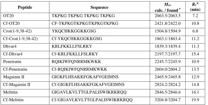
Therefore, to be able to evaluate surface activity of the peptides they were applied at a higher concentration: 200 μM. The surface tension of peptide solutions was followed by pendant drop profile analysis for 1 h (Fig. 1a). Within this time interval some solutions presented a slight gradual decrease, such as OT20 or Crot(1-9,38-42). Other peptides, like Magainin or Melittin were proved to be more surface active showing a fast decrease then the levelling of the surface tension. The change of surface tension of Penetratin solution as a function of time was unique, starting with an induction period and then turning into a steady reduction. Transportan was the most surface active among the seven peptides for which constant value of surface tension was not reached within the time of investigation. The values of surface tension reduction, ∆γ measured after 1 h of drop formation are given in Table 2. The range of ∆γ is quite large indicating the substantial difference in hydrophobicity of the peptides studied.
7 Fig. 1 Time dependence of surface tension (γ) of aqueous solutions of peptides (a) and respective Cf-peptides (b) at
concentration of 200 μM. (Five of the studied peptides are presented for clarity)
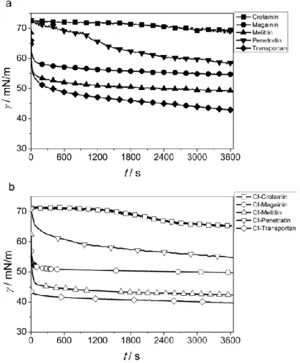
Similar experiments were performed with the solutions of Cf-labelled peptides. Significant increase in surface activity (more reduced surface tension) was obtained for all the peptides (Fig. 1b). It is expected that the carboxyfluorescein molecule coupled to the N-terminus of the peptide results in an increased hydrophobicity of the whole molecule. This difference is clearly reflected in the Rt values (Table 1) where retention time on a reversed-phase coloumn is consistently higher for the Cf-peptides than for the original ones. That increased hydrophobicity is responsible for the enhanced surface activity of Cf-peptides. The difference in the reduction of surface tension comparing the Cf-peptides to the original ones amounts 3–6 mN/m at the given concentration (200 μM) as the data in Table 2 show.
As we can see in Figure 1 the surface activity of the various peptides is highly influenced by their amino acid composition.
The overall polarity of the peptides can be estimated by using the average of hydrophilicity values of each amino acids (Hopp and Woods 1981). The order of surface activity of the seven peptides studied here agrees with the estimated hydrophilicity/hydrophobicity order (Table 2). The most hydrophilic OT20 has the lowest surface activity, while the less hydrophilic Transportan reduces the surface tension of water at the highest degree. Within this range the surface activity is in line with the estimated hydrophobicity of the given peptide except Penetratin which was found to be more surface active than expected from its amino acid composition.
As the surface activity measurements reveal the character of the labelled peptides is consistently shifted into the more hydrophobic direction while the order of peptides according to their hydrophilicity was not influenced by the Cf-coupling.
That effect was found to be valid for each peptide studied here. The relative increase of hydrophobicity is reasonably larger for the originally more hydrophilic ones.
Table 2 Structural properties and surface activity (∆γ) of the peptides and their Cf-labelled derivatives
Peptide na Zb Z/n Hydrophilicityc
∆γd (mN/m) Helicitye (I TASSER) Peptide Cf-peptide %
OT20 20 9+ 0.45 +1.1 2.0 6.5 0
8
Crot(1-9,38-42) 14 6+ 0.43 +0.8 3.4 7.7 29
Dhvar4 14 7+ 0.5 +0.3 4.3 8.8 71
Penetratin 16 8+ 0.5 +0.5 14.2 18.1 0
Magainin II 23 4+ 0.17 -0.1 18.4 22.5 87
Melittin 26 6+ 0.23 -0.2 24.0 30.1 73
Transportan 21 5+ 0.24 -0.3 30.0 33.0 48
a n: number of amino acids
b Z: net charge at neutral pH. Calculated by the number of (K+R)-(E+D). Positive charge at the N-terminus increases Z by 1 unit
c Hydrophilicity calculated using values of amino acids expressing hydrophilicity of each amino acid (Hopp and Woods 1981)
d Decrease of surface tension of water at peptide concentration of 200 μM after 1 h adsorption
e Calculated by I-TASSER
Secondary structure of the peptides
The cell penetrating and antibacterial peptides are classified into three groups according to the degree and mechanism of interaction with cell membranes (Madani 2011). Amphipathic peptides containing more than 20 amino acids belong to the first group. These peptides are able to directly cross the membrane by pore or inverse micelle formation such as transportane or melittin with a high antibacterial effect and membrane lytic property (Asthana 2004). This intense structural perturbance of membrane is usually caused by helical peptide sequence.
The second group is formed by smaller peptides with less amphiphilicity e.g. Penetratin. Highly charged peptides such as polyarginine or the OT20 make the third group presenting weak membrane interaction but the case of negatively charged lipids.
The various types of interaction are suggested to be associated with the amino acid sequence and membrane induced structural change during the interaction (Zorko 2005). Therefore to understand the difference in membrane affinity it is important to estimate the possible secondary structure of the peptides.
Small peptides are usually considered to be in a random coil conformation in aqueous medium. There is however an extensive experimental evidence that cpps can easily fold into helical structures (Lindberg et al. 2001; Nikawa et al.
2004). In many cases these structural changes are induced by adsorption to an interface (Avitabile et al. 2014).
9 Fig. 2 Predicted secondary structure of the peptides using I-TASSER and PEP-FOLD methods
The possible secondary structure of the peptides was predicted using two online applications. The I-TASSER algorithm provides the most probable structural motif for each amino acid in the sequences along with a confidence score for the
10 prediction on a scale of 0 to 9. The predicted structures along with the confidence scores are shown in Fig. 2. The helicity of the peptides in % calculated from these predictions is collected in Table 2.
PEP-FOLD algorithm was also used to make an estimation on the secondary structure of the peptides. The structural results of the PEP-FOLD algorithm along with a predicted 3D conformation are also shown in Fig. 2. The PEP-FOLD results can be interpreted as the probability of an amino acid in the sequence to be in a helical, random coil or extended strand structure.
The structural information provided by the two methods is similar in all cases. According to the prediction OT20 is a random coil and does not form helical structure at all, while the probability of helix formation is quite low for Penetratin and Crot(1-9,28-42). On the other hand, helix formation is highly feasible for Melittin, Magainin, Transportan and somewhat for Dhvar according to both predictions in a coherent way.
Membrane affinity
The peptide-lipid interactions were studied in a Langmuir-balance using floating monolayer of highly packed, ordered DPPC and DPPG molecules. DPPC is one of the main component of membrane forming lipids. External leaflet of plasma membrane contains mostly zwitterionic phospholipids with a small amount of anionic lipids. DPPC (similarly to POPC) represents the noncharged component of the lipid double layer allowing the evaluation of hydrophobic type interaction.
20 mN/m was selected as surface pressure of lipid monolayer for the penetration experiments. This value is lower than the generally accepted 30 mN/m characterising the lipid density of real cell membrane however is especially suitable to demonstrate the differences and effects of molecular structure of the penetrating species. Alhakamy reported that maximum penetration of the cpps was observed for phospholipid films compressed to a surface pressure of 20 mN/m (Alhakamy 2013). The peptides were applied at 2 μM concentration in the aqueous phase which provides sufficient molecules to achieve the maximum interaction with lipids according to previous studies (Haedicke and Blume 2015).
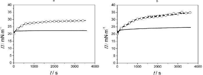
The change of surface pressure was recorded for 1 h which proved to be sufficient to get a saturation value of penetration in most cases except Melittin. Two examples of penetration curve for Transportan and Melittin are displayed in Figure 3 comparing the lipid interaction of the peptide and its fluorescently labelled derivative. Small degree of membrane affinity was observed for the two peptides, while the penetration of Cf-labelled ones are increased significantly resulting in 10 mN/m or higher rise in surface pressure of the layer.
Fig. 3 Surface pressure (Π) of DPPC monolayer during the interaction with Transportan (a) and Melittin (b) comparing to their Cf-labelled derivatives (○)
The results of membrane affinity to DPPC monolayer for the peptides and their Cf-derivatives are summarized in Fig. 4.
The error bars in Fig. 4 represent the confidence intervals of the determined mean values, calculated using Student’s t- test at 95% confidence level.
11 Fig. 4 Degree of penetration (ΔΠ) of peptides (black) and Cf-peptides (grey) into DPPC lipid monolayer determined at
1 h of interaction. Error bars represent the confidence intervals (95%) of the measured mean values.
Considering the membrane affinity of the various peptides we see that the binding to DPPC layer is modest for the most hydrophilic OT20 and Crot(1-9,38-42), while higher for Penetratin. According to the ANOVA test of the results the difference between OT20 and Crot are insignificant, while significant for all other peptides at a significance level of 0.05.
Membrane affinity then gradually increased in the order of peptides Dhvar4 < Transportan < Magainin < Melittin. This behaviour reflects the hydrophobic/hydrophilic character of the peptides although the hydrophilicity values given in Table 2 do not correspond exactly to the order of penetration. That implies that besides the amino acid composition additional structural effect might have an important contribution to the binding of the molecules.
This influence is accounted for in another approach developed by Wimley and White (White and Wimley 1998; Hristova and White 2005; Almeida and Pokorny 2009). They introduced a scale to describe the membrane affinity of cpps using fractional values of amino acids to estimate the interfacial free enthalpy of the binding of a peptide to the membrane surface. The binding process is a coupled binding-folding event whereby a peptide mostly disordered in aqueous solution, binds and folds to an α-helix on the membrane surface. It is well documented that peptides readily change their conformation in the neighbourhood of membrane surface and α-helix formation was detected by different instrumental techniques. When an aliphatic helix forms at an interface, ∆G of peptides is reduced by 0.4 kcal/mol per residue (Ladokhin and White 1999). We used I-TASSER algorithm to approximate the extent of helix formation of the given peptides (Table 2). The calculated ∆G values for binding of our peptides perfectly corresponds to the order of the experimentally determined degree of binding/penetration of our peptides calculated ∆G values. (That order of peptides was used in the plots of Figures 4 and 6.)
The degree of peptide interaction with lipid layer for the Cf-peptides is almost the same as the original peptides for OT20, Crot(1-9,38-42), Penetratin, and Dhvar4. For the Cf-labelled Transportan, Magainin and Melittin, however a much higher membrane affinity was found comparing to the corresponding unlabelled peptides. According to ANOVA test the changes in membrane affinity due to Cf-labelling can be considered significant (at 0.05 significance level) with the notable exception of OT20, Penetratin and Dhvar4. We recognized that the coupling of fluorescein to the peptides results in more hydrophobic character of the whole molecule supported by retention time and surface tension experimental data.
Considering that the zwitterionic DPPC molecular layer will sense mainly hydrophobic interactions, enhanced membrane affinity of Cf-labelled peptides was expected. This was found to be valid for the most hydrophobic peptides. The increase of surface pressure caused by binding was almost doubled for Magainin while became even larger for Transportan and Melittin due to Cf-labelling. That influence of Cf-labelling was not realized for the other four peptides. The possible explanation can be that those more hydrophilic peptides are the highly charged ones, presenting Z/n values close to 0.5 (Table 2), whereas this value is <0.25 for the three previously discussed hydrophobic peptides (Transportan, Magainin, Melittin). The strong hydrophilicity of OT20, Crot(1-9,38-42), Penetratin, and Dhvar4 cannot be reversed or compensated by the Cf-coupling, therefore resulting in no detectable enhancement in the hydrophobic interaction with DPPC layer.
Membrane affinity of the peptides and their Cf-labelled derivatives was also characterized using the other membrane model which is formed from DPPC+DPPG mixture. The mean results are presented in Fig. 6, where the error bars represent the confidence intervals of the determined mean values, calculated using Student’s t-test at 95% confidence level. Since the cpps are cationic peptides in general it is reasonable to study their membrane affinity with such model lipid layer which provides the possibility of electrostatic interaction.
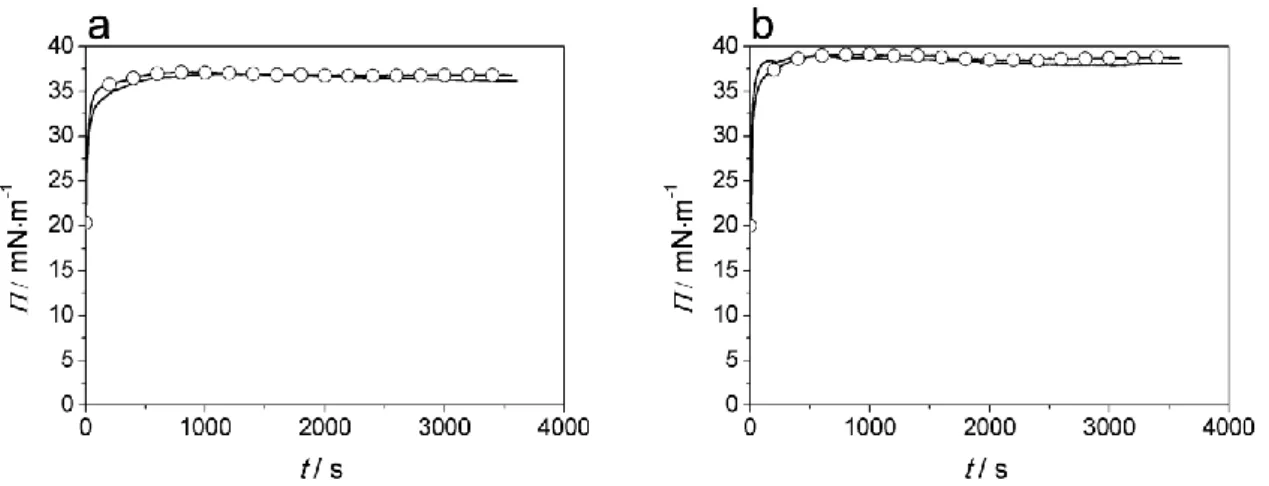
According to the expectation, higher degree of binding was observed to DPPC+DPPG layer than to pure DPPC for all peptides studied here (Fig. 6). Two characteristic penetration curves of the Transportan and Melittin clearly demonstrate this high membrane affinity (Figure 5). Surface pressure increases suddenly from 20 mN/m to a value above 35 mN/m after introduction of the peptides below the lipid layer.
12 Fig. 5 Surface pressure (Π) of DPPC+DPPG monolayer during the interaction with Transportan (a) and Melittin (b)
comparing to their Cf-labelled derivatives (○)
In the case of charged lipid layer, the most hydrophobic peptides (Transportan, Magainin, and Melittin) bind to the lipid layer with the highest affinity (Fig. 6). The electrostatic interaction however, offers a significant contribution to the binding for the less hydrophobic peptides, too. Especially the Penetratin shows intense interaction with charged lipid layer. For the peptides where high membrane affinity was observed helix formation appears to be favoured although no direct correlation could be found between the penetration and helicity. Low probability of helix formation for Penetratin is suggested by the PEP-FOLD algorithm we used. It is also known that Penetratin enters and transfers the lipid bilayer by different mechanism than the highly effective pore forming cpps such as Melittin or Magainin do. This increased penetration ability of Cf-conjugated Penetratin corresponds to previous findings of Illien et al. (2016) where internalization efficiency dependent on the hydrophilic/hydrophobic nature of the chemical moiety positioned at the N- terminal extremity of the peptide was reported.
Fig. 6 Degree of penetration (ΔΠ) of peptides (black) and Cf-peptides (grey) into DPPC+DPPG lipid monolayer determined at 1 h of interaction. Error bars represent the confidence intervals (95%) of the measured mean values.
The most remarkable result relating the membrane affinity of Cf-modified peptides is that both the original and Cf- peptides present similar degree of penetration except Penetratin and Dhvar4. The one-way ANOVA test of the experimental values supported that in these two cases the Cf-coupling induced penetration increase considered significant.
It seems to be valid that the coupling of Cf has no significant influence on the combined hydrophobic and electrostatic interactions. A slightly increased membrane affinity was obtained for Cf-Dhvar4 and somewhat higher in the case of Cf- Penetratin due to labelling.
According to the classification of cell penetrating peptides (Ziegler 2008; Madani et al. 2011; Reissmann 2014) primary amphipathic cpps contain typically more than 20 amino acids and have sequentially hydrophilic and hydrophobic amino acids in their primary structure enabling their direct penetration. Magainin and Melittin, the two typical examples of amphipathic amps also possess the structural feature that the N-terminal region of the sequence is composed mainly of hydrophobic amino acids while the C-terminal region is rich in charged or polar amino acids. It is reasonable to assume that this character contributes to the surfactant like activity of such peptides.
Secondary amphipathic cpps with the typical member of Penetratin commonly have less than 20 amino acids, and can form their α-helical or β-sheet conformations upon interaction with phospholipid membranes. The interaction of Penetratin is weak with neutral membrane and does not form helical structure (Almeida and Pokorny 2009). With the negatively charged membrane surface however the interaction is more intense (Castanho 2009) inducing significant conformational change from random coil to either β-sheet (Bellet-Amalric et al. 2000) or α-helix (Christiaens et al 2002) structures. Regarding the primary sequence in Penetratin (and Dhvar4) it is interesting to see that both the N- and C-
13 terminal regions compile charged and polar amino acids yielding a rather symmetrical distribution of polarity. The coupling of Cf-moiety at the N-terminal introduces asymmetry in polarity possibly rendering these peptides more amphiphilic. That can be the possible explanation of the enhanced membrane affinity observed in the case of Penetratin and Dhvar4.
In vitro biological activity of peptides in MonoMac6 cells; cellular uptake, cell viability and membrane integrity studies
Determination of cell viability, internalisation rate and membrane integrity is critical in compounds screening when evaluating the in vitro response. Flow cytometry and fluorescent microscopy are reliable methods to study these parameters. Human MonoMac6 cells were chosen as an excellent and useful model for investigating cytotoxicity, uptake and membrane damage following treatment with synthetic cationic peptides, cpps and amps and their Cf-labelled derivatives. MonoMac6 cell line represents monocytic cells with a closely related pattern of surface, phenotypic, functional features and adhesion properties of mature monocytes and macrophages (Ziegler-Heitbrock et al. 1988; Erl et al. 1995). In our previous studies these model cells were used to determine uptake profile, cytotoxicity and membrane interactions of promising drug carrier peptides (Horváti et al. 2017; Baranyai et al. 2017; Horváti et al. 2018; Ábrahám et al. 2016; Horváti et al 2014).
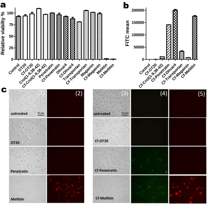
Fig. 7 Comparison of internalisation and cytotoxicity of selected peptides and their Cf-derivatives using flow cytometry (BD LSRII) and microscopy (Olympus CKX41) performed as described in the Experimental section. Panel a represents the relative viability of unlabelled and Cf-labelled peptides following treatment (10 μM, 2 h) measured on MonoMac6
Control Cf-OT
20
Cf-Crot(1-9,38-42) Cf-Pe
net ratin Cf-D
hvar4 Cf-Tran
sport an Cf-M
againin Cf-M
elittin 0
50000 100000 150000 200000
FITC mean
Control OT
20 Cf-OT
20
Crot(1- 9,38-42)
Cf-Crot(1-9,38-42) Penet
ratin Cf-Pe
net ratin
Dhva r4 Cf-D
hvar4 Transportan
Cf-Tran sport
an Magainin
Cf-M againin
Melittin Cf-M
elittin 0
25 50 75 100
Relative viabilty %
a b
c (1) (2) (3) (4) (5)
14 human monocytes. Spontaneous cell death due to sample preparation was determined for untreated control cells (3-5 % of death). Panel b shows the mean intracellular fluorescence intensity of Cf-peptide treated MonoMac6 cells (10 μM, 2 h; FITC LP505; BP 530/30). Error bars are SEM of three independent measurements. Panel c displays representative images of internalisation and membrane integrity studies with selected unlabelled and Cf-labelled peptides. Columns (1) bright field images of untreated and unlabelled OT20, Penetratin and Melittin, 20 μM; 2h (2) membrane integrity; PI staining, SuperWideGreen Filter (3) bright field images, Cf-labelled cpps, 20 μM. 2 h (4) internalisation, WideBlue Filter (5) membrane integrity; PI staining, SuperWideGreen Filter. After PI staining non-viable cells have bright red fluorescence, while viable cells remain non-fluorescent.
Relative viability of the cells was above 80 % for each peptide except the bee venom Melittin. It is important to note that no significant differences were measured comparing Cf-labelled and unlabelled peptides however, the cytotoxic effect of Transportan is somewhat higher than Cf-Transportan (Fig. 7/a). Internalisation rate of Cf-Penetratin, Cf-Dhvar4 and Cf- Melittin was the highest but in the case of Cf-Melittin, Cf-positive cells were mainly membrane-damaged non-viable cells (Fig. 7/b). This observation was in accordance with the microscopic images, where dramatic changes in the cell morphology and severe damage on the cell membrane was captured for MonoMac6 cells treated with Melittin and Cf- Melittin. In the assay described in this study, a red fluorescent dye, PI was used for the identification of membrane damaged, permeabilised cells. Propidium-iodide (PI) is a polar, fluorescent compound and can only enter cells that lack membrane integrity. PI penetrates the membrane of dead and dying cells, and this method is widely used for determination membrane integrity and quantification cell viability. After treatment with the other peptides, intact MonoMac6 cells were observed (as representatives, OT20 and Penetratin-treated cells are shown in Fig. 7/c). Again, there were no differences in the cell membrane integrity for Cf-labelled and unlabelled peptides.
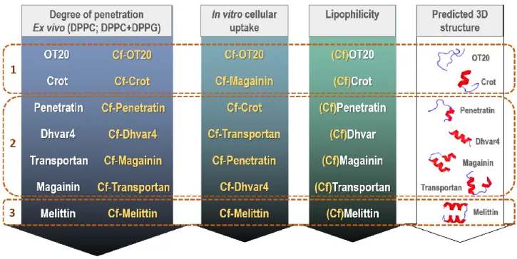
The ex vivo membrane penetration and in vitro cellular uptake profile of the seven membrane active peptides are summarized in Fig.8. The order of lipophilicity and the predicted 3D structure is also displayed to evaluate the influence of these properties on the in vitro cellular interaction. According to these properties three groups were defined: Group 1:
no significant differences between unlabelled and Cf-labelled peptides; Group 2: Penetration rate, cellular uptake slightly affected by Cf-labelling; Group 3: Highly membrane disrupting peptide; not distinguishable effect of Cf-labelling.
Fig. 8 Summary of ex vivo membrane penetration, in vitro cellular uptake profile and predicted lipophilicity, helicity of cationic synthetic (OT20, Crot(1-9,38-42) and Dhvar4), cell penetrating (Penetratin, Transportan) and antimicrobial (Magainin, Melittin) peptides. Coloured arrows represent behavioral tendencies (arrows point in the direction “low –
high”) in case of unlabelled and/or Cf-labelled peptides. The membrane penetration was determined on lipid monolayers, the cellular internalisation and membrane integrity was measured in MonoMac6 cells. As an indirect approach the lipophilic-hydrophilic character of the peptides and Cf-peptides was estimated using retention time (Rt)
obtained by high performance liquid chromatography on a reverse phase, C18 column and surface activity measurements. Helicity was predicted by PEP-FOLD methods. Group 1: no significant differences between unlabelled
15 and Cf-labelled peptides; Group 2: Penetration rate, cellular uptake slightly affected by Cf-labelling; Group 3: Highly
membrane disrupting peptide; not distinguishable effect of Cf-labelling.
Conclusions
In this study a group of cell penetrating, antimicrobial and cationic synthetic peptides was investigated based on their ability to bind to lipid model membranes. The seven peptides represent different chemical properties and biological specificities. Membrane affinity of the original and Cf-labelled peptides were compared.
The coupling of peptides with 5(6)-carboxyfluorescein resulted in remarkable increase of hydrophobicity indicated by retention time on reversed phase chromatographic column (C18) and surface activity determined by surface tension measurements of their aqueous solutions.
Model lipid monolayers were used to characterise the membrane affinity of the various peptides and their Cf-labelled compounds. The hydrophobic interaction is mainly responsible for membrane affinity to zwitterionic lipid monolayer (DPPC). Considering the effect of composition of peptides it was found that the degree of interaction with DPPC monolayer is increasing as it is favoured by the free energy of surface binding calculated by the Wimley-White approach.
The increased hydrophobicity of the Cf-labelled peptides led to higher membrane affinity only for the most hydrophobic peptides with smaller ratio of charged amino acids.
The other lipid monolayer composed of DPPC and DPPG simulates the charged character of outer leaflet of membrane.
An increased penetration was found using this negatively charged model membrane for both unlabelled and their Cf- labelled derivatives resulting from the combined hydrophobic and electrostatic interactions. Cf-labelling had negligible influence on the membrane affinity of most peptides studied here with the exception of Cf-Penetratin and Cf-Dhvar4 where an increase in penetration was observed comparing to the original peptides. The possible explanation for this behaviour might be that according to their amino acid sequence these peptides are less amphiphilic in the original form, while the Cf-labelling enhances such property.
Cytotoxicity of the selected peptides and internalisation of their Cf-modified derivatives were comparatively studied. The response on membrane integrity was also investigated. Flow cytometry and fluorescent microscopy are sensitive methods to determine compound-induced cytotoxic effects and membrane damaging. Cellular uptake by human MonMac6 cells was the highest for Cf-Dhvar4, Cf-Penetratin and Cf-Melittin. The uptake depends on penetration abilities, charge and structural features. The small differences in the lipophilicity of the peptides seem to have no effect on the uptake at the applied experimental conditions. The morphology of the cells treated with most of the peptides and Cf-peptides is normal, similar to control cells. The morphology of the cells treated with Melittin and Cf-Melittin is different, the cells were membrane damaged. The more intense interaction with membranes presented by Melittin can be related to its cytotoxic and/or membrane disrupting properties. Flow cytometric data with microscope imaging were in agreement in case of all peptides and their Cf-derivatives.
These types of peptides are widely applied as part of carrier/drug delivery systems. However, membrane interaction (due to differed charge, lipophilicity, penetration profile) can be affected by small molecule cargos (fluorophores, drug molecules, etc.), which alteration need to be considered when choosing cationic peptide carriers.
The internalisation pathway of cationic peptides, cpps and amps is widely studied and often lead to controversial results investigating the same peptide in different experimental approaches. The comparative analysis of the modifying effect of fluorescent moiety on peptides, which were performed in the present work applying model systems, might contribute to the better understanding of in vitro data.
Acknowledgements
Valuable assistance of Mrs. I. Hórvölgyi and H. Szatmári in Langmuir experiments are acknowledged. This work was financially supported by National Research Development and Innovation Office, Hungary (OTKA 104275, 115431, 124077) and VEKOP-2.3.2-16-2017-00014 European Union and the State of Hungary, co-financed by the European Regional Development Fund. K. Horváti was supported by the János Bolyai Research Scholarship of the Hungarian Academy of Sciences. G. Gyulai was supported by the Hungarian Academy of Sciences Postdoctoral Research Program.
Compliance with ethical standards Conflict of interest
The authors declare that they have no conflict of interest.
16 Research involving human participants and/or animals
This article does not contain any studies with human participants or animals performed by any of the authors.
References
Ábrahám Á, Baranyai Z, Gyulai G, Pári E, Horváti K, Bősze S, Kiss É (2016) Comparative analysis of new antitubercular drug peptide conjugates–Model membrane and in vitro studies. Colloid Surface B 147:106–115.
https://doi.org/10.1016/j.colsurfb.2016.07.054
Ábrahám Á, Katona M, Kasza G, Kiss É (2017) Amphiphilic polymer layer – cell membrane interaction studied by QCM and AFM in model systems. Eur Polym J 93C:212–221. https://doi.org/10.1016/j.eurpolymj.2017.05.047
Alhakamy NA, Kaviratna A, Berkland CJ, Dhar P (2013) Dynamic Measurements of Membrane Insertion Potential of Synthetic Cell Penetrating Peptides. Langmuir 29:15336−15349. https://doi.org/10.1021/la403370p
Almeida PF, Pokorny A (2009) Mechanism of Antimicrobial, Cytolytic and Cell-penetrating peptides: From Kinetics to Thermodynamics. Biochemistry 48:8083–8093. https://doi.org/10.1021/bi900914g
Asthana N, Yadav SP, Ghosh JK (2004) Dissection of antibacterial and toxic activity of Melittin. J Biol Chem 279 53:55042-55050. https://doi.org/10.1074/jbc.M408881200
Avitabile C, D’Andrea LD, Romanelli A (2014) Circular Dichroism studies on the interactions of antimicrobial peptides with bacterial cells. Sci Rep 4:4293. https://doi.org/10.1038/srep04293
Baranyai Z, Krátký M, Vosátka R, Szabó E, Senoner Z, Dávid S, Stolaříková J, Vinšová J, Bősze S (2017) In vitro biological evaluation of new antimycobacterial salicylanilide-tuftsin conjugates. Eur J Med Chem 133:152-173.
https://doi.org/10.1016/j.ejmech.2017.03.047
Bellet-Amalric E, Blaudez D, Desbat B, Graner F, Gauthier F, Renault A (2000) Interaction of the third helix of Antennapedia homeodomain and a phospholipid monolayer, studied by ellipsometry and PM-IRRAS at the air–water interface. Biochim Biophys Acta 1467:131–143. https://doi.org/10.1016/S0005-2736(00)00218-2
Birch D, Christensen MV, Staerk D, Franzyk H, Nielsen HM (2017) Fluorophore labeling of a cell-penetrating peptide induces differential effects on its cellular distribution and affects cell viability. BBA-Biomembranes 1859:2483–2494 https://doi.org/10.1016/j.bbamem.2017.09.015
Brezesinski G, Möhwald H (2003) Langmuir Monolayers to Study Interactions at Model Membrane Surfaces. Adv Colloid Interface Sci 100:563−584. https://doi.org/10.1016/S0001-8686(02)00071-4
Brockman H (1999) Lipid Monolayers: Why Use Half a Membrane to Characterize Protein-Membrane Interactions? Curr Opin Struct Biol 9:438−443. https://doi.org/10.1016/S0959-440X(99)80061-X
Castanho MARB (Ed.) (2009) Membrane-active Peptides: Methods and Results on Structure and Function. International University Line, La Jolla, California, IUL Biotechnology Series ISBN 978-0972077453
Christiaens B, Symoens S, Verheyden S, Engelborghs Y, Joliot A, Prochiantz A, Vandekerckhove J, Rosseneu M, Vanloo B, Vanderheyden S (2002) Tryptophan fluorescence study of the interaction of penetratin peptides with model membranes. Eur J Biochem 269:2918–2926. https://doi.org/10.1046/j.1432-1033.2002.02963.x
Cranfield CG, Henriques ST, Martinac B, Duckworth PA, Craik DJ, Cornell B (2017) Kalata B1 and Kalata B2 Have a Surfactant-Like Activity in Phosphatidylethanolomine Containing Lipid Membranes. Langmuir 33:6630–6637.
https://doi.org/10.1021/acs.langmuir.7b01642
Erl W, Weber C, Wardemann C, Weber PC (1995) Adhesion properties of Mono Mac 6, a monocytic cell line with characteristics of mature human monocytes. Atherosclerosis 113:99–107. https://doi.org/10.1016/0021-9150(94)05434- K
Flasinski M, Hac-Wydro K, Wydro P, Dynarowicz-Łatka P (2014) Influence of platelet-activating factor, lyso-platelet- activating factor and edelfosine on Langmuir monolayers imitating plasma membranes of cell lines differing in susceptibility to anti-cancer treatment: the effect of plasmalogen level. J R Soc Interface 11:20131103.
https://doi.org/10.1098/rsif.2013.1103
Guo Z, Peng H, Kang J, Sun D (2016) Cell-penetrating peptides: Possible transduction mechanisms and therapeutic applications. Biomed Rep 4:528–534. https://doi.org/10.3892/br.2016.639
Haedicke A, Blume A (2015) Binding of Short Cationic Peptides (KX)4K to Negatively Charged DPPG Monolayers:
Competition between Electrostatic and Hydrophobic Interactions. Langmuir 31:12203−12214.
https://doi.org/10.1021/acs.langmuir.5b02882
17 Hedegaard SF, Derbas MS, Lind TK, Kasimova MR, Christensen MV, Michaelsen MH, Campbell RA, Jorgensen L, Franzyk H, Cárdenas M, Nielsen HM (2018) Fluorophore labeling of a cell-penetrating peptide significantly alters the mode and degree of biomembrane interaction. Sci Rep. 20; 8(1):6327. https://doi.org/10.1038/s41598-018-24154-z Hegedüs R, Manea M, Orbán E, Szabó I, Kiss É, Sipos É, Halmos G, Mező G (2012) Enhanced cellular uptake and in vitro antitumor activity of short-chain fatty acid acylated daunorubicin-GnRH-III bioconjugates. Eur J Med Chem 56:155–165. https://doi.org/10.1016/j.ejmech.2012.08.014
Henriques ST, Melo MN, Castanho MARB (2006) Cell-penetrating peptides and antimicrobial peptides: how different are they? Biochem J 99:1–7. https://doi.org/10.1042/BJ20061100
Hill K, Pénzes CB, Schnöller D, Horváti K, Bősze S, Hudecz F, Keszthelyi T, Kiss É (2010) Characterization of the membrane affinity of an isoniazide peptide-conjugate by tensiometry, atomic force microscopy and sum-frequency vibrational spectroscopy, using a phospholipid Langmuir monolayer model. Phys Chem Chem Phys 12:11498–11506.
https://doi.org/10.1039/C002737E
Horváti K, Bacsa B, Kiss E, Gyulai G, Fodor K, Balka G, Rusvai M, Szabó E, Hudecz F, Bősze S (2014) Nanoparticle encapsulated lipopeptide conjugate of antitubercular drug isoniazid: in vitro intracellular activity and in vivo efficacy in a Guinea pig model of tuberculosis. Bioconjug Chem 12:2260-8. https://doi.org/10.1021/bc500476x
Horváti K, Bacsa B, Mlinkó T, Szabó N, Hudecz F, Zsila F, Bősze S (2017) Comparative analysis of internalization, haemolytic, cytotoxic and antibacterial effect of membrane-active cationic peptides: aspects of experimental setup. Amino Acids 49:1053–1067. https://doi.org/10.1007/s00726-017-2402-9
Horváti K, Gyulai G, Csámpai A, Rohonczy J, Kiss É, Bősze S (2018) Surface Layer Modification of Poly(D,L-lactic- co-glycolic acid) Nanoparticles with Targeting Peptide: A Convenient Synthetic Route for Pluronic F127-Tuftsin Conjugate. Bioconjug Chem 5:1495-1499. https://doi.org/10.1021/acs.bioconjchem.8b00156
Horvath R, Kobzi B, Keul H, Möller M, Kiss É (2013) Molecular Interaction of a New Antibacterial Polymer with a Supported Lipid Bilayer Measured by an in situ Label-Free Optical Technique. Int J Mol Sci 14:9722–9736.
https://doi.org/10.3390/ijms14059722
Hopp TP, Woods KR (1981) Prediction of protein antigenic determinants from amino acid sequences. Proc Natl Acad Sci USA 78:3824–3828. https://doi.org/10.1073/pnas.78.6.3824
Hristova K, White SH (2005) An Experiment-Based Algorithm for Predicting the Partitioning of Unfolded Peptides into Phosphatidylcholine Bilayer Interfaces. Biochemistry 44:12614–12619. https://doi.org/10.1021/bi051193b
Illien F, Rodriguez N, Amoura M, Joliot A, Pallerla M, Cribier S, Burlina F, Sagan S (2016) Quantitative fluorescence spectroscopy and flow cytometry analyses of cell-penetrating peptides internalization pathways: optimization, pitfalls, comparison with mass spectrometry. Sci Rep 6:36938. https://doi.org/10.1038/srep36938
Jensen EC (2012) Use of Fluorescent Probes: Their Effect on Cell Biology and Limitations. Anat Rec 295:2031–2036.
https://doi.org/10.1002/ar.22602
Kapus A, Grinstein S, Wasan S, Kandasamy R, Orlowski J (1994) Functional characterization of three isoforms of the Na+/H+ exchanger stably expressed in Chinese hamster ovary cells. ATP dependence, osmotic sensitivity, and role in cell proliferation. J Biol Chem 269:23544–23552. PMID: 8089122
Keszthelyi T, Hill K, Kiss É (2013) Interaction of phospholipid Langmuir monolayers with an antibiotic peptide conjugate. J Phys Chem B 117:6969–6979. https://doi.org/10.1021/jp401533c
Kiss É, Heine ET, Hill K, He Y-C, Keusgen N, Pénzes CB, Schnöller D, Gyulai G, Mendrek A, Keul H, Moeller M (2012) Membrane affinity and antimicrobal properties of polyelectrolytes with different hydrophobicity. Macromolec Biosci 12:1181–1189. https://doi.org/10.1002/mabi.201200078
Kitchens KM, Kolhatkar RB, Swaan PW, Eddington ND, Ghandhari H (2006) Transport of Poly(Amindoamine) Dendrimers across Caco-2 Cell Monolayers: Influence of Size, Charge and Fluorescent Labeling. Pharm Res 23:2818–
2826. https://doi.org/10.1007/s11095-006-9122-2
Ladokhin AS, White SH (1999) Folding of amphipathic α-helices on membranes: Energetics of helix formation by melittin. J Mol Biol 285:1363–1369. https://doi.org/10.1006/jmbi.1998.2346
Lindberg M, Jarvet J, Langel Ü, Gräslund A (2001) Secondary Structure and Position of the Cell-Penetrating Peptide Transportan in SDS Micelles As Determined by NMR. Biochemistry 40:3141–3149. https://doi.org/10.1021/bi0008985 Liu X, Peng J, He J, Li Q, Zhou J, Liang X, Tang S (2018) Selection and identification of novel peptides specifically targeting human cervical cancer. Amino Acids 50: 577-592. https://doi.org/10.1007/s00726-018-2539-1
18 Lohr M, Bernier SC, Horchani H, Bussières S, Cantin L, Desbat B, Salesse C (2014) Comparison between the behavior of different hydrophobic peptides allowing membrane anchoring of proteins. Adv Colloid Interface Sci 207:223–239.
https://doi.org/10.1016/j.cis.2014.01.015
Madani F, Lindberg S, Langel Ü, Futaki S, Gräslund A (2011) Review article: mechanisms of cellular uptake of cell- penetrating peptides. J Biophys 2011:414729. https://doi.org/10.1155/2011/414729
Marek T, Szeles C, Süvegh K, Kiss É, Vértes A, Lynn KG (1999) Characterisation of arachidate Langmuir-Blodgett films by variable-energy positron beams. Langmuir 15:8189–8196. https://doi.org/10.1021/la990109o
Michanek A, Yanez M, Wacklin H, Hughes A, Nylander T, Sparr E (2012) RNA and DNA Association to Zwitterionic and Charged Monolayers at the Air-Liquid Interface. Langmuir 28:9621-9633. https://doi.org/10.1021/la204431q Mohai M, Kiss É, Tóth A, Szalma J, Bertóti I (2002) Preparation and Characterization of Langmuir-Blodgett Type Arachidate Films. Surface Interface Anal 34:772–776. https://doi.org/10.1002/sia.1408
Nikawa H, Fukushima H, Makihira S, Hamada T, Samaranayake LP (2004) Fungicidal effect of three new synthetic cationic peptides against Candida albicans. Oral Dis 10:221–228. https://doi.org/10.1111/j.1601-0825.2004.01010.x Pénzes CB, Schnöller D, Horváti K, Bősze S, Mező G, Kiss É (2012) Membrane affinity of antituberculotic drug conjugate using lipid monolayer containing mycolic acid. Colloid Surface A 413:142–148.
https://doi.org/10.1016/j.colsurfa.2012.02.013
PEP-FOLD server, Paris Diderot University (FR) (cited 2018) De novo peptide structure prediction [Internet]
http://bioserv.rpbs.univ-paris-diderot.fr/services/PEP-FOLD
Reissmann S (2014) Cell penetration: scope and limitations by the application of cell-penetrating peptides. J Pept Sci 20:760–784. https://doi.org/10.1002/psc.2672
Roy A, Kucukural A, Zhang Y (2010) I-TASSER: a unified platform for automated protein structure and function prediction. Nat Protoc 5:725–738. https://doi.org/10.1038/nprot.2010.5
Schöne A-C, Roch T, Schulz B, Lendlein A (2017) Evaluating polymeric biomaterial–environment interfaces by Langmuir monolayer techniques. J. R. Soc. Interface 14:20161028. https://doi.org/10.1098/rsif.2016.1028
Shen Y, Maupetit J, Derreumaux P, Tufféry P (2014) Improved PEP-FOLD approach for peptide and miniprotein structure prediction. J Chem Theor Comput 10:4745–4758. https://doi.org/10.1021/ct500592m
Thévenet P, Shen Y, Maupetit J, Guyon F, Derreumaux P, Tufféry P (2012) PEP-FOLD: an updated de novo structure prediction server for both linear and disulfide bonded cyclic peptides. Nucleic Acids Res 40:W288–W293.
https://doi.org/10.1093/nar/gks419
Weingart CL, Broitman-Maduro G, Dean G, Newman S, Peppler M, Weiss AA (1999) Fluorescent Labels Influence Phagocytosis of Bordetella pertussis by Human Neutrophils. Infect Immun 67:4264–4267. PMID: 10417202
White SH, Wimley WC (1998) Hydrophobic interactions of peptides with membrane interfaces. Biochim Biophys Acta 1376:339–352. https://doi.org/10.1016/S0304-4157(98)00021-5
Więcek A, Dynarowicz-Łątka P, Miñones J, Conde O, Casas M (2008) Interactions between an anticancer drug- edelfosine-and cholesterol in Langmuir monolayers. Thin Solid Films 516:8829–8833.
https://doi.org/10.1016/j.tsf.2007.11.054
Zhang Lab, University of Michigan (cited 2018) I-TASSER Protein Structure and Function Predictions [Internet]
https://zhanglab.ccmb.med.umich.edu/I-TASSER/
Zhang Y (2008) I-TASSER server for protein 3D structure prediction. BMC Bioinformatics 9:40.
https://doi.org/10.1186/1471-2105-9-40
Ziegler A (2008) Thermodynamic studies and binding mechanisms of cell penetrating peptides with lipids and glycosaminoglycans. Adv Drug Deliv Rev 60:580–597. https://doi.org/10.1016/j.addr.2007.10.005
Ziegler-Heitbroc, HW, Thiel E, Futterer A, Herzog V, Wirtz A, Riethmuller G (1988) Establishment of a human cell line (MonoMac6) with characteristics of mature monocytes. Int. J. Cancer 41, 456–461.
https://doi.org/10.1002/ijc.2910410324
Zorko M, Langel Ü (2005) Cell-penetrating peptides: mechanism and kinetics of cargo delivery. Adv Drug Deliver Rev 57 4:529-545. https://doi.org/10.1016/j.addr.2004.10.010
19 Table captions
Table 1 Analytical characteristics of the peptides Table 1 Notes:
a Measured average molecular mass by Bruker Esquire 3000+ ESI-MS.
bRetention time using analytical RP-HPLC, Agilent ZORBAX StableBond 80Å C18, 4.6 mm × 150 mm, 3.5 µm HPLC column, gradient: 5% B, 2 min; 5-100% B, 20 min.
Table 2 Structural properties of the seven peptides and surface activity (∆γ) of those and their Cf-labelled counterparts Table 2 Notes:
a n: number of amino acids
b Z: net charge at neutral pH. Calculated by the number of (K+R)-(E+D). Positive charge at the N-terminus increases Z by 1 unit
c Hydrophilicity calculated using values of amino acids expressing hydrophilicity of each amino acid (Hopp and Woods 1981)
d Decrease of surface tension of water at peptide concentration of 200 μM after 1 h adsorption
e Calculated by I-TASSER
Figure captions
Fig. 1 Time dependence of surface tension (γ) of aqueous solutions of peptides and respective Cf-peptides at concentration of 200 μM. (Five of the studied peptides are presented for clarity)
Fig. 2 Predicted secondary structure of the peptides using I-TASSER and PEP-FOLD methods
Fig. 3 Surface pressure (Π) of DPPC monolayer during the interaction with Transportan (a) and Melittin (b) comparing to their Cf-labelled derivatives (○)
Fig. 4 Degree of penetration (ΔΠ) of peptides (black) and Cf-peptides (grey) into DPPC lipid monolayer determined at 1 h of interaction. Error bars represent the confidence intervals (95%) of the measured mean values.
Fig. 5 Surface pressure (Π) of DPPC+DPPG monolayer during the interaction with Transportan (a) and Melittin (b) comparing to their Cf-labelled derivatives (○)
Fig. 6 Degree of penetration (ΔΠ) of peptides (black) and Cf-peptides (grey) into DPPC+DPPG lipid monolayer determined at 1 h of interaction. Error bars represent the confidence intervals (95%) of the measured mean values.
Fig. 7 Comparison of internalisation and cytotoxicity of selected peptides and their Cf-derivatives using flow cytometry (BD LSRII) and microscopy (Olympus CKX41) performed as described in the Experimental section. Panel a represents the relative viability of unlabelled and Cf-labelled peptides following treatment (10 μM, 2 h) measured on MonoMac6 human monocytes. Spontaneous cell death due to sample preparation was determined for untreated control cells (3-5 % of death). Panel b shows the mean intracellular fluorescence intensity of Cf-peptide treated MonoMac6 cells (10 μM, 2 h; FITC LP505; BP 530/30). Error bars are SEM of three independent measurements. Panel c displays representative images of internalisation and membrane integrity studies with selected unlabelled and Cf-labelled peptides. Columns (1) bright field images of untreated and unlabelled OT20, Penetratin and Melittin, 20 μM; 2h (2) membrane integrity; PI staining, SuperWideGreen Filter (3) bright field images, Cf-labelled cpps, 20 μM. 2 h (4) internalisation, WideBlue Filter (5) membrane integrity; PI staining, SuperWideGreen Filter. After PI staining non-viable cells have bright red fluorescence, while viable cells remain non-fluorescent.
Fig. 8 Summary of ex vivo membrane penetration rate, in vitro cellular uptake profile and predicted lipophilicity, helicity of cationic synthetic (OT20, Crot(1-9,38-42) and Dhvar4), cell penetrating (Penetratin, Transportan) and antimicrobial (Magainin, Melittin) peptides. Coloured arrows represent behavioral tendencies (arrows point in the direction “low – high”) in case of unlabelled and/or Cf-labelled peptides. The membrane penetration was determined on lipid monolayers, the cellular internalisation and membrane integrity was measured in MonoMac6 cells. As an indirect approach the