CHAPTER 7
The Effect of Proteins and Amino Acids on the Growth of Adult Tissue in Vitro
HENRY S. SIMMS AND MARY S. PARSHLEY1
Department of Pathology, College of Physicians and Surgeons, Columbia University, New York, New York
Page
I. Introduction 144 A. Tissue Cultures as a Means for Studying Nutritional Requirements
of Adult Cells 144 B. Studies on Adult Chicken Aorta Fibroblasts 144
C. Maintenance versus Dormancy—Adult Tissue Inhibitor 145
II. Methods of Obtaining Data 145
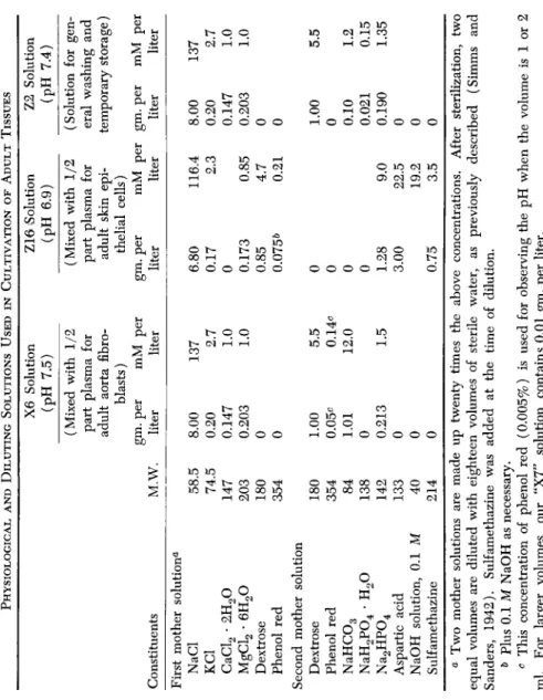
A. Tissues 145 B. Culture Media 145 C. Composition of Physiological Solutions and Diluting Solutions
(Table I) 145 D. Serum Ultrafiltrate 147 E. Methods of Treatment of Tissues with Test Substances 147
F. Obtaining Sterile Plasma and Tissues 148
G. Preparation of Aorta Tissue 148 H. Preparation of Skin Tissue 149 I. Evaluation of Growth 149 III. Explanation of Tables 149
A. Significance of Data Given in the Tables 149 B. Abbreviations and Symbols Used in the Tables 150 IV. Effect of Proteins, Physiological Materials, and Other Substances on
Growth 151 A. Blood and Tissue Constituents, Including Hormones (Table II) 151
B. Proteins and Digested Proteins (Table III) 155
C. Amino Acids (Table IV) 158 D. Vitamins (Table V) 163 E. Enzymes (Table VI) 163 F. Other Organic Substances (Table VII) 163
G. Sulfur Compounds (Table VIII) 165 H. Plant Materials (Table IX) 165 I. Inorganic Compounds (Table X) 165 V. Summary of Substances Favoring Adult Cell Growth (Table XI) 165
VI. Substances Related to Wound Healing (Table XII) 171 A. Water Soluble Ointment Bases (Table XIII) 175 B. Water Insoluble (Greasy) Bases (Table XIV) 177 C. Stearates and Greasy Bases Containing Stearates (Table XV) . . . . 181
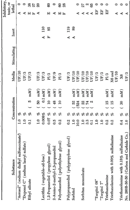
D. Detergents, Emulsifiers, Penetrants, etc. (Table XVI) 181
E. Antibacterial Substances 181 VII. Discussion and Summary 192
References 192 1 The research reported here was completed previous to 1949 and was aided by
143
144 HENRY S. SIMMS AND MARY S. PARSHLEY
I. INTRODUCTION
A. TISSUE CULTURES AS A MEANS FOR STUDYING NUTRITIONAL REQUIREMENTS OF ADULT CELLS
This chapter deals with the nutritional requirements of adult cells in tissue culture. It should be pointed out that most tissue culture work in the past has dealt with embryonic tissue, rather than with adult tissue.
This is true of the early work on chick embryo tissue in the laboratories of Carrel, of Fischer, and of many others. The role of embryo extract in cultures of embryo tissue has been given considerable attention. De- velopments have been reviewed by Morgan (1950) and by Waymouth
(1954).
It has been our impression that the requirements of adult cells differ from those of the embryo cell. Embryo extract is foreign to adult tissue and while stimulating, is not -needed by it (Parshley et al, 1953; Las- fargues, 1956). On the other hand, the "A" Factor (Simms and Stillman, 1937c), which is produced by leucocytes and is found in blood plasma, is essential for the life of adult cells—but not for embryo cells.
In recent years strains of adult tissue, including the "L" strain (of adult mouse subcutaneous connective tissue) from Earle and his co- workers' laboratory (1943) have been used for nutritional experiments by Earle, and also by Healy et al. (1955), by Eagle (1955a,b), and by Waymouth (1956). A discussion of long term cultivation of adult cells can be found in the reports of the Tissue Culture Conference of 1956.
As will be shown later in this chapter, in overcoming the dormancy of adult aorta tissue and in supporting subsequent growth, embryo ex- tract is inferior to extracts of adult spleen and heart.
The nutritional requirements of different adult cell types in the same animal are not the same (Parshley and Simms, 1950). This confirms earlier work on embryo cells by Baker and Carrel (see Morgan, 1950).
Nutritional requirements may also vary with the species and with the state of physiological activity of the cells.
B. STUDIES ON ADULT CHICKEN AORTA FIBROBLASTS
For a number of years the authors of this chapter studied the con- ditions which were favorable for the growth of adult tissues in vitro in order to determine what substances are necessary for the maintenance of adult cell life, and what substances serve to overcome the dormancy grants from the Josiah Macy, Jr. Foundation, the John and Mary R. Markle Founda- tion, W. R. Warner and Co., Dr. Marvin R. Thompson, The Office of Scientific Research and Development (in contract with Columbia University), The Life In- surance Medical Research Fund, and the Wendell L. Willkie Memorial Grant from the Albert and Mary Lasker Foundation.
PROTEINS AND ADULT TISSUE GROWTH 145 of normal adult tissue. For this purpose, special techniques were de- veloped for making quantitative determination of the stimulating or inhibitory effect of any given substance on adult tissues in vitro. The methods were applied during the second World War to the testing of materials which might be used to promote wound healing. Some of those observations will be reported here.
Several hundred substances have been tested for their effect on the growth of adult tissues in vitro. These include proteins, digested pro- teins, amino acids, plant materials, vitamins, hormones, enzymes, tissue extracts, sulfur compounds, and various other organic and inorganic compounds, including antibacterial agents and ointment bases.
C. MAINTENANCE VERSUS DORMANCY—ADULT TISSUE INHIBITOR
It should be explained that by "maintenance" we mean the nutritional requirements necessary to keep a cell alive and functioning—without cell division. But when we "overcome the dormancy" of adult tissue, we must not only maintain the cell in a living state, but we must also eliminate or counteract the "adult tissue inhibitor" which is present in adult tissues (Simms and Stillman, 1937b) and is even present in cul- tures of adult cells. This inhibitor protects adults from unrestrained growth or malignancy. It is a highly important material.
II. METHODS OF OBTAINING DATA
A brief summary of the technique follows. This technique was de- veloped over a period of years (Simms and Stillman, 1937a,b,c,d, and e;
Simms and Sanders, 1942).
A. TISSUES
Adult chicken aorta and adult chicken skin were used for most of the experiments reported here. The aorta cultures produced a clearly distinguishable growth of new fibroblasts. The adult chicken skin was cultured in a special medium which gave sheets of epithelial cells rela- tively free from fibroblasts.
B. CULTURE MEDIA
The media consisted of chicken plasma diluted with two parts of a suitable "diluting solution" (see below). No heparin or other anti- coagulant was used.
C. COMPOSITION OF PHYSIOLOGICAL SOLUTIONS AND DILUTING SOLUTIONS (TABLE I)
The composition of the solutions which were used for bathing or storing tissues and for diluting the culture media are given in Table I.
TABLE I PHYSIOLOGICAL AND DILUTING SOLUTIONS USED IN CULTIVATION OF ADULT TISSUES Constituents First mother solutiona NaCl KC1 CaCl2 · 2H20 MgCl2 · 6H20 Dextrose Phenol red Second mother solution Dextrose Phenol red NaHC03 NaH2P04 · H20 Na2HP04 Aspartic acid NaOH solution, 0.1 M Sulfamethazine
M.W. 58.5 74.5 147 203 180 354 180 354 84 138 142 133 40 214
X6 Solution (pH 7.5) (Mixed with 1/2 part plasma for adult aorta fibro- blasts) gm. per mM per liter liter 8.00 0.20 0.147 0.203
0 0 1.00 0.05^ 1.01
0 0.21
3
0 0 0
137 2.7 1.0 1.0 5.5 0.14c 12.0 1.5
Z16 Solution (pH 6.9) (Mixed with 1/2 part plasma for adult skin epi- thelial cells) gm. per liter 6.80 0.17
0 0.17
3 0.85 0.075&
0 0 0 0 1.28 3.00 0.75
mM per liter 116.4 2.3 0.85 4.7 0.21 9.0 22.5 19.2 3.5
Z2 Solution (pH 7.4) (Solution for gen- eral washing and temporary storage) gm. per liter 8.00 0.20 0.147 0.203
0 0 1.00
0 0.1
0 0.021 0.190
0 0 0
mM per liter 137 2.7 1.0 1.0 5.5 1.2 0.15 1.35 a Two mother solutions are made up twenty times the above concentrations. After sterilization, two equal volumes are diluted with eighteen volumes of sterile water, as previously described (Simms and Sanders, 1942). Sulfamethazine was added at the time of dilution. b Plus 0.1 M NaOH as necessary. c This concentration of phenol red (0.005%) is used for observing the pH when the volume is 1 or 2 ml. For larger volumes, our "X7" solution contains 0.01 gm. per liter.
2 Hi > Ö ^
£
PROTEINS AND ADULT TISSUE GROWTH 147 The "X6 Solution" was developed for the cultivation of adult chicken aorta fibroblasts (Simms and Sanders, 1942). It may be used by itself as a physiological solution for bathing or storing tissue. It is used also as a "diluting solution" for diluting plasma or serum ultrafiltrate.
The "Z16 Solution" is used only as a diluting solution. When used with plasma, it gives a medium favorable for the growth of adult skin epithelial cells. It contains neither bicarbonate nor calcium, but has a high concentration of phosphate. It will be noted that aspartic acid is included because of its growth stimulating action. Sulfamethazine was added for its antibacterial effect.
The "Z2 Solution" is used for washing or for temporary storage of tissues.
D. SERUM ULTRAFILTRATE
Serum ultrafiltrate contains a growth stimulating agent, which we call the "A factor," which is essential for adult cell maintenance and for the growth of adult tissues. It is used in fluid media diluted with a suit- able physiological or diluting, solution (Simms and Stillman, 1937c;
Simms and Sanders, 1942). Serum ultrafiltrate must always be kept under an atmosphere containing about 5% C02. Otherwise, the activity of the A factor is immediately lost due to the change in pH. Ultra- filtrate is not species specific.
E. METHODS OF TREATMENT OF TISSUES WITH TEST SUBSTANCES
We have two methods for testing the effect of an agent on the growth of adult tissue, the "fluid medium method" and the "plasma medium method." In the fluid medium method, the tissue was given a prelimi- nary incubation at 37 °C. for 3 days in a suitable fluid medium con- taining the test substance after which it was planted in a plasma clot (without the test substance) and the subsequent growth was evaluated daily. This was the more satisfactory method for aorta fibroblast tests and was used almost invariably for them.
The fluid medium consisted of X6 solution, or other physiological solution, or of serum ultrafiltrate diluted with two parts of X6 or other diluting solution. In general, when stimulating substances were tested, a nonstimulating medium was used. But when toxic or inhibitory sub- stances were tested, a stimulating medium (containing serum ultra- filtrate) was used. In each case, the subsequent growth was compared with control solutions containing no test substances.
In the "plasma medium method," the fresh tissue was planted direct- ly in a plasma medium containing the test substances. This method was used in tests on skin epithelial cells. When agents were insoluble in water,
148 HENRY S. SIMMS AND MARY S. PARSHLEY
they were floated on the surface of the medium. In such cases the volume of the medium was usually doubled or tripled.
F. OBTAINING STERILE PLASMA AND TISSUES
In each experiment an anesthetized chicken (preferably 2 or 3 years of age) was bled aseptically through a glass cannula inserted in the carotid artery. The blood was collected in a cold centrifuge tube. After centrifugation the plasma was stored in paraffined tubes. Skin tissue, when desired, was removed from the pectoral region, after cleaning with 50% alcohol (Parshley and Simms, 1950), and was stored tem- porarily in about 10 ml. of serum ultrafiltrate diluted with two parts of Z16 solution.
The thoracic aorta was removed aseptically and was stored tempo- rarily in 10 ml. of Z2 solution. The tissues can be stored for several days in the refrigerator, but best results were obtained when they were used within 24 hours.
G. PREPARATION OF AORTA TISSUE
Each aorta was washed in Z2 solution and the adventitia was stripped off. A section of the aorta was placed in a watch glass containing Z2 solution and was cut open lengthwise. It was then split through the media in such a way that the outer layer of the media could be dis- carded. The remaining tissue, consisting of the inner media and intima, was washed again in Z2 solution and then sliced into strips about 1.2 by 2.4 mm. in area. Four of these strips were used in each test.
A series of test tubes, 13 by 100 mm. were prepared, each containing 2.0 ml. of fluid medium as well as a test substance (except for the control tubes). Into each tube four strips of aorta tissue were placed and were allowed to rest on the bottom of the tube. The pH was adjusted to 7.4, as indicated by phenol red in the medium, by introducing a suitable sterile mixture of C 02 in air, usually 5%, as previously described (Simms and Stillman, 1937a). The tubes were closed with rubber stoppers and were incubated at 37°C. for 3 days.
After incubation the four strips of tissue from each tube were placed in Z2 solution in a watch glass, and were cut into a total of thirty-two pieces, 0.6 mm. square. These were immediately planted in Carrel flasks, sixteen pieces in each of two flasks. Special flasks, 32 mm. in diameter, were used. Before the tissue was introduced, about 0.06 ml. of chicken plasma, diluted with two parts of X6 solution, was spread over the bottom of each flask. After the planting and after this first plasma had clotted, a second layer of 0.8 ml. of diluted plasma was spread over the
PROTEINS AND ADULT TISSUE GROWTH 149 surface. The pH was adjusted to 7.4 with sterile 4% C 02 in air. The flasks were closed with rubber stoppers and incubated at 37 °C.
The subsequent growth of tissue was observed daily under the microscope.
H. PREPARATION OF SKIN TISSUE
Each piece of skin, having been placed temporarily in ultrafiltrate diluted with Z16 solution at pH 6.9, was afterwards washed repeatedly with this solution. Any feather follicles were removed and discarded.
Subcutaneous connective tissue and fat were dissected away. The pieces were cut under Z2 solution into strips about 1.5 by 2.0 mm. These were either incubated in tubes in a fluid medium as described for the aorta (but at pH 6.9), or were cut up into smaller pieces about 0.6 to 0.8 mm.
and planted directly in chicken plasma diluted with Z16 solution.
I. EVALUATION OF GROWTH
Daily observations were made of the extent of growth of test cultures as compared with control cultures. The number of cells around each of the explants in a given flask was estimated and the growth of each colony was rated according to the following system:
0 = no growth
0.25 = one to six new cells 0.5 = about ten new cells 1 = about thirty new cells
2 = extensive growth, about ninety cells
3 = abundant growth, not visible to the naked eye 4 = new growth clearly visible to naked eye Intermediary ratings of 1.5, 2.5, and 3.5 were sometimes used.
The sum of the ratings of the sixteen colonies in each flask was taken as the "growth rating" for that flask. The ratings for the two duplicate flasks in each test were averaged. On the third or fourth day the relative growth of test cultures was compared with that of the con- trols, where the latter value was taken as 100.
III. EXPLANATION OF TABLES
A. SIGNIFICANCE OF DATA GIVEN IN THE TABLES
In the tables in this Chapter, the following information is given:
in the first column is the name of the substance that was tested; in the second and third columns are the concentration and the medium in which it was tested. (The identical medium was always tested as a control.) In the other three columns the results are given. Data on substances
150 HENRY S. SIMMS AND MARY S. PARSHLEY
that were stimulating are placed in column 4, inert in column 5, and so on, according to the criteria indicated in the tabulation.
Category Stimulating Inert Inhibitory Toxic Lethal
zr z=z z=z
~
=
Relative growth rating over 125 — third column 125 to 75 — fourth column
75 to 25 — last column 25 to 1 — last column 0 — last column
The type of tissue or cell is indicated by a symbol (see below). This is followed by the relative growth rating (100 times the ratio of the growth rating with the test substance, divided by that of the control solution).
B. ABBREVIATIONS AND SYMBOLS USED IN THE TABLES
1. Type of Tissue and Cells A = aorta fibroblasts F = skin fibroblasts E := skin epithelial cells Thy = thyroid epithelial cells 2. Composition of Fluid Media
In tests using a fluid medium, the agent was added to the designated fluid medium in which the tissue was incubated for 3 days before it was planted in a plasma clot (not containing the agent).
UF = Serum ultrafiltrate (diluted with two or nine parts of physiological solution and designated "UF/3" or "UF/10,"
respectively).
D.UF = Decarbonated serum ultrafiltrate (diluted).
S = Serum (usually diluted with three parts of physiological solution and designated "S/4").
X6 = A special physiological, or diluting, solution (see Table I) which is favorable for adult fibroblasts.
Z16 = A special diluting solution (see Table I) favorable for adult skin epithelial cells.
Z8 = A special diluting solution similar to Z16 but containing one-third more phosphate—but no aspartic acid.
Ty = Tyrode solution (not used in this laboratory after March 1937 because it has been replaced by X6 which is more favorable for adult fibroblast growth).
PROTEINS AND ADULT TISSUE GROWTH 151 3. Solid Medium
In these tests the agent was added to the plasma clot in which tissue was planted directly.
PI = Plasma medium: Blood plasma diluted with two parts of
"X6" physiological solution for aorta tissue, or with "Z8"
or "Z16" solution for skin tissue.
4. Physical State of Substance in Medium
When no symbol appears, the agent was in solution.
G = The agent formed a gel.
D — The agent was dispersed as a suspension or an emulsion.
P = The agent was poorly soluble and settled as a precipitate;, in whole or in part.
S = The agent was insoluble and was floated on the surface.
IV. EFFECT OF PROTEINS, PHYSIOLOGICAL MATERIALS, AND OTHER SUBSTANCES ON GROWTH
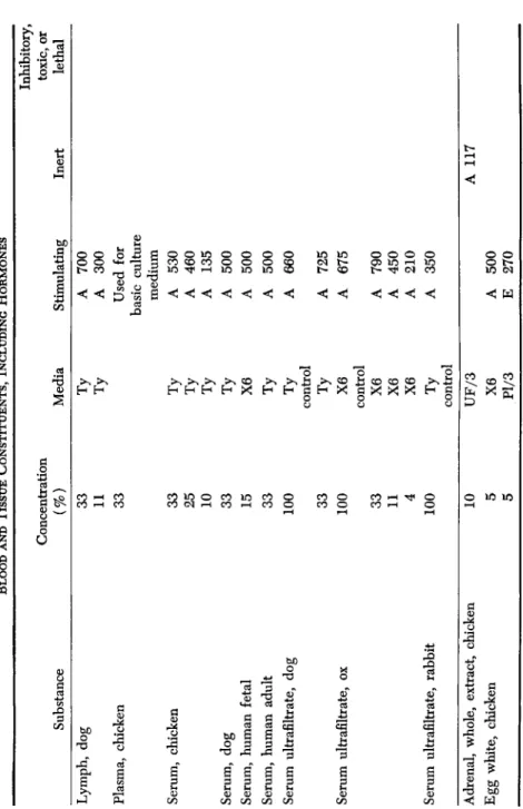
A. BLOOD AND TISSUE CONSTITUENTS, INCLUDING HORMONES ( TABLE II)
Several constituents of blood are important for growth and main- tenance of adult cells in vitro. One of these, the "A Factor" (Simms and Stillman, 1937c; Simms and Sanders, 1942), appears to be indis- pensable to the maintenance of adult cells. It is present in plasma and serum. Serum ultrafiltrate contains the A Factor and is free from pro- teins.2 This is used as a basis for a standard medium for the main- tenance of adult cells. However, serum ultrafiltrate alone is not satis- factory for the initiation of growth. For this reason, plasma proteins were usally added to the tissue culture medium.
A material contained in the red cells was highly stimulating to the growth of both fibroblasts and epithelial cells when present in a very low concentration. The active substance cannot be hemoglobin, for both hemoglobin and hemin were inert when tested in purified form.
Red cell extract was included in certain of our media. An extract of leucocytes was also very stimulating, and since this is the only agent which has served as a substitute for serum ultrafiltrate in the main- tenance of adult cells over a period of time, we have assumed that the A Factor is produced by leucocytes. An extract made from blood plate- lets was also found to improve the growth of adult fibroblasts.
2 The serum ultrafiltrate was obtained from Microbiological Associates, Bethesda, Maryland. Unfortunately, serum ultrafiltrate must be kept and used under an atmosphere containing 3 to 5% C 02. Otherwise, the loss of C 02 causes a rise in.
pH, with immediate complete destruction of the A Factor.
TABLE II BLOOD AND TISSUE CONSTITUENTS, INCLUDING HORMONES Substance Concentration (%) Media Stimulating Inert
Inhibitory, toxic, or lethal Lymph, dog Plasma, chicken Serum, chicken Serum, dog Serum, human fetal Serum, human adult Serum ultrafiltrate, dog Serum ultrafiltrate, ox Serum ultrafiltrate, rabbit Adrenal, whole, extract, chicken Egg white, chicken
33 11 33 33 25 10 33 15 33 100 33 100 33 11 4 100
IÖ
5 5Ty Ty Ty Ty Ty Ty X6 Ty Ty control Ty X6 control X6 X6 X6 Ty
control UF/3 X6 Pl/3
A A
700 300 Used for basic culture medium
A A A A A A A A A A A A A A E
530 460 135 500 500 500 660 725 675 790 450 210 350 500 270
A 117
TABLE II (Continued) Substance Concentration (%) Media Stimulating Inert
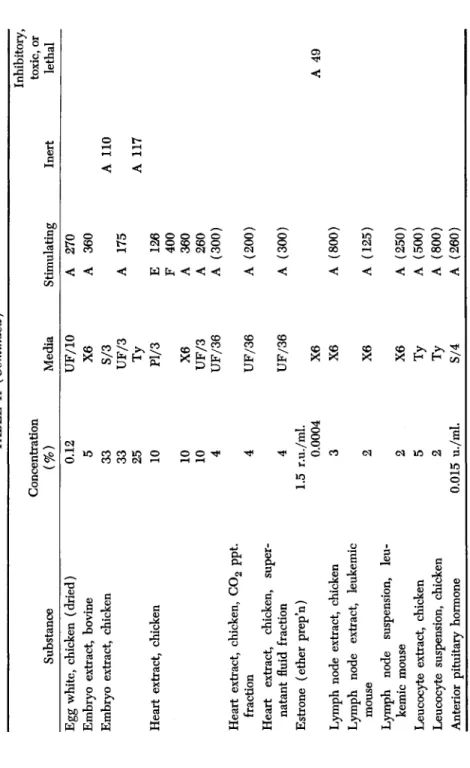
Inhibitory, toxic, or lethal Egg white, chicken (dried) Embryo extract, bovine Embryo extract, chicken Heart extract, chicken Heart extract, chicken, C02 ppt. fraction Heart extract, chicken, super- natant fluid fraction Estrone (ether prep'n) Lymph node extract, chicken Lymph node extract, leukemic mouse Lymph node suspension, leu- kemic mouse Leucocyte extract, chicken Leucocyte suspension, chicken Anterior pituitary hormone
0.12 5 33 33 25 10 10 10 4 4 4 1.5 r.u./ml. 0.0004 3
UF/10 X6 S/3 UF/3
Ty Pl/3 X6 UF/3 UF/36 UF/36 UF/36 X6 X6 2 5 2 0.015 u./ml.
X6 X6 Ty Ty S/4
A 270 A 360 A 175
E F A A
126 400 360 260 A (300) A (200) A (300) A (800) A (125) (250) (500) (800) (260)
A 110 A 117 A 49
TABLE II (Continued) Substance Concentration (%) Media Stimulating Inert
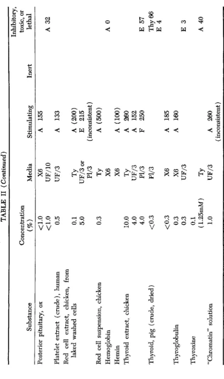
Inhibitory, toxic, or lethal Posterior pituitary, ox Platelet extract (crude), human Red cell extract, chicken, from laked washed cells Red cell suspension, chicken Hemoglobin Hemin Thyroid extract, chicken Thyroid, pig (crude, dried) Thyroglobulin Thyroxine "Chromatin" solution
<1.0 <1.0 0.5 0.1 5.0 0.3 10.0 4.0 4.0 <0.3 <0.3 0.3 0.3 0.1 (1.25mM) 1.0
X6 UF/10 UF/3 Ty UF/3 or Pl/3
Ty X6 X6
Ty UF/3 Pl/3 Pl/3 X6 X6 UF/3
Ty UF/3
A A A E
155 133 (200) 215 (inconsistent) A A A A F A A A (500) (100) 260 152 250 185 160 260 (inconsistent)
A 32 A 0 E 57 Thy 66 E 4 E 3 A 40
PROTEINS AND ADULT TISSUE GROWTH 155 Most of the hormones had little effect on adult cells. A fresh thyroid extract, crude dried thyroid, and thyroglobulin were stimulating to fibroblasts but inhibited the growth of skin epithelium and thyroid epithelium. Thyroxine has no stimulating effect. Anterior pituitary ex- tract (a standardized aqueous preparation) greatly stimulated the growth of aorta fibroblasts. Other hormones, including crude posterior pituitary extract, and a fresh adrenal extract, had slight if any stimulat- ing effect. A purified preparation of estrone was inhibitory to fibroblasts.
Extracts from some adult organs stimulated a growth of new cells when added to explanted aorta. Whole heart extract and fractions of it were extremely stimulating to both fibroblasts and epithelial cells. On the other hand, chicken embryo extract was inconsistent in its action on adult tissue. We found it to be unsatisfactory for use in adult tissue culture. Furthermore, it is a medium unnatural for adult cells. Doljanski, et al. (1942) state that embryo extract is far less stimulating for adult fibroblasts and epithelial cells than extracts of adult brain, heart, and smooth muscle. However, for embryo tissues, Davidson and Waymouth
(1945) find sheep embryo extract superior to adult tissue extracts.
Egg white (both fresh and dried) contains a substance which is very stimulating to the growth of both fibroblasts and epithelial cells.
It has been used as a substitute for serum in one of our basic culture media but does not give as consistently good results. It appears also to contain the factor which produces cohesiveness of cells (Simms, 1936).
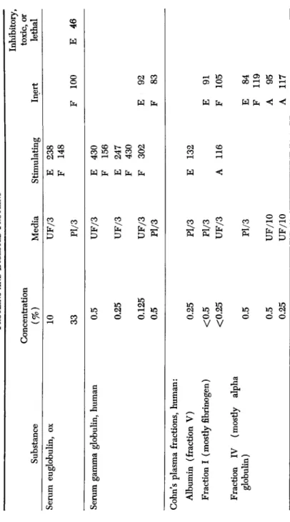
B. PROTEINS AND DIGESTED PROTEINS ( TABLE III)
Hemoglobin and certain tissue proteins have already been discussed under "blood and tissue constituents" (Table II).
The importance of proteins and their split products to embryo cell growth was observed by the Lewises (1911), who found that the addi- tion of amino acid and polypeptide mixtures to their media enhanced the growth of cells. Carrel and Baker (1926) observed that proteoses and peptones from the peptic digestion of proteins were superior to lower split products as growth-stimulants. Baker and Carrel (1928) obtained embryo cell stimulation by hydrolyzates of egg albumin, casein, edestin, and fibrin. However, the addition of glycine improved the stimulation. Willmer and Kendal (1932) found that embryo cells re- quire plasma or embryo extract in addition to the proteoses.
Adult cells, as seen in Table III, were stimulated by certain of the plasma proteins, especially thrombin, euglobulin, gamma globulin, and fibrinogen.3 Casein, after digestion by pepsin, had a marked growth-
3 These serum proteins all contain lipfanogens and are not suitable for tissue cultures where the prevention of fat granules is desired.
Substance Serum euglobulin, ox
TABLE III PROTEINS AND DIGESTED PROTEINS Concentration (%) 10 33
Media UF/3 Pl/3
Stimulating E 238 F 148
Inert F 100
Inhibitory, toxic, or lethal E 46 Serum gamma globulin, human 0.5 0.25 0.125 0.5
UF/3 UF/3 UF/3 Pl/3
E 430 F 156 E 247 F 430 F 302 E F 92 83
s
> Öl
inCohn's plasma fractions, human: Albumin (fraction V) Fraction I (mostly fibrinogen) Fraction IV (mostly alpha globulin)
0.25 <0.5 <0.25 0.5 0.5 0.25
Pl/3 Pl/3 UF/3 Pl/3 UF/10 UF/10
E 132 A 116 E F E F A A
91 105
84 119 95 117
TABLE III (Continued) Substance Gamma globulin Gamma globulin with penicil- lin (170 u./ml.) Thrombin Plasma fibrinogen, chicken Casein, digested by pepsin Lung protein Placental protein
Concentration (%) 0.5 0.25 0.5 0.25 0.5 0.25 0.5 0.5 0.25 33 33 33 1.7 0.25 0.25
Media Pl/3 Pl/3 UF/3 UF/10 UF/10 Pl/3 UF/10 UF/10 S/10 S/3 Pl/3 Pl/3 X6 X6
Stimulating E A F A A A E E E F
212 142 175 200 274 184 176 160 216 450
Inert E 91 F 86 E 82 A 92 E 112 A 80
Inhibitory, toxic, or lethal A 71 A 0
hi
8 a i § £ g
H9
Cfls §
Hj158 HENRY S. SIMMS AND MARY S. PARSHLEY
promoting effect on adult aorta fibroblasts and was also very stimulating to the growth of skin epithelium. Hanks (1955) reported that the eu- globulin fraction of serum contains growth stimulants. Serum albumin was not particularly stimulating in our tests, although Waymouth (1955) reported that albumin (Armour fraction V) added to a simple medium, enhanced growth of L strain of adult cells.
The growth promoting properties of tissue extracts have been as- scribed to the nucleoproteins by Fischer (1946) and by Davidson and Waymouth (1945).
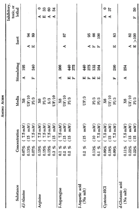
C. AMINO ACIDS (TABLE IV)
Most of the amino acids which were tested, with the exception of arginine and an impure preparation of glycine, were found to have a mild stimulating effect when added to a medium consisting of a physio- logical solution but gave no added stimulation when they were tested in serum ultrafiltrate. The small concentration of amino acids present in serum ultrafiltrate (only 1.4 millimolar) should not account for this difference. It seems likely that the stimulating action of most amino acids is inconspicuous in comparison with that of the A Factor which is present in the ultrafiltrate. However, aspartic acid, asparagine, and proline greatly increased the rate of growth of skin epithelial cells when added to a plasma medium. Aspartic acid and proline had some stimu- lating effect on fibroblasts in the same medium.
Efforts have been made by other workers to concoct synthetic tissue culture media free from proteins or other substances of unknown chem- ical structure. Such media have contained amino acids in addition to salts, glucose, and vitamins (Evans, and associates in Earle's laboratory, 1956a,b). The amino acid composition of such synthetic media has varied widely (White, 1946; Morgan, et al., 1950); Eagle, 1955; Healy et ah, 1955). Media having no other source of nitrogen than the amino acids can be sufficient for cell survival. However, protein molecules appear to be needed for cell division. Amino acid mixtures are invari- ably inferior to protein digests as growth stimulants.
The dialysis of plasma, serum, and tissue extracts was found (Fischer and co-workers, 1946, 1948) to leave a residue inadequate for growth.
They concluded that this was due to a loss of amino acids and other nutrients—especially cystine. White (1949) and Harris (1952) obtained more favorable results in their tests with dialyzed serum on embryo cul- tures. However, it has been our experience that adult tissues require the
"A Factor" that is removed by dialysis (Simms and Stillman, 1937c;
Simms and Sanders, 1942).
PROTEINS AND ADULT TISSUE GROWTH 159 Baker and Carrel (1926) considered that amino acids stimulate em- bryo cell division and migration. Their results with the addition of glycine to digested proteins have already been mentioned. On the other hand, Vogelaar and Erlichman (1936) found that glycine inhibited human thyroid fibroblasts, while Burrows and Neyman (1917) reported- toxic action on embryo cells of a number of amino acids. Carrel and Ebeling (1924) stated that 20-50 mg. of glycine per 100 ml. had no effect on growth, but accelerated migration.
Eagle (1955) reported that 13 amino acids were essential for growth of two cell strains, Earle's "L" strain of fibroblasts from the subcutaneous connective tissue of an adult mouse and Gey's HeLa strain isolated from a human uterine carcinoma. The amino acids listed, all L-isomeric, were arginine, cysteine, glutamine, histidine, isoleucine, lycine, methionine, phenylalanine, threonine, tryptophan, tyrosine, and valine. It is of in- terest that although the author states that the amino acid requirements of these two extremely different cell types proved remarkably similar, there was a marked difference in concentration for optimum growth.
Glutamine is considered to play an essential metabolic role. Fischer and co-workers (1946, 1948) had stressed earlier the importance of glu- tamine as a growth stimulant. Waymouth (1955) observed also that growth was improved by the addition of glutamine hydroxanthine to a modification of the semi-synthetic medium of Wilson et al. (1942) in which Whites' amino acid mixture was substituted for Witte's peptone.
Greatly increasing the phosphate and NaHC03 further improved the medium. Glutamic acid at any concentration failed to permit growth of the "L" strain of fibroblasts. It was effective for the HeLa cell in con- centrations ten to twenty times that of glutamine.
Eagle (1955) noted that a medium containing these 13 amino acids plus 7 vitamins, found to be essential, and glucose did not permit growth unless a small amount of serum protein (serum 1-5%) was present. Attempts to analyze the function of the serum residue sug- gested to the author that trace elements bound to the protein might be of importance. Human plasma fractions I, II, and III were inert and IV and V, only weakly active, separately or in combination. Exhaus- tively dialyzed serum was inactive, while fractions obtained by simple
(NH4)2S04 salting out followed by 24-hour dialysis were all more or less equally active. More recently Lockart and Eagle (1959) have re- ported that this minimum growth medium supplemented with dialyzed serum, which supported the propagation of heavily inoculated cultures of cell strains, was not satisfactory for regular or optimum growth of small numbers of cells or cloning from single cells. The deficiency was overcome by the addition of the 7 nonessential amino acids (alanine, as-
Substance d,Z-Alanine Ar ginine Z-Asparagine Z-Aspartic acid (Na salt) Cysteine-HCl cZ-Glutamic acid (Na salt)
Concentration 0.07% 0.13% 0.07% 0.15% 0.15% 0.15% 0.3 % 0.1 % 0.2 % 0.2 % 0.2 % 0.13% 0.1 % 0.09% 0.18% 0.09% 0.11% 0.22% 0.22%
( 7.5mM) (15 mM) ( 7.5mM) ( 7.5mM) ( 7.5mM) ( 7.5mM) (15 mM) ( 7.5mM) (15 mM) (15 mM) (15 mM) (10 mM) ( 7.5mM) ( 7.5mM) (15 mM) ( 7.5mM) ( 7.5mM) (15 mM) (15 mM)
TABLE IV AMINO ACIDS Media X6 UF/10 Pl/3 X6 Pl/3 S/3 UF/10 X6 UF/10 Pl/3 UF/3 Pl/3 Pl/3 X6 UF/10 Pl/3 X6 UF/10 Pl/3
Stimulating A F A E F E F E E F A
195 240 266 440 375 440 375 132 164 290 254
Inert A E A A F F E A E
94 89 87 95 66 100 83 82 >100
Inhibitory, toxic, or lethal A E A A A A F
0 33 60 54 0 37 50
OS o
s
3 Cfl </5 M 2 3I
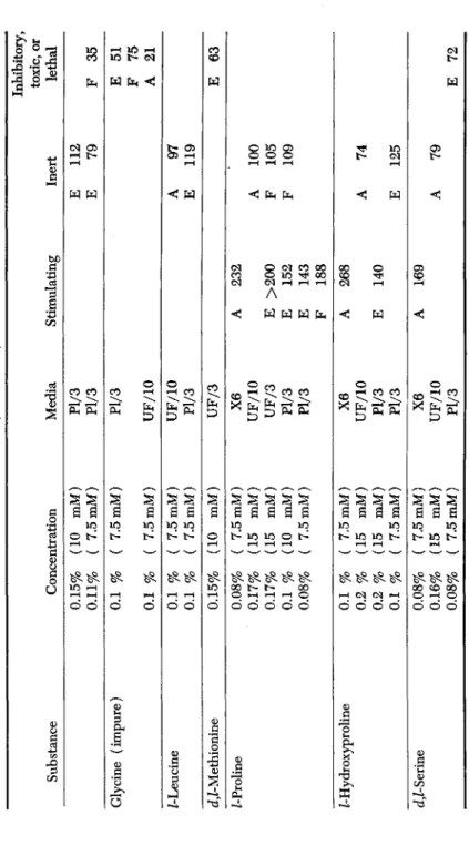
>"4TABLE IV (Continued) Inhibitory, toxic, or Substance Concentration Media Stimulating Inert lethal Glycine (impure) Z-Leucine d,Z-Methionine Z-Proline Z-Hydroxyproline d^-Serine
0.15% 0.11% ( 0.1 % 0.1 % 0.1 % 0.1 % 0.15% 0.08%
0.17% 0.17
% 0.1 % 0.08% 0.1 % ( 0.2 % 0.2 % 0.1 % ( 0.08% ( 0.16% ( 0.08% {
[10 mM) f 7.5mM) [ 7.5mM) ; 7.5mM) [ 7.5mM) [ 7.5mM) [10 mM) [ 7.5mM) [15 mM) [15 mM) [10 mM) [ 7.5mM) ' 7.5mM) [15 mM) [15 mM) ' 7.5mM) 7.5 mM) 15 mM) 7.5 mM)
Pl/3 Pl/3 Pl/3 UF/10 UF/10 Pl/3 UF/3 X6 UF/10 UF/3 Pl/3 Pl/3 X6 UF/10 Pl/3 Pl/3 X6 UF/10 Pl/3
A E E E F A E A
232 >200 152 143 188 268 140 169
E E A E A F F A E A
112 79 97 119 100 105 109 74 125 79
F E F A E E
35 51 75 21 63 72
162 HENRY S. SIMMS AND MARY S. PARSHLEY
partic acid, asparagine, glycine, glutamic acid, proloine, and serine). In most experiments serine alone was sufficient. These authors conclude also that specific metabolites may be required for the cultivation of mam- malian cells isolated directly from the animal, which are not necessary for the propagation of established cell lines.
Furthermore, Waymouth's (1956) serum-free nutrient for "L" strain mouse fibroblasts contains (in addition to salts, sugars, vitamins, 0.5%
Difco Bacto-Peptone, and 0.05% Armour's bovine albumin fraction V) the amino acids listed by Eagle as essential, but in different con- centrations. L-aspartic acid, L-glutamic acid, glycine, and L-proline were also present and the total amino acid content was ten times that in Eagle's medium (in which serum is present). The presence of pep- tone was of paramount importance. It was suggested by Winnick and Winnick (1953) that the peptides themselves may be assimilated by the cells, and perhaps even whole protein molecules.
In cultures of chick tissues there was substantial utilization of proteins in the presence of simultaneous amino acid uptake and incorporation
(Francis and Winnick, 1953). Harris (1958) cites a number of experi- mental studies which indicate the penetration of cells by proteins with- out preliminary breakdown, and evidence that in chick embryo tissue cultures the proteins are the preferred precursors of chick embryo tis- sues. He felt that a minimal synthesis of protein from amino acid pre- cursors may be necessary for the utilization of media proteins by direct incorporation. Thus a deficiency of lysine or other essential amino acid may block both.
Workers in Earle's laboratory have kept alive for several years strains of cells from adult mouse, monkey, and human tissue in the protein-free media of known chemical composition mentioned above (Evans, 1957).
However, the addition of 10% gross globulin residue or 0.25% of horse serum greatly increased proliferation and improved the condition of the cells. These workers reported that all the protein fractions of horse serum possessed growth-stimulating properties inversely proportional to the amount of manipulation required to separate the fractions. They sug- gested that the growth-promoting factor of serum is nonspecific. How- ever, ovalbumin was far less stimulating for these mammalian cell strains than fractions of horse serum. Evans in a discussion of the factor or fac- tors important to the growth of animal cells associated with serum, pro- posed that the serum proteins may provide additional amounts of an agent present in growth-limiting amounts in synthetic media, or may have a protective action against toxic agents present such as traces of heavy metals. The chemically defined media in use today, although they have been satisfactory for the continuous proliferation of some cell
PROTEINS AND ADULT TISSUE GROWTH 163 strains, have proved inadequate for in vitro maintenance of highly spe- cialized functioning cells. There is also mounting evidence that the cells of stable cell strains are not representative of cells freshly isolated from the animal.
D. VITAMINS (TABLE V)
The only vitamin we found to have appreciable stimulating action on adult tissue was ascorbic acid. This stimulated both fibroblasts and epithelial cells. However, its effect was not sufficiently beneficial to war- rant its incorporation in our standard media.
Working with embryo tissues, Chambers and Cameron (1943) con- cluded that ascorbic acid is essential to epithelial growth and should be added to cultures in Tyrode's solution or scorbutic plasma. Von Jeney and Toro (1936) claimed that ascorbic acid aided argyrophil fiber formation.
As for other vitamins, tests on embryo cultures by Gordonoff and Ludwig (1937) indicate that Vitamins A, Bx, and B2 stimulate growth, whereas deficiency or excess of C, D, and E, had no effect. Baker
(1935) found no effect of carotene on embryo fibroblasts. However, she found stimulation from a small concentration of serum which had been saturated with Vitamin A (from halibut liver oil.) Greater con- centrations proved toxic. Our tests with similar material failed to stim- ulate adult fibroblasts.
Yeast extract, reported by Heaton (1929) to inhibit the growth of fibroblasts but not of epithelium, had the opposite effect in our cultures.
E. ENZYMES (TABLE VI)
The stimulating action on fibroblasts of trypsin and papain was very marked when used to partially digest fresh adult aorta tissue. The ad- dition of sulfadiazine appears to increase the stimulating effect. How- ever, conditions had to be so carefully controlled (to prevent over- digestion or underdigestion) that it was not practical as a routine pro- cedure. Previous work (Simms and Stillman, 1937b) showed that the proteolytic digestion removed an inhibitory material contained in adult tissue. Digestion of skin inhibited the growth of epithelial cells.
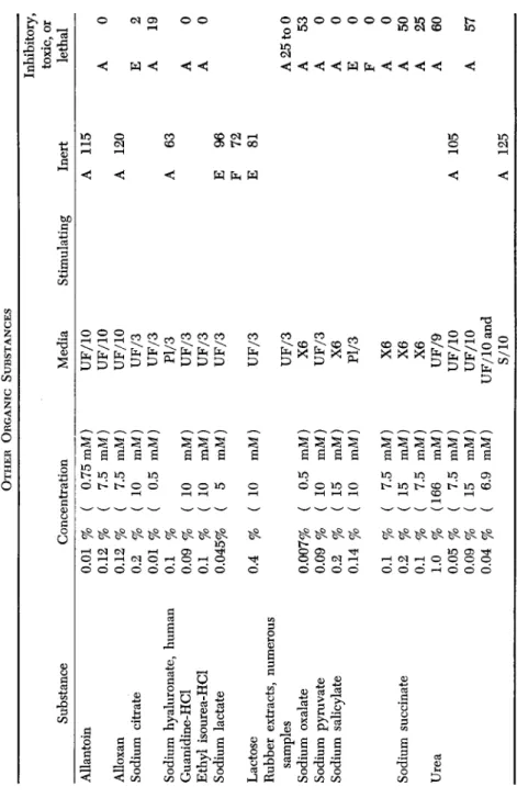
F. OTHER ORGANIC SUBSTANCES (TABLE VII)
This group includes several substances of interest in relation to wound healing. The toxicity of rubber extracts presents a problem for tissue culture workers, since it necessitates that rubber stoppers should never come in contact with solutions.