ContentslistsavailableatScienceDirect
Journal of Chromatography A
journalhomepage:www.elsevier.com/locate/chroma
Bioactive clerodane diterpenes of giant goldenrod ( Solidago gigantea Ait.) root extract
Ágnes M. Móricz
a,∗, Dániel Krüzselyi
a, Péter G. Ott
a, Zsófia Garádi
b, Szabolcs Béni
b, Gertrud E. Morlock
c, József Bakonyi
aaPlant Protection Institute, Centre for Agricultural Research, Herman O. Str. 15, 1022 Budapest, Hungary
bDepartment of Pharmacognosy, Faculty of Pharmacy, Semmelweis University, Üll ˝oi Str. 26, 1085 Budapest, Hungary
cChair of Food Science, Institute of Nutritional Science, and TransMIT Center of Effect-Directed Analysis, Justus Liebig University Giessen, Heinrich-Buff-Ring 26-32, 35392 Giessen, Germany
a rt i c l e i nf o
Article history:
Received 27 September 2020 Revised 13 November 2020 Accepted 16 November 2020 Available online 19 November 2020 Keywords:
High-performance thin-layer
chromatography – effect directed analysis High-performance thin-layer
chromatography – high-resolution mass spectrometry
Fusarium avenaceum
Giant goldenrod ( Solidago gigantea Ait.) Clerodane diterpenes
Antibacterials and antifungals
a b s t r a c t
Giant goldenrod (Solidago gigantea Ait.) root extract was screened for bioactive compounds by high- performancethin-layerchromatography(HPTLC),coupledwitheffect-directedanalysisincludingantibac- terial(BacillussubtilisF1276,B.subtilissubsp.spizizenii,AliivibriofischeriandXanthomonaseuvesicatoria), antifungal (Fusariumavenaceum)and enzymeinhibition(acetyl-and butyrylcholinesterases,α- and β- glucosidasesandα-amylase)assays.Compoundsofsixmultipotentzones(Sg1-Sg6)werecharacterized by HPTLC-heatedelectrospray ionization-high-resolution massspectrometry (HRMS) and HPTLC-Direct AnalysisinRealTime-HRMS. ApartfromzoneSg3,containingthree compounds,asingle characteristic compoundwasdetectableineachbioactivezone.Thebioassay-guidedisolationusingpreparative-scale flashchromatographyandhigh-performanceliquidchromatographyprovidedeightcompoundsthatwere identifiedbyNMRspectroscopyasclerodanediterpenes.Allisolatespossessedinhibitingactivityagainst atleastoneofthetestedmicroorganisms.
© 2020 The Author(s). Published by Elsevier B.V.
ThisisanopenaccessarticleundertheCCBYlicense(http://creativecommons.org/licenses/by/4.0/)
1. Introduction
A continuously higher qualitative and quantitative supply of agricultural raw materials can meet the increasing demand of the food and feedindustry. To improveplant production,appro- priate pest management is needed, using effectiveagrochemicals that are more and more difficult to find due to the emerging (multi)resistance in pathogens against the generally used agents [1,2]. Therefore, many research projects are aimed at discover- ing effectiveagrochemicalswithnewchemicalbasestructureand possibly with low toxicity and fast biodegradability. In the last decade syntheticapproaches havenot resulted innewantibacte- rial agents,andthetarget-baseddrugdiscovery alsobroughtdis- appointment [3]. Purposeful tracking, characterization and isola- tionofbioactivecompoundsfromnaturalsourcescanbeachieved by bioassay-guided processes,comprisingextraction,fractionation andpurificationsteps,allassociatedwithbiomonitoring[4,5].The high-throughput, relatively cheap effect-directedanalysis(EDA)is
∗Corresponding author.
E-mail address: moricz.agnes@atk.hu (Á.M. Móricz).
enabled by high-performance thin-layer chromatography(HPTLC) combined with bioactivity assays [4,6,7]. EDA is a usefultool to pointtoindividualbioactivecompounds(accordingtotheselected assay) separated from a complex matrix, e.g., plant extract. The characterization of potent compounds can easily be achieved by HPTLC-massspectrometry(MS)usingvariousionizationinterfaces [8].
The genus Fusarium contains more than twenty species that are among the most important filamentous fungal pathogens on crops,causingeconomiclossesviasignificantyieldreductionsand mycotoxin contaminations by thisharmful secondary metabolites [9].InEurope,Fusariumavenaceum isone ofthedominantFusar- iumspecies,causingdiseasesincludingheadblightofcereals,root rot of legumes and dry rot of potato [10,11]. The pathogen pro- duces several harmful mycotoxins, such as moniliformin, beau- vericinandenniatins [12].SeveralFusarium specieshavebeenin- troducedforTLC-bioautography.Amongthem,TLC-directbioautog- raphy,inwhichdiffusionofbioactivesubstancesthroughagarlayer is eliminated, F. culmorum [13],F. sambucinum [14], F. oxysporum [15,16], F. lateritium [17], F. virguliforme [17], F. solani [15] and F.
proliferatum[15]havebeenexploited. Asaninoculum,their spore (conidium) suspensions were used for the detection of the ger-
https://doi.org/10.1016/j.chroma.2020.461727
0021-9673/© 2020 The Author(s). Published by Elsevier B.V. This is an open access article under the CC BY license ( http://creativecommons.org/licenses/by/4.0/ )
mination and/or mycelial growth inhibition by separated bioac- tive compounds.The inhibitionzone wasindicated bythelack of visible fungalhyphae, however,vital dyes[15,16] oriodinevapor [18]significantlyimprovedthedetectability.Conidialsuspensionof F. avenaceum hasbeenused fordot blottest [18],which issimi- lartodirectbioautography,buttheTLCadsorbentisonlyusedto hold thesample, not to separate its components.Spore germina- tion andhyphal growthare distinct biological processes, thus,to assess inhibition ofthefungal growthindependentlyof germina- tion,ahyphalsegments(practically,theirsuspension)arerequired.
Theuseofmycelialgrowthinhibitorsareusefulinplantprotection astheypreventlocalspreadofthefungalpathogen.Sofar,neither an HPTLCadsorbentnora mycelialsuspensionhavebeenapplied fordirectbioautographyofFusariumspecies.
Solidago gigantea (giantgoldenrod) isnativetoNorth America.
About250yearsago,itwasintroducedtoEuropeasanornamental andhasbecomeanexceptionallysuccessfulinvasiveandcompeti- tivespeciesinmostEuropeancountrieswithanabundantbiomass [19]. It is widespread in whole Europe and a serious invader of abandoned fields,forest edges andriverbanks[20].Goldenrodis also amedicinalplantandlisted inthe EuropeanPharmacopoeia as Solidaginisherba(the whole orcut dried floweringaerial part ofeitherS.giganteaAit.and/orS.canadensis L.)usedtotreat dis- ordersoftheurinarytract,prostateandkidney.Thegoldenrodex- tractwasshowntodisplayfavourableantibacterial[21],antifungal [22], insecticidal [23] and anti-obesity [24] activities that can be attributedtoitsessentialoil[25],phenolics[26],saponins[27]and diterpenes[28].
Recently, S. gigantea root extract was reported to have anti- hyperglycaemic (
α
- andβ
-glucosidaseandα
-amylase inhibitory) andcholinesteraseinhibitoryeffectsinascreeningoffivegolden- rodspecies[29].Thepresentstudytargetedthedetailedcharacter- ization andbioprofiling ofthe giant goldenrodroot extract using HPTLC-EDA. The discovered active compounds against enzymes, bacteria(Bacillussubtilis,AliivibriofischeriandXanthomonaseuvesi- catoria) and fungus (F. avenaceum) were characterized by HPTLC- heatedelectrospray ionization (HESI)-highresolution (HR)MSand HPTLC-direct analysis in real time (DART)-HRMS. The bioassay- guidedisolated compoundswereidentifiedby NMRandtheiran- timicrobialactivitywasconfirmedbyHPTLC-antimicrobialtests.2. Materialsandmethods 2.1. Materials
HPTLC or TLC silica gel 60 F254 plates or foils and MS-grade methanol were suppliedby Merck (Darmstadt, Germany).Formic acid, vanillin, potassium hydroxide, calcium carbonate, sodium chloride and analytical grade solvents used for layer and flash chromatographywerepurchasedfromReanal(Budapest,Hungary), Th. Geyer (Renningen, Germany) or Sigma-Aldrich (Steinheim, Germany). Gradient grade methanol, acetonitrile (Molar Chemi- cals, Budapest, Hungary) and pure water produced by a Milli- pore Direct-Q3UV system (Merck) were usedforHPLC. Gentam- icin, methanol-d4 (99.8%), acetylcholinesterase lyophilisate (from Electrophorus electricus, AChE), butyrylcholinesterase (from horse serum, BChE), Fast Blue Salt B (95%),
α
-glucosidase solution (from Saccharomyces cerevisiae), 2-naphthyl-β
-D-glucopyranoside,α
-amylase (from pig pancreas) and 2-chloro-p-nitrophenyl-α
-D-maltotrioside (CNP-G3) were from Sigma. 2-Naphthyl-
α
-D-glucopyranoside was from Fluorochem (Karlsruhe, Germany).
α
-Naphthyl acetate was from Panreac (Barcelona, Spain).β
-Glucosidase (from almond) was purchased from Carl Roth (Karl- sruhe, Germany). Gram-positive Bacillus subtilis subsp. spizizenii soil bacterium (DSM 618) wasfrom Merck and B. subtilis (strain F1276) from József Farkas, Central Food Research Institute, Bu-
dapest, Hungary. Gram-negative, naturally luminescent marine bacterium Aliivibriofischeri(DSM 7151) were obtainedfromLeib- niz Institute DSMZ, German Collection of Microorganisms and Cell Cultures, Berlin, Germany. The Hungarian paprika pathogen Xanthomonas euvesicatoria was obtained by János Szarka, Pri- mordiumKft., Budapest, Hungary.Fusarium avenaceum strain IMI 319947 was from CABI-IMI Culture Collection, Egham, UK. 3- [4,5-Dimethylthiazol-2-yl]-2,5-diphenyltetrazoliumbromide (MTT), 2,3,5-triphenyl-tetrazoliumchloride(TTC)and2-(4-iodophenyl)-3- (4-nitrophenyl)-5-phenyl-2H-tetrazoliumchloride(INT)were from CarlRothandSigma,respectively.Vegetablejuice(V8)wasbought atthelocalmarket.TryptonewasfromMicrotrade(Budapest,Hun- gary),yeast extractformScharlau(Barcelona,Spain)andbenomyl (Fundazol50WP)fromChinoinZRT.(Budapest,Hungary).
2.2. Samplepreparation
RootsofSolidago giganteaAit.were collectedinFebruary2017 (young shoots), August 2017 (full flowering) and July 2019 (full flowering)in theGreatPlain, Hungary (N 46°41 52.1"E19°03 3.6" Alt. 90 m). The fresh root was gently washed with water, chopped,driedatroomtemperatureandground(BoschMKM6000, Stuttgart,Germany).Powderedsamplesweremaceratedinethanol (150 mg/mL) for 24 h. The filtered crude extract (17 mg dry weight/mL)wasusedforHPTLCandisolation.Isolatedcompounds weredissolvedinethanol(2.5mg/mL).
2.3. HPTLCmethod
Root extracts (1–2 μL/band for antimicrobial tests and 5 μL/band for enzyme tests) and isolated compounds (0.2–0.5 μL/band for antimicrobial testsand 2 μL/bandfor reagent) were applied as 6-mm bands with 8-10 mm track distance onto the HPTLCplate(AutomatedTLCSamplerATS4orATS3,CAMAG,Mut- tenz,Switzerland)at8mmdistancefromthebottom.HPTLCsep- aration wascarried out withn-hexane– isopropylacetate– ace- tone 16:3:1, V/V/V (MP1)[29] or n-hexane – isopropyl acetate – acetic acid40:9:1, V/V/V (MP2) up to a migration distance of70 mm (Twin Trough Chamber TTC, CAMAG). Plates were dried in a cold stream ofair (5 min). Residues of aceticacid were elimi- nated bya 20-mindrying(Automatic DevelopingChamberADC2, CAMAG) or by potassium hydroxide in the opposite TTC trough for 2 h [30]. The excess of potassium hydroxide was evaporated by a stream ofcold airfor 15 min. The plate wascut (bladeor smartCUT Plate Cutter, CAMAG)into segments forvarious bioac- tivityassaysorderivatization withvanillinsulphuricacidreagent (40mgvanillin,10mLethanoland200μLconcentratedsulphuric acid, heatedat110°C for5 minanddocumented atUV 365 nm and white light illumination in transmittance mode). The chro- matograms were detected by a UV lamp anddigital camera (Cy- bershotDSC-HX60,Sony,Neu-Isenburg,Germany)orTLCVisualizer DocumentationSystemorTLCScanner4(bothCAMAG).
2.4. HPTLC-EDA
Bacterial cellsuspensions were prepared andthe antibacterial effectwasdetected,asdescribedinpreviousmethodsusingB.sub- tilis F1276 [31], B.subtilis subsp. spizizenii[32],A. fischeri[33,34] andX.euvesicatoria[31].Briefly,thedeveloped,neutralizedplates wereimmersedintothecellsuspensionsfor6s.Thedarkantibac- terial zones in the bioautograms of luminescent A. fischeri were instantly documented (BioLuminizer, CAMAGor iBrightTM FL1000 Imaging System, Thermo Fisher Scientific, Budapest,Hungary). In thecases ofnon-luminescentbacteria, bioautograms werevisual- izedaftera2-hincubation(100%humidityatappropriatetemper- ature) by staining withan aqueousMTT solution(1 mg/mL) fol-
lowedbya 0.5-hincubation.Colorless(bright)antibacterial zones wereobtainedagainstabluishbackground.
The reported HPTLC-AChE/BChE [35], HPTLC-
α
/β
-glucosidase andHPTLC-α
-amylase[29]inhibitionassayswereapplied.Fortheα
-amylaseassay,theplatewasimmersedintothesubstratesolu-tion (1.4mg/mL CNP-G3inethanol),dried for2min inastream of cold air, immersed in the buffered
α
-amylase enzyme solu-tion, incubatedat 37 °C for15 min anddried. Forthe other en- zyme assays, each plate wasdipped into the respective buffered enzyme solution and incubated for 15-20 min at 37 °C. The AChE/BChE and
α
/β
-glucosidase autograms were immersed in a solutionofthesubstrate(α
-naphthylacetateand2-naphthyl-α
/β
-D-glucopyranoside,respectively, followedbya 10-minincubation) and the chromogenic reagent (Fast Blue Salt B) and dried. Doc- umentation wasperformedat whitelight illumination in there- flectance mode.Colorless(bright) zonesindicated theactive com- pounds against a yellow (
α
-amylase) or violet (AChE/BChE andα
/β
-glucosidase)background. The detailedenzyme inhibition as- saymethodsarepresentedinthesupplementarydata.TheF.avenaceumstrainwasgrownonV8agarmedium(80mL water, 20 mL Campbell’s V8vegetable juice, 100
μ
L 1 M potas-sium hydroxideto adjusttopH6.5, 0.4m/m%calciumcarbonate, 1.8 g agar) at 20°C in the dark.Lysogenic broth(LB, 20mL; 10 g/Ltryptone, 5g/Lyeast extractand10g/Lsodiumchloride)was inoculated in a 100 mL Erlenmeyer flask withthe fungalculture grownontheV8agarplateandshakenat120rpmat21°Cfor3 daysinthedark.Themycelium waswashedwithLBtoeliminate theconidiaandcuttosmallpieceswithasterilerotor-statortype homogenizer(ModelTR-10,Tekmar,Cincinnati,OH)for2mintwo times. The mycelium suspension was diluted to OD600 0.4 - 0.8.
Developed HPTLCplates were dippedinto the mycelium suspen- sionfor6sandincubatedinavaporchamberat21°Cfor48-72 h.Thelackofvisiblewhitefungalhyphaeindicatedtheinhibition zones.BysprayingaqueousINTsolution(1mg/mL;cca2mL/plate) on the bioautograms followed by a 1-h incubation, the contrast was enhanced by revealingbrightinhibition zones against a vio- let background. An aqueous solution of benomyl (1 μL/band; 25 mg/mL,active ingredientofFundazol50WP)wasappliedasposi- tive control(attheedgeofthedevelopedplatebeforeimmersion inthemyceliumsuspension).
ToevaluatetheeffectsonB.subtilissubsp.spizizeniiandAChE, the(bio)autogramswerescannedinthefluorescencemodeat546 and533nmusingthemercuryandwolframlamp(TLCScanner4, CAMAG),respectively.ImageswereprocessedwithImageJsoftware (NIH,Bethesda,MA,USA).
2.5. HPTLC-HRMSandHRMS-MS
Prewashed plates(withmethanol−water4:1,V/Vdried at100
°C for 20 min) were used andseparated zonesof interest were marked. For HPTLC-HESI-HRMS, the quaternary pump (Ultimate LPG-3400 XRS, Dionex Softron, Germering, Germany) guided the methanol at a flow rate of 0.1 mL/min through the ovalelution head(4mmx2mm)ofthePlateExpressinterface(Advion,Ithaca, NY,USA)totheHESI-IIinstalledatthehybridquadrupole-orbitrap massspectrometer(QExactivePlus,ThermoFisherScientific,Bre- men, Germany). Spray voltage was3.5 kV, capillary temperature was270 °C,and nitrogenassheathandauxiliary gas (20and10 arbitraryunits,respectively) wasproduced by an SF2compressor (AtlasCopcoKompressorenundDrucklufttechnik,Essen,Germany).
FullscanHRMSspectrawererecordedinthenegativeandpositive ionization mode in the range of m/z 50 - 750 witha resolution of280,000,automaticgaincontroltargetof3×106 andmaximum injectiontimeof100ms.Isolatedcompoundsandflashchromato- graphic fractions were directlyinjected by flowinjection analysis (FIA)inthementionedHRMSsystem(sprayvoltage3.5kV,capil-
lary temperature270°C,sheathandauxiliary gasesat20and10 arbitraryunits,respectively).Tandem massspectrawere acquired as parallel reaction monitoring with mass isolationof the target molecule(fragmentation/collision energyof40-60eV,resolution of17,500,automaticgaincontroltargetof2×105,maximuminjec- tiontimeof100msandisolationwindowofm/z2form/z50).Op- eration anddataprocessingwere performedwithXcalibur 3.0.63 software(Thermo).
A DART interface (IonSense, Saugus, MA, USA) modified for scanning of HPTLC plates [36] was coupled to the mentioned HRMSsystem.Theionsourcewasoperatedwithan initialneedle voltageof4kVandgridvoltageof50Vinthepositive ionization mode using helium (99.999%) ata flow rateof 3.0 L min−1 and temperatureof500°C.Spectrawererecordedinthefullscan(m/z 100−750)ata resolutionof280,000,automatic gain controltar- get of5×104 andmaximum injectiontime of 50 ms. The DART scanning speed was0.2mm s−1.The extracted ioncurrent(EIC) chromatogramswere processedwitha Gauss smoothing function widthof11points.
2.6. Preparativecolumnchromatography
The extract(10 mL, dried andre-suspendedin n-hexane)was fractionatedwithCombiflashNextGen300(TeledyneIsco,Lincoln, NE,USA)flashchromatographysystem.Separationwasperformed on a RediSep Rf Gold silica gel column (20-40 μm, 12 g; Tele- dyne Isco) at a flow rate of 30 mL/min with a gradient of n- hexane(A) and acetone(B): 0-0.5 min,0% B; 0.5-8.5 min 0-30%
B.Thechromatogramwasmonitoredbyabsorbancemeasurement at215 nm.Compoundsin thecollectedactivefractions were fur- therfractionatedandpurified.HPLCseparations(conditionsarein Table1)werecarriedoutusinganLC–MS-2020system(Shimadzu, Kyoto,Japan),includingbinarygradientsolventpump,vacuumde- gasser,thermostatedautosampler,columnoven,photodiodedetec- torandelectrosprayionization(ESI)-MSsystem.Instrumentcontrol anddataacquisition wereperformedwiththeLabSolutions 5.42v program(Shimadzu).First,analyticalmethodsweredevelopedthat were scaled up using a semi-preparative column. The analytical separation of the fractions (5 μL) on a Gemini C18 column (250 mm x4.6mm ID,5μm particlesize, Phenomenex, Torrance,CA, USA)at35°Cwithastep-wise gradientwasdetected byMS (ni- trogenasnebulizergas,flow rate1.5L/min,dryinggas(nitrogen) flow rate15 L/min,interface temperature350 °C, heatblocktem- perature 400 °C, desolvation line temperature250 °C anddetec- tor voltage 4.5kV). Full scan mass spectra were recorded in the positive andnegativeionization mode in the rangeof m/z 150 – 900withascanspeedof5000amu/s.Thesemi-preparativesepa- rationofthefractions(100μL)onaGeminiC18column(250mm x 10 mm ID, 10 μm particle size, Phenomenex) wasdetected in theUV.CompoundSg6(90μL)waspurifiedonaKinetexpentaflu- orophenyl (PFP) column (100 mm x 4.6 mm ID, 2.6 μm particle size,Phenomenex)at35°C.The fractionation/purificationwasre- peated up to 10-times.The combinedfractions were investigated byHPTLC-assays,driedwitharotary evaporator(RotavaporR-134, Büchi,Flawil,Switzerland)at40°C andtransferred toNMRspec- troscopy.
2.7. NMR
NMRspectrawererecordedindeuteratedmethanol(Methanol- d4,99.8atom%D,containing0.05%tetramethylsilane)onaBruker Avance III HD 600 (600/150 MHz) instrument equipped with a Prodigycryo-probeheadat295K.Thepulseprograms weretaken fromtheBrukersoftwarelibrary(TopSpin3.5).Chemicalshifts(
δ
)and coupling constants (J) are given in ppm and in Hz, respec- tively.1Hchemicalshiftsaregiveninppmrelativetotetramethyl-
Table 1
Detailed description of preparative and analytical scale HPLC methods
Method Extract Column Eluent A Eluent B Gradient
Flow rate (mL/min) 0.8 ; 3.5 enter 0.7 ; 3.5
enter 0.7 ; 4 enter 0.8 Collected peaks 1 flash fractions Gemini C18, 250 mm x
10 mm, 10 μm Gemini C18, 250 mm x 4.6 mm, 5 μm
5% aqueous methanol
acetonitrile 0-14 min 75% B 14-20 min 100% B 20-26 min 75% B
4 0.8 Sg1, Sg2,
Sg3a + Sg3b, Sg4 + Sg3c, Sg5
2 Sg3a + Sg3b 5% aqueous
methanol methanol 0-18 min 75% B 18-21 min
100% B 21-25 min 75% B 3.5 0.7 Sg3a, Sg3b
3 Sg4 + Sg3c 5% aqueous
methanol
methanol 0-18 min 80-88% B 18-21 min 100% B 21-25 min 80% B
3.5 0.7 Sg4, Sg3c
4 Sg5 5% aqueous
methanol
acetonitrile + 0.1%
formic acid
0-14 min 75% B 14-20 min 100% B 20-26 min 75% B
4 0.8 Sg5
5 flash fraction 13
Kinetex PFP, 100 mm x 4.6 mm, 2.6 μm
5% aqueous methanol
methanol 0-12.5 min 65-77% B 12.5-15.5 min 100% B 15.5-19.5 min 65% B
0.6 Sg6
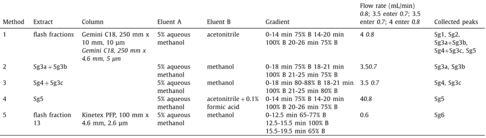
Fig. 1. HPTLC chromatograms and autograms of the S. gigantea root extract, developed with n -hexane – isopropyl acetate – acetone 16:3:1 ( V / V / V , MP1) and detected at UV 254 nm (a), UV 365 nm (b), after derivatization with vanillin sulphuric acid reagent at UV 365 nm (c) and white light illumination (d; also e-g and i-k) AChE (e), BChE (f), B. subtilis subsp. spizizenii (g), A. fischeri (h, greyscale image of the bioluminescence), α- glucosidase (i), β-glucosidase (j) and α- amylase (k).
silane (
δ
=0.00 ppm). 13C chemical shifts are given in ppm rela- tive totheNMRsolvent(δ
=49.00ppm).Thecomplete1Hand13C assignments were deduced using conventional 1D (1H NMR, 13C NMR, DeptQ)and2D (1H-1HCOSY, 1H-13CedHSQC 1H-13CHMBC and1H-1HNOESY)measurements.3. Resultsanddiscussion 3.1. HPTLC-EDA
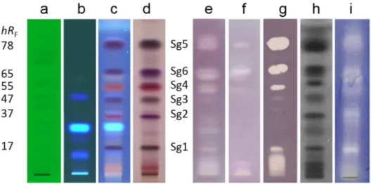
Two methods were investigated for the separation of the gi- ant goldenrod root compounds (Figs. 1 and 2) on HPTLC plates silica gel 60 with either n-hexane – isopropyl acetate – acetone 16:3:1 V/V/V (MP1, [29]) orn-hexane– isopropylacetate – acetic acid40:9:1 V/V/V (MP2).Bothseparations detected atwhitelight illumination after derivatization with the universal vanillin sul- phuricacidreagent,providedthesixdistinguishablecoloredzones Sg1-Sg6 that were hardly detectable without the derivatization (Figs.1a,1b,2aand2b).MP2resultedinhigherhRFvalues,which wasobviousforSg5andSg6, indicatingapotential acidiccharac- ter.MP2chromatogramsrequiredaneutralizationstepbeforeEDA.
Compoundswithantidiabetic(anti-hyperglycaemic)effectcouldbe usedforthetreatmentoftype2diabetesandobesitythatarethe mostcommonandincreasingchronicdiseasesintheworld.Inthe HPTLC-EDA screeningwithMP1(Fig.1e-k),the zonesSg5(athRF
58)andSg6(athRF 37)showed
α
-glucosidase,β
-glucosidaseandα
-amylaseinhibition. The application zone up to hRF 9 wasalso effectiveinα
-glucosidaseandα
-amylaseassays.Cholinesterasein- hibitors are of therapeutic interest in the Alzheimer disease, the most common cause of dementia and one of the major public healthissues. CompoundsinthezonesSg1(hRF9), Sg4(hRF 51), Sg5 and Sg6 inhibited AChE and BChE (Fig. 1e and f). However, with MP2 and neutralization, BChE inhibitory zones of Sg1 (hRF 17)andSg4(hRF55)werelacking(Fig.2f).Multipotentcompounds aregenerallynotfavorableforpharmaceuticaluse,however,these compoundscanbecomeleadcompoundstowardthedevelopment ofmorespecific,thusmoreusefulderivatives.Antibacterialagentsarewidelyrequiredforthefightagainstin- fectious plant, animal and humandiseases caused by pathogenic bacterialandfungalstrains.Intheantibacterialassays,theGram- positive B. subtilis subsp. spizizenii soil bacterium, the Gram- negative, naturally luminescent marine A. fischeri andthe Gram- negativepaprikapathogenX. euvesicatoria were employed.In the B.subtilis assay, Sg1and compounds at an even lower hRF were only active in the MP2 separation (Figs. 1g and 2g). All marked zoneswere active against A. fischeri (Figs. 1h and2h) andX. eu- vesicatoria (Fig. 2i), except for Sg3 atthe given amount applied.
Additionally,Sg6 showedactivityagainst A. fischerionly onacid- freebioautograms.The positionsofthebioactivezoneswerecon-
Fig. 2. HPTLC chromatograms and autograms of the S. gigantea root extract, developed with n -hexane – isopropyl acetate – acetic acid 40:9:1 ( V/V/V , MP2) and detected at UV 254 nm (a), UV 365 nm (b), after derivatization with vanillin sulphuric acid reagent at UV 365 nm (c) and white light illumination (d; also e-g and i), AChE (e), BChE (f), B. subtilis subsp. spizizenii (g), A. fischeri (h, greyscale image of the bioluminescence) and X. euvesicatoria (i) assays.
Fig. 3. HPTLC- F. avenaceum bioautogram of giant goldenrod root extract (E, 2 and 3 μL/band) and positive control benomyl (B, 25 μg/band) developed with n -hexane – isopropyl acetate – acetone 16:3:1 ( V / V / V , MP1) and detected at white light illumi- nation after the bioassay (a) and subsequent staining with INT (b).
firmedbydensitometryorvideodensitometryoftheMP2bioauto- grams(Fig.S1).Theresultsoftheenzymeassaysareinagreement withourprevious observations[29],however,theinfluenceofan acidic development and its neutralization on the outcome needs furtherstudy.
AnHPTLC-antifungalassayusingplantpathogenicfungusF.ave- naceumwasnewlydeveloped.F.avenaceumgrewwell onV8solid agarmediumandthesurfaceofa5cmpetridish wastotallyoc- cupied by its white hyphae within 2-3 days after inoculation by an agar block (5 mm x 5 mm) in the center. Similarly, a dense mycelium suspension wasobtainedwithin 3daysby shakingthe liquidinoculum(LBmedium)at21°C.After cuttingthemycelium to small fragments, the suspension was initially diluted to reach anOD600of0.4.Dippingaplateintothissuspension,anappropri- atemyceliumgrowthwasopticallyreachedaftera3-daysincuba- tion inavapor chamber at21°C (Fig.3), indicatingasmycelium growthinhibitiontheantifungalactivityofzonesSg1,Sg2,Sg3,Sg4 and Sg5. For detection, the three different tetrazolium dyes INT, MTTandTTCwereinvestigated,wherebythefastestcolorationwas achieved with INT, which colored the hyphae violet (Figs. 3 and S2). Comparedtothe3-days-oldmyceliumsuspension,the1-day- old inoculum did not grow as fast on the HPTLC plate, andthe
5-days-oldwasfoundtobelesssensitive(Fig.S3).Inagreementto ourpreviousstudy,nocharacteristicinhibitionwasobservedusing oldermicrobial(bacterial)cellsinthestationaryanddeathphases [37].Thestainingprocedure enabledthereductionoftheincuba- tiontimeto2days.Theincubationtimecould notbeshortened- byincreasing theOD600ofthe mycelialinoculumfrom0.2to0.8 -whithout a loss indiscernability ofthe inhibitionzones. In the finalprotocol,themyceliumsuspensionatanOD600of0.4wasin- cubatedfor2 daysonthe HPTLCplateandthendetected by INT staining, followedby a 1-h incubation anddocumentation under whitelightilluminationinthereflectancemode(Fig.S4).
3.2. HPTLC-MS
The bioactive compound zones Sg1-Sg6 were separated with MP1 and characterized by HPTLC-HESI-HRMS and HPTLC-DART- HRMS(Table2,Fig.S5).HPTLC-HESI+-HRMSsignalswereobtained for all six compound zones, but the signal intensities were very low forSg1atm/z 341.2088 [M+Na]+ (C20H30O3Na+) andSg2at m/z 355.1879 [M+Na]+ (C20H28O4Na+). However, it still allowed the assignment of their molecular formulae. The recorded mass signalsfortheSg3zoneatm/z 337.1776[M+Na]+ (C20H26O3Na+) andat very low intensity m/z 325.2140 [M+Na]+ (C20H30O2Na+) indicated the coelution of atleast two compounds. As discussed later, even three compounds Sg3a-cwere identified inthis zone.
An intense mass signal was recorded at m/z 339.1931 [M+Na]+ (C20H28O3Na+) forSg4.ForSg5and Sg6, prominentmasssignals were obtainedinbothionization modeatm/z 339.1931[M+Na]+ (C20H28O3Na+)andm/z437.2304[M+Na]+ (C25H34O5Na+)aswell as at m/z 315.1956 [M-H]− and m/z 413.2304 [M-H]−, respec- tively. These zone assignments were successfully confirmed by separation withMP2 andHPTLC-ESI-MS (Fig. S6). In the positive andnegativeionization mode, alsothe sodium-methanol-adducts [M+CH3OH+Na]+ and sodium-acetate-adducts of the respective compounds were observed, which wascaused by a toolow dry- inggasflow.
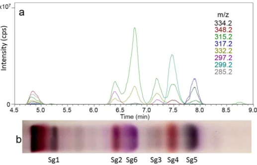
WithexceptionofSg1,allothercompoundzonesgavemasssig- nals by HPTLC-DART-HRMS (Table 2, Figs. 4and S7). In the pos- itive ionization mode, the protonatedmolecule [M+H]+ was de- tected for Sg3 and Sg5. Further, ammonium adducts [M+NH4]+ and dehydroxylatedmolecular ions [M-OH]+ were prominent. In the negative ionization mode, the deprotonated molecules were
detected for the Sg5and Sg6 zones, and also the dimer for Sg5 andaC5H7O2−fragmentforSg6.
3.3. Isolationoftheactivecompounds
The rootextract(10 mL) wasfractionatedby flashchromatog- raphy on a silicagel column, resultingin nineteen fractions(fr1- fr19), which were studied by HPTLC-Vis afterderivatization with the vanillin sulphuric acidreagent(Fig. S8a and b). The sixfrac- tionscontainingthepreviouslycharacterizedbioactivecompounds (fr10-fr14 andfr17)weresubjected toRP-HPLC-DAD-ESI-MSanal-
ysis(Fig. S8c,Table 2). The HPLC method 1 (Table 1) was found to be suitable for compound isolation in the Sg1, Sg2 and Sg5 zones,wherebythelatterzonewastailing.Acid-freemobilephases wereselectedtoavoidacid-analytereactions,whichmightimpair the bioactivity result. However, in the case ofSg5, the collected peakwasfurtherpurifiedwithanacidicconditioningintheHPLC method4.TheSg6compoundzone waselutedfromthe C18col- umnduringthewashingstep.Thus,itsretentionwasachievedon a PFP column (HPLC method 5). The HPTLC separation revealed two co-eluting compounds in each of the HPLC peaksat 10-min (Sg3aandSg3b)and12.5-minretentiontimes(Sg3candSg4)(Fig.
Table 2
Chemical structures, molecular formulae and assignments (using orthogonal techniques) of the eight discovered bioactive compounds in S. gigantea root extract.
( continued on next page )
Table 2 ( continued )
Fig. 4. EIC chromatograms of scanning HPTLC-DART-MS in the positive ionization mode (a) and respective HPTLC chromatogram of the bioactive giant goldenrod root components separated with n -hexane – isopropyl acetate – acetone 16:3:1 ( V / V / V , MP1) detected at white light illumination after derivatization with vanillin sulphuric acid reagent (b).
Fig. 5. Confirmation of purity and bioactivity: HPTLC chromatogram and autograms of the bioactive compounds Sg1-Sg6 isolated from giant goldenrod root extract (E) separated with n -hexane – isopropyl acetate – acetone 16:3:1 ( V / V / V , MP1) and detected at white light illumination (a, b and d) after derivatization with the vanillin–
sulphuric acid reagent (a), B. subtilis F1276 (b), A. fischeri (c, greyscale image of the bioluminescence) and F. avenaceum (d) assays.
S8c).Fortheirseparation,HPLCmethods2and3weredeveloped, respectively. Eight compounds were isolated using a preparative C18column(Fig.S9)andananalyticalPFPcolumn(Fig.S10).These were assignedasSg1(1.1mg), Sg2(1.6mg),Sg3a(0.9 mg),Sg3b (1.0 mg),Sg3c (1.5mg), Sg4(2.1 mg),Sg5 (2.2mg) andSg6(6.2 mg)accordingtotheirrespectiveHPTLCzones.
3.4. Identificationandcharacterizationoftheisolatedcompounds
The isolates were analyzed by FIA-HESI-HRMS/MSto success- fullyconfirmthattheiridentitywasnotchangedduringtheisola- tion procedure,including fractionationandpurification.Fragmen- tationoftherespectivesodiumadductofthecompoundswasnot observed exceptforSg6 (Table2), andthus, alsothe respectively lessintense protonatedor deprotonatedmolecules wereselected.
The intense deprotonated molecules of Sg5 and Sg6 were easily fragmented.
The HPTLC-Vis analysis of the isolates (separated with MP1, Fig. 5a, and MP2, Fig. S11and detected afterderivatization with thevanillinsulphuricacidreagent)showedthatonlyasinglecom- pound was present in all previously assigned zones, except for three compounds (Sg3a-c) detected in Sg3. All isolates were ac- tive against B. subtilis, whereby Sg3b had only a weak activity (Fig. 5b). Most isolated compounds strongly affected A. fischeri, wherebySg3bshowedamildeffect,andSg3candSg6noresponse atall(Fig.5c).AllcompoundsexceptSg3bsuppressedthegrowth of F. avenaceum mycelium with varying degrees of effectiveness (Fig.5d).AlthoughfortheSg3andSg6zonesoftherootextract,no activity againstB.subtilis andF.avenaceum,respectively,wasevi- dent,theirrespectiveisolatesdidhaveanantimicrobialeffect.This wasexplainedbythefactthattheirconcentrationsintherootex- tractweretoolowtoreachtheminimuminhibitoryconcentration.
Theeightisolatedcompounds weremostprobablyresponsiblefor theenzymeinhibitingactivity,butthiswasnotconfirmed.Thepu- rityoftheeightisolateswasadequate,asconfirmedbyNMRanal- ysis (Table S1, Figs. S12-S59). The eight compounds were identi- fied asthefuran-containingclerodane diterpenoids:aglycol(Sg1, known also as kingidiol, white solid), an epoxy-hemiacetal (Sg2, oil), adialdehyde(Sg3a,whitesolid),theclerodanelactone (Sg3b, oil, also known as hautriwaic lactone), an alcohol (Sg3c, oil), a hemiacetal (Sg4, oil),the solidagoicacid A(Sg5,white solid)and thesolidagoicacidB(Sg6,whitesolid),whichhavebeendescribed ascomponentsofS.gigantearoots[38-40](Table2). NMRdatain literature supported the identificationof the isolated compounds [28,38,39,41,42].Antimicrobial[43-46],cytotoxic[45-47]andanti- inflammatory effect [48] of severalclerodane diterpenes isolated from plants have been described. Among Solidago species,clero- dane diterpenes of European goldenrod (S. virgaurea) were pub-
lishedtoinhibitStaphylococcusaureus[42].Theactivityofthreeof ourisolated compounds hasalreadybeen studied mainlyagainst insects.Sg5wasinactive,Sg1causedtheretardationoffolliclede- velopmentinmosquitoovaries [28]anddisplayedmoderatetoxi- city againstbrineshrimp (Artemia salina) [49].Incontrastto Sg1, Sg3bwasfoundasanti-feedantandrepellentagentinastudyof mealworms (Tenebrio molitor) [50]. Antiplasmodial (antimalarial) andantileishmanial (inhibitionof the intracellular parasiteLeish- mania donovani) effects of Sg3b were also observed, whereby it didnotshowcytotoxicityagainstVerocells(Africangreenmonkey kidneyfibroblast)[51].Exceptforthenewantifungalprofiling,the HPTLC-bioprofilingoftherootextracthasbeenpreviouslyreported byus,however,itlackedinidentificationoftheactivecompounds byHRMSandNMR.Tothebestofourknowledge,thisisthefirst reportabouttheantimicrobialprofileoftheeightisolatedS.gigan- tea rootditerpenoids.Furtherinvestigations areintended to eval- uatetheir possibleuse assafeandeffective pesticides,especially fungicides.
Conclusions
The newly developed HPTLC-F. avenaceum assay was demon- strated for the first time using mycelium suspension. It allowed acost-effective screeningofcomplexplantextracts forantifungal (mycelium growth inhibiting) compounds in S. gigantea root ex- tract. Further bioprofiling of the extract against various bacterial strainsandenzymesby HPTLC-EDApointedtomulti-potentcom- pounds.Thecomparisonoftheuseofaneutralversusacidicmo- bilephasedemonstratedthatthelattercaninfluencethebioassay result. The state of the art combination of HPTLC-bioactivityas- says,HPTLC-HESI-HRMS,HPTLC-DART-HRMS,preparative-scalecol- umnchromatography(flashchromatographyandHPLC)andNMR provided eight multi-potent clerodane diterpenoidsin goldenrod.
It is the first report on the antifungal, antibacterial andenzyme inhibiting activity ofthe multipotent isolates, which showed po- tential aslead compounds especially forvarious infectious plant diseases.
DeclarationofCompetingInterest
Theauthorsdeclarethattheyhavenoknowncompetingfinan- cialinterestsorpersonalrelationshipsthatcouldhaveappearedto influencetheworkreportedinthispaper.
Acknowledgment
Á.M.Móricz thanks the OECDfor thescholarship JA00092484 thatallowed her tostayatJLUGiessen. Instrumentationwaspar-
tially fundedby theDeutsche Forschungsgemeinschaft(DFG,Ger- manResearchFoundation)-INST162/471-1FUGG;INST162/536-1 FUGG. Thiswork wasalso funded by the National Research, De- velopmentandInnovationOfficeofHungary(NKFIHK128921)and partiallysupportedbytheJánosBolyaiResearchScholarshipofthe Hungarian AcademyofSciences andBolyai+New NationalExcel- lence Program(ÚNKP-19-4-SE-53) of the Ministry of Human Ca- pacities. The authors thank SalimHage,Tim Häbe, Imanuel Yüce andTamaraSchreiner fortheir supportintheperformance ofthe ChEassay,DART-andHESI-HRMSexperiments,respectively.
Supplementarymaterials
Supplementary material associated with this article can be found,intheonlineversion,atdoi:10.1016/j.chroma.2020.461727. References
[1] B.A. Lorsbach, T.C. Sparks, Innovations in agrochemical discovery and the role of metabolism, bioavailability and formulations, Pest Manag. Sci. 73 (2017) 655–657, doi: 10.1002/ps.4533 .
[2] M. Hahn, The rising threat of fungicide resistance in plant pathogenic fungi: Botrytis as a case study, J. Chem. Biol. 7 (2014) 133–141, doi: 10.1007/
s12154- 014- 0113- 1 .
[3] P.S. Hoffman, Antibacterial Discovery: 21st Century Challenges, Antibiotics 9 (2020) 213, doi: 10.3390/antibiotics9050213 .
[4] Á.M. Móricz, P.G. Ott, T.T. Häbe, A. Darcsi, A. Böszörményi, Á. Alberti, D. Krüzselyi, P. Csontos, S. Béni, G.E. Morlock, Effect-directed discovery of bioactive compounds followed by highly targeted characterization, isolation and identification, exemplarily shown for Solidago virgaurea , Anal. Chem. 88 (2016) 8202–8209, doi: 10.1021/acs.analchem.6b02007 .
[5] W. Jonker, M.H. Lamoree, C.J. Houtman, J. Kool, Methodologies for effect- directed analysis: environmental applications, food analysis, and drug dis- covery, in: Anal. Biomol. Interact. by Mass Spectrom, Wiley-VCH Verlag GmbH & Co. KGaA, Weinheim, Germany, 2015, pp. 109–163, doi: 10.1002/
9783527673391.ch4 .
[6] G. Morlock, W. Schwack, Hyphenations in planar chromatography, J. Chro- matogr. A 1217 (2010) 6600–6609, doi: 10.1016/j.chroma.2010.04.058 . [7] G.E. Morlock, Chromatography combined with bioassays and other hyphen-
ations – the direct link to the compound indicating the effect, in: 2014: pp.
101–121. 10.1021/bk-2014-1185.ch005.
[8] G. Morlock, W. Schwack, Coupling of planar chromatography to mass spec- trometry, TrAC Trends Anal. Chem. 29 (2010) 1157–1171, doi: 10.1016/j.trac.
2010.07.010 .
[9] J. Yu, R.H. Proctor, D.W. Brown, K. Abe, K. Gomi, M. Machida, F. Hasegawa, W.C. Nierman, D. Bhatnagar, T.E. Cleveland, Genomics of economically signif- icant Aspergillus and Fusarium species, in: 2004: pp. 249–283. 10.1016/S1874- 5334(04)80013-3.
[10] S. Vogelgsang, M. Sulyok, A. Hecker, E. Jenny, R. Krska, R. Schuhmacher, H.- R. Forrer, Toxigenicity and pathogenicity of Fusarium poae and Fusarium ave- naceum on wheat, Eur. J. Plant Pathol. 122 (2008) 265–276, doi: 10.1007/
s10658- 008- 9279- 0 .
[11] A.T. Pollard, P.A. Okubara, Real-time PCR quantification of Fusarium avenaceum in soil and seeds, J. Microbiol. Methods 157 (2019) 21–30, doi: 10.1016/j.mimet.
2018.12.009 .
[12] S. Uhlig, A.C. Gutleb, U. Thrane, A. Flåøyen, Identification of cytotoxic princi- ples from Fusarium avenaceum using bioassay-guided fractionation, Toxicon 46 (2005) 150–159, doi: 10.1016/j.toxicon.20 05.03.0 05 .
[13] A .L. Homans, A . Fuchs, Direct bioautography on thin-layer chromatograms as a method for detecting fungitoxic substances, J. Chromatogr. A 51 (1970) 327–
329, doi: 10.1016/S0021-9673(01)96877-3 .
[14] K.D. Burkhead, D.A. Schisler, P.J. Slininger, Pyrrolnitrin Production by biological control agent Pseudomonas cepacia B37w in culture and in colonized wounds of potatoes, Appl. Environ. Microbiol. 60 (1994) 2031–2039, doi: 10.1128/AEM.
60.6.2031-2039.1994 .
[15] N. Kasanah, L.L. Farr, A. Gholipour, D.E. Wedge, M.T. Hamann, Metabolism and resistance of Fusarium spp. to the manzamine alkaloids via a putative retro Pictet-Spengler reaction and utility of the rational design of antimalar- ial and antifungal agents, Mar. Biotechnol. 16 (2014) 412–422, doi: 10.1007/
s10126-014-9557-0 .
[16] R.E. Beale, D. Pitt, The antifungal properties of Minimedusa polyspora , Mycol.
Res. 99 (1995) 337–342, doi: 10.1016/S0953-7562(09)80910-6 .
[17] G.L. Gallardo, N.I. Peña, G.M. Cabrera, Neric acid derivatives produced by the honey bee fungal entomopathogen Ascosphaera apis , Phytochem. Lett. 1 (2008) 155–158, doi: 10.1016/j.phytol.20 08.07.0 08 .
[18] F. Hadacek, H. Greger, Testing of antifungal natural products: methodologies, comparability of results and assay choice, Phytochem. Anal. 11 (20 0 0) 137–147, doi: 10.1002/(SICI)1099-1565(200005/06)11:3 137:AID-PCA514 3.0.CO;2-I . [19] M. Szymura, T.H. Szymura, Interactions between alien goldenrods ( Solidago and
Euthamia species) and comparison with native species in Central Europe, Flora - Morphol. Distrib. Funct. Ecol. Plants. 218 (2016) 51–61, doi: 10.1016/j.flora.
2015.11.009 .
[20] G. Jakobs, E. Weber, P.J. Edwards, Introduced plants of the invasive Solidago gi- gantea (Asteraceae) are larger and grow denser than conspecifics in the native range, Divers. Distrib. 10 (2004) 11–19, doi: 10.1111/j.1472-4642.20 04.0 0 052.x . [21] Barbara Kołodziej, Antibacterial and antimutagenic activity of extracts above-
ground parts of three Solidago species: Solidago virgaurea L., Solidago canaden- sis L. and Solidago gigantea Ait, J. Med. Plants Res. 5 (2011), doi: 10.5897/
JMPR11.1098 .
[22] D. Webster, P. Taschereau, R.J. Belland, C. Sand, R.P. Rennie, Antifungal activity of medicinal plant extracts; preliminary screening studies, J. Ethnopharmacol 115 (2008) 140–146, doi: 10.1016/j.jep.2007.09.014 .
[23] G. Benelli, R. Pavela, K. Cianfaglione, D.U. Nagy, A. Canale, F. Maggi, Evalua- tion of two invasive plant invaders in Europe ( Solidago canadensis and Solidago gigantea ) as possible sources of botanical insecticides, J. Pest Sci. 92 (2019) 805–821 (2004), doi: 10.1007/s10340- 018- 1034- 5 .
[24] Z. Wang, J.H. Kim, Y.S. Jang, C.H. Kim, J.-Y. Lee, S.S. Lim, Anti-obesity effect of Solidago virgaurea var. gigantea extract through regulation of adipogenesis and lipogenesis pathways in high-fat diet-induced obese mice (C57BL/6N), Food Nutr. Res. 61 (2017) 1273479, doi: 10.1080/16546628.2016.1273479 .
[25] D. Kalemba, B. Thiem, Constituents of the essential oils of four micropropagat- edSolidago species, Flavour Fragr. J. 19 (2004) 40–43, doi: 10.1002/ffj.1271 . [26] J. Radusiene, M. Marska, L. Ivanauskas, V. Jakstas, B. Karpaviciene, Assessment
of phenolic compound accumulation in two widespread goldenrods, Ind. Crops Prod. 63 (2015) 158–166, doi: 10.1016/j.indcrop.2014.10.015 .
[27] G. Reznicek, M. Freiler, M. Schader, U. Schmidt, Determination of the content and the composition of the main saponins from Solidago gigantea Ait. using high-performance liquid chromatography, J. Chromatogr. A 755 (1996) 133–
137, doi: 10.1016/S0 021-9673(96)0 0571-7 .
[28] S.-H. Lee, H.-W. Oh, Y. Fang, S.-B. An, D.-S. Park, H.-H. Song, S.-R. Oh, S.-Y. Kim, S. Kim, N. Kim, A.S. Raikhel, Y.H. Je, S.W. Shin, Identification of plant com- pounds that disrupt the insect juvenile hormone receptor complex, Proc. Natl.
Acad. Sci. 112 (2015) 1733–1738, doi: 10.1073/pnas.1424386112 .
[29] Á.M. Móricz, M. Jamshidi-Aidji, D. Krüzselyi, A. Darcsi, A. Böszörményi, P. Cson- tos, S. Béni, P.G. Ott, G.E. Morlock, Distinction and valorization of 30 root extracts of five goldenrod ( Solidago ) species, J. Chromatogr. A 1611 (2020) 460602, doi: 10.1016/j.chroma.2019.460602 .
[30] Á.M. Móricz, P.G. Ott, I. Yüce, A. Darcsi, S. Béni, G.E. Morlock, Effect-directed analysis via hyphenated high-performance thin-layer chromatography for bio- analytical profiling of sunflower leaves, J. Chromatogr. A 1533 (2018) 213–220, doi: 10.1016/j.chroma.2017.12.034 .
[31] Á.M. Móricz, T.T. Häbe, A. Böszörményi, P.G. Ott, G.E. Morlock, Tracking and identification of antibacterial components in the essential oil of Tanacetum vulgare L. by the combination of high-performance thin-layer chromatogra- phy with direct bioautography and mass spectrometry, J. Chromatogr. A 1422 (2015) 310–317, doi: 10.1016/j.chroma.2015.10.010 .
[32] M. Jamshidi-Aidji, G.E. Morlock, Bioprofiling of unknown antibiotics in herbal extracts: Development of a streamlined direct bioautography using Bacillus subtilis linked to mass spectrometry, J. Chromatogr. A 1420 (2015) 110–118, doi: 10.1016/j.chroma.2015.09.061 .
[33] S. Krüger, O. Urmann, G.E. Morlock, Development of a planar chromatographic method for quantitation of anthocyanes in pomace, feed, juice and wine, J.
Chromatogr. A 1289 (2013) 105–118, doi: 10.1016/j.chroma.2013.03.005 . [34] Á.M. Móricz, T.T. Häbe, P.G. Ott, G.E. Morlock, Comparison of high-performance
thin-layer with overpressured layer chromatography combined with direct bioautography and direct analysis in real time mass spectrometry for tansy root, J. Chromatogr. A 1603 (2019) 355–360, doi: 10.1016/j.chroma.2019.03.068 . [35] S. Hage, G.E. Morlock, Bioprofiling of Salicaceae bud extracts through high- performance thin-layer chromatography hyphenated to biochemical, micro- biological and chemical detections, J. Chromatogr. A 1490 (2017) 201–211, doi: 10.1016/j.chroma.2017.02.019 .
[36] T.T. Häbe, G.E. Morlock, Improved desorption/ionization and ion transmission in surface scanning by direct analysis in real time mass spectrometry, Rapid Commun. Mass Spectrom. 30 (2016) 321–332, doi: 10.1002/rcm.7434 . [37] Á. Móricz, N. Adányi, E. Horváth, P. Ott, E. Tyihák, Applicability of the BioArena
system to investigation of the mechanisms of biological effects, J. Planar Chro- matogr. – Mod. TLC 21 (2008) 417–422, doi: 10.1556/JPC.21.2008.6.4 . [38] M.S. Henderson, R. McCrindle, D. McMaster, Constituents of Solidago species.
Part V. Non-acidic diterpenoids from Solidago gigantea var. serotina , Can. J.
Chem. 51 (1973) 1346–1358, doi: 10.1139/v73-201 .
[39] T. Anthonsen, M.S. Henderson, A. Martin, R.D.H. Murray, R. McCrindle, D. Mc- Master, Constituents of Solidago species. Part IV. Solidagoic acids A and B, diterpenoids from Solidago gigantea var. serotina , Can. J. Chem. 51 (1973) 1332–
1345, doi: 10.1139/v73-200 .
[40] T. Anthonsen, M.S. Henderson, A. Martin, R. McCrindle, R.D.H. Murray, Furan- containing diterpenoids from Solidago serotina Ait, Acta Chem. Scand. 22 (1968) 351–352, doi: 10.3891/acta.chem.scand.22-0351 .
[41] T.G. Payne, P.R. Jefferies, The chemistry of Dodonaea spp - IV, Tetrahedron 29 (1973) 2575–2583, doi: 10.1016/0040-4020(73)80176-0 .
[42] C.M. Starks, R.B. Williams, M.G. Goering, M. O’Neil-Johnson, V.L. Norman, J.- F. Hu, E. Garo, G.W. Hough, S.M. Rice, G.R. Eldridge, Antibacterial clerodane diterpenes from goldenrod ( Solidago virgaurea ), Phytochemistry 71 (2010) 104–
109, doi: 10.1016/j.phytochem.2009.09.032 .
[43] V.K. Gupta, N. Tiwari, P. Gupta, S. Verma, A. Pal, S.K. Srivastava, M.P. Darokar, A clerodane diterpene from Polyalthia longifolia as a modifying agent of the re- sistance of methicillin resistant Staphylococcus aureus , Phytomedicine 23 (2016) 654–661, doi: 10.1016/j.phymed.2016.03.001 .
[44] A . Bisio, A .M. Schito, S.N. Ebrahimi, M. Hamburger, G. Mele, G. Piatti, G. Ro- mussi, F. Dal Piaz, N. De Tommasi, Antibacterial compounds from Salvia adenophora Fernald ( Lamiaceae ), Phytochemistry 110 (2015) 120–132, doi: 10.
1016/j.phytochem.2014.10.033 .
[45] P. Chawengrum, J. Boonsombat, P. Kittakoop, C. Mahidol, S. Ruchirawat, S. Thongnest, Cytotoxic and antimicrobial labdane and clerodane diterpenoids from Kaempferia elegans and Kaempferia pulchra , Phytochem. Lett. 24 (2018) 140–144, doi: 10.1016/j.phytol.2018.02.009 .
[46] A.L. Pfeifer Barbosa, A. Wenzel-Storjohann, J.D. Barbosa, C. Zidorn, C. Peifer, D. Tasdemir, S.S. Çiçek, Antimicrobial and cytotoxic effects of the Copaifera reticulata oleoresin and its main diterpene acids, J. Ethnopharmacol. 233 (2019) 94–100, doi: 10.1016/j.jep.2018.11.029 .
[47] P.M.P. Ferreira, G.C.G. Militão, D.J.B. Lima, N.D. de, J. Costa, K.da C. Machado, A .G. dos Santos, A .J. Cavalheiro, V.da S. Bolzani, D.H.S. Silva, C. Pessoa, Mor- phological and biochemical alterations activated by antitumor clerodane diter- penes, Chem. Biol. Interact. 222 (2014) 112–125, doi: 10.1016/j.cbi.2014.10.015 .
[48] Y. Wang, J. Lin, Q. Wang, K. Shang, D.-B. Pu, R.-H. Zhang, X.-L. Li, X.-C. Dai, X.- J. Zhang, W.-L. Xiao, Clerodane diterpenoids with potential anti-inflammatory activity from the leaves and twigs of Callicarpa cathayana , Chin. J. Nat. Med. 17 (2019) 953–962, doi: 10.1016/S1875-5364(19)30118-9 .
[49] C. Labbe, M. Castillo, M. Hernandez, Diterpenoids from Baccharis lejía , Phyto- chemistry 30 (1991) 1607–1611, doi: 10.1016/0031- 9422(91)84217- G . [50] M.E. Sosa, C.E. Tonn, O.S. Giordano, Insect antifeedant activity of clerodane
diterpenoids, J. Nat. Prod. 57 (1994) 1262–1265, doi: 10.1021/np50111a012 . [51] A .A .da S. Filho, D.O. Resende, M.J. Fukui, F.F. Santos, P.M. Pauletti, W.R. Cunha,
M.L.A. Silva, L.E. Gregório, J.K. Bastos, N.P.D. Nanayakkara, In vitro antileish- manial, antiplasmodial and cytotoxic activities of phenolics and triterpenoids from Baccharis dracunculifolia D. C. ( Asteraceae ), Fitoterapia 80 (2009) 478–482, doi: 10.1016/j.fitote.20 09.06.0 07 .