Goldenrod Root Compounds Active against Crop Pathogenic Fungi
Dániel Krüzselyi, József Bakonyi, Péter G. Ott, András Darcsi, Péter Csontos, Gertrud E. Morlock, and A ́ gnes M. Móricz*
Cite This:https://doi.org/10.1021/acs.jafc.1c03676 Read Online
ACCESS
Metrics & More Article Recommendations*
sı Supporting InformationABSTRACT:
Root extracts of three goldenrods were screened for antimicrobial compounds. 2Z,8Z- and 2E,8Z-matricaria esters from European goldenrod (Solidago virgaurea) and E- and Z-dehydromatricaria esters from grass-leaved goldenrod (Solidago graminifolia) and
first from showy goldenrod (Solidago speciosa) were identi
fied by high-performance thin-layer chromatography combined with e
ffect-directed analysis and high-resolution mass spectrometry or nuclear magnetic resonance spectroscopy after liquid chromatographic fractionation and isolation. Next to their antibacterial e
ffects (against Bacillus subtilis, Aliivibrio
fischeri, and Pseudomonas syringae pv. maculicola), they inhibited the crop pathogenic fungi Fusarium avenaceum and Bipolaris sorokiniana with half maximal inhibitory concentrations (IC
50) between 31 and 107
μg/mL. Benzyl 2-hydroxy-6-methoxybenzoate, for the
first time found in showy goldenrod root, showed the strongest antifungal e
ffect, with IC
50of 25
−26
μg/mL for both fungal strains.
KEYWORDS:
Fusarium avenaceum, Bipolaris sorokiniana, HPTLC
−EDA, antifungal assay, Solidago species
■
INTRODUCTIONThe phytopathogenic fungal species Fusarium avenaceum (teleomorph: Gibberella avenacea) and Bipolaris sorokiniana (teleomorph: Cochliobolus sativus) are in the phylum of the Ascomycetes and can cause signi
ficant economic losses. F.
avenaceum is a generalist plant pathogen that mainly causes head blight of cereals, root rot of legumes, and dry rot of potato.
1,2It produces di
fferent terpenes, indole-diterpene-type compounds, alkaloids, and various mycotoxins harmful for humans, such as fusarin C, moniliformin, enniatins, and beauvericin.
3,4B. sorokiniana has a wide host range in the Poaceae family, with the greatest economic importance as the causal agent of common root rot, seedling blight, and foliar spot blotch of barley and wheat in warm and humid environments.
5,6There is a wide spectrum of secondary metabolites of B. sorokiniana, including many biologically active compounds, like terpenes, quinones, and xanthones.
Several of them are speci
fic for this pathogen, such as the phytotoxins sorokiniol and sorokinianin as well as the crop- destroying helmintosporal acid.
7−10As a result of the increasing concerns about fungicide resistance, the continued e
ffort to improve the disease control practices with more e
ffective and environmentally friendly but less harmful agents is imperative. Hyphenated analytical
−microbiological techniques are very useful for e
ffect screen- ing.
11−14High-performance thin-layer chromatography (HPTLC) combined with microbiological detection (direct bioautography) is widely used, but the number of introduced microscopic fungal species is limited.
15Thus, there is still room for improvement in this
field that could lead to the discovery of new antifungal agents. For examination of agriculturally prominent fungal strains, several direct bioautography methods have been introduced
15and also for F. avenaceum.
16The latter method is suitable for non-targeted and e
ffect-directed analysis (EDA) of antimicrobial compounds with considerable time
and cost savings, if compared to status quo analyses for detection and isolation of individual antimicrobial compounds in complex plant extracts.
12,16,17In the past decade, HPTLC analyses have bene
fited from their hyphenation to high- resolution mass spectrometry (HRMS
n), particularly through the use of elution head-based TLC
−MS interfaces.
13,18,19This supports the characterization and identi
fication of the separated compounds of interest, e.g., the compounds responsible for the desired e
ffect.
17,20Most of the about 120 goldenrod (Solidago) species are native in North America, and three of them, i.e., Solidago canadensis L., Solidago gigantea Ait., and Solidago graminifolia (L.) Salisb., were introduced as ornamental
flowers in Europe in the 19th century and have been naturalized. S. graminifolia [synonym for Euthamia graminifolia (L.) Nutt., grass-leaved goldenrod] is a less successful invader occurring only sporadically in Central Europe.
21,22Its
first spontaneous occurrence in Hungary was reported in 2008 without the identification of its source.
23Showy goldenrod (Solidago speciosa Nutt.) is widely merchandised and generally not considered as an invasive species. Both S. graminifolia and S.
speciosa have been used as traditional medicine by Native Americans. The decoction of their roots has been applied to treat lung ailments, and their aboveground parts have been applied to cure fever and pains.
24Solidago virgaurea L.
(European goldenrod) is native in Eurasia and North Africa, and its aerial part is listed in the European Pharmacopoeia as
Received: June 20, 2021 Revised: September 27, 2021 Accepted: September 27, 2021
© XXXX American Chemical Society A
https://doi.org/10.1021/acs.jafc.1c03676 J. Agric. Food Chem.XXXX, XXX, XXX−XXX
Downloaded via Ágnes Móricz on October 19, 2021 at 20:42:23 (UTC). See https://pubs.acs.org/sharingguidelines for options on how to legitimately share published articles.
for natural, plant-derived pesticides. Recently, the enzyme inhibitory pro
file of S. graminifolia and S. virgaurea root extract was reported by us.
17On the basis of this, polyacetylene esters (2Z,8Z-matricaria ester and 2E,8Z-matricaria ester) in S.
virgaurea root were identi
fied as antimicrobials and inhibitors of cholinesterase, glucosidase, and
α-amylase.
12,17To our knowledge, there is no report yet on the antibacterial and antifungal activities of S. graminifolia and S. speciosa roots.
Hence, in this study, the antimicrobial pro
files of S. virgaurea, S. graminifolia, and S. speciosa roots were compared with focus on the characterization of the antifungal components. For the
first time, HPTLC was combined with the B. sorokiniana assay.
HPTLC
−ultraviolet/visible (UV/vis)/
fluorescence detection (FLD)
−EDA, HPTLC
−heated electrospray ionization (HESI)
−HRMS
n,
flash chromatography, and high-perform- ance liquid chromatography
−ESI
−quadrupole
−time-of-
flight MS (HPLC
−ESI
−qTOFMS) were used as techniques. The main antifungal compounds were isolated and identi
fied by nuclear magnetic resonance (NMR) spectroscopy. Their antifungal activity was speci
fied as the half maximal inhibitory concentration (IC
50) in the liquid phase.
■
MATERIALS AND METHODSChemicals.The 20×10 cm silica gel 60 F254HPTLC plates were from Merck (Darmstadt, Germany). The analytical-grade solvents used for HPTLC and flash chromatography and gradient-grade methanol were from Molar Chemicals (Halásztelek, Hungary) or Th.
Geyer (Renningen, Germany). Distilled water was produced by a Millipore Direct-Q 3 UV system (Merck). Methanol-d4(99.8 atom % D) was purchased from VWR (Budapest, Hungary). 3-[4,5- Dimethylthiazol-2-yl]-2,5-diphenyltetrazolium bromide (MTT) was from Sigma (Budapest, Hungary). Sodium chloride was acquired from Reanal (Budapest, Hungary), tryptone from Microtrade (Budapest, Hungary), yeast extract from Scharlau (Barcelona, Spain), and benomyl (Fundazol 50WP, Chinoin, Budapest, Hungary) from Chinoin (Budapest, Hungary).
Microorganisms. The Gram-positive Bacillus subtilis soil bacterium (F1276) was from József Farkas, Central Food Research Institute, Budapest, Hungary; the Gram-negative, naturally lumines- cent marine bacterium Aliivibrio fischeri (DSM 7151) was from Leibniz Institute DSMZ (German Collection of Microorganisms and Cell Cultures, Berlin, Germany); and the Gram-negative luminescent reporter gene-tagged paprika pathogen Pseudomonas syringae pv.
maculicola was from Jun Fan (John Innes Center, Department of Disease and Stress Biology, Norwich, U.K.).F. avenaceum(Fr.) Sacc.
IMI 319947 was from the CABI-IMI Culture Collection, Egham, U.K., andB. sorokiniana(Sacc.) Shoemaker H-299 (NCBI GenBank accession number MH697869) was collected from barley in Hungary.
Sample Preparation.Roots of fullyflowered goldenrod species were collected in July 2019 in Hungary: S. virgaureain a wood of sessile oaks and Turkey oaks in the Buda hills,S. graminifoliain the Bükk Mountains at Eger-Almár, and S. speciosa in the National Botanical Garden, Vácrátót. Voucher plant specimens were deposited at the Herbarium of the Hungarian Natural History Museum, Budapest, Hungary, under the accession numbers HNHM-
a RediSep Rf Gold silica gel column (20−40μm, 12 g, Teledyne Isco) at aflow rate of 30 mL/min with a gradient ofn-hexane (A) and acetone (B): 0−1 min, 0% B; 1−2 min, 0−5% B; 2−9 min, 5% B; 9−
14 min, 5−60% B; and 14−15 min, 100% B. The absorbance was measured at 200 nm. The active fractions (selected by HPTLC−
EDA) were further purified by HPLC using a LC-MS-2020 system (Shimadzu, Kyoto, Japan) with a diode array detector (DAD), ESI−
MS, and installed Gemini C18 columns (Phenomenex, Torrance, CA, U.S.A.). The separation on the analytical column (250 ×4.6 mm inner diameter, 5μm particle size) used an isocratic elution (76.25%
aqueous methanol) at 35 °C at a flow rate of 0.8 mL/min. MS conditions were as follows: nitrogen as nebulizer gas,flow rate of 1.5 L/min, drying gas (nitrogen) flow rate of 15 L/min, interface temperature of 350°C, heat block temperature of 400°C, desolvation line temperature of 250°C, and detector voltage of 4.5 kV. Full scan mass spectra in the range of m/z 100−900 were recorded in the positive and negative ionization modes. The analogous separation on the semi-preparative column (250×10 mm inner diameter, 10μm particle size) used aflow rate of 4 mL/min. Instrument control and data acquisition were achieved with the LabSolutions 5.42v program (Shimadzu). The methanol was evaporated from the collected fractions with a rotary evaporator (Rotavapor R-134, Büchi, Flawil, Switzerland) at 40 °C. Then, using liquid−liquid extraction, each aqueous residue was transferred into ethyl acetate (2×5 mL).
HPTLC−UV/vis−EDA. Another part of the n-hexane crude extracts was dried with a rotary evaporator and redissolved in ethanol (1 mg/mL). These still crude extracts (1−5 μL) and the HPLC- isolated compounds in ethyl acetate (0.2−1μL/band for antibacterial and 2−3 μL/band for fungal assays) were applied as 5 mm bands onto the HPTLC plate at a 8 mm distance from the bottom and 8−10 mm track distance using the Automated TLC Sampler (ATS3, CAMAG, Muttenz, Switzerland) or applied manually with a 10 μL microsyringe (Hamilton, Bonaduz, Switzerland). HPTLC plates were developed with n-hexane−acetone (17:3, v/v) up to a migration distance of 70 mm (Twin Trough Chamber 20×10 cm, CAMAG).
The dried chromatograms were detected under UV/vis (UV lamp and digital camera Cybershot DSC-HX60, Sony, Neu-Isenburg, Ger- many), cut into identical segments, and subjected to the various assays as follows.
Bacterial cell suspensions were prepared, and the direct bioauto- graphic assays were performed as described previously for the B.
subtilisF1276,32A.fischeri,33andP. maculicolabioassays.34Briefly, the developed and dried HPTLC plates were immersed into the bacterial suspensions. The bioautograms of the luminescent bacterial strains of A. fischeri and P. maculicola were immediately recorded with exposition times of 40−80 s (iBright FL1000 Imaging System, Thermo Fisher Scientific, Budapest, Hungary). On the bioautograms, the antibacterially active zones appear dark against a bright bioluminescent background.B. subtilisbioautograms were incubated in a humidity chamber at 28°C for 2 h, then dipped into an aqueous MTT solution (1 mg/mL), and further incubated for 30 min. The antibacterial compounds produce bright zones against a purple background.
The HPTLC−antifungal assays were carried out with F.
avenaceum16andB. sorokinianaplant pathogens. Briefly,F. avenaceum andB. sorokinianawere grown in lysogeny broth (LB, 20 mL; 10 g/L tryptone, 5 g/L yeast extract, and 10 g/L sodium chloride) in the dark
https://doi.org/10.1021/acs.jafc.1c03676 J. Agric. Food Chem.XXXX, XXX, XXX−XXX B
at 120 rpm and 21°C for 3 days. The washed mycelium in LB was cut to small pieces with a FastPrep-24 Classic homogenizer (MP Bio, Beograd, Serbia), using 7 2 mm glass beads in a 2 mL Eppendorf tube at 4.5 m/s speed for 20 s. The mycelium suspension was diluted with LB to an optical density (OD600) of 0.4 forF. avenaceumor 0.2 forB.
sorokiniana. HPTLC plates were dipped in the respective mycelium suspension and incubated in a vapor chamber at 21°C for 48−72 h.
The lack of visible white (F. avenaceum) or dark gray (B. sorokiniana) fungal mycelia indicated the inhibition zones on the bioautograms.
HPTLC−HRMSn. Bioactive zones of interest were eluted with methanol (MS-grade) via an elution head-based interface (PlateEx- press, Advion, Ithaca, NY, U.S.A., or TLC-MS Interface 2, CAMAG, equipped with an oval elution head of 4 × 2 mm) coupled to a quaternary pump (Ultimate LPG-3400 XRS, Dionex Softron, Germering, Germany, with a flow rate of 0.1 mL/min) into the heated electrospray probe (HESI-II) of a hybrid quadrupole-Orbitrap mass spectrometer (Q Exactive Plus, Thermo Fisher Scientific, Bremen, Germany). The HESI-II was set to a spray voltage of 3.5 kV, capillary temperature of 270°C, probe heater temperature of 200°C, N2 sheath gas flow of 20 arbitrary units (au), and N2 auxiliary (drying) gas flow of 10 au. Nitrogen was produced by a SF2 compressor (Atlas Copco Kompressoren and Drucklufttechnik, Essen, Germany). HRMS spectra were recorded in the positive ionization mode as full scan in the range ofm/z50−750, and the automatic gain control (AGC) target, maximum injection time (IT), and resolution were 3×10⁶, 100 ms, and 280 000, respectively. HRMSnspectra were acquired as parallel reaction monitoring with mass isolation of the target molecule, fragmentation energy (collision energy) of 15−100 eV, resolution of 35 000, AGC target at 1×10⁵, maximum IT of 100 ms, and isolation window of m/z0.4 fromm/z50. Operation and data processing were performed by Xcalibur 3.0.63 software (Thermo).
HPLC−ESI−qTOFMS. For the exact mass determination of the isolated compound Ss3, an Agilent 1200 Series HPLC system was used with an Agilent 6520A qTOFMS (Agilent Technologies, Santa Clara, CA, U.S.A.) equipped with a dual ESI ion source operated in negative ionization mode. The mobile phase consisted of 0.1 vol % formic acid (A) and acetonitrile (B). The solventflow rate was 0.5 mL/min, and the column temperature was set to 25°C. The injection volume was 5 μL. Nitrogen was applied as drying gas at the temperature of 325°C at 5 L/min, and the nebulizer pressure was 30 psi. Full-scan mass spectra were recorded in the negative ion mode in the range ofm/z45−1700. For collision-induced dissociation (CID), the collision energy varied between 10 and 30 eV. As collision gas, high-purity nitrogen was used. The fragmentor voltage was set to 175 V, and the capillary voltage was 3500 V. Product ion mass spectra were recorded in the negative ion mode in the range ofm/z45−700.
Reference masses ofm/z112.985587 and 1033.988109 were used to calibrate the mass axis during analysis. Mass spectra were processed using the MassHunter Qualitative Analysis 10.0 software (Agilent).
Determination of IC50.The 96-well microtiter plates were used for the determination of IC50of the isolates (2 mg/mL in ethanol)
against the mycelial growth of F. avenaceum and B. sorokiniana.
Benomyl (25 mg/mL in ethanol) was used as the positive control, and ethanol was used as the negative control. A 2-fold dilution series of 10 μL of the isolates in ethanol was prepared in microtiter plates. After the evaporation of the ethanol in a sterile laminarflow, 70μL of LB and then 50 μL of a mycelium suspension (OD600 of 0.2 in LB prepared as mentioned for EDA above) were added to each well.
OD600 of the suspensions was measured by a spectrophotometer (Labsystems Multiscan MS 4.0, Thermo Scientific) immediately and after 72 h of incubation at 21°C. Each experiment was repeated on different days, and the results were averaged (n= 3).
NMR Spectroscopy. The isolated fractions were dried with a rotary evaporator and redissolved in deuterated methanol for NMR measurement. All NMR experiments were carried out on a 600 MHz Varian DDR NMR spectrometer equipped with a 5 mm inverse- detection gradient probehead maintained at 298 K using standard 5 mm NMR tubes. Standard pulse sequences and processing routines available in the VnmrJ 3.2 C/Chempack 5.1 software were used for structure identifications. The complete resonance assignments were established from direct1H−13C, long-range1H−13C, and scalar spin− spin connectivities using one-dimensional (1D) 1H, 13C, 1H−1H gradient correlation spectroscopy (gCOSY),1H−1H total correlation spectroscopy (TOCSY, NOESY), 1H−13C gradient heteronuclear single-quantum correlation with adiabatic pulses (gHSQCAD) (J = 140 Hz), and 1H−13C gradient heteronuclear multiple-bond correlation with adiabatic pulses (gHMBCAD) (J = 8 Hz) experiments, respectively. The1H chemical shifts were referenced to the applied NMR solvent CD3OD [δ(CD2HOD) = 3.310 ppm], and
13C chemical shifts were referenced to 49.00 ppm.
■
RESULTS AND DISCUSSIONIn our previous work,
17the root extracts of S. virgaurea and S.
graminifolia provided distinct chemical HPTLC pro
files.
However, the compounds at higher hR
Fvalues showed similar chromatographic properties and detectabilities. For HPTLC separation, n-hexane−acetone (17:3, v/v) was used to obtain an appropriate resolution of compounds
Sv1(hR
Fof 48),
Sv2(hR
Fof 70),
Sgr1(hR
Fof 39), and
Sgr2(hR
Fof 61) (Figure
1). Meanwhile, the root of another goldenrod species,S.
speciosa, was found to contain the compounds
Ss1and
Ss2with the same hR
Fvalues as those in S. graminifolia (Sgr1 and
Sgr2, respectively). To screen for antimicrobial compounds,HPTLC was combined with antibacterial (B. subtilis, P.
maculicola, and A.
fischeri) and antifungal (B. sorokiniana and F. avenaceum) assays (Figures 1 and
2). In HPTLC−antibacterial assays, the zones of the mentioned compounds showed inhibitory activity; however,
Sgr2and
Ss2had very weak activity against Gram-positive B. subtilis. The zones
Sgr1, Ss1,Sv1, andSv2also displayed antifungal e
ffects against both
Figure 1.HPTLC chromatogram and bioautograms ofS. graminifolia(1),S. speciosa(3), andS. virgaurea(7) root extracts and isolated compounds Sgr1(2),Ss1 (4),Ss2(5),Ss3(6), andSv1(8), developed withn-hexane−acetone (17:3, v/v) and detected (a) at UV of 254 nm and after applying the antibacterial (b)B. subtilis, (c)P. maculicola, and (d)A.fischeribioassays.https://doi.org/10.1021/acs.jafc.1c03676 J. Agric. Food Chem.XXXX, XXX, XXX−XXX C
test organisms (Figure 2). It has to be noted that the marked compound
Ss3at hR
Fof 43 was hardly detected at UV of 254 nm, and in the separated S. speciosa extract, a clear inhibition zone (above compound
Ss1) was not evident.HPTLC
−HRMS and the application of the previously isolated S. virgaurea root compound con
firmed the identity of compounds
Sv1and
Sv2as 2Z,8Z-matricaria ester and 2E,8Z- matricaria ester, respectively (Figure 3). Both isomers were detectable by HPTLC
−HESI
−HRMS in the positive ioniza- tion mode, and their sodium adducts at m/z 197.0575 and 197.0571 (calculated m/z 197.0573 for C
11H
10O
2Na
+) were dominant; however, the protonated molecules at m/z 175.0756 and 175.0755 (calculated m/z 175.0754 for
C
11H
11O
2+) and sodium adduct of the dimers at m/z 371.1257 and 371.1248 (calculated m/z 371.1254 for C
22H
20O
4Na
+) were also recorded, respectively. Similarly, in the spectra of compounds
Sgr1and
Sgr2, the sodium adductsat m/z 195.0424 and 195.0425 (calculated m/z 195.0417 for C
11H
8O
2Na
+) were dominant and the protonated molecules at
andS. virgaurea(7) root extracts and isolated compoundsSgr1(2),Ss1 (4), Ss3 (6), and Sv1 (8), developed with n-hexane−acetone (17:3, v/v) and detected after applying the antifungal (a) B.
sorokinianaand (b)F. avenaceumbioassays.
Figure 3. Chemical structures of the identified S. virgaurea root compounds, 2Z,8Z-matricaria ester (Sv1) and 2E,8Z-matricaria ester (Sv2).
Figure 4.HPTLC−HESI−HRMSnspectra of the determined protonated molecule of compoundsSgr1andSv1(blue diamonds), obtained via the elution head-based TLC−MS interface 2.
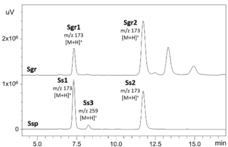
Figure 5.HPLC−DAD chromatograms at UV of 254 nm of theflash fractions ofS. graminifolia (Sgr) andS. speciosa(Ssp) root extracts (labeled with the corresponding m/z values obtained by HPLC− DAD−ESI+−MS).
Figure 6.UV spectra of the bioactive goldenrod compounds obtained by HPLC−DAD.
https://doi.org/10.1021/acs.jafc.1c03676 J. Agric. Food Chem.XXXX, XXX, XXX−XXX D
m/z 173.0604 and 173.0598 (calculated m/z 173.0597 for C
11H
9O
2+) and the sodium adduct of the dimers at m/z 367.0953 and 367.0952 (calculated m/z 367.0941 for C
22H
16O
4Na
+) had low signal intensity. Furthermore, the HPTLC
−HESI
−HRMS
ninvestigation provided a very similar fragmentation pattern of the protonated molecules of compounds
Sv1and
Sgr1with the sequential loss of CH
4O, CO, C
2H
2, and C
2groups (Figure 4). After
flash chromatog- raphy, each fraction with the concentrated compounds of interest of S. speciosa and S. graminifolia root extracts were compared. Their HPLC
−DAD
−ESI
−MS chromatograms showed the same retention time for the pairs of compounds
Ss1and
Sgr1as well as compounds
Ss2and
Sgr2(Figure 5), while the MS and UV spectra of compounds
Sgr1,Sgr2,Ss1,and
Ss2were very similar (Figure 6). Thus, it was assumed that compounds
Sgr1and
Ss1as well as compounds
Sgr2and
Ss2are identical and compounds
Sgr1and
Sgr2are isomers.
Together with the already mentioned compound
Ss3,altogether
five compounds (Sgr1,
Sgr2, Ss1, Ss2, and Ss3)were isolated as
flash chromatographic fractions by semi- preparative HPLC. The isolated compounds were subjected to NMR spectroscopy (Figures S-1−S-36 of the Supporting Information) and compound
Ss3were also subjected to HPLC
−qTOF
−MS analysis. Compound
Ss3was detected in both the positive and negative ionization modes as sodium adduct at m/z 281.0822 and deprotonated molecule at m/z 257.0828 corresponding to the compound with the sum formula C
15H
14O
4. The MS/MS spectrum (Figure 7) recorded at 20 eV collision energy was helpful to elucidate the structure based on the NMR spectrum. The NMR analysis proved that compounds
Sgr1and
Ss1as well as compounds
Sgr2and
Ss2were identical and determined as 2Z-dehydromatricaria ester and 2E-dehydromatricaria ester, respectively (Table 1). The NMR spectral data of the isomers were essentially identical with those reported before.
35−38The NMR assignment of compound
Ss3allowed its identi
fication as benzyl 2-hydroxy-6- methoxybenzoate (Table 1) that was also con
firmed by comparison to reported spectral data.
39,40The antibacterial activity of the pure isolated compounds was tested via HPTLC
−direct bioautography. On the basis of the HPTLC
fingerprints (Figure 1a) of the isolates, none of the isomeric forms of matricaria and dehydromatricaria esters could be obtained in pure form because they transformed into each other, even though the isolated forms dominated the respective isolates. It was con
firmed that the isolates were responsible for the observed antibacterial e
ffects. The benzyl 2-
hydroxy-6-methoxybenzoate
Ss3showed a pronounced activity against A.
fischeri, weak activity against B. subtilis, and no activity against P. maculicola (Figure 1). In the bioautograms, the 2Z,8Z-matricaria ester
Sv1and the Z-dehydromatricaria ester
Ss1inhibited both B. sorokiniana and F. avenaceum, while compound
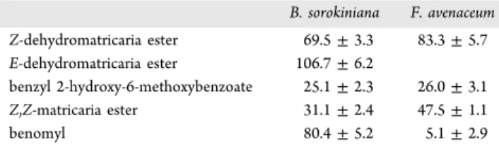
Ss3inhibited B. sorokiniana (Figure 2). IC
50determined by the microdilution assay of the isolates against fungal (mycelial) growth of both species was lowest for compound
Ss3, followed by compoundsSv1and
Ss1(Table
2). Note that theE-dehydromatricaria ester
Ss2did not reach IC
50at the maximum concentration used for F. avenaceum. The previous comparison of the bioactivity of the isomers indicated similarly that the 2Z,8Z-matricaria ester was more active than the 2E,8E-matricaria ester against plant pathogenic fungal species.
41The Z,Z form also had a similar or stronger antimycobacterial e
ffect than the E,Z form.
42C
10-polyacetylenes, like the identi
fied esters, are allelochem- icals with strong inhibitory activities on other plants.
43Matricaria and dehydromatricaria esters have been found in the roots of several goldenrod species, including S. virgaurea and S. graminifolia.
12,44−46However, to our knowledge, this is the
first report about the presence of dehydromatricaria estersin the root of S. speciosa. There are few literature data regarding the bioactivity of the isolated compounds. Essential oils of Conyza species rich in matricaria ester (74 and 88%) were found to be antifungal against Cryptococcus neoformans, Rhodotorula glutinis, Microsporum canis, and Candida and Trichophyton species, antibacterial against Staphylococcus aureus, and phytotoxic against Raphanus sativus, with inhibition of the seed germination.
47,48Both Z,Z- and E,Z-matricaria esters inhibited Mycobacterium tuberculosis and Mycobacterium avium
42and showed remarkable nematicidal
49and
α- glucosidase,
β-glucosidase,
α-amylase, acetylcholinesterase, and butyrylcholinesterase inhibitory activity.
17Z,Z-Matricaria ester exhibited antifungal effects against Botrytis cinerea, Colletotrichum acutatum, Colletotrichum fragariae, Colletotri- chum gloesporioides, Cladosporium cucumerinum, and Pyricularia oryzae plant pathogenic fungi,
41,50antibacterial activity against B. subtilis, P. syringae maculicola, Xanthomonas euvesicatoria, Lactobacillus plantarum, and A.
fischeri,
12and molluscicidal activity against Planobdella trivolvis.
41E,Z-Matricaria ester displayed antileishmanial activity.
51Moreover, Z- and E- dehydromatricaria esters exhibited nematicidal activity and cytotoxicity against human cancer cell lines,
46,49,52but Z- dehydromatricaria ester also inhibited the growth of normal mammalian cells.
46Furthermore, Z-dehydromatricaria ester
Figure 7.HPLC−ESI−qTOFMS spectra of the deprotonated molecule of compoundSs3(blue diamond) obtained at 20 eV collision energy.https://doi.org/10.1021/acs.jafc.1c03676 J. Agric. Food Chem.XXXX, XXX, XXX−XXX E
Table1.NMRSpectralDataandStructuresofCompoundsSs1,Ss2,andSs3 number1Hδ(ppm)13Cδ(ppm)1Hδ(ppm)13Cδ(ppm)1Hδ 1166.13167.05 26.37(d,3JH2,H3(cis)=11.5Hz,1H)133.926.44(d,3JH2,H3(trans)=15.9Hz,1H)135.14 36.32(dd,3JH2,H3(cis)=11.5Hz,9JH10,H3=0.5Hz,1H)122.446.79(dd,3JH2,H3(trans)=15.9Hz,9JH10,H3=0.5Hz,1H)124.206.53(d,J=8.3Hz,1H) 472.2072.537.28(t,J=8.3Hz,1H) 585.9882.866.51(dd,3JH5,H4=8.3, 658.7858.35 772.3271.75 864.8864.713.81(brs,3H) 982.2482.17 102.030(d,9JH10,H3=0.5Hz,3H)4.022.026(d,9JH10,H3=0.5Hz,3H)3.98 113.761(s,3H)52.183.755(s,3H)52.56 1′ 2′,6′7.47(d,J=7.5Hz,2H) 3′,5′7.37(t,J=7.3Hz,2H) 4′7.31(t,J=7.6Hz,1H) 7′5.37(s,2H)
https://doi.org/10.1021/acs.jafc.1c03676 J. Agric. Food Chem.XXXX, XXX, XXX−XXX F
showed antimycobacterial activity against M. tuberculosis and M. avium.
42However, without the improvement of the chemical stability of polyacetylenes, e.g., by formulation or modi
fication, their application as pesticides or drugs is limited.
41Benzyl 2-hydroxy-6-methoxybenzoate has been isolated from various plants, among others, from roots of Lindera fruticosa,
53Securidaca longipedunculata,
54and Secur- idaca diversifolia
40and aerial parts of Ageratina deltoidea
55but not from Solidago species so far. This benzyl benzoate exhibited cytotoxic (against HeLa cell lines), antiviral (against Herpes simplex virus type 1 and poliovirus Sabin 1), and human acyl-CoA:cholesterol acyltransferase inhibitory ef- fects,
40,53but it was not active against S. aureus, Escherichia coli, and Candida albicans.
55In conclusion, HPTLC bioassays developed for crop pathogenic fungi enabled the screening and selection of fungicidal goldenrod root compounds. The targeted, effect- directed HPTLC
−HESI
−HRMS
ncharacterization, isolation, and NMR analysis resulted in the identification of Z,Z- and E,Z-matricaria esters, Z- and E-dehydromatricaria esters, and benzyl 2-hydroxy-6-methoxybenzoate. To our knowledge, dehydromatricaria esters and benzyl 2-hydroxy-6-methoxyben- zoate were found in S. speciosa for the
first time. All
five compounds inhibited the mycelial growth of F. avenaceum and B. sorokiniana. Benzyl 2-hydroxy-6-methoxybenzoate was the most e
ffective, followed by Z,Z-matricaria and Z-dehydroma- tricaria esters. These showed potential to become agrochemical agents or lead compounds after appropriate modi
fication and formulation.
■
ASSOCIATED CONTENT*
sı Supporting InformationThe Supporting Information is available free of charge at
https://pubs.acs.org/doi/10.1021/acs.jafc.1c03676.NMR spectroscopy of the isolated compounds (Figures S-1−S-36) (PDF)
■
AUTHOR INFORMATION Corresponding AuthorÁgnes M. Móricz−
Plant Protection Institute, Centre for Agricultural Research, E
ötv
ös Lor
ánd Research Network (ELKH), 1022 Budapest, Hungary;
orcid.org/0000- 0002-4330-9396; Phone: +3614877515;Email:
moricz.agnes@atk.hu AuthorsDániel Krüzselyi−
Plant Protection Institute, Centre for Agricultural Research, E
ötv
ös Lor
ánd Research Network (ELKH), 1022 Budapest, Hungary
József Bakonyi −
Plant Protection Institute, Centre for Agricultural Research, E
ötv
ös Lor
ánd Research Network (ELKH), 1022 Budapest, Hungary
Péter G. Ott −
Plant Protection Institute, Centre for Agricultural Research, E
ötv
ös Lor
ánd Research Network (ELKH), 1022 Budapest, Hungary
András Darcsi−
Pharmaceutical Chemistry and Technology Department, National Institute of Pharmacy and Nutrition, 1051 Budapest, Hungary
Péter Csontos −
Institute for Soil Sciences, Centre for Agricultural Research, E
ötv
ös Lor
ánd Research Network (ELKH), 1022 Budapest, Hungary
Gertrud E. Morlock−
Chair of Food Science, Institute of Nutritional Science, and TransMIT Center of E
ffect-Directed Analysis, Justus Liebig University Giessen, 35392 Giessen, Germany;
orcid.org/0000-0001-9406-0351Complete contact information is available at:
https://pubs.acs.org/10.1021/acs.jafc.1c03676 Author Contributions
Dániel Kru
̈zselyi, experiments and writing the original draft;
József Bakonyi, methodology, mycological work, resources, and writing review and editing; Péter G. Ott, methodology, bacteriological work, resources, and writing review and editing;
András Darcsi, NMR and LC-qTOF experiments and writing the original draft; Péter Csontos, plant material collection and identi
fication; Gertrud E. Morlock, conceptualization, resour- ces, and writing review and editing; and A
́gnes M. Móricz, conceptualization, supervision, methodology, resources, experi- ments, data analysis, and writing the original draft and review and editing.
Funding
This work was supported by the National Research, Develop- ment and Innovation O
ffice of Hungary (NKFIH K128921 and PD134467). A
́gnes M. Mo
́ricz thanks the Organisation for Economic Co-operation and Development (OECD) for Scholarship JA00092484 that allowed her to stay at Justus Liebig University Giessen. Instrumentation was partially funded by the Deutsche Forschungsgemeinschaft (DFG, German Research Foundation), INST 162/471-1 FUGG and INST 162/536-1 FUGG.
Notes
The authors declare no competing
financial interest.
■
ACKNOWLEDGMENTSThe authors thank Imanuel Yu
̈ce, Food Science, Justus Liebig University Giessen, for HESI
−HRMS
nrecording. The authors are grateful to András Schmotzer, Directorate of Bu
̈kk National Park, Eger, Hungary, and Erzsébet Fráter, National Botanical Garden, Vácrátót, Hungary, for supporting the collection of S.
graminifolia and S. speciosa, respectively. The authors gratefully acknowledge the Faculty of Pharmacy, Semmelweis University, Budapest, Hungary, for providing access to the NMR system.
■
(1) Vogelgsang, S.; Sulyok, M.; Hecker, A.; Jenny, E.; Krska, R.;REFERENCES Schuhmacher, R.; Forrer, H.-R. Toxigenicity and pathogenicity of Fusarium poaeandFusarium avenaceumon wheat.Eur. J. Plant Pathol.2008,122(2), 265−276.
(2) Pollard, A. T.; Okubara, P. A. Real-time PCR quantification of Fusarium avenaceumin soil and seeds.J. Microbiol. Methods2019,157, 21−30.
(3)The Fusarium Laboratory Manual; Leslie, J. F., Summerell, B. A., Eds.; Blackwell Publishing: Ames, IA, 2006; DOI: 10.1002/
9780470278376.
Table 2. IC50Values of the Isolated Compounds and the Fungicide Benomyl against Fungal Strains inμg/mL
B. sorokiniana F. avenaceum Z-dehydromatricaria ester 69.5±3.3 83.3±5.7 E-dehydromatricaria ester 106.7±6.2
benzyl 2-hydroxy-6-methoxybenzoate 25.1±2.3 26.0±3.1
Z,Z-matricaria ester 31.1±2.4 47.5±1.1
benomyl 80.4±5.2 5.1±2.9
https://doi.org/10.1021/acs.jafc.1c03676 J. Agric. Food Chem.XXXX, XXX, XXX−XXX G
(7) Berestetskiy, A. O.; Dalinova, A. A.; Dubovik, V. R.; Grigoryeva, E. N.; Kochura, D. M.; Senderskiy, I. V.; Smirnov, S. N.;
Stepanycheva, E. A.; Turaeva, S. M. Analysis and isolation of secondary metabolites of Bipolaris sorokiniana by different chroma- tography techniques and the spectrum of their biological activity.
Appl. Biochem. Microbiol.2020,56(5), 569−582.
(8) Ali, L.; Khan, A. L.; Hussain, J.; Al-Harrasi, A.; Waqas, M.; Kang, S.-M.; Al-Rawahi, A.; Lee, I.-J. Sorokiniol: A new enzymes inhibitory metabolite from fungal endophyte Bipolaris sorokinianaLK12.BMC Microbiol.2016,16(1), 103.
(9) Nakajima, H.; Isomi, K.; Hamasaki, T.; Ichinoe, M.
Sorokinianin: A novel phytotoxin produced by the phytopathogenic fungusBipolaris sorokiniana.Tetrahedron Lett.1994,35(51), 9597−
9600.
(10) Qader, M. M.; Kumar, N. S.; Jayasinghe, L.; Araya, H.;
Fujimoto, Y. Bioactive Sesquiterpenes from an endophytic fungus Bipolaris sorokinianaisolated from a popular medicinal plant Costus speciosus.Mycology2017,8(1), 17−20.
(11) Morlock, G.; Schwack, W. Hyphenations in planar chromatog- raphy.J. Chromatogr. A2010,1217(43), 6600−6609.
(12) Móricz, Á. M.; Ott, P. G.; Häbe, T. T.; Darcsi, A.; Böszörményi, A.; Alberti, Á.; Krüzselyi, D.; Csontos, P.; Béni, S.; Morlock, G. E.
Effect-directed discovery of bioactive compounds followed by highly targeted characterization, isolation and identification, exemplarily shown forSolidago virgaurea.Anal. Chem.2016,88(16), 8202−8209.
(13) Morlock, G. E. High-performance thin-layer chromatography combined with effect-directed assays and high-resolution mass spectrometry as an emerging hyphenated technology: A tutorial review.Anal. Chim. Acta2021,1180, 338644.
(14) Schreiner, T.; Morlock, G. E. Non-target bioanalytical eight- dimensional hyphenation including bioassay, heart-cut trapping, online desalting, orthogonal separations and mass spectrometry. J.
Chromatogr. A2021,1647, 462154.
(15) Móricz, Á. M.; Ott, P. G. Conventional and modern bioassaysDetection, isolation, identification. In Forced-Flow Layer Chromatography; Tyihák, E., Ed.; Elsevier: Amsterdam, Netherlands, 2016; Chapter 6, pp 347−395, DOI: 10.1016/B978-0-12-420161- 3.00006-X.
(16) Móricz, Á. M.; Krüzselyi, D.; Ott, P. G.; Garádi, Z.; Béni, S.;
Morlock, G. E.; Bakonyi, J. Bioactive clerodane diterpenes of giant goldenrod (Solidago giganteaAit.) root extract.J. Chromatogr. A2021, 1635, 461727.
(17) Móricz, Á. M.; Jamshidi-Aidji, M.; Krüzselyi, D.; Darcsi, A.;
Böszörményi, A.; Csontos, P.; Béni, S.; Ott, P. G.; Morlock, G. E.
Distinction and valorization of 30 root extracts of five goldenrod (Solidago) species.J. Chromatogr. A2020,1611, 460602.
(18) Morlock, G.; Schwack, W. Coupling of planar chromatography to mass spectrometry. TrAC, Trends Anal. Chem. 2010, 29 (10), 1157−1171.
(19) Morlock, G. E.; Brett, N. Correct assignment of lipophilic dye mixtures? A case study for high-performance thin-layer chromatog- raphy−mass spectrometry and performance data for the TLC−MS interface.J. Chromatogr. A2015,1390, 103−111.
(20) Krüger, S.; Hüsken, L.; Fornasari, R.; Scainelli, I.; Morlock, G.
E. Effect-directed fingerprints of 77 botanical extracts via a generic high-performance thin-layer chromatography method combined with assays and mass spectrometry.J. Chromatogr. A2017,1529, 93−106.
Uses of Native Plants; Minnesota Historical Society: Saint Paul, MN, 2005.
(25) Fursenco, C.; Calalb, T.; Uncu, L.; Dinu, M.; Ancuceanu, R.
Solidago virgaurea L.: A review of its ethnomedicinal uses, phytochemistry, and pharmacological activities. Biomolecules 2020, 10(12), 1619.
(26) Toiu, A.; Vlase, L.; Vodnar, D. C.; Gheldiu, A.-M.; Oniga, I.
Solidago graminifolia L. Salisb. (Asteraceae) as a valuable source of bioactive polyphenols: HPLC profile, in vitro antioxidant and antimicrobial potential.Molecules2019,24(14), 2666.
(27) Kołodziej, B. Antibacterial and Antimutagenic Activity of extracts aboveground parts of three Solidago species: Solidago virgaureaL.,Solidago canadensisL. andSolidago giganteaAit.J. Med.
Plants Res.2011,5(31), 6770−6779.
(28) Anžlovar, S.; Koce, J. D. Antibacterial and antifungal activity of aqueous and organic extracts from indigenous and invasive species of goldenrod (Solidagospp.) grown in Slovenia.Phyton (Horn, Austria) 2014,54(1), 135−147.
(29) Konovalov, D. A. Polyacetylene compounds of plants of the Asteraceae family (Review).Pharm. Chem. J.2014,48(9), 613−631.
(30) Negri, R. Polyacetylenes from terrestrial plants and fungi:
Recent phytochemical and biological advances.Fitoterapia2015,106, 92−109.
(31) Veneziani, R. C. S.; Ambrósio, S. R.; Martins, C. H. G.; Lemes, D. C.; Oliveira, L. C. Antibacterial potential of diterpenoids.Stud. Nat.
Prod. Chem.2017,54, 109−139.
(32) Móricz, Á. M.; Häbe, T. T.; Böszörményi, A.; Ott, P. G.;
Morlock, G. E. Tracking and identification of antibacterial components in the essential oil of Tanacetum vulgare L. by the combination of high-performance thin-layer chromatography with direct bioautography and mass spectrometry.J. Chromatogr. A2015, 1422, 310−317.
(33) Móricz, Á. M.; Häbe, T. T.; Ott, P. G.; Morlock, G. E.
Comparison of high-performance thin-layer with overpressured layer chromatography combined with direct bioautography and direct analysis in real time mass spectrometry for tansy root.J. Chromatogr.
A2019,1603, 355−360.
(34) Móricz, Á. M.; Tyihák, E.; Ott, P. Usefulness of transgenic luminescent bacteria in direct bioautographic investigation of chamomile extracts. J. Planar Chromatogr.–Mod. TLC2010,23(3), 180−183.
(35) Hearn, M. T. W.; Turner, J. L. The carbon-13 nuclear magnetic resonance spectra of the antibiotic polyacetylenic nitrile, diatretyne 2 (7-cyanohept-trans-2-ene-4,6-diynoic acid), and related compounds.J.
Chem. Soc., Perkin Trans. 21976, No. 9, 1027.
(36) Wang, Q.; Hao, J.; Gong, J.; Bao, W. Isolation and structure elucidation of two new compounds fromArtemisia ordosica Krasch.
Nat. Prod. Res.2020,34(13), 1862−1867.
(37) Drake, D. Polyacetylenes ofArtemisia vulgaris.Phytochemistry 1974,13(2), 455−457.
(38) Tsao, R.; Eto, M. Light-activated plant growth inhibitory activity of cis-dehydromatricaria ester, rose bengal and fluoren-9-one on lettuce (Lactuca salivaL.).Chemosphere1996,32(7), 1307−1317.
(39) Kodpinid, M.; Sadavongvivad, C.; Thebtaranonth, C.;
Thebtaranonth, Y. Benzyl benzoates from the root ofUvaria purpurea.
Phytochemistry1984,23(1), 199−200.
https://doi.org/10.1021/acs.jafc.1c03676 J. Agric. Food Chem.XXXX, XXX, XXX−XXX H
(40) Casu, L.; Solinas, M. N.; Saba, A. R.; Cottiglia, F.; Caboni, P.;
Floris, C.; Laconi, S.; Pompei, R.; Leonti, M. Benzophenones from the roots of the popoluca amerindian medicinal plant Securidaca diversifolia(L.) S.F. Blake.Phytochem. Lett.2010,3(4), 226−229.
(41) Meepagala, K. M.; Sturtz, G.; Wise, D.; Wedge, D. E.
Molluscicidal and antifungal activity of Erigeron speciosus steam distillate.Pest Manag. Sci.2002,58(10), 1043−1047.
(42) Lu, T.; Cantrell, C.; Robbs, S.; Franzblau, S.; Fischer, N.
Antimycobacterial matricaria esters and lactones from Astereae species.Planta Med.1998,64(7), 665−667.
(43) Kobayashi, A.; Morimoto, S.; Shibata, Y.; Yamashita, K.;
Numata, M. C10-Polyacetylenes as allelopathic substances in dominants in early stages of secondary succession. J. Chem. Ecol.
1980,6(1), 119−131.
(44) Lam, J.; Christensen, L. P.; Färch, T.; Thomasen, T. Acetylenes from the roots of Solidago species. Phytochemistry 1992, 31 (12), 4159−4161.
(45) Lam, J. Polyacetylenes of Solidago virgaurea: Their seasonal variation and NMR long-range spin coupling constants.Phytochem- istry1971,10(3), 647−653.
(46) Matsunaga, H.; Katano, M.; Tasaki, M.; Yamamoto, H.; Mori, M.; Takata, K. Inhibitory effect of cis-dehydromatricaria ester isolated from Solidago altissima on the growth of mammalian cells. Chem.
Pharm. Bull.1990,38(12), 3483−3484.
(47) Veres, K.; Csupor-Löffler, B.; Lázár, A.; Hohmann, J. Antifungal activity and composition of essential oils ofConyza canadensisherbs and roots.Sci. World J.2012,2012, 1−5.
(48) Mabrouk, S.; Salah, K. B. H.; Elaissi, A.; Jlaiel, L.; Jannet, H. B.;
Aouni, M.; Harzallah-Skhiri, F. Chemical composition and antimicro- bial and allelopathic activity of TunisianConyza sumatrensis(Retz.) E.
Walker essential oils.Chem. Biodiversity2013,10(2), 209−223.
(49) Kimura, Y.; Mori, M.; Suzuki, A.; Kobayashi, A. Isolation and identification of two nematicidal substances from roots of Erigeron philadelphicusL. and nematicidal activities of their related compounds.
Agric. Biol. Chem.1981,45(12), 2915−2917.
(50) Vidari, G.; Abdo, S.; Gilardoni, G.; Ciapessoni, A.; Gusmeroli, M.; Zanoni, G. Fungitoxic metabolites from Erigeron apiculatus.
Fitoterapia2006,77(4), 318−320.
(51) Chandra Pandey, S.; Dhami, D. S.; Jha, A.; Chandra Shah, G.;
Kumar, A.; Samant, M. Identification of trans−2- cis−8-matricaria- ester from the essential oil ofErigeron multiradiatusand evaluation of its antileishmanial potential by in vitro and in silico approaches.ACS Omega2019,4(11), 14640−14649.
(52) Lone, S. H.; Bhat, K. A.; Naseer, S.; Rather, R. A.; Khuroo, M.
A.; Tasduq, S. A. Isolation, cytotoxicity evaluation and HPLC- quantification of the chemical constituents fromArtemisia amygdalina Decne.J. Chromatogr. B: Anal. Technol. Biomed. Life Sci. 2013,940, 135−141.
(53) Song, M.-C.; Nigussie, F.; Jeong, T.-S.; Lee, C.-Y.; Regassa, F.;
Markos, T.; Baek, N.-I. Phenolic compounds from the roots of Lindera f ruticosa.J. Nat. Prod.2006,69(5), 853−855.
(54) Dibwe, D. F.; Awale, S.; Kadota, S.; Morita, H.; Tezuka, Y.
Heptaoxygenated xanthones as anti-austerity agents from Securidaca longepedunculata.Bioorg. Med. Chem.2013,21(24), 7663−7668.
(55) Arciniegas, A.; Pérez-Castorena, A. L.; Meléndez-Aguirre, M.;
Ávila, J. G.; García-Bores, A. M.; Villaseñor, J. L.; Romo de Vivar, A.
Chemical composition and antimicrobial activity of Ageratina deltoidea.Chem. Biodiversity2018,15(3), No. e1700529.
https://doi.org/10.1021/acs.jafc.1c03676 J. Agric. Food Chem.XXXX, XXX, XXX−XXX I