Article
Stress Echo 2030: The Novel ABCDE-(FGLPR) Protocol to Define the Future of Imaging
Eugenio Picano1,*, Quirino Ciampi2 , Lauro Cortigiani3, Adelaide M. Arruda-Olson4,
Clarissa Borguezan-Daros5 , JoséLuis de Castro e Silva Pretto6,7 , Rosangela Cocchia8, Eduardo Bossone8 , Elisa Merli9, Garvan C. Kane4, Albert Varga10, Gergely Agoston10, Maria Chiara Scali11, Doralisa Morrone12 , Iana Simova13, Martina Samardjieva13, Alla Boshchenko14, Tamara Ryabova14, Alexander Vrublevsky14, Attila Palinkas15, Eszter D. Palinkas16 , Robert Sepp16, Marco A. R. Torres17, Hector R. Villarraga4, Tamara Kovaˇcevi´c Preradovi´c18 , Rodolfo Citro19, Miguel Amor20, Hugo Mosto20, Michael Salamè20, Paul Leeson21, Cristina Mangia22, Nicola Gaibazzi23, Domenico Tuttolomondo23 , Costantina Prota24, Jesus Peteiro25 , Caroline M. Van De Heyning26 , Antonello D’Andrea27 , Fausto Rigo28,
Aleksandra Nikolic29, Miodrag Ostojic29, Jorge Lowenstein30, Rosina Arbucci30 , Diego M. Lowenstein Haber30, Pablo M. Merlo30, Karina Wierzbowska-Drabik31 , Jaroslaw D. Kasprzak31, Maciej Haberka32,
Ana Cristina Camarozano33, Nithima Ratanasit34, Fabio Mori35, Maria Grazia D’Alfonso35, Luigi Tassetti35 , Alessandra Milazzo35, Iacopo Olivotto35 , Alberto Marchi35, Hugo Rodriguez-Zanella36 , Angela Zagatina37, Ratnasari Padang4, Milica Dekleva38, Ana Djordievic-Dikic39, Nikola Boskovic39, Milorad Tesic39 ,
Vojislav Giga39, Branko Beleslin39, Giovanni Di Salvo40 , Valentina Lorenzoni41, Matteo Cameli42 , Giulia Elena Mandoli42 , Tonino Bombardini18, Pio Caso27, Jelena Celutkiene43, Andrea Barbieri44 , Giovanni Benfari45 , Ylenia Bartolacelli46 , Alessandro Malagoli47 , Francesca Bursi48,
Francesca Mantovani49 , Bruno Villari2, Antonello Russo50, Michele De Nes1, Clara Carpeggiani1, Ines Monte51 , Federica Re52, Carlos Cotrim53 , Giuseppe Bilardo54, Ariel K. Saad55, Arnas Karuzas56 , Dovydas Matuliauskas56, Paolo Colonna57,58 , Francesco Antonini-Canterin58,59, Mauro Pepi58,60 , Patricia A. Pellikka4and The Stress Echo 2030 Study Group of the Italian Society of Echocardiography and Cardiovascular Imaging (SIECVI)†
Citation: Picano, E.; Ciampi, Q.;
Cortigiani, L.; Arruda-Olson, A.M.;
Borguezan-Daros, C.; de Castro e Silva Pretto, J.L.; Cocchia, R.; Bossone, E.; Merli, E.; Kane, G.C.; et al. Stress Echo 2030: The Novel
ABCDE-(FGLPR) Protocol to Define the Future of Imaging.J. Clin. Med.
2021,10, 3641. https://doi.org/
10.3390/jcm10163641
Academic Editors: Ernesto Di Cesare and Alberto Signore
Received: 9 July 2021 Accepted: 13 August 2021 Published: 17 August 2021
1 CNR, Biomedicine Department, Institute of Clinical Physiology, 56100 Pisa, Italy; denesm@ifc.cnr.it (M.D.N.);
claracarpeggiani@gmail.com (C.C.)
2 Cardiology Division, Fatebenefratelli Hospital, 82100 Benevento, Italy; qciampi@gmail.com (Q.C.);
villari.bruno@gmail.com (B.V.)
3 Cardiology Department, San Luca Hospital, 55100 Lucca, Italy; lacortig@tin.it
4 Department of Cardiovascular Medicine, Mayo Clinic, Rochester, MN 55905, USA;
ArrudaOlson.Adelaide@mayo.edu (A.M.A.-O.); kane.garvan@mayo.edu (G.C.K.);
Villarraga.Hector@mayo.edu (H.R.V.); Padang.Ratnasari@mayo.edu (R.P.);
pellikka.patricia@mayo.edu (P.A.P.)
5 Cardiology Division, Hospital San José, Criciuma 88801-250, Brazil; clarissabdaros@cardiol.br
6 Hospital Sao Vicente de Paulo e Hospital de Cidade, Passo Fundo 99010-080, Brazil; jlpretto@cardiol.br
7 Hospital de Cidade, Passo Fundo 99010-080, Brazil
8 Azienda Ospedaliera Rilevanza Nazionale A. Cardarelli Hospital, 80100 Naples, Italy;
rosangelacocchia@hotmail.com (R.C.); ebossone@hotmail.com (E.B.)
9 Department of Cardiology, Ospedale per gli Infermi, Faenza, 48100 Ravenna, Italy; elisamerli@libero.it
10 Institute of Family Medicine, Szeged University Medical School, University of Szeged, 6720 Szeged, Hungary;
varga.albert@med.u-szeged.hu (A.V.); drgergoagoston@gmail.com (G.A.)
11 Campostaggia Cardiology Division, Montepulciano, 53045 Siena, Italy; chiara_scali@yahoo.it
12 Cardiothoracic Department, University of Pisa, 56100 Pisa, Italy; doralisamorrone@gmail.com
13 Heart and Brain Center of Excellence, Cardiology Department, University Hospital, Medical University, 5800 Pleven, Bulgaria; ianasimova@gmail.com (I.S.); martina_vl@abv.bg (M.S.)
14 Cardiology Research Institute, Tomsk National Research Medical Centre of the Russian Academy of Sciences, 634009 Tomsk, Russia; allabosh@mail.ru (A.B.); rtr@cardio-tomsk.ru (T.R.); avr@cardio-tomsk.ru (A.V.)
15 Internal Medicine Department, Elisabeth Hospital, 6800 Hódmez˝ovásárhely, Hungary;
palinkasa@hotmail.com
16 Albert Szent-Gyorgyi Clinical Center, Department of Internal Medicine, Division of Non-Invasive Cardiology, University Hospital, 6725 Szeged, Hungary; sepprobert@gmail.com (R.S.); palinkaseszti@hotmail.com (E.D.P.)
17 9th July Hospital, DASA, San Paolo 04122-000, Brazil; mtorres.mt10@gmail.com
J. Clin. Med.2021,10, 3641. https://doi.org/10.3390/jcm10163641 https://www.mdpi.com/journal/jcm
Publisher’s Note:MDPI stays neutral with regard to jurisdictional claims in published maps and institutional affil- iations.
Copyright: © 2021 by the authors.
Licensee MDPI, Basel, Switzerland.
This article is an open access article distributed under the terms and conditions of the Creative Commons Attribution (CC BY) license (https://
creativecommons.org/licenses/by/
4.0/).
18 Clinic of Cardiovascular Diseases, University Clinical Centre of the Republic of Srpska, 78 000 Banja Luka, Bosnia and Herzegovina; tamara.kovacevic@medicolaser.info (T.K.P.);
tbombardini@yahoo.it (T.B.)
19 Echocardiography Laboratory, Cardiology Department, University Hospital “San Giovanni di Dio e Ruggi d’Aragona”, 84100 Salerno, Italy; rodolfocitro@gmail.com
20 Cardiology Department, Ramos Mejia Hospital, Buenos Aires C1221, Argentina;
miguelamor68@gmail.com (M.A.); hmosto@gmail.com (H.M.); michael.f.salame@gmail.com (M.S.)
21 RDM Division of Cardiovascular Medicine, Cardiovascular Clinical Research Facility, University of Oxford, Oxford OX3 9DU, UK; paul.leeson@cardiov.ox.ac.uk
22 CNR, ISAC-Institute of Sciences of Atmosphere and Climate, 73100 Lecce, Italy; c.mangia@isac.cnr.it
23 Cardiology Department, Parma University Hospital, 43100 Parma, Italy; ngaibazzi@gmail.com (N.G.);
d.tuttolomondo@hotmail.it (D.T.)
24 Cardiology Department, Vallo della Lucania Hospital, 84100 Salerno, Italy; costantinaprota@gmail.com
25 CHUAC-Complexo Hospitalario Universitario A Coruna, CIBER-CV, University of A Coruna, 15070 La Coruna, Spain; Jesus.Peteiro.Vazquez@sergas.es
26 Department of Cardiology, Antwerp University Hospital, 2650 Edegem, Belgium; carovdh@msn.com
27 UOC Cardiologia/UTIC/Emodinamica, PO Umberto I, Nocera Inferiore (ASL Salerno)—UniversitàLuigi Vanvitelli della Campania, 84014 Salerno, Italy; antonellodandrea@libero.it (A.D.); pio.caso@tin.it (P.C.)
28 Department of Cardiology, Dolo Hospital, 30031 Venice, Italy; faustorigo@alice.it
29 Department of Noninvasive Cardiology, Institute for Cardiovascular Diseases Dedinje, School of Medicine, Belgrade 11000, Serbia; nikolicdrsasa@gmail.com (A.N.); mostojic2011@gmail.com (M.O.)
30 Cardiodiagnosticos, Investigaciones Medicas Center, Buenos Aires C1082, Argentina;
lowensteinjorge@hotmail.com (J.L.); rosinaarbucci@hotmail.com (R.A.); lowediego@hotmail.com (D.M.L.H.);
pablommerlo@gmail.com (P.M.M.)
31 Department of Cardiology, Bieganski Hospital, Medical University, 91-347 Lodz, Poland;
wierzbowska@ptkardio.pl (K.W.-D.); kasprzak@ptkardio.pl (J.D.K.)
32 Department of Cardiology, SHS, Medical University of Silesia, 40-752 Katowice, Poland; mhaberka@op.pl
33 Medicine Department, Hospital de Clinicas UFPR, Federal University of Paranà, Curitiba 80000-000, Brazil;
a.camarozano@yahoo.com.br
34 Department of Medicine, Division of Cardiology, Siriraj Hospital, Mahidol University, Bangkok 10700, Thailand; nithimac@hotmail.com
35 SOD Diagnostica Cardiovascolare, DAI Cardio-Toraco-Vascolare, Azienda Ospedaliera-Universitaria Careggi, 50139 Firenze, Italy; morif@aou-careggi.toscana.it (F.M.); mariagrazia.dalfonso@gmail.com (M.G.D.);
luigi.tassetti.1990@gmail.com (L.T.); ales.milazzo@gmail.com (A.M.); iacopo.olivotto@unifi.it (I.O.);
alb.marchi@yahoo.com (A.M.)
36 Instituto Nacional de Cardiologia Ignacio Chavez, Mexico City 14080, Mexico; drzanella@gmail.com
37 Cardiology Department, Saint Petersburg State University Hospital, 199034 Saint Petersburg, Russia;
zag_angel@yahoo.com
38 Clinical Cardiology Department, Clinical Hospital Zvezdara, Medical School, University of Belgrade, Belgrade 11000, Serbia; dekleva.milica@gmail.com
39 University Clinical Centre of Serbia, Medical School, Cardiology Clinic, University of Belgrade, 11000 Belgrade, Serbia; skali.ana7@gmail.com (A.D.-D.); belkan87@gmail.com (N.B.); misa.tesic@gmail.com (M.T.);
voja2011@yahoo.com (V.G.); branko.beleslin@gmail.com (B.B.)
40 Division of Pediatric Cardiology, University Hospital, 35100 Padua, Italy; giodisal@yahoo.it
41 Institute of Management, Scuola Superiore Sant’Anna, 56100 Pisa, Italy; v.lorenzoni@sssup.it
42 Division of Cardiology, University Hospital, 53100 Siena, Italy; matteo.cameli@yahoo.com (M.C.);
giulia_elena@hotmail.it (G.E.M.)
43 Centre of Cardiology and Angiology, Clinic of Cardiac and Vascular Diseases, Faculty of Medicine, Institute of Clinical Medicine, Vilnius University, LT-03101 Vilnius, Lithuania; Jelena.Celutkiene@santa.lt
44 Noninvasive Cardiology, University Hospital, 43100 Parma, Italy; olmoberg@libero.it
45 Cardiology Department, University of Verona, 37121 Verona, Italy; giovanni.benfari@gmail.com
46 Paediatric Cardiology and Adult Congenital Heart Disease Unit, S. Orsola-Malpighi Hospital, 40100 Bologna, Italy; ylenia.bartolacelli@gmail.com
47 Nephro-Cardiovascular Department, Division of Cardiology, Baggiovara Hospital, University of Modena and Reggio Emilia, 41126 Modena, Italy; ale.malagoli@gmail.com
48 ASST Santi Paolo e Carlo, Presidio Ospedale San Paolo, 20100 Milano, Italy; francescabursi@gmail.com
49 Azienda UnitàSanitaria Locale—IRCCS di Reggio Emilia, Cardiology, 42100 Reggio Emilia, Italy;
francy_manto@hotmail.com
50 Association for Public Health “Salute Pubblica”, 72100 Brindisi, Italy; antonellorusso72@hotmail.com
51 Echocardiography Laboratory, Cardio-Thorax-Vascular Department, “ Policlinico Vittorio Emanuele”, Catania University, 95100 Catania, Italy; inemonte@gmail.com
52 Ospedale San Camillo, Cardiology Division, 00100 Rome, Italy; re.federica77@gmail.com
53 Heart Center, Hospital da Cruz Vermelha, Lisbon, and Medical School of University of Algarve, 1549-008 Lisbon, Portugal; carlosadcotrim@hotmail.com
54 UOC di Cardiologia, ULSS1 DOLOMITI, Presidio Ospedaliero di Feltre, 32032 Belluno, Italy;
bilardogiuseppe@gmail.com
55 División de Cardiología, Hospital de Clínicas Joséde San Martín, Buenos Aires C1120, Argentina;
arielsaad@gmail.com
56 Ligence Medical Solutions, 49206 Vilnius, Lithuania; a.karuzas@ligence.io (A.K.);
d.matuliaskas@ligence.io (D.M.)
57 Cardiology Hospital, Policlinico University Hospital of Bari, 70100 Bari, Italy; colonna@tiscali.it
58 Italian Society of Echocardiography and Cardiovascular Imaging, 20138 Milan, Italy;
antonini.canterin@gmail.com (F.A.-C.); Mauro.Pepi@cardiologicomonzino.it (M.P.)
59 Cardiac Prevention and Rehabilitation Unit, Highly Specialized Rehabilitation Hospital Motta di Livenza, Motta di Livenza, 31045 Treviso, Italy
60 Centro Cardiologico Monzino, IRCCS, 20138 Milan, Italy
* Correspondence: picano@ifc.cnr.it; Tel.: +39-050-315-2246; Fax: +39-050-315-2374
† Membership of the Stress Echo 2030 Study Group is provided in the Acknowledgments.
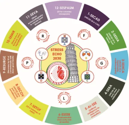
Abstract:With stress echo (SE) 2020 study, a new standard of practice in stress imaging was devel- oped and disseminated: the ABCDE protocol for functional testing within and beyond CAD. ABCDE protocol was the fruit of SE 2020, and is the seed of SE 2030, which is articulated in 12 projects: 1-SE in coronary artery disease (SECAD); 2-SE in diastolic heart failure (SEDIA); 3-SE in hypertrophic cardiomyopathy (SEHCA); 4-SE post-chest radiotherapy and chemotherapy (SERA); 5-Artificial intelligence SE evaluation (AI-SEE); 6-Environmental stress echocardiography and air pollution (ESTER); 7-SE in repaired Tetralogy of Fallot (SETOF); 8-SE in post-COVID-19 (SECOV); 9: Recovery by stress echo of conventionally unfit donor good hearts (RESURGE); 10-SE for mitral ischemic regur- gitation (SEMIR); 11-SE in valvular heart disease (SEVA); 12-SE for coronary vasospasm (SESPASM).
The study aims to recruit in the next 5 years (2021–2025)≥10,000 patients followed for≥5 years (up to 2030) from≥20 quality-controlled laboratories from≥10 countries. In this COVID-19 era of sustainable health care delivery, SE2030 will provide the evidence to finally recommend SE as the optimal and versatile imaging modality for functional testing anywhere, any time, and in any patient.
Keywords:effectiveness; registry; stress echocardiography; sustainability
1. Introduction
Stress echo (SE) 2020 is an international, multicenter, prospective, effectiveness study started in 2016 that conceptualized, disseminated, and validated a new approach for func- tional testing within and beyond coronary artery disease (CAD). As originally planned, the study created the cultural, informatic, and scientific infrastructure connecting high-volume, accredited SE labs, sharing common criteria of indication, execution, reporting, and archiv- ing SE. This approach allowed acquisition of original safety, feasibility, and outcome data in evidence-poor diagnostic fields, beyond the established core application of SE in CAD based on regional wall motion abnormality (RWMA) assessment. SE2020 standardized procedures, validated emerging signs, and integrated new information with established knowledge, helping to build a next-generation SE lab adopting the ABCDE protocol [1].
Each and every step of ABCDE-SE provides independent and incremental prognostic information building on the prior steps and identifies distinct patient phenotypes and vulnerabilities possibly outlining different therapeutic targets: myocardial ischemia in step A, pulmonary congestion with B-lines in step B, preload reserve and left ventricular contractile reserve (LVCR) in step C, coronary microcirculation with coronary flow velocity reserve (CFVR) or real-time myocardial contrast echocardiography in step D, and cardiac autonomic balance with heart rate reserve (HRR) in step E [2]. This shared practice can now be used as a new standard of care [3] and a suitable platform for the next wave of studies converging towards SE 2030—sharing with the older SE2020 study some distinct features: effectiveness study, performed in the real world with real doctors facing real clini-
cal problems in real consecutive patients; upstream quality control of reading and direct entering of data from peripheral centers in the data bank so that evidence is obtained inside and outside highly specialized academic centers; identification of simple yet innovative objectives relevant to change the clinical practice. These features are completely different from efficacy studies, as when highly specialized centers recruit highly selected patients, the resulting data may be difficult to translate in clinical practice. For these reasons, the American Society of Echocardiography has identified already in 2013 as a top research need “the development of a registry of echocardiographic information (and eventually images) that can serve as a platform for quality improvement and clinical research. Such registry data would be accessible to the research community facilitating a broad range of clinical research on the effectiveness of echocardiography for the improvement of patient management and outcome” [4].
SE2030 will establish the platform of evidence to build the perfect SE test, suit- able for all patients, anywhere, anytime, that is also quantitative and operator indepen- dent. The need for such an ideal test is especially vital in our times, when economic crises, the increased awareness of cancer and non-cancer radiation damage, the press- ing need for climate-neutral choices in health care, and the unavoidable trend to exter- nalize health care are potent propelling forces, boosted by COVID pandemics, for the diffusion of a low cost, radiation-free, climate-friendly, and portable technique such as cardiovascular ultrasound [5].
2. Materials and Methods
Five important aspects will be shared by SE2030 in full continuity with SE2020, with minor adaptations and implementations.
2.1. Upstream Quality Control
The study is a prospective registry, but it is necessary to have an upstream quality control with a certified reader from each center. Peripheral reading from each center is necessary for the effectiveness study to provide a snapshot of the real world. On the other side, a multicenter study is like a fish soup. The more the fish or contributing centers, the tastier the soup, but a single rotten fish will render the entire soup uneatable. Therefore, a mandatory quality control is necessary for conventional and innovative parameters, since the volume of activity is necessary but not sufficient to ensure the quality of reading [6].
2.2. Peripheral Reading and Inclusivity
Once the reader has been certified, the peripheral reading will be directly entered in the data bank via the Redcap program property of the Italian Society of Echocardiography and Cardiovascular Imaging. This will allow a more flexible and rapid platform compared to the standard excel approach, which requires greater human resources and has a greater chance of error in data inputting. In addition, Redcap has better compliance with new regulations strictly protecting privacy in clinical studies. Another feature of SE2030 is inclusivity, so that any center meeting the selection criteria can be enrolled, allowing centers traditionally outside the editorial stage but producing high quality clinical activity to contribute to generate data relevant for the scientific community. The inclusion of centers from many sites contains potential to recognize similarities and differences between countries, continents, cultures, and ethnicities. Inclusivity will also allow to assess how the proposed protocol will materialize in different scenarios (private practice settings, public hospitals, academic institutions) with different reimbursement policies and variable direct costs and commercial availability for drugs (such as dipyridamole or adenosine) and ultrasound-enhancing agents.
2.3. Uniform Methodology
Each laboratory will adopt the preferred choice of stress among physical, pharma- cologic, or pacing stress according to standardized protocol in line with guideline rec-
ommendations. Physical exercise includes semi-supine or upright bicycle exercise, and peak or post-treadmill exercise. Pharmacologic testing will be with dobutamine or va- sodilators (dipyridamole, adenosine, or regadenoson) according to physician preferences, patients’ contraindications, local availability, and cost. Pacing stress can be performed with transesophageal atrial pacing or with external programming of a permanent pacemaker.
Independent of the chosen form of stress, execution, performance, archiving, and interpre- tation of testing will follow a standardized format with the ABCDE protocol. From the technical viewpoint of success rate, a limiting step is step D. Step D is easy and feasible with vasodilator, less easy but still highly feasible with dobutamine, not easy and less feasible with semi-supine exercise, and virtually impossible with (peak or post) treadmill exercise.
Therefore, our recommendation is to use semi-supine exercise, capturing coronary flow signal in early or intermediate stages of exercise when most flow increase occurs and feasibility is still high, before it drops at higher levels of exercise. When treadmill is used, step D is skipped; if information is deemed important, a vasodilator test can be performed at 300after the end of exercise focused on CFVR and heart rate response.
All laboratories will be granted with free artificial intelligence (AI) software and en- couraged to use ultrasound enhancing agents when needed to help leading edge technology upgrade and uniformity of methods across all study laboratories [7].
2.4. The Full Spectrum of Enrolled Patients Evaluated for Clinically Relevant Endpoints
The various projects will include patients with known or suspected CAD (project 1), known or suspected heart failure with preserved ejection fraction (project 2), hyper- trophic cardiomyopathy (HCM, project 3), status post-chest radiotherapy and chemother- apy (project 4), repaired Tetralogy of Fallot (project 7), cardio-pulmonary involvement post-COVID 19 (project 8), post-ischemic (project 10) and primary valvular heart disease (project 11), and suspected coronary vasospasm, a diagnosis frequently missed but impor- tant to recognize as a possible cause of life-threatening disease, which is easy to treat when promptly identified (project 12). The 12 protocols running on the SE-ABCDE platform are spread all over the spectrum of cardiovascular disease, from severe valvular heart disease to suspected CAD in patients with normal LV function. Potential heart donors with brain death will be evaluated to assess the suitability for donation of hearts currently dismissed on the basis of clinical history criteria but in the absence of a cardiac functional evaluation (project 9). The study will exploit and possibly contribute to upgrading the leading edge quantitative and operator-independent technology of AI-SE and cardiac strain (project 5) for image interpretation and data analysis and will also evaluate the results of SE parameters in the context of powerful environmental modulators of stress results and/or long-term outcome such as air pollutants and medical radiation exposure analyzed through big data mining with AI (project 6). The overarching aim of the study is to make SE practice more uniform, versatile, standardized, quantitative, and evidence-rich, producing data potentially relevant to change clinical practice (Figure1).
When clinically indicated, SE will be repeated comparing results before and after treatment. Treatment will include medical treatment, percutaneous coronary interventions, transcatheter aortic valve implantation, surgical treatment, device therapy, and others.
2.5. Sponsored by a Professional Scientific Society
The study is investigator-driven and not industry-driven. It is endorsed by an independent-not for profit professional society (Italian Society of Echocardiography and Cardiovascular Imaging) and not sponsored by industry, although some materials use- ful for project completion such as AI-software will be donated by industrial partners for recruiting centers.
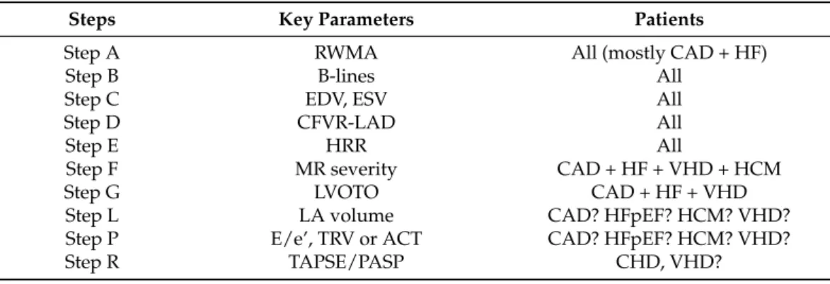
Figure 1. SE 2030 at a glance. The core protocol of SE2030 is the same as in SE2020 with ABCDE:
epicardial coronary artery stenosis (with RWMA), step A; lung water (with B-lines), step B;
myocardial function (with left ventricular end-systolic volume for contractile reserve and end- diastolic volume for preload reserve), step C; coronary microvascular dysfunction (with CFVR), step D; cardiac autonomic balance (with HRR), step E. Ancillary steps (necessary in some but not all patients) are step F (regurgitant flows), step L (left atrial volume and function), step P (pulmonary and LV pressures) and step R (right ventricular function). The study is endorsed by the Italian Society of Echocardiography and Cardiovascular Imaging and initiated in Pisa, Italy, as shown by the Leaning Tower present in the logo. SE protocols are indicated from 1 to 12 clockwise, and cover a wide spectrum of clinical conditions within and beyond CAD.
When clinically indicated, SE will be repeated comparing results before and after treatment. Treatment will include medical treatment, percutaneous coronary interventions, transcatheter aortic valve implantation, surgical treatment, device therapy, and others.
2.5. Sponsored by a Professional Scientific Society
The study is investigator-driven and not industry-driven. It is endorsed by an independent-not for profit professional society (Italian Society of Echocardiography and Cardiovascular Imaging) and not sponsored by industry, although some materials useful for project completion such as AI-software will be donated by industrial partners for recruiting centers.
2.6. ABCDE-SE in CAD (SECAD) 2.6.1. Background
The cornerstone of diagnosis with SE is the finding of reversible RWMA. This is the only sign established through over 40 years of clinical experience and endorsed in general cardiology guidelines [8]. However, the valuable diagnostic and prognostic information provided by SE extends well beyond RWMA [9] and today includes all the steps of the comprehensive ABCDE protocol, including B-lines [10], LVCR [11], CFVR [12], and HRR [13]. Functional mitral regurgitation (step F) is also important in patients with mitral regurgitation at least mild at rest since it may significantly worsen during stress affecting prognosis and possibly driving specific treatment [14–16], especially in patients with dilated cardiomyopathy. During inotropic stress (dobutamine and exercise, especially in
Figure 1.SE 2030 at a glance. The core protocol of SE2030 is the same as in SE2020 with ABCDE: epi- cardial coronary artery stenosis (with RWMA), step A; lung water (with B-lines), step B; myocardial function (with left ventricular end-systolic volume for contractile reserve and end-diastolic volume for preload reserve), step C; coronary microvascular dysfunction (with CFVR), step D; cardiac auto- nomic balance (with HRR), step E. Ancillary steps (necessary in some but not all patients) are step F (regurgitant flows), step L (left atrial volume and function), step P (pulmonary and LV pressures) and step R (right ventricular function). The study is endorsed by the Italian Society of Echocardiography and Cardiovascular Imaging and initiated in Pisa, Italy, as shown by the Leaning Tower present in the logo. SE protocols are indicated from 1 to 12 clockwise, and cover a wide spectrum of clinical conditions within and beyond CAD.
2.6. ABCDE-SE in CAD (SECAD) 2.6.1. Background
The cornerstone of diagnosis with SE is the finding of reversible RWMA. This is the only sign established through over 40 years of clinical experience and endorsed in general cardiology guidelines [8]. However, the valuable diagnostic and prognostic in- formation provided by SE extends well beyond RWMA [9] and today includes all the steps of the comprehensive ABCDE protocol, including B-lines [10], LVCR [11], CFVR [12], and HRR [13]. Functional mitral regurgitation (step F) is also important in patients with mitral regurgitation at least mild at rest since it may significantly worsen during stress affecting prognosis and possibly driving specific treatment [14–16], especially in patients with dilated cardiomyopathy. During inotropic stress (dobutamine and exercise, especially in upright position) step G will be assessed to identify dynamic left ventricular outflow tract obstruction as a cause of chest pain, dyspnea, or syncope [14–16].
2.6.2. Aims
The primary aim is to evaluate the feasibility of rest and stress-induced integrated ap- proach during SE with the ABCDE protocol with different stress modalities. The secondary aim is to assess the prognostic value of the different steps for predicting outcome.
2.6.3. Methods
All patients (“all-comers”) referred to the SE lab with known or suspected CAD will be evaluated with ABCDE-SE. Presenting patients referred to SE lab according to existing 2020 guidelines indications and contraindications will be recruited (Table1).
Table 1.General inclusion/exclusion criteria.
Inclusion Criteria Exclusion Criteria
Age > 18 years * √
Acceptable acoustic window at rest (≥14 out of 17 LV segments)
√
Clinically indicated test √
Informed consent √
Acute coronary syndromes or acute heart failure
√ Acute pulmonary embolism, myocarditis,
pericarditis, or aortic dissection
√ Serious cardiac arrhythmias
(ventricular tachycardia, complete atrio-ventricular block)
√
Prognosis-limiting (survival <1 year) extra-cardiac disease
√ Resting systolic blood pressure >180 mmHg
or significant hypotension
√
* Except for project 7 that may include younger patients with repaired tetralogy of Fallot after parental consent.
Patients with at least 14 readable segments on resting echocardiogram will be re- ferred for: (a) assessment of chest pain or dyspnea; (b) risk stratification of (ischemic or non-ischemic) dilated cardiomyopathy; (c) for reassessment after an abnormal test or as- sessment of known CAD (previous acute coronary syndrome and/or previous myocardial revascularization, prior CAD by invasive or noninvasive coronary angiography); (d) for risk stratification prior to high or intermediate risk non-cardiac vascular surgery (such as liver transplant or major non-cardiac vascular surgery) in patients with poor functional capacity (<4 METS) and/or suspected cardiac symptoms, in presence of a revised cardiac risk index (Lee criteria)≥2 (high risk surgery; CAD; congestive heart failure; cerebrovas- cular disease; diabetes mellitus on insulin; serum creatinine > 2 mg/mL); (e) status post heart transplant; (f) pediatric patients and congenital heart disease (Kawasaki, transpo- sition of the great arteries/status post-arterial switch operation, anomalous origin of a coronary artery, familial homozygous hypercholesterolemia); (g) peri-partum cardiomy- opathy. Usual contraindications will apply to all forms of stress testing as recommended by major scientific societies [7,9]: 1-unstable or complicated acute coronary syndromes;
2-severe cardiac arrhythmias (ventricular tachycardia, ventricular fibrillation, complete atrio-ventricular block). The presence of moderate to severe systemic hypertension (resting systolic blood pressure >180 mmHg) is a contraindication to exercise or dobutamine; the presence of a hemodynamically significant LV outflow tract obstruction (LV intraventricular gradient >30 mmHg) is a specific contraindication to dobutamine; significant hypotension (resting systolic blood pressure <90 mmHg) and pronounced active bronchospastic disease are a specific contraindication to vasodilator stress (dipyridamole or adenosine or regadeno- son). Information on demographics (sex, age, body mass index, and body surface area), lifestyle (smoking and physical activity), other risk factors and ongoing therapy will be collected. From resting echocardiography and vascular carotid scan evaluation data related to carotid disease and cardiac calcification will also be collected, when available, since both may contribute significantly to atherosclerosis phenotyping and comprehensive risk strati- fication independently and incrementally to SE results [14]. All patients will enter a regular clinical follow-up program with annotation of cardiovascular and non-cardiovascular end- points, since the biomarkers under investigation may precede and predict conditions other
than cardiovascular disease, such as cancer or neurodegenerative disease, characterized by low grade inflammation, somatic DNA instability, endothelial dysfunction (step D positivity) and autonomic dysfunction (step E positivity) which are associated with SE positivity and are considered biomarkers of systemic, not merely cardiovascular, disease.
2.6.4. Sample Size Calculation
The expected incidence of SE positivity (by at least one of the ABCDE criteria) is around 35% [10–13]. For prognostic end-point, we conservatively assume a 5% yearly incidence of composite end-points (all-cause death, myocardial infarction, stroke, pro- gression of chronic heart failure which requires hospitalization, evident intensification of diuretic therapy, new hospital admission for heart failure, heart transplant, ventricular assist device implantation, aborted sudden death, or new onset atrial fibrillation or atrial flutter), with doubling of likelihood of events in the presence of a positive SE (for composite criteria). Assuming that the hypothesis of proportionality of hazard holds, as required for Cox proportional hazards regression, a sample size of about 2430 patients with a 5-year follow-up is required to provide 90% power with an alpha error of 5% to detect a differ- ence for the primary endpoint of all-cause mortality among those with positive versus negative SE also considering a 20% drop-out. The estimated sample size will also allow detection of differences in secondary endpoints such as coronary revascularization for clinically refractory angina (predicted by A); readmission for decompensated heart failure or development of de novo heart failure (step B and step C); development of heart failure (step D); sudden death, de novo atrial fibrillation, severe ventricular arrhythmias such as sustained ventricular tachycardia causing syncope or ventricular fibrillation (step E). The newly recruited patients will add-up to the patients already recruited in years 2016–2020 with the same methodology as a part of a similar project (named DITSE) in SE2020 (1).
2.6.5. Study Hypothesis
ABCDE-SE is highly feasible with excellent success rate in all patients with all stress modalities.
The five steps of ABCDE protocol have independent and incremental value in predict- ing outcome, and each one selectively predicts some endpoints providing an integrated assessment of the many possible vulnerabilities of the patient. Each phenotype (ischemic, congestive, failing, microcirculatory, autonomic) is especially responsive to specific ther- apies which are left to the decision of referring physician and will be recorded in the follow-up to allow exploratory analysis.
2.7. ABCDE-Stress Echo in Diastolic Heart Failure (SEDIA Project) 2.7.1. Background
Diastolic heart failure is also called heart failure with preserved ejection fraction and is a challenging target of diastolic SE [15,16]. The diagnosis remains difficult, and the previous European Society of Cardiology criteria, based upon low sensitivity criteria such as echocardiographic data and plasma natriuretic peptides [17], have been recently revised with a new stepwise approach and a pretest assessment (including resting transthoracic echocardiography) in any patient with symptoms or signs compatible with heart failure, progressing to diastolic SE for intermediate score values and finally moving to invasive rest and exercise hemodynamic study for final confirmation of diagnosis. A pulmonary capillary wedge pressure≥15 mm Hg at rest or≥25 mm Hg at peak exercise is diagnostic of heart failure with preserved ejection fraction [18].
This stepwise approach has some potential limitations in the reliance on costly, risky, and time-consuming techniques such as rest and exercise invasive hemodynamic testing.
They were not practiced in any of the 30 laboratories of SE2020 network as a further demonstration of the dissociation between the aerial world of guidelines and ground-based clinical practice populated by restrictions due to economic, logistic, and medico-legal con- cerns. A much simpler clinical score can be used with parameters such as age, obesity, atrial
fibrillation, anti-hypertensive treatment, and resting echocardiographic parameters such as echocardiographic E/e’ ratio >9 and systolic pulmonary artery pressure >35 mm Hg, which however misses the heterogeneity of clinical and echocardiographic responses dur- ing stress of patients with identical clinical presentation and resting echocardiographic variables [19]. Indeed, more than one-half of patients with heart failure with preserved ejection fraction have normal/near normal left ventricular filling pressures at rest and invariably require stress to bring out the heart failure phenotype [20]. The 2019 European Society of Cardiology algorithm does not provide any indication for patients unable to exercise, and yet exercise is not feasible in about 50% of patients with the epidemiological profile of heart failure with preserved ejection fraction (age > 70 years, obesity/overweight, hypertensive, and diabetic). The algorithm does not include the recently developed pa- rameters which focus on key aspects of diastolic heart failure physiology such as reduced preload reserve [21], coronary microvascular disease as a trigger and amplifier of myocar- dial fibrosis in the natural history of the disease [22], and blunted chronotropic reserve with Step E of stress testing [23]. It emphasizes the value of indices such as E/e’ and tricuspid regurgitant jet velocity during exercise with known limitations of feasibility (50%) and accuracy, with unsatisfactory correlations with invasively measured parameters that they are supposed to mirror such as LV end-diastolic pressure for E/e’ and pulmonary artery systolic pressure for tricuspid regurgitant jet velocity [24].
Patients with unexplained dyspnea often have occult cardiac causes which can be easily unmasked by SE such as inducible ischemia, severe mitral regurgitation, or dynamic LV outflow tract obstruction [16]. These patients should be removed from the basket of heart failure with preserved ejection fraction since they exemplify a different pathogenesis and require a different therapy [16]. While E/e’ and tricuspid regurgitant jet velocity may play a role in characterizing changes in hemodynamics during stress, SE has much more to offer in the screening, diagnosis, risk stratification, and therapy in patients with known or suspected heart failure with preserved ejection fraction. Emerging data suggest left atrial mechanics may play a significant role in proportion of patients with heart failure with preserved ejection fraction, particularly in those with atrial dysrhythmias. While left atrial volume index at rest may convey risk of heart failure with preserved ejection fraction, changes in left atrial volume with stress may better risk stratify patients with 1 out of 5 patients seeing a reduction in left atrial volume with stress, but another 1 out of 5 with normal left atrial size dilating during stress [25]. Left atrial strain, particularly reservoir strain is also feasible, reproducible, and important to characterize left atrial function during stress. Pulmonary artery systolic pressure can be measured in virtually all patients during exercise if we use acceleration time of pulmonary flow velocity for patients with unreadable tricuspid regurgitant jet velocity [26]. A steep increase of left ventricular filling pressure and pulmonary capillary wedge pressure during exercise is a typical hemodynamic response in heart failure with preserved ejection fraction [27] This elevated pressure then backwardly transmitted to pulmonary circulation results in pulmonary congestion. This, in turn, overloads the right ventricle as well, causing right ventricular dysfunction and failure. In heart failure patients right ventricular free wall strain performs better (in terms of diagnostic and prognostic power) than conventional echocardiographic measures in the detection of right ventricular dysfunction [28]. Volumetric SE allows to measure preload reserve as the increase in end-diastolic volume and the contractile reserve through the reduction in end-systolic volume. Rest and stress B-lines are essential to recognize a pulmonary congestion phenotype [29] which may require B-lines driven decongestion therapy [30]. Therefore, ABCDE-SE during exercise or pharmacological SE deserves a systematic assessment in this challenging cohort with three major objectives:
1-to screen and exclude cardiac causes of dyspnea mimicking diastolic dysfunction; 2-to identify the reduction of functional reserve in cardiac output and its underlying separate but not mutually exclusive mechanisms (reduction in chronotropic, preload, or contractile reserve); 3-to characterize the underlying heterogeneous phenotypes potentially allowing a targeted therapy, from pulmonary congestion to coronary microvascular dysfunction.
2.7.2. Aims
The primary aim is to evaluate the feasibility and value of a SE-centered approach to diagnose heart failure with preserved ejection fraction in patients who can exercise, after screening for common mimickers of this condition such as severe mitral regurgitation, inducible ischemia, and dynamic LV outflow tract obstruction. The secondary aim is to evaluate the feasibility of a SE-centered approach with pharmacological stress in patients who are unable to exercise, which represent a high proportion of the total population of heart failure with preserved ejection fraction patients. The tertiary aim is to assess the prognostic value of SE indices (ABCDE plus pulmonary artery systolic pressure with tricuspid regurgitant jet velocity or acceleration time of pulmonary flow velocity, right ven- tricular free wall strain, left atrial volume and strain) for outcome stratification, compared to standard predictors such as plasma cardiac natriuretic peptides levels.
2.7.3. Methods
Patients with dyspnea and known or suspected heart failure with preserved ejection fraction by 2019 European Society of Cardiology criteria will be enrolled and studied with cycle-ergometer in semi-supine SE (or treadmill). A score of at least 1 according to the criteria proposed by Pieske et al. is required for inclusion [18]. The score (from 0 to 9) proposed by Reddy et al. on the basis of simple clinical and resting echocardiographic parameters will be also assessed [19]. In patients unable to exercise or when exercise is not feasible or did not allow sampling of CFVR, pharmacological test (vasodilator or dobutamine) is recommended. The diastolic assessment should be included into all exercise SE tests by measuring standard Doppler-derived mitral inflow velocity, pulsed Tissue Doppler of mitral annulus, and retrograde tricuspid gradient of tricuspid regurgitation.
These measurements can be performed at intermediate load of exercise and/or 1–2 min after the end of the exercise, after obtaining wall motion acquisitions, when the heart rate decreases and mitral inflow E and A velocities appear to be well separated. As a part of the “diastolic package”, we will also assess, at baseline, intermediate load (50 watts) and peak-post stress (11): end-diastolic left ventricular volume index (to evaluate left ventricular diastolic volume reserve, impaired in stiff hearts, which are less dilated for any given filling pressure); end-systolic left ventricular volume index (for assessment of left ventricular force, which may unmask occult systolic dysfunction with normal ejection fraction increase); ejection fraction and both stroke volume and cardiac output (to assess conventional contractile reserve) from 2D images; mitral regurgitation and left ventricular outflow tract obstruction; pulmonary artery systolic pressure (from velocity of tricuspid regurgitation or acceleration time of antegrade systolic pulmonary flow); B-lines with stress (to provide a direct imaging of extra-vascular lung water accumulation as a direct cause of dyspnea); right ventricular free wall strain to assess the presence of right ventricular dysfunction; left atrial volume index (with an abnormal response identified as a dilated left atrium exceeding the physiologic preload reserve or a stiff heart failing to dilate with increased pressures witnessed by B-lines increase); peak atrial longitudinal strain (a highly feasible and reproducible index of atrial reservoir function); and mitral inflow E velocity and mitral annulus e’ tissue Doppler velocity. Global longitudinal strain (GLS) will be determined as the average of the regional longitudinal strain measured in a 16 or 17 segment model from the apical long-axis, 4-chamber, and 2-chamber view.
2.7.4. Sample Size Calculation
The expected incidence of composite end-points (as defined in project 1) is around 20%
per year. We assume a positivity rate to SE (by composite criteria) of 30%, with doubling of likelihood of events in presence of SE positivity (by any criteria). Assuming that the hypothesis of proportionality of hazard holds, as required for Cox proportional hazards regression, with a power of 90%, an attrition rate of 10% and a 5-year follow-up period of a sample size of 181 patients is required. The newly recruited patients will add-up to the
patients already recruited in years 2018–2020 with the same methodology as a part of the same project in SE2020 [1].
2.7.5. Study Hypotheses
The invasive hemodynamic-based diagnosis of heart failure with preserved ejection fraction is not feasible for routine practice. The current clinical diagnostic criteria are of variable quality and not well tested in large patient populations. A comprehensive non- invasive stress-based algorithm have should be feasible, safe, simple, and prognostically relevant, thereby leading to “replace, reduce, and refine” (the 3 R’s approach) the current criteria: Replace invasive with noninvasive, ionizing with nonionizing, and rest with stress evaluation; Reduce the number of patients labelled heart failure with preserved ejection fraction by identifying at the outset patients with inducible ischemia presenting as dyspnea as the main symptom or with stress-induced mitral regurgitation or left ventricular outflow tract obstruction; Refine the current sub-setting identifying different phenotypes on the basis primarily of cardiac functional reserve (possibly impaired for chronotropic, preload, or contractile reserve) and associated phenotypes such as pulmonary congestion or coronary microvascular disease.
2.8. Stress Echo in Hypertrophic Cardiomyopathy (SEHCA) 2.8.1. Background
HCM is a heterogeneous inherited cardiomyopathy with variable phenotypic expres- sion [31–33]. The assessment of mortality risk in asymptomatic or mildly symptomatic patients is a challenging task, and several approaches targeted on different physiologic variables have been proposed. Resting transthoracic and SE are especially attractive for the purpose of identification of different phenotypes in HCM, risk stratification and serial follow-up examinations are often needed in the same patient to assess natural history and effects of interventions [15,16]. As a consequence, facilities and skills for exercise SE are usually available in specialist HCM centers [32,33]. Exercise SE provides compre- hensive information on the different vulnerabilities of the HCM patient with the ABCDE protocol: RWMA due to myocardial ischemia [34], pulmonary congestion due to diastolic dysfunction, preload reserve and contractile reserve impairment, coronary microcirculatory dysfunction [35], and blunted HRR which is a marker of cardiac autonomic dysfunction and reduced sympathetic reserve [36]. In addition to this standard ABCDE protocol adapted to HCM, at least two other parameters can be added in the assessment of HCM: evaluation of regurgitant mitral flow (step F) and dynamic left ventricular outflow tract gradient (step G) [37]. Of note, exercise limitation and breathlessness may be due to a number of different causes. Despite similar clinical manifestations, management may differ substan- tially based on the mechanisms [38]. SE is the only test with the potential to discriminate the various components, allowing a targeted treatment driven by pathophysiology.
2.8.2. Aims
The primary aim is to evaluate the feasibility of comprehensive ABCDEFG-SE in the evaluation of HCM. The secondary aim is to assess the value of each of these parameters in predicting response to specific therapy and other interventions. The tertiary aim is to assess the prognostic value of SE indices for prognostic stratification in the medium-long-term.
2.8.3. Methods
Diagnosis of HCM will be based on existing guidelines [31]. Phenocopies such as infiltrative/storage disease (e.g., Fabry, amyloid) will be excluded. All patients will be followed-up and the prognostic value of different rest and SE parameters (also com- pared to standard prognostic indices) will be assessed. For each patient new or changing therapies will be recorded and symptomatic status reassessed every year as unchanged (same New York Heart Association class), improved (class decrease≤1), or worsened (class increase≥1). In patients and first-degree relatives with genetic characterization
already performed as a part of the routine work-up different phenotypes will be correlated with specific genotypes. Non-imaging or routine imaging non-ultrasound exams such as EKG, cardiovascular magnetic resonance (myocardial fibrosis with delayed enhancement), and other available examinations will be collected and analyzed also with neural network analysis techniques developed in project 5.
2.8.4. Sample Size Calculation
The composite outcome end-points of the study are defined as in project 1 plus myectomy and percutaneous transluminal septal myocardial ablation. A pilot study showed an incidence of events around 8% per year [37]. Hypothesizing assumptions behind the Cox proportional hazards regression are met, if we assume a positivity rate to SE (by composite criteria) of 40%, a 5-year follow-up doubling of likelihood of events in presence of SE positivity (by any criteria), with a power of 90%, an alpha error of 5%, and an attrition rate of 10%, a sample size of 338 patients is required. The newly recruited patients will add-up to the patients already recruited in years 2018–2020 with the same methodology as a part of the same project in SE2020 [1].
2.8.5. Study Hypothesis
HCM patients have different phenotypes with a spectrum of different underlying functional alterations and therapeutic correlates. SE-ABCDEFG is essential to identify the pathophysiologic and prognostic heterogeneity underlying similar clinical manifestations allowing targeted therapeutic actions.
2.9. Stress Echo Post-Radiotherapy (SERA) 2.9.1. Background
Radiation-induced heart disease is associated with a significantly higher morbidity and mortality in cancer patients [39]. It affects cancer survivors who received chest radi- ation therapy as an adjuvant or exclusive treatment for cancer. The most frequent forms treated with chest radiation therapy are breast, lung, and esophageal cancers or lymphoma.
Less frequently, pleural mesothelioma and thymic malignancies are treated with chest radiotherapy. Radiotherapy is based on photon therapy (with conventional or advanced protocols) or proton therapy [40]. The chances of developing radiation-induced heart disease increase with higher cumulative doses (>30 Gray) in anterior or left sided irradia- tion, concomitant chemotherapy (especially cardiotoxic anthracycline therapy), presence of cardiovascular risk factors, and with increased distance from time of irradiation. SE is recommended in these patients for several reasons. First, alternative excellent imaging tests are available but they require exposure to ionizing radiation or potentially toxic agents. It is especially important to use safe and nonionizing diagnostic modalities in these cancer patients who need serial follow-up examinations. These patients already developed a primary cancer, and they previously received radiotherapy which predisposes to second cancer. In all patients, and in these patients in particular, every dose counts to determine cumulative exposures and therefore extra-risk of cancer. Second, the presentation is often vague, years or even decades after chest radiation exposure, and requires a high index of suspicion with comprehensive assessment to allow early detection which may allow timely treatment often with percutaneous interventions. Third, there is not a single target of radiation-induced heart disease but many different pathophysiological targets which can be recognized with a comprehensive SE approach. Radiation-induced coronary atheroscle- rosis is the major clinical effect in post-radiotherapy patients. The estimated incidence of major cardiac events related to ischemic heart disease is 30% at 10 years post-treatment in female patients with radiotherapy post-breast cancer [41]. Radiation-induced inflammatory response and direct DNA damage are associated with endothelial dysfunction and smooth muscle cell proliferation leading to macrovascular damage and accelerated atherosclerosis with inflammatory plaques with high collagen and fibrin content. The resulting epicardial artery stenosis may be identified also at a pre-symptomatic or asymptomatic stage as
possible abnormalities of step A of SE. Low grade inflammation determines increased permeability of the alveolar capillary barrier favoring lung congestion and abnormalities of step B, also possible for rarefaction and fibrosis of lung lymphatic vessels and lung fibro- sis [42]. Progressive myocardial fibrosis may lead to systolic and/or diastolic dysfunction altering step C [43,44], which is also impaired in case of pericardial constriction which selectively alters preload reserve. Microvascular injury and reduced myocardial capillary density decrease coronary microvascular vasodilatory capacity [45] and therefore may alter step D. Neuronal cell inflammation and degeneration of extrinsic and intrinsic (in- trapericardial) autonomic nervous system can determine alterations in autonomic balance with inappropriate sinus tachycardia or reduced sympathetic reserve detectable with step E [46]. The standard ABCDE protocol is also further expanded in these patients to F, G, P, and R steps to face the complexity of multifaceted damage. Valve leaflets, fibrosis, and accelerated calcification can lead to significant, mostly mitral, regurgitant flows (step F) and mostly aortic stenosis with transvalvular gradients (step G) [47]. Pulmonary circulation can show a significant rarefaction and increased reactivity assessed with pulmonary vascular resistances (step P) and right ventricle (step R) can show significant alterations in structure and function [48]. The same clinical manifestation of dyspnea can recognize extremely heterogeneous conditions which may limit quality of life and survival in these patients, and therefore a comprehensive assessment is needed for a targeted therapy.
The application of SE in post-radiotherapy patients is recommended by scientific societies [49–51]. For asymptomatic patients with a history of mediastinal chest radiation, major imaging societies recommend a screening transthoracic echocardiography and SE at 10 years after mediastinal radiation therapy and serial exams every 5 years thereafter. The National Comprehensive cancer network has similar period recommendations for SE [50].
As stated by European Society of Medical Oncology recommendations 2020, “nonionizing modalities may be most appropriate due to concern regarding cumulative radiation dose in cancer patients”, who are already highly exposed for oncology diagnosis and follow-up programs [51]. Additionally, SE has a role in the assessment of potentially cardiotoxic chemotherapies. It can be helpful for the detection of subclinical LV dysfunction, in addition to allowing detection of accelerated atherosclerosis.
2.9.2. Aims
The primary aim is to assess the feasibility of an integrated ABCDEFG (+PR) approach in these patients. The secondary aim is to evaluate other SE parameters in populations stratified according to radiotherapy (type, location, cumulative dose, combined chemother- apy), chemotherapy, and clinical variables (age at exposure, cardiovascular risk factors, genetic substrate when available). The tertiary aim is to assess the prognostic value of individually considered or combined SE indices in prognostic modeling using traditional risk factors and radiotherapy variables.
2.9.3. Methods
ABCDEFG (+PR) SE will be performed and analyzed according to the general stan- dardized protocol. Inclusion criteria: 1-history of photon or proton radiotherapy 10 years or more in asymptomatic patients; 2-history of chest radiation therapy in symptomatic patients (dyspnea, chest pain, palpitations); 3-history of chest radiation therapy in asymptomatic patients with significant alterations (>mild) in resting TTE (such as ejection fraction <40%, diastolic dysfunction, constrictive physiology). The relevant radiotherapy parameters will be collected and analyzed under the coordination of a radiation oncologist. For each patient, the minimum data set will include 8 factors generating a composite radiotherapy score (each item absent, score 0, to present, score 1, with overall values from 0, least cardiac vulnerability, to 8, most cardiac vulnerability). The 8 items are [45]: 1-younger age at expo- sure (<40 years, score 1); 2-overall dose (>30 Gray, score 1); 3-division into fractions >2 Gray (present, score 1); 4-the heart was exposed to ionizing radiation (yes, score 1); 5-Use of cytotoxic therapies (yes, score 1); 6-Irradiation technique (tele-radiotherapy, score 1, since
it is more toxic than brachytherapy or proton therapy); 7-Radiation in the morning hours between 6 a.m. and noon (yes, score 1); 8-Longer time since exposure (>10 years, score 1).
2.9.4. Sample Size Calculation
The composite outcome end-points of the study are defined as in project 1. A pilot study showed an incidence of events around 8% per year [6]. If we assume a positivity rate to SE (by composite criteria) of 20%, with doubling of likelihood of events in presence of SE positivity (by any criteria), with a power of 90%, an alpha error of 5%, and an attrition rate of 10%, a sample size of 507 patients is required.
2.9.5. Study Hypothesis
Post-radiotherapy patients have different phenotypes with a spectrum of different underlying functional alterations and therapeutic correlates. SE-ABCDEFG is essential to identify the pathophysiologic and prognostic heterogeneity underlying similar clinical manifestations allowing targeted therapeutic actions.
2.10. Artificial Intelligence Stress Echo: AI-SEE 2.10.1. Background
AI in echocardiography describes the applications of the overlapping fields of machine learning, deep learning, and network analysis to the processing and analysis of cardiac ultrasound images [52]. This field holds great potential in the development of self-learning algorithms capable of stratifying disease by providing clinicians with real-time analysis of medical images. AI may provide a solution for automated and in-depth handling of imaging and non-imaging information, with two main aims: 1-to make objective what is currently done by the ‘naked eye’, for instance, regional wall motion analysis, or by ‘hand measurements’, for instance, ejection fraction from left ventricular volumes (subproject:
AI-SEE images); 2-to uncover complex clinical relationships that redefine disease and previously unseen, are undetectable by “natural” intelligence and conventional analysis models, can be extracted from data set through data mining and can be made readily available for clinical use (subproject: AI-SEE data) [53].
2.10.2. AI-SEE Images
At present, operator-dependence remains a leading limitation in the interpretation and analysis of SE. An expert reader is substantially more accurate than a beginner reader [54].
While substantial training and a high volume of SE is required to reach [55,56] and maintain competence [57,58], low accuracy has been reported in high volume centers [59]. At present, current training and competency requirements are focused on the challenging analysis of RWMA, with contemporary practice requiring additional expertise, such as lung sonogra- phy. AI algorithms provide a platform with a high capacity for the automated analysis of complex and multifactorial data, which can identify pertinent and prognostic information relevant for the clinician. As such, AI has the capacity to mine image and non-image data to identify inter-related variables, which may optimize risk stratification for individual patients [60]. AI in SE (AI-SEE) may rapidly change the daily practice of echocardiogra- phy laboratories and likely the practice of cardiology, allowing integration, quantification, and operator-independence, so that echocardiography and SE can be established as the definitive, quantitative, unsupervised, and objective imaging test [61]. For individual centers, enhanced precision and reproducibility derived from AI means volumes of activity will no longer be necessary to guarantee the quality of the laboratory. For the individual reader, echocardiographic analysis time will be shifted from tedious measurements and time-consuming training to integration and innovation. As such, research into the value of AI for clinicians, echocardiographers, and importantly, the impact on patient care and outcomes in the real world is required. The SE2020 and SE2030 effectiveness study is a valuable platform to perform this work.
2.10.3. AI-SEE Data
The overwhelming depth of information that can now be extracted from SE can be confusing for the cardiologist. In a 15 to 30-min examination we obtain unique data on cardiovascular anatomy, function, flow, structure, coronary supply, and lung appearances, with different techniques including M-mode, 2D, color-, continuous wave-, pulsed, tissue- Doppler, contrast, 3D, and deformation (strain) imaging. AI may help to mine big data, extracting information now hidden under the overflow of data with techniques such as machine learning and network analysis to identify inter-related variables, and thereby optimize risk stratification for individual patients.
2.10.4. Aims
The two methodologically and conceptually interconnected projects have two separate aims:
AI-SEE images. Evaluate the prediction of patient outcomes from fully automated echocardiographic imaging algorithms in a large multicenter population undergoing SE.
AI-SEE data. To identify the hidden links between clinical imaging and stress variables and develop a tailored personalized model for risk prediction with specific biomarkers linked to specific endpoints.
2.10.5. Methods
AI-SEE images. A total of 5000 consecutive studies will be enrolled from 20 centers over 3 years (250 from each center), between 2017–2023. All images (rest and peak stress) will be acquired in DICOM format and sent to core laboratory for analysis. Recruiting centers will have access to CE-marked AI driven software, providing clinicians with on- line risk stratification for CAD using algorithms in a cloud-based system. Data from SE2020 and SE2030 will be used to develop and refine existing AI algorithms for a two part statistical analysis; (1) the incremental value of machine learning algorithms to predict patient outcomes in comparison to routine clinical and SE data; (2) the incremental value of machine learning algorithms to predict patient outcomes with the 5-step ABCDE path- way [6] (A—regional wall motion; B—lung sonography, C—contractile reserve; D—rest and stress pulsed-wave Doppler tracings [with at least 3 beats]; and E—rest and stress echo EKG), with data provided by centers. For each parameter assessment (positivity versus negativity), the area under the receiver-operating characteristic curve produced by the automated parameters will be compared to that produced by the experienced cardiologist (cross-sectional analysis). Core lab will analyze data obtained with the ABCDE protocol in 5000 patients provided from the SE2020 (n = 2500, years 2017–2020) and SE2030 (n = 2500, years 2021–2023) projects in patients with known or suspected CAD. Data analysis will be performed with standard Cox multivariate analysis to assess the independent and incremental value of any AI variables compared to clinical and routine echocardiographic parameters as per the two-part statistical analysis. The input function will be the excel file with 5000 patients with follow-up information of at least 1 year. The analysis will evaluate independent predictors of all-cause mortality as the primary endpoint.
2.10.6. Sample Size Calculation
AI-SEE images. Using the C-index of the Cox analysis, a sample of 626 will be sufficient to detect an incremental change in model performance of 0.05. Furthermore, assuming 5%
mortality, the proposed sample size will be sufficient to avoid overfitting of the regression coefficients in the proposed analysis.
AI-SEE data. From previous similar experiences [62], a set of data (with ABCDE information) from 2500 patients acquired from at least 10 laboratories will be sufficient to develop the algorithm (modeling set) subsequently prospectively tested on a different set of 2500 patients (validation set).