Organization and functional effects of perisomatic inhibition on principal cell activity in the basolateral
amygdala
Ph.D. thesis
Judit Veres
Semmelweis University
János Szentágothai Doctoral School of Neurosciences
Supervisor: Norbert Hájos, PhD, DSc
Official Reviewers of the Ph.D. Dissertation: Zita Puskár, PhD
Zoltán Kisvárday, PhD, DSc
Members of the Final Examination Board: Anita Kamondi, PhD, DSc Lucia Wittner, PhD
Tibor Zelles, PhD
Budapest
2016
1
Table of Contents
List of Abbreviations ... 4
1. Introduction ... 6
1.1. Interneuron diversity in cortical networks ... 7
1.1.1. Perisomatic region-targeting interneurons ... 8
1.1.2. Dendrite-targeting interneurons ... 11
1.1.3. Interneuron-selective interneurons ... 13
1.2. Neuronal circuits of the rodent basolateral amygdala involved in fear memory formation ... 14
1.2.1. Basic structure of the amygdala and its role in fear memory learning... 14
1.2.2. Principal cells of the BLA ... 17
1.2.3. Interneurons of the BLA: types, firing during network activity and role in network function ... 18
1.2.4. Wiring properties of the neural networks in the BLA, emerging questions .. 25
2. Objectives ... 26
3. Methods ... 27
3.1. Electrophysiological experiments ... 27
3.1.1. Experimental animals and slice preparation ... 27
3.1.2. Whole-cell recordings ... 27
3.1.3. Perforated patch recordings ... 29
3.1.4. Extracellular axon and whole-cell axon bleb recordings ... 31
3.1.5. Morphological analysis of the recorded pairs ... 31
3.2. Anatomical experiments ... 33
3.2.1. Principal cell reconstructions and analysis of the inputs on the perisomatic region in vitro ... 33
3.2.2. Evaluation of GABAergic inputs on the perisomatic region of principal cells in vivo ... 34
3.2.3. Neurochemical marker detection in biocytin filled INs ... 36
3.2.4. Target distribution analysis of biocytin filled INs ... 36
3.2.5. Reconstruction of in vivo labeled AISs ... 37
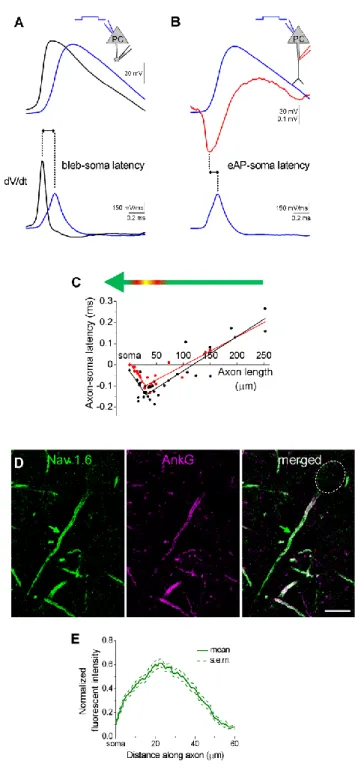
3.2.6. Nav 1.6 and ankyrin G staining of AISs ... 38
2
3.3. Statistical analysis ... 38 3.4. Personal contribution to the results ... 39 4. Results ... 40
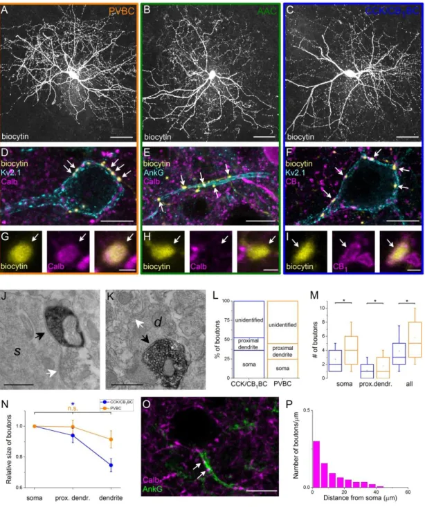
4.1. Part I: Sources and organization of the GABAergic inputs onto the perisomatic region of principal cells in the BLA ... 40
4.1.1. The extent of the perisomatic region along the dendrites of principal cells . 40 4.1.2. Quantification of GABAergic inputs onto the soma and the proximal
dendrites of amygdalar principal cells... 43 4.1.3. Distribution of GABAergic boutons along the AIS ... 44 4.1.4. The vast majority of GABAergic inputs onto the soma and proximal dendrites of amygdalar principal cells originates from two distinct types of GABAergic interneurons ... 46 4.1.5. Interneurons innervating the perisomatic region of amygdalar principal cells are neurochemically distinct ... 47 4.1.6. Target distribution of the two types of basket cells in the BLA ... 48 4.1.7. Boutons of CCK/CB1BCs targeting the perisomatic region are larger than those targeting the distal dendrites ... 51 4.1.8. PVBCs target the proximal part of the AISs ... 52 4.2. Part II: Electrophysiological and morphological properties of the output synapses of AACs ... 53
4.2.1. Axo-axonic cells innervate the axon initial segments of principal cells in the BLA ... 53 4.2.2. Fast and large synaptic inhibition characterizes the connections between axo-axonic cells and PCs ... 53 4.2.3. Axo-axonic cells potently regulate principal cell spiking ... 56 4.2.4. Higher efficacy of inhibition is promoted by multiple synapses ... 59 4.2.5. GABAergic innervation of AISs overlaps with the site of the action potential generation in PCs ... 62 4.2.6. AACs strategically position their synapses at the site of action potential generation... 65 4.3. Part III: Comparison of the physiological and morphological properties of the output synapses of CCK/CB1BCs and PVBCs ... 67
4.3.1. CCK/CB1BCs and PVBCs provide inhibitory input onto PCs with similar properties ... 67 4.3.2. CCK/CB1BCs and PVBCs inhibit PC spiking with similar efficacy ... 70
3
4.3.3. PV and CCK/CB1R+ INs target the same somato-dendritic compartments of
PCs ... 72
4.3.4. Innervation pattern of single CCK/CB1BCs and PVBCs is variable on different postsynaptic cells ... 77
5. Discussion ... 81
5.1. GABAergic inputs onto the axon initial segments of PCs in the BLA ... 82
5.2. Synaptic inputs received by the soma and proximal dendrites of PCs in the BLA ... 86
5.3. Functional implications ... 89
6. Conclusions ... 93
7. Summary ... 94
8. Összefoglalás ... 95
9. References... 96
10. List of publications ... 113
11. Acknowledgements ... 114
4
List of Abbreviations
5-HT 5-hydroxytryptamine AAC Axo-axonic cell
ACSF Artificial cerebrospinal fluid AIS Axon initial segment
BAC Bacterial artificial chromosome
BC Basket cell
BDA Biotinylated dextran amine BLA Basolateral amygdala BMA Basomedial amygdala Calb Calbindin
CB1 Type 1 cannabionoid receptor CCK Cholecystokinin
CeA Central amygdala CeL Centrolateral amygdala CeM Centromedial amygdala
CR Calretinin
CS Conditioning stimulus
Ctx Cortex
DAB Diaminobenzidine
DSI Depolarization-induced suppression of inhibition GABA Gamma-amino butyric acid
GAD Glutamic acid decarboxylase GFP Green fluorescent protein
HC Hippocampus
IC Current clamp
IN Interneuron
IPSC Inhibitory postsynaptic current IPSP Inhibitory postsynaptic potential ISI Interneuron-selective interneuron ITC Intercalated cell
5
LA Lateral amygdala
NGFC Neurogliaform cell NMDA N-methyl-D-aspartate NPY Neuropeptide Y
PB Phosphate buffer
PC Principal cell PFA Paraformaldehyde PP Perforated patch
PTI Perisomatic region-targeting interneuron
PV Parvalbumin
SOM Somatostatin
Str Striatum
TBS Tris buffered saline Thal Thalamus
US Unconditioned stimulus
VC Voltage clamp
VGAT Vesicular GABA transporter
VGLUT1 Type 1 vesicular glutamate transporter VGLUT2 Type 2 vesicular glutamate transporter VGLUT3 Type 3 vesicular glutamate transporter VIP Vasoactive intestinal polypeptide WCR Whole cell recording
6
1. Introduction
Information processing in cortical networks relies on the precise, spatiotemporally coordinated interaction of billions of neurons. The accurate activity of the ensemble is ensured and controlled by various mechanisms at subcellular, cellular, synaptic and network levels. At network levels, the intricate wiring and precisely orchestrated excitatory and inhibitory interactions between the components are crucial for proper cortical functions, and even seemingly slight changes in the connections can lead to severe pathological states like epileptic seizures or schizophrenic symptoms (Gonzalez-Burgos et al., 2010). Unraveling the basic principles of connectivity patterns and the effects of different connections on the activity of the network components is indispensable for understanding cortical information processing.
Cortical ensembles consist of two main neuronal cell types: 80-90% of the neurons are excitatory principal cells (PCs) and 10-20% are inhibitory interneurons (INs). Cells belonging to the two main categories are thought to play different roles in cortical functions: PCs are proposed to be responsible for information processing, storage and recall, whereas INs control and tune the electrical activity of PCs at both subcellular and cellular levels as well as synchronize their rhythmic firing (Buzsaki and Chrobak, 1995, Paulsen and Moser, 1998, Engel et al., 2001). PCs use glutamate as neurotransmitter and give rise to both local collaterals and projecting axons out of the local circuit to form the main output of the given cortical area. PCs have either spherical or pyramidal-like somata from which 3-7 spiny dendrites can emerge , while their axon can emanate from the soma or the proximal dendrites (Faber et al., 2001, Thome et al., 2014). These distinct compartments have different basic functions: the dendrites receive the majority of the incoming inputs, and can locally integrate and propagate the signals towards the soma, where the inputs arriving from various dendritic branches are integrated. The proximal part of the axon, called the axon initial segment (AIS) has the lowest threshold for action potential generation, therefore here the inputs of the cell are translated into all-or-none output signals. In cortical networks, the distinct membrane domains of PCs are controlled by a massive inhibitory input originated from heterogeneous local interneuronal populations. In general, although there are some exceptions, interneurons have aspiny dendrites, use ɣ-amino butiryc acid (GABA) as
7
main neurotransmitter, and their axon collaterals are restricted to the local circuits. The remarkable heterogeneity of cortical interneurons observed in their morphology, electrophysiological properties, postsynaptic target distribution and cell type-specific expression of calcium binding proteins or neuropeptides may all serve to fulfil their different functions in cortical processing.
The majority of our knowledge on cortical networks comes from the investigation of the hippocampus and several neocortical areas like the somatosensory or visual cortices. Based on comparative studies we know that the main features of cortical wiring principles are similar across various cortical areas, often referring to this notion as canonical microcircuits (Douglas and Martin, 1991). Therefore, the characteristics of these well-described wiring structures- taking into account the possibility of some regional differences- can be regarded as common properties of cortical networks. The basolateral amygdala (BLA), which is in the focus of this study, belongs structurally and developmentally to cortical areas and this region is known to play a critical role in fear and extinction memory formation (Aggleton, 1993, Sah et al., 2003). Examination of the building blocks of the networks and the interactions among the elements in this region therefore can contribute to understanding of information processing and learning mechanisms in cortical areas. In the next section I will summarize our recent knowledge on the different cortical interneuronal types in general, then I will describe and compare the properties of the network in the basolateral amygdala.
1.1. Interneuron diversity in cortical networks
As mentioned above, the different subcellular compartments of PCs serve for different physiological roles, which may imply that the inhibitory inputs arriving to the distinct compartments should have different regulatory effects on PC function (Cobb et al., 1995, Miles et al., 1996). Therefore, a functional approach to IN classification can be based on the specificity in the innervation of separate subcellular domains of PCs.
Accordingly, the majority of the INs in distinct cortical regions can be divided into three groups (Freund and Buzsáki, 1996): (I) perisomatic region-targeting interneurons, which form synapses on the soma, proximal dendrites and axon initial segment of PCs,
8
(II) dendrite-targeting interneurons, which preferentially target PC dendrites and (III) interneuron-selective interneurons, which selectively innervate other GABAergic cells.
To provide a complete view of cortical interneurons, I will present the different interneuron types with a special emphasis on perisomatic region-targeting interneurons, which are in the focus of the experimental work of this thesis.
1.1.1. Perisomatic region-targeting interneurons
Perisomatic region-targeting interneurons (PTIs) predominantly innervate the cell body, proximal dendrites and axon initial segment of excitatory neurons, membrane domains of PCs where the inputs are finally integrated and the action potentials are generated. Thus, these interneurons are in the critical position to powerfully regulate the output of individual PCs as well as to synchronize the spiking activity of large neural ensembles (Buhl et al., 1994b, Miles et al., 1996). PTIs can be further subdivided into axo-axonic cells and at least two types of basket cells.
The proximal part of the axon, the axon initial segment (AIS) plays a crucial role in the generation of action potentials, therefore any input, which targets this region is thought to have a profound influence on the firing of the cells. In cortical regions a type of interneuron is specialized to innervate the AIS of PCs, the so-called chandelier or axo-axonic cell (AAC) (Somogyi, 1977). Specific innervation of the AIS in various cortical regions by AACs is a general phenomenon, since these interneurons have been identified in paleo-, archi- and neocortical areas (Inda et al., 2009). In the cortical regions investigated so far, it has been found that AACs show the highest target specificity: they form 2-30 synapses almost exclusively on AISs of PCs and avoid other cellular compartments as well as other cell types (Somogyi, 1977, Soriano et al., 1990, Han et al., 1993). AACs show typical fast spiking characteristics: they fire action potentials at high rates and there is only a moderate accommodation in their firing pattern evoked by a depolarizing current step pulse. In may cortical regions, AACs express the calcium binding protein parvalbumin (PV), a potent calcium buffer, which can also facilitates the temporally precise, fast release of neurotransmitters from their axon terminals (Eggermann and Jonas, 2012). AACs influence their postsynaptic partners via A-type GABA receptors. The intracellular chloride concentration, which
9
primarily sets the reversal potential for GABAergic responses, varies in distinct developmental (Ben-Ari, 2002) and pathological states (Cohen et al., 2002), and it can be controlled locally in different subcellular compartments (Szabadics et al., 2006, Foldy et al., 2010). Previous results show that the effects of AACs on their postsynaptic partners are also variable and can either inhibit or promote firing in PCs depending on several factors, including the membrane potential of the postsynaptic cell and the Cl- reversal potential in the AIS (Buhl et al., 1994b, Szabadics et al., 2006, Glickfeld et al., 2009, Woodruff et al., 2011). Thus, to understand the role of AACs in different network states, it is indispensable to have information both on their firing activity as well as their instantaneous effect on their postsynaptic partners.
The other types of PTIs are basket cells. This cell type targets predominantly the soma and proximal dendrites of PCs, and occasionally also forms few synapses on axon initial segments (Somogyi et al., 1983a, Halasy et al., 1996). Their name refers to the characteristic, basket like axon terminal plexus around PC cell bodies formed by the convergent terminals of this cell type. An individual basket cell forms on average 1-10 boutons on the perisomatic region of single PCs (Gulyás et al., 1993, Miles et al., 1996, Kubota et al., 2015), however they also innervate more distal dendritic segments in varying proportions. In the neocortex, 25-30% of the basket cell terminals contact the perisomatic region, whereas in the hippocampus this number can reach up to 80%, indicating higher target specificity in this archicortical area. In contrast to AACs, basket cells also innervate other interneurons, mainly other basket cells (Galarreta and Hestrin, 2002, Pfeffer et al., 2013). The two main type of basket cells, PV-immunopositive, fast spiking basket cells (PVBCs) and cholecystokinin (CCK) and type 1 cannabinoid receptor (CB1)-expressing, regular spiking basket cells (CCK/CB1BCs) differ in various morphological and electrophysiological properties as well as in protein expression patterns. PVBCs have low input resistance and short membrane time constant, which enables them to respond fast to incoming inputs and faithfully monitor the neural activity in excitatory networks (Galarreta and Hestrin, 2001, Papp et al., 2013).
Similarly to AACs, PVBCs generate narrow action potentials at high rates, show moderate- or non-accommodating firing (Papp et al., 2013, Povysheva et al., 2013). In their presynaptic terminals, the calcium influx upon spike invasion is mediated by P/Q type (Cav2.1) channels. These voltage gated Ca2+ channels and Ca2+ sensors at their
10
synapses are tightly coupled, forming nanodomain release machinery, which facilitates the fast, temporally precise and reliable GABA release from their axon varicosities (Poncer et al., 1997, Bucurenciu et al., 2008). CCK/CB1BCs on the other hand have different intrinsic membrane properties: they show higher input resistance and longer membrane time constant than PVBCs enabling them to integrate incoming multiple inputs more efficiently (Glickfeld and Scanziani, 2006). Their peak firing rate is lower than those of PVBCs, and show marked accommodation in their firing (Szabó et al., 2010). As shown in the hippocampus, various subtypes of these cells can be formed based on the expression of the type 3 vesicular glutamate transporter (VGLUT3) and the vasoactive intestinal polypeptide (VIP), but the presence and functional significance of these subtypes in other cortical regions has not been clarified yet (Freund and Buzsáki, 1996). In contrast to PVBCs, the calcium influx into the presynaptic terminals of CCK/CB1BCs is mediated by N-type calcium channels (Cav2.2), and the calcium sensors are loosely coupled to these voltage gated Ca2+ channels, which provides a less precise timing in transmitter release, but may enable presynaptic forms of synaptic plasticity (Hefft and Jonas, 2005, Ahmed and Siegelbaum, 2009, Lenkey et al., 2015).
There is growing evidence supporting the concept that the two types of basket cells have distinct inhibitory roles during network activities (Freund and Katona, 2007).
Most of this knowledge comes from results obtained in the hippocampus, a brain region, in which the network activities and the underlying synaptic mechanisms are the most investigated among cortical areas. This is due to the fact that its circuit is relatively simple and thoroughly described, the characteristic network activities can be induced in in vitro preparations, and it is accessible for in vivo recordings. It has been shown in the hippocampus that PVBCs can effectively inhibit the firing of their postsynaptic partners and precisely regulate the function of large populations of PCs; i.e. they are well suited to operate as high-precision clockworks during network oscillations. In contrast, due to the different input integration and release properties, CCK/CB1BCs provide a less precise synaptic inhibition (Freund and Katona, 2007). Remarkably, by expressing specific receptors, the activity of this latter cell type can be influenced by the ascending subcortical pathways, like serotoninergic (via 5-HT3 receptors) and cholinergic (α7 nicotinic receptors) afferents, therefore it is proposed to be involved in fine-tuning of local network activities. Thus, PVBCs can effectively synchronize PC activity
11
generating rhythmic patterns, while CCK/CB1BCs appear to be able to modulate network dynamics conveying information from subcortical areas about emotional, motivational and general physiological states. In line with this concept, distinct PTI types may contribute distinctly to different pathological conditions: PVBCs are implicated in epilepsy; an abnormal rhythmic synchrony in cortical networks, whereas CCK/CB1BCs may be rather involved in mood disorders like excessive anxiety (Freund, 2003, Cossart et al., 2005).
1.1.2. Dendrite-targeting interneurons
Dendrites of PCs in the cortex are complex structures specialized to receive the vast majority of inputs arriving to the cell. There is emerging evidence showing that PC dendrites have not a mere passive role as input receivers, but in various brain regions they can non-linearly integrate the incoming inputs by active local electrogenesis. This capacity is due to the expression of voltage-gated channels present in specific dendritic regions, which profoundly influence their integration properties. The all-or-none activation of voltage-gated Ca2+ and Na+ channels, as well as NMDA type of glutamate receptors can evoke local dendritic spikes in excitable segments called "hotspots", which propagate, sometimes in a regenerative manner, along the dendritic tree. This enables specific dendritic branches to summate excitatory inputs supralinearly, e.g. the synchronous activation of neighbouring inputs can evoke higher voltage responses than the mathematical sum of their individual responses, thereby performing space and time specific input integration (Magee, 2000, Magee and Cook, 2000). The supralinear integration of inputs has deep influence on the activity of the cell: dendritic spikes can trigger action potentials and facilitate burst firing at the soma (Golding and Spruston, 1998, Larkum et al., 1999, Ariav et al., 2003, Losonczy and Magee, 2006), and the local dendritic depolarization and Ca2+ influx can be sufficient to induce synaptic plasticity (Golding et al., 2002, Remy and Spruston, 2007). Therefore, any inhibitory input targeting these specific dendritic domains - besides shunting the excitatory inputs - is in a strategic position to control dendritic integration linearity by changing the gain and offset of the input-output function (Mitchell and Silver, 2003, Isaacson and Scanziani, 2011, Mullner et al., 2015). Thereby, dendrite-targeting interneurons may even be able
12
to control the action potential output in an all-or-none manner (Buzsaki et al., 1996, Takahashi and Magee, 2009, Lovett-Barron et al., 2012, Muller et al., 2012). The expression pattern of the voltage gated ion channels along the PC dendritic tree and therefore the signal integration mode of dendrites show high degree of cell type specificity and even single cell types can switch between integration modes according to specific inputs (Lavzin et al., 2012, Jia et al., 2014).
In cortical structures most of the incoming inputs arriving from other cortical or subcortical regions is arranged in a layer-specific manner, therefore dendritic branches present in different layers can receive source specific input(s). Dendrite-targeting inhibitory cells, which arborize in specific layers are in the position to control the input and integration mode of PCs in an input source-selective manner. In the neocortex and hippocampus, a large variety of dendrite-targeting inhibitory cells is present to inhibit the distinct dendritic segments of PCs (Somogyi and Klausberger, 2005). Some of the interneurons have axonal arborization restricted to well-defined layers, which receive a specific input. For example, in the hippocampus, oriens-lacunosum moleculare cells restrict their innervation to the layer where the entorhinal and thalamic inputs contact the apical tuft of PCs (Gulyás et al., 1993, McBain et al., 1994). On the other hand, there are dendrite-targeting interneurons, which are less specific for dendritic subregions, like Martinotti cells in the neocortex, which innervate both the apical trunk, oblique dendrites and apical tuft of PCs (Wang et al., 2004). Overall, dendrite-targeting interneurons in the neocortex and hippocampus show a large heterogeneity across cortical regions regarding their layer (and target) specificity and neurochemical marker expression.
However, it should be noted that a recent theoretical study, which investigated the impact of inhibition on local excitatory inputs proposed a concept that the maximal effect of inhibition can be far from the actual site of the synapses (Gidon and Segev, 2012). Based on experimental data these computational modelling studies showed that due to the passive cable properties of dendrites, the centripetal (from distal to proximal) spread of the shunting impact of inhibition is more effective than the centrifugal (from proximal to distal). This implies that distal inhibitory conductances can more effectively dampen excitatory inputs on proximal dendritic regions than locally at their contact sites. Interestingly, they also proposed that this condition holds only if the inhibition
13
consists of at least three synchronously activated synapses. Importantly, it was also shown, that the distal "off-path" inhibition could inhibit the spread of dendritic calcium spikes from the hotspot to the soma more effectively than the proximal, "on-path"
inhibition. I think this modelling data has an extreme importance, if it turns out to be true in real neurons, because it shows that if an IN type targets a PC dendrite region, which receives a specific excitatory input, this does not necessarily imply that the given IN type is the best suited to control that particular input. Therefore, to elucidate the effect of distinct IN populations on PC input processing we need an integrated view, which takes into account the exact inhibitory contact pattern (e.g. the number and location of synapses), the release properties of those synapses, as well as the firing activity and synchronization level of that particular IN population in line with the network activity and the incoming inputs.
1.1.3. Interneuron-selective interneurons
The main target of the majority of local GABAergic interneurons are PCs, although many INs innervate other INs at about the frequency of their occurrence (Gulyás et al., 1993). However, it was described first in the hippocampus and later in many other cortical circuits that there are GABAergic cell types, which preferentially, if not selectively innervate other INs in the local circuits (Acsády et al., 1996, Gulyas et al., 1996, David et al., 2007). Most of the described interneuron-selective interneurons (ISI) in the cortex express the calcium binding protein calretinin and/or the neuropeptide VIP, which can be therefore used as a potential marker for these neurons (although VIP expression is not restricted to this IN group, some other GABAergic cells, e.g. a subpopulation of CCK/CB1BCs also expresses this protein). ISIs possess rarely branching local axonal tree with irregular bouton distribution. A group of ISIs besides innervating local interneurons, can give rise to long-range projections targeting other cortical or sucbortical areas (Gulyas et al., 2003) (but see (Jinno et al., 2007) for in vivo filled cells). Locally, ISIs tend to target predominantly somatostatin or calbindin immunopositive dendrite-targeting interneurons both in the hippocampus and the cortex (Acsády et al., 1996, Pi et al., 2013). By innervating local interneurons, the effect of ISIs on PCs is disinhibitory: it has been shown in vivo that upon their activation, local
14
INs are inhibited, and the activity of PCs is elevated due to the release of their inhibitory input (Pi et al., 2013). Despite their relatively low occurrence their impact on the local networks has been shown to be impressive: although they represent only 1-2% of the local cells, their simultaneous activity can alter the firing of 20% of the cells in a given microcircuit (Pi et al., 2013). The dendrites of these cells are often restricted to specific layers, which receive source specific excitatory inputs (Freund and Buzsáki, 1996).
Therefore, by the specific innervation of local interneurons, these cells are in the position to ensure the input specific dendritic disinhibiton of large PC populations.
1.2. Neuronal circuits of the rodent basolateral amygdala involved in fear memory formation
1.2.1. Basic structure of the amygdala and its role in fear memory learning
An approach to understand the cellular mechanisms of cortical learning processes is to investigate model networks, which have well-defined roles in specific learning tasks. One of the most investigated learning paradigm is the classical Pavlovian conditioned fear learning, where the subject learns to associate a neutral stimulus (conditioned stimulus-CS) with a noxious stimulus (unconditioned stimulus, US). Fear conditioning is a relatively easily inducible, reliable memory test, which makes it an excellent model for studying the cellular mechanisms of memory formation. The amygdaloid complex is a medial temporal lobe structure, which has a central role in emotional processing (Aggleton, 1993, Scott et al., 1997). This brain region has been proved to be essential for learning the emotional significance of a stimulus in the environment, recognizing emotions on faces, especially fear and anger, and has a dynamic role in interpreting the emotions during social interactions (Phelps and LeDoux, 2005, Adolphs, 2010). Abnormal development and activity of the amygdala is associated with various mental disorders e.g. autism, Williams syndrome, social phobia and epilepsy (Schumann et al., 2011). In general, in these disorders the hyperactivity of the amygdala leads to increased anxiety states and impairment of social interactions (Schumann et al., 2011). It has been proved that this region is indispensable for
15
learning, storage and retrieval of conditioned fear and extinction memories (Armony et al., 1995, Herry et al., 2008, Pape and Pare, 2010). To understand the cellular mechanisms of emotional information processing in this region in health and disease, it is necessary to uncover the organization of the local connectivity and the features of synaptic communication between the neural elements.
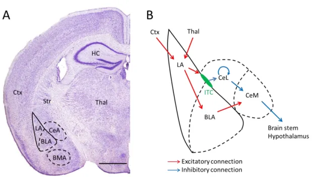
The amygdaloid complex comprises different nuclei, which can be divided into three main groups based on their structure, main input sources and developmental origin (Sah et al., 2003) (Fig. 1A). (I) The basolateral amygdala complex (or deep cortical nuclei) has a cortex-like network structure, shares its developmental origin with cortical structures and it is composed of several nuclei: the lateral (LA), basal(BA)(or also called basolateral; BLA) and the accessory basal amygdala nucleus (AB)(or basomedial amygdala (BMA). The LA is traditionally considered as the input site of the amygdala receiving various specific and polymodal sensory inputs from the cortex and thalamus.
Figure 1. Location of the amygdala and the main routes of information flow
A, Location of the main amygdala nuclei in coronal mouse brain section (Bregma -1.94 mm). Adopted from (Paxinos G, 2001). B, The main information flow routes involved in fear memory learning in a schematic amygdala. Predominantly excitatory connections shown in red, inhibitory connections in blue.
BLA: basolateral amygdala, BMA: basomedial amygdala, CeA: central amygdala, Ctx: cortex, HC:
hippocampus, ITC: intercalated cell mass, LA: lateral amygdala, Str: striatum, Thal: thalamus. Scale bar:
1 mm
16
(II) The superficial cortical like nuclei, as the name suggests, also have cortical structure and origin, and comprise the nucleus of the lateral olfactory tract, the bed nucleus of the accessory olfactory tract, the anterior and posterior cortical nucleus and the periamygdaloid cortex. The main input of this complex arrives from the olfactory system and other amygdalar nuclei. (III) The central amygdala has a striatum-like network structure, derives from striatal progenitors, and includes the centrolateral (CeL), centromedial (CeM) nuclei, the medial nucleus and the amygdaloid part of the bed nucleus of stria terminalis. The CeM is considered as the main output region of the amygdala by innervating various regions responsible for autonomic responses in the hypothalamus, brain stem, and by modulating the ascending monoaminergic and cholinergic systems. In addition to these three main groups of nuclei, there are separate sets of nuclei that don't belong to any of these three groups: the intercalated cell masses (ITCs), which are small groups of GABAergic cells with striatal origin, located in the capsules surrounding the basolateral nuclei; the anterior amygdala area and the amygdalohippocampal area. Besides the classically described main inputs and outputs, almost all amygdala nuclei receive other cortical and subcortical inputs and give rise to efferent projections to various brain regions, which might further enable the amygdala to integrate, associate and process a large variety of information during emotional memory formation.
The basolateral amygdala complex is proposed to play a crucial role in the fear conditioning learning paradigm. The neutral CS and noxious US are associated in the LA via the integration of the converging multisensory and higher order cortical inputs, and fear learning induces the potentiation of these excitatory synapses (Rogan et al., 1997). Thalamic inputs are proposed to deliver rapid, unprocessed multisensory information to the LA, whereas processed information arrives from visual, auditory or somatosensory cortices (Li et al., 1996, LeDoux, 2000, Szinyei et al., 2000). The integrated information from the LA reaches the effector area of the amygdala, the CeM indirectly, through several routes (Fig. 1B). Projection cells of the LA excite PCs in the BLA, which in turn excite CeM cells. Within the BLA the information is further processed by the interaction of the local interneurons and PCs: local INs can provide feedforward inhibition due to their excitation by LA neurons, or they can be recruited by the activity of BLA PCs thereby forming a feedback inhibitory loop. The second
17
route of information flow from the LA to the CeM is through the ITC cells, which also produce disinhibition of CeM projection neurons. Numerous studies confirmed that information processing in these parallel routes is involved in fear memory processes (Anglada-Figueroa and Quirk, 2005, Herry et al., 2008, Likhtik et al., 2008, Haubensak et al., 2010, Busti et al., 2011). In the last two decades, information processing in the BLA has received special attention. Much evidence shows that the sophisticated network structures and the plastic changes in the inhibition within the BLA can control fear learning. Local inhibitory circuits in the amygdala have been shown to play major roles in controlling the acquisition, expression and extinction of fear memories (Herry et al., 2010, Tye et al., 2011, Wolff et al., 2014). Similar to other cortical regions, local interneurons of the BLA show large diversity regarding their electrophysiological properties and postsynaptic target distribution, which might endow them with specific role during information processing. Recent advancements in recording techniques enabled the targeted investigation of different IN populations, which significantly facilitates the exploration of the function of diverse INs in fear memory processes. In the next section, I will summarize our recent knowledge on the physiological and anatomical properties of BLA PCs and INs and their proposed role during network activities.
1.2.2. Principal cells of the BLA
As in other cortical networks, PCs in the BLA form about 80% of the neuronal population. Morphologically, they have either triangular pyramidal-like or rounded somata with 15-20 µm diameter and 3-7 rapidly tapering spiny dendrites (McDonald, 1992, Washburn and Moises, 1992). As the amygdala is not a layered structure, the dendrites are not arranged and oriented strictly in one plane, but emerge from the soma at random directions (Faber et al., 2001). However, some PCs have one or two thicker, apical-like dendrites, but the difference in their inputs or electrophysiological properties compared to other dendrites has not been showed yet. According to the presence or absence of an apical-like dendrite, PCs can have pyramidal-cell like or stellate cell-like appearance. However, it is difficult to group the cells into these two types, since they rather form a morphological continuum. As in other cortical structures (Megias et al.,
18
2001), the glutamatergic synaptic inputs arrive predominantly on their spines or thin dendritic shafts, whereas GABAergic synapses are formed mainly on their somata, AIS and dendritic shafts and spine necks (Carlsen, 1988, McDonald et al., 2002). Their axon emerges from the soma or from a proximal dendrite, and forms local collaterals before leaving the BLA to innervate e.g. the CeA, hippocampus, prefrontal cortex, nucleus accumbens and thalamus. They use glutamate as neurotransmitter and express the type 1 vesicular glutamic acid transporter (VGLUT1), as other cortical projecting cells. These neurons produce broad action potentials, relatively low spontaneous firing rate (0.1-0.3 Hz) and often fire spike bursts (Gaudreau and Pare, 1996, Bienvenu et al., 2012). It has been shown in cats that the membrane potential of PCs intrinsically oscillates at theta frequency in vivo, which shapes the cell's firing probability (Lang and Pare, 1997, Pape and Driesang, 1998). Moreover, as shown in rats in vitro, large periodic IPSPs arrive onto PCs inhibit and/or synchronize their firing. Interestingly, these periodic IPSPs might be induced by the activity of a small subset of local PCs which excite local INs, suggesting that there might be a subpopulation of PCs, which orchestrates the synchronous activity in local inhibitory networks (Popescu and Pare, 2011). This implies the inhomogeneity in amygdalar PC populations, however, clear morphological or electrophysiological differences between the subgroups in this study could not be shown. Another source of inhomogeneity in BLA PCs is the difference in the input and projection of individual cells. It has been shown that there are at least two different PC populations, which might have different roles in amygdala functions: one group, receiving information from the hippocampus is involved in fear memory formation, whereas another, receiving input and projecting to the medial prefrontal cortex is implicated in fear extinction. In summary, although PCs are usually considered as a homogenous cell population, there is emerging evidence that separate sub-networks can be present, which may have different functions in amygdala processes.
1.2.3. Interneurons of the BLA: types, firing during network activity and role in network function
GABAergic interneurons form about 20% of the BLA cell population (McDonald, 1985, McDonald and Augustine, 1993, Sah et al., 2003). The first
19
approaches to classify BLA INs were based on the expression of calcium binding proteins or neuropeptides. Early studies described two main non-overlapping groups:
50% of the INs expresses calbindin (Calb) and ~20% calretinin (CR) (Kemppainen and Pitkanen, 2000, McDonald and Mascagni, 2001). These groups can be further divided by the expression of PV, VIP, CCK and somatostatin (SOM). Immunocytochemical analysis using electron microscopy showed that axon terminals containing different markers contact specific compartments of BLA PCs: e.g. PV+ terminals often form synapses with PC somata, large caliber dendrites and axon initial segments, CCK+
terminals contact soma and large caliber dendrites, whereas SOM+ terminals often synapse on small caliber dendrites (Katona et al., 2001, McDonald and Betette, 2001, Muller et al., 2006, 2007a). These data imply that INs expressing a specific combination of markers might restrict their input to specific compartments on the PCs. As mentioned in the previous sections, INs targeting distinct subcellular compartments of PCs may have different effects on their activity, therefore a functional approach to classify INs can be based on their postsynaptic target distribution.
However, to precisely define the target distribution of different IN groups, it is necessary to label individual INs from each group and analyse their targets, preferably at the electron microscopic level. This can be achieved by various methods. Golgi staining can label the dendritic and axonal processes of few random cells in the preparation, which can be analyzed at the light and electron microscopic levels. This technique, however, doesn't allow targeted labeling of specific cell types. In addition, cells can be labeled after single cell in vivo recordings, which enables the investigation of multiple features of the recorded neuron: basic electrophysiological properties, which can be informative about the type of the cell (e.g., shape of the action potential, resting membrane potential, passive and active membrane properties), it's firing pattern upon stimulation and during induced or spontaneous network activity. Importantly, the labeled cells can be processed for various anatomical investigations: multiple immunolabeling with fluorescent microscopy, reconstruction of the cell's morphology and analysis of its targets at the electron microscopical level. This method, therefore, is perfectly suited to collect information for the classification of interneurons, however, it has several drawbacks. First, these experiments are very time consuming, and in an ideal case, only one cell is labeled in every animal, therefore the data productivity of
20
this method is very slow, and collecting statistically sufficient data to characterize cell groups can take years. Second, the targeted recordings from specific cell groups is still difficult, although there are several methods to identify cells in vivo based on the shape of the action potentials or with optogenetic tagging of specific interneuron types. These disadvantages can be overcome by labeling single cells in in vitro slice preparations.
Various transgenic animal strains exist, in which different IN types are labeled with fluorescent proteins due to their controlled expression by cell type specific protein promoters. By using an in vitro recording setup equipped with a fluorescent microscope, the different IN types can be targeted very specifically and effectively. The labeled cells then can be processed for the same anatomical investigations as the in vivo filled cells.
Naturally, while using this technique, one always has to keep in mind that due to the slicing procedure and the artificial environment, the electrophysiological and anatomical properties of the cells can change, therefore the interpretation of the results should be done with caution and their in vivo confirmation is desired. Based on the data obtained with these methods in the last two decades, local interneurons in the BLA can be functionally categorized into (at least) seven groups.
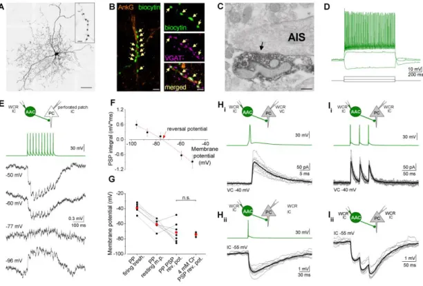
Axo-axonic cells (AACs)
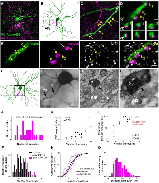
The first study, which suggested the existence of AACs in the BLA described Golgi stained spine-free neurons forming characteristic cartridges presumably surrounding axon initial segments (McDonald, 1982). Later, electron microscopic studies revealed that axon terminals immunostained for the calcium binding protein PV formed symmetrical synapses on the AIS of PCs in the BLA (Muller et al., 2006), suggesting the presence of AACs in this cortical region. A recent in vivo study obtained in the rat (Bienvenu et al., 2012) finally undoubtedly proved that there is a GABAergic interneuron type in the BLA specialized to target exclusively AISs. In this study they showed that AACs are PV+ and Calb-, and can fire action potentials with narrow half width at high frequencies. Despite the recognition of AACs in the BLA, it is still uncertain how effective the regulation of PC firing by single AACs is, and how many presynaptic AACs have to discharge synchronously to veto PC action potential generation. In addition, it is also unknown that how the distribution of AAC output synapses along the AIS relates to the action potential generation site. These basic
21
questions are unanswered not only in the BLA but in other cortical regions as well. The above mentioned in vivo study also demonstrated that AACs in the BLA can be effectively activated by noxious stimulation, which suggests their role in regulating PC spiking during fear-memory related information processing. Therefore, the investigation of how this cell type is embedded in the local networks and its effect on the output of the BLA can help us to understand the mechanisms of fear memory processes in health and disease.
Parvalbumin-positive basket cells (PVBCs)
PV+ terminals in the BLA were described to surround PC somata forming characteristic, basket like structures, which indicated the presence of basket cells in the BLA (McDonald and Betette, 2001). Numerous studies investigated the electrophysiological properties of PV+ cells in the BLA. These studies showed that PV+ cells can generate action potentials with narrow width at high frequencies with little or no accommodation, and can be clustered into groups based on their firing patterns (Rainnie et al., 2006, Woodruff and Sah, 2007b). An in vitro study showed that a population of PV+ cells in the BLA is capable to inhibit PC spiking, and synchronize the firing of their postsynaptic partners (Woodruff and Sah, 2007a). Moreover, a recent study has proved that optogenetic manipulation of PV+ cells can effectively modulate the acquisition of fear memories in an auditory fear learning task (Wolff et al., 2014).
Unfortunately, in these studies the various PV+ IN types (e.g. AACs, BCs and other PV+ GABAergic cells) were not identified anatomically, thus the correlation between the electrophysiological parameters characterizing a subgroup and cell type specificity is not clarified yet. Recently, it has been shown that the firing of anatomically identified PVBCs in vivo were found to be weakly coupled to theta oscillation recorded in dorsal CA1 or some of these cells’ firing was not even coupled to this hippocampal rhythm.
Interestingly, this cell type displayed heterogeneous and generally moderate responses to noxious stimuli (Bienvenu et al., 2012). Thus, their role -instead of pacing theta rhythm in the BLA as speculated previously (Ehrlich et al., 2009)- might be the tonic inhibition of PCs and setting their baseline activity level, i.e. the gain control. The synaptic output of PVBCs might contribute to the source of large amplitude IPSPs recorded in PCs during both spontaneous and evoked synaptic activity, which inhibitory
22
input can set the characteristic low firing rate of PCs in the BLA (Lang and Pare, 1997).
Similarly to AACs, there are many questions still unanswered about the effectiveness of the inhibition provided by individual PVBCs, and that how individual cells can influence the timing of PC firing. Moreover, the basic anatomical features of individual PVBC-PC connections are also unknown.
Cholecystokinin and type 1 cannabinoid receptor-positive basket cells (CCK/CB1BCs) Early studies described that CCK-positive GABAergic neurons in the BLA can be divided into two groups according to the size of the cell body (McDonald and Pearson, 1989). Those cells, which have small soma size, lack the CB1 receptor immunolabeling from their cell body and are proposed to innervate distal dendrites of PCs, whereas cells with large soma express CB1 receptor in their terminals and often form basket-like multiple contacts around PC somata (Katona et al., 2001, Mascagni and McDonald, 2003), therefore called CCK/CB1 basket cells. CCK+ cells in the rat colocalize with Calb, but not with VIP and calretinin (Mascagni and McDonald, 2003).
In vitro this cell type discharges relatively broad action potentials, can have marked AHP and regular spiking or adapting firing pattern. A recent study showed that in rats large CCK cells can be further divided into two subgroups based on their electrophysiological properties, although the functional difference between these groups has not been shown (Jasnow et al., 2009). Unfortunately, there is no in vivo data about the firing activities of amygdalar CCK/CB1BCs during hippocampal theta oscillation or noxious stimuli, probably because the shape of their action potential is similar to PCs, thus they usually are not separated from PCs during in vivo recordings. However, the role of CCK/CB1BCs in the normal function of the amygdala in fear memory processes can be crucial, as their synapses can undergo CB1 receptor dependent depolarization- induced suppression of inhibition (DSI) (Zhu and Lovinger, 2005) and endocannabinoid-mediated long-term depression (Marsicano et al., 2002), which has been shown to be necessary for the extinction of fear memories (Chhatwal et al., 2009).
In addition, a population of CCK/CB1BCs expresses 5HT-3 type serotonin receptor (Muller et al., 2007b) suggesting that 5-HT may exert its anxiolytic effects by modulation of the transmitter release from this cell type. To assess their role in the
23
amygdalar network functions, it is necessary to unravel the basic properties of their synapses, their ability to control local principal cells and the underlying anatomical structures of their connections.
Dendrite-targeting INs
Previous studies described that SOM-positive terminals often form symmetrical synapses on thin caliber dendrites and spines. Besides these compartments, terminals containing SOM also target perikarya and large caliber dendrites, but in a much less extent (37% on thin dendrites, 51% on spines, 4% on perikarya and 6% on thick dendrites) (Muller et al., 2007a). Therefore, SOM+ cells, which represent 11-18% of the GABAergic neurons of the BLA, are proposed to be dendrite-targeting interneurons (McDonald and Mascagni, 2002). The majority (90%) of SOM+ cells are Calb-positive, but does not express PV or calretinin. A recent publication using optogenetic manipulation showed a crucial role of SOM+ dendrite targeting interneurons in fear learning (Wolff et al., 2014).
A detailed study using electron microscopy on the inhibitory input of BLA PCs showed that about half of the symmetrical synapses arriving onto the dendritic shafts, and occasionally onto spines is PV-immunopositive (Muller et al., 2006), which suggested the presence of another, PV+ dendrite-targeting IN type in the BLA. There has been only 3 cells described in the literature, labelled in vivo, which has proved to be selectively targeting dendrites (Bienvenu et al., 2012). Like PVBCs, these cells were Calb+, however their behavior was remarkably different from the perisomatic region- targeting interneurons: their firing was deeply modulated by hippocampal theta oscillations, which might enable this cell type to modulate the integration of incoming dendritic inputs and synaptic plasticity processes in accordance with the ongoing oscillation. These cells, however, did not contain SOM, which clearly separates them from the above mentioned dendrite-targeting cell group.
Unfortunately there is no data yet about the role of Calb+ dendrite-targeting cells in fear learning, therefore there is no information whether the two major dendrite- targeting cells play different, maybe complementary role in BLA network activities.
24 AStria cells
A special PV+ cell type was recently described in the BLA (Bienvenu et al., 2012), which -besides targeting the soma and dendrites of local PCs- project out from the BLA to the amygdalostriatal transition area, where they innervate the soma and dendrites of medium spiny neurons. These cells have dense axonal arbor and profoundly branching, very tortuous dendrites. Interestingly, and in contrast to other BLA INs, these cells are robustly inhibited by noxious stimuli. These properties clearly separate them from other PV+INs, but their role in BLA network function is presently unknown.
Neurogliaform cells
Most of the INs mentioned so far are known to provide fast, phasic GABAergic inputs, however, there is a special interneuron type, called the neurogliaform cell (NGFC), which provides 5-10 times slower inhibition on the PCs than the above mentioned INs (Manko et al., 2012). NGFCs in the BLA are neuropeptide Y (NPY) and SOM positive, and likely express nitric oxide synthase (McDonald et al., 1993). NGFCs target somata and dendrites mainly with non-synaptic juntions (77%), and only the minority of their terminals form synapses (23%) (Manko et al., 2012). Like PV- and Calb-positive dendrite-targeting INs, the firing of NGFCs is strongly coupled to the hippocampal theta oscillation. This two cell types together can provide a rhythmic inhibition on the dendrites with complementary temporal profiles, which might be necessary for the coordination of hippocampal-amygdala theta oscillations emerging during fear memory retrieval (Seidenbecher et al., 2003).
Interneuron selective interneurons
As already mentioned, VIP expressing interneurons in the hippocampus or in the neocortex selectively inhibit other IN types, thereby providing a disinhibitory effect on local PCs. The presence of VIP+ cells which target local INs has been described in the BLA circuits, however, their interneuron target selectivity has not been proven yet (Muller et al., 2003). However, there is evidence showing that a strong interneuron specific inhibitory source exists in the BLA, which provide phasic inhibition on local PV+ and SOM+ cells during auditory fear conditioning. The suppression of interneuron firing leads to a phasic disinhibition of the entire somatodendritic domain of PCs in the
25
BLA (Wolff et al., 2014). Possible candidates for this disinhibitory source are local VIP cells, in analogy to the auditory cortex (Pi et al., 2013) however this theory in the BLA is not proven yet.
1.2.4. Wiring properties of the neural networks in the BLA, emerging questions
To understand the mechanisms of different network activities in health and disease, a basic step is to describe the elements of the neural ensembles, the logic of their connections and their effects on each other. To this end, besides describing the properties of connected neuronal pairs, it also necessary to estimate the convergence and divergence of the connections established by different IN types to be able to predict their impact at the network level. In the BLA networks, many basic fundamental questions regarding the build-up of the network are still unanswered. How many PCs are controlled by single INs? How many INs converge on single PCs? What is the ratio of the inhibitory inputs on single PCs originating from the different IN types? How effectively can INs control PC firing? During my PhD studies, I described the basic electrophysiological and anatomical properties of the perisomatic inhibitory inputs of BLA PCs, the functional effects of the inhibition on their firing, and the organization of the different perisomatic inputs in the BLA networks.
26
2. Objectives
The main goal of our studies was to investigate the organization and impact of the perisomatic inputs on the PC spiking in the BLA. Therefore, we focused on three main topics with the following specific questions:
I. To uncover the sources of the perisomatic inputs and the ratio of the innervation emerging from different cell types.
What is the extent of the perisomatic region along the dendrites of BLA PCs?
What is the density of inhibitory inputs arriving to this region?
Which IN types give rise to the perisomatic GABAergic inputs and what is the ratio of their innervation?
How many PCs are controlled by single INs? How many INs converge on individual PCs?
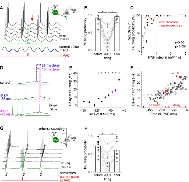
II. To determine the electrophysiological and morphological properties of the AAC inputs on the AIS and its effects on PC firing activity.
What are the effects on the membrane potential of AAC inputs on BLA PCs?
How efficiently can this input shape the spiking activity of PCs?
What is the spatial distribution of the GABAergic inputs along the AIS and how it relates to the site of the action potential generation?
III. To compare the electrophysiological and morphological properties of the inputs of PCs originating from basket cells and their effects on PC firing activity.
What electrophysiological properties characterize the connections established by CCK/CB1BCs and PVBCs?
Is there any difference in the impact of their inputs on PC spike generation?
What is the distribution of the basket cell synapses along the somato-dendritic axis of PCs?
27
3. Methods
3.1. Electrophysiological experiments
3.1.1. Experimental animals and slice preparation
All experiments were approved by the Committee for the Scientific Ethics of Animal Research (22.1/360/3/2011) and were carried out according to the guidelines of the institutional ethical code and the Hungarian Act of Animal Care and Experimentation (1998. XXVIII. section 243/1998, renewed in 40/2013.). For recording AACs and PVBCs, transgenic mice of both sex (P18-24) expressing enhanced green fluorescent protein (eGFP) under the control of the PV promoter (Meyer et al., 2002) were used. For targeted patching of CCK/CB1BCs, transgenic mice expressing red fluorescent protein under the control of cholecystokinin (CCK) promoter were used (BAC-CCK-DsRed) (Mate et al., 2013). Mice were deeply anaesthetised with isoflurane and decapitated. The brain was quickly removed and placed into ice-cold cutting solution containing (in mM): 252 sucrose, 2.5 KCl, 26 NaHCO3, 0.5 CaCl2, 5 MgCl2, 1.25 NaH2PO4, 10 glucose, bubbled with 95% O2/5 % CO2 (carbogen gas). Horizontal slices of 200 µm thickness containing the basolateral amygdala were prepared with a Leica VT1000S or VT1200S Vibratome (Wetzlar, Germany), and kept in an interface- type holding chamber containing artificial cerebrospinal fluid (ACSF) at 36 ºC that gradually cooled down to room temperature. ACSF contained (in mM) 126 NaCl, 2.5 KCl, 1.25 NaH2PO4, 2 MgCl2, 2 CaCl2, 26 NaHCO3, and 10 glucose, bubbled with carbogen gas.
3.1.2. Whole-cell recordings
After at least one hour of incubation, slices were transferred individually into a submerged type of recording chamber perfused with ACSF at 32±2 ºC with a flow rate of 2-3 ml/min. Recordings were performed under visual guidance using differential interference contrast microscopy (Olympus BX61W). EGFP or DsRed in cells was excited by a UV lamp, and the fluorescence was visualized by a CCD camera (Hamamatsu Photonics, Japan). Patch pipettes were pulled from borosilicate glass capillaries with inner filament (Hilgenberg, Germany) using a DMZ-Universal Puller
28
(Zeitz-Instrumente GmbH, Germany). For somatic whole-cell recordings, pipettes with 0.188 mm wall thickness were used and had a resistance of around 3-5 MΩ when filled with the intrapipette solution. K-gluconate-based intrapipette solution used in all recordings contained (in mM): 110 K-gluconate, 4 NaCl, 2 Mg-ATP, 20 HEPES, 0,1 EGTA, 0.3 GTP (sodium salt) and 10 phosphocreatine adjusted to pH 7.3 using KOH and with an osmolarity of 290 mOsm⁄L. For recording the presynaptic interneurons 10 mM GABA and 0.2% biocytin were added, whereas for the postsynaptic PC 100 μM Alexa Fluor 488 hydrazide sodium salt (Invitrogen) was included. Recordings were made with a Multiclamp 700B amplifier (Molecular Devices, Foster City, CA, USA), low-pass filtered at 2 kHz, digitized at 10 kHz and recorded with in-house data acquisition and stimulus software (Stimulog, courtesy of Prof. Zoltán Nusser, Institute of Experimental Medicine, Hungarian Academy of Sciences, Budapest, Hungary).
Recordings were analyzed with EVAN 1.3 (courtesy of Prof. Istvan Mody, Department of Neurology and Physiology, UCLA, CA), the in-house analysis software SPIN 1.0.1 (courtesy of Prof. Zoltán Nusser) and Origin 8.6 and 9.2 (Northampton, MA).
Recordings were not corrected for junction potential. To record the firing characteristics, cells were injected with 800-ms-long hyperpolarizing and depolarizing square current pulses with increasing amplitudes from 10 to 600 pA. PC identity was characterized by the broad action potential waveform, accommodating firing pattern and slow afterhyperpolarzing current as well as the post hoc morphological analysis of their spiny dendrites. For recording postsynaptic inhibitory currents (IPSCs), the presynaptic IN was held around a membrane potential of -65 mV in current clamp mode, and stimulated by brief square current pulses (2 ms, 1.5–2 nA) to evoke action potentials, and the PC was clamped at a holding potential of -40 mV. Series resistance was monitored (range: 6-20 MΩ) and compensated by 65%. To record postsynaptic inhibitory potentials (IPSPs), the presynaptic cell was stimulated in the same way, and the postsynaptic PC was held in current clamp mode around -55 mV. Bridge balance was adjusted throughout the recordings. The kinetic properties of IPSCs and IPSPs were analysed on averaged events that were calculated with excluding the transmission failures. The latency of synaptic transmission was calculated by subtracting the time of the action potential peaks from the onset of the postsynaptic currents.
29
To test the ability of INs to inhibit PC firing, theta frequency (3.53 Hz) sinusoidal current pulses with peak-to-peak amplitudes of 30 pA and 50 pA were injected into the postsynaptic PC. The membrane potential of PCs was set (approximately around -55 mV) such to evoke a spike at the peak of the sinusoidal current pulses with the amplitude of 50 pA, but not of 30 pA. This adjustment maintained the membrane potential of PCs near the spiking threshold. One trial consisted of 7 sinusoidal waves (5x50pA and 2x30pA), repeated 10-20 times in each pair. Three action potentials at 30 Hz were evoked in the interneuron by brief square current pulses (2 ms, 1.5–2 nA) before the 4th sinusoidal wave (50 pA) in each trial. To calculate the reduction in firing probability, the firing probability of PCs under control conditions was calculated from the average of the responses to 50 pA currents (1st, 3rd, 5th and 6th sinusoidal wave), which was compared to that obtained during the 4th cycle.
To evoke firing in PCs by synaptic input, electrical stimulation of external capsule fibers was delivered via a theta glass electrode filled with ACSF using a Supertech timer and isolator (Supertech Ltd., Pécs, Hungary).
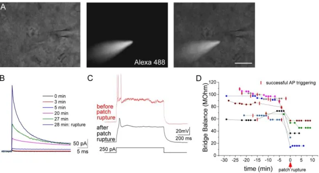
3.1.3. Perforated patch recordings
For perforated patch recordings 100 mg/ml gramicidin (Sigma) stock solution was prepared in DMSO daily and kept at 4 ºC. Before the recordings gramicidin stock solution was diluted to the concentration of 100 µg/ml in the same K-gluconate based intrapipette solution as used for whole-cell recordings, containing (in mM): 110 K- gluconate, 4 NaCl, 2 Mg-ATP, 20 HEPES, 0,1 EGTA, 0.3 GTP (sodium salt) and 10 phosphocreatine adjusted to pH 7.3 using KOH and with an osmolarity of 290 mOsm⁄L.
In addition, the solution contained 100 μM Alexa 488 hydrazide sodium salt and 1 mM QX-314 (Sigma). The solution was sonicated for a short period of time in an ultrasound bath several times before use. Pipettes with 0.188 mm wall thickness were used and had a resistance of around 3-5 MΩ when filled with the intrapipette solution. The tip of the pipette was filled with gramicidin free solution and then backfilled with the gramicidin containing intrapipette solution. The series resistance was monitored throughout the experiment, and recordings were started when the resistance fell below 100 MΩ. Pipette capacitance was neutralized and bridge balance was carefully adjusted throughout the recordings. Patch rupture was detected by i) the inability to evoke action potentials with
30
depolarizing current steps (the consequence of the Na+/K+ channel blocker QX-314 diffusion into the cell), ii) with the penetration of Alexa 488 dye into the cell, or iii) sudden drop in the access resistance, and in such cases the experiment was terminated (Fig. 2).
Figure 2: The three approaches to test the integrity of the perforated patch configuration A, Presence of the Alexa 488 in the recording pipette but not inside the cell was monitored throughout the recordings. B, The current changes in response to a depolarizing 5mV pulse (seal test) during a representative recording.
Note the sudden increase in the current after the rupture of the perforated patch induced by negative pressure application. C, The cell capability to produce action potential in response to an intracellular current pulse injection was also tested throughout the recordings. Note the inability to evoke firing after the rupture of the patch, in a consequence of the QX-314 diffusion into the cell, a drug, which blocks voltage gated sodium and potassium channels applied in the intrapipette solution for the perforated patch recordings. D, Changes in the bridge balance values during the recordings, indicating the series resistance in current clamp mode. Each color represents a recorded pair (n=7), each dot represents a current clamp recording preceded by an Alexa 488 test (see A). Red vertical lines represent a successful test for the firing ability of the recorded cell (see C). In two cases (magenta and grey) the cell didn’t survive the rupture of the patch by negative pressure application. Scale (A): 20 µm
To estimate the reversal potential of the evoked postsynaptic responses (Fig.7 E- G and Fig. 12 E-G), we plotted the area (integral) of IPSPs as a function of membrane potential and obtained the value, where the second order polynomial fit crossed the x