Excitatory synaptic inputs onto parvalbumin-
positive perisomatic region-targeting interneurons in the hippocampus
PhD Thesis
Orsolya Papp
János Szenthágotai Doctoral School of Neurosciences Semmelweis University
Supervisor: Norbert Hájos, D.Sc.
Official Reviewers: Tibor Zelles, Ph.D.
Gábor Molnár, Ph.D.
Chair of the Final Examination Board: Béla Halász, D.Sc.
Members of the Final Examination Board: Tibor Wenger, D.Sc.
István Tarnawa, Ph.D.
Budapest
2013
2
TABLE OF CONTENTS
TABLE OF CONTENTS ... 2
LIST OF ABBREVIATIONS ... 6
INTRODUCTION ... 8
I. The hippocampus ... 9
I.1. Cytoarchitecture ... 9
I.1.1. Principal cells ... 10
I.1.2. Intrahippocampal connectivity ... 11
I.1.3. Afferent-efferent connectivity of the hippocampal formation ... 12
I.1.4. GABAergic cells ... 13
I.1.4.1. Perisomatic region-targeting interneurons ... 14
I.1.4.2. Dendritic layer-targeting interneurons ... 15
I.1.4.3. Interneuron-selective interneurons ... 16
I.1.4.4. Long-range projecting GABAergic neurons ... 17
II. Long-term synaptic changes in hippocampal neurons ... 17
II.1. Induction mechanisms ... 18
II.2. Long-term plasticity of excitatory synapses onto principal cells ... 19
II.3. Long-term plasticity of excitatory synapses onto interneurons ... 20
II.4. Endocannabinoid-dependent long-term plasticity ... 20
II.5. Long-term plasticity at excitatory synapses onto PV+ interneurons ... 21
III. Oscillations in the hippocampus ... 22
III.1. Theta oscillations ... 24
III.1.1. Theta rhythm-related place coding ... 25
III.2. Gamma oscillations ... 25
III.2.1. Cooperative action of theta and gamma oscillations in the formation of working memory ... 27
III.3. Sharp wave-ripples ... 27
III.3.1. Sharp wave-ripples in memory consolidation ... 27
III.4. In vitro oscillation models ... 28
III.4.1. Carbachol-induced in vitro gamma oscillations ... 28
3
III.5. Parvalbumin-positive perisomatic region-targeting interneurons in oscillations ... 29
III.5.1. Unique properties of PV+ cells that make them suitable to govern oscillations .. 29
III.5.2. Emerging evidences for the role of PV+ cells in oscillations ... 31
III.5.3. Distinct entrainment of AACs and FS BCs during oscillations ... 32
IV. Parvalbumin-positive interneurons in schizophrenia ... 33
AIMS OF THE THESIS ... 35
MATERIALS AND METHODS ... 37
I. Animals ... 37
II: Electrophysiological measurements ... 37
II.1. Slice preparation ... 37
II.2. Electrophysiological recordings ... 38
II.3. Investigation of single-cell and synaptic properties ... 38
II.4. Comparison of axo-axonic cells and fast-spiking basket cells ... 39
II.5. Investigation of LTD ... 41
II.6. Studying gamma oscillation ... 42
III. Anatomy ... 43
III.1. Identification of the cell types ... 43
III.2. Reconstruction of cells with drawing tube ... 44
III.3. Estimating the density of VGluT1-expressing synapses onto biocytin-labeled dendrites ... 44
III.4. Neurolucida analysis ... 45
RESULTS ... 46
Differentiation between parvalbumin-positive axo-axonic and fast-spiking basket cells using Ankyrin-G immunostaining ... 46
I. Quantitative differences in the convergence of local pyramidal cells onto parvalbumin- positive axo-axonic- and basket cells in the hippocampal CA3 subfield ... 48
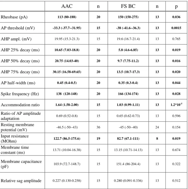
I.1. Membrane properties of AACs and FS BCs in the hippocampal CA3 region ... 48
I.2. FS BCs receive a higher number of proximal excitatory synaptic inputs than AACs ... 51
I.3. The density of excitatory synapses on the proximal dendrites of AACs and FS BCs is similar ... 54
I.4. FS BCs have significantly longer dendrites with a more extensive proximal arborization compared to AACs ... 56
4
II.: Investigating long-term depression of excitatory synaptic inputs onto parvalbumin- positive interneurons ... 62
II.1. Spike timing-dependent LTD at excitatory synapses has higher induction threshold in FS INs compared to pyramidal cells ... 62 II.2. Properties of spike timing-dependent LTD at excitatory synapses onto pyramidal cells and FS INs ... 65 II.3. Spike timing-dependent LTD at excitatory synapses onto both pyramidal cells and FS INs is mediated by endocannabinoid signaling ... 66 II.4. Chemical LTD at excitatory synapses also requires endocannabinoid signaling ... 67 III. The role of excitatory synaptic inputs onto fast-spiking basket cells in cannabinoid- mediated suppression of gamma oscillations ... 71
III.1. Effect of CB1R activation on cholinergically-induced oscillations in the hippocampus ... 71 III.2. CB1R activation suppresses the firing rate of CA3 pyramidal cells and fast-spiking basket cells during gamma oscillations ... 74 III.3. CB1R activation suppresses monosynaptically evoked EPSCs in CA3 pyramidal cells and fast-spiking basket cells in the presence of carbachol ... 76 III.4. CB1R activation has no effect on monosynaptically-evoked IPSCs recorded in CA3 pyramidal cells in the presence of carbachol... 79
DISCUSSION ... 82 I. Quantitative differences underlie the different properties of excitatory synaptic inputs received by axo-axonic and fast-spiking basket cells ... 83
I.1. Single-cell properties of PV+ interneurons targeting the perisomatic region in cortical areas ... 83 I.2. Distinct innervation of AACs and FS BCs by glutamatergic afferents ... 84 I.3. Firing behavior of AACs and FS BCs during different network states ... 85 II. Endocannabinoid-mediated long-term depression of excitatory synapses onto fast- spiking interneurons require higher induction threshold compared to pyramidal cells . 86
II.1. Endocannabinoid-mediated LTD in hippocampal pyramidal neurons ... 86 II.2. Endocannabinoid-mediated LTD in fast-spiking interneurons ... 88 III. Cannabinoid effects on gamma oscillations is primarily mediated by the reduction of excitatory synaptic inputs onto fast-spiking basket cells ... 89
5
III.1. Influencing gamma oscillations with different drugs have dissimilar effects reflecting
distinct mechanisms for impairing rhythmicity ... 89
III.2. Role of excitatory synaptic transmission in the reduction of gamma oscillation power ... 90
III.3. Selective reduction of intrahippocampal excitatory synaptic connections may contribute to cannabinoid-mediated weakening of gamma oscillation ... 91
IV. Functional implications ... 92
CONCLUSIONS ... 94
SUMMARY ... 96
ÖSSZEFOGLALÁS ... 97
REFERENCES ... 98
LIST OF PUBLICATIONS ... 124
Publications related to the dissertation ... 124
Other publications ... 124
ACKNOWLEDGEMENTS ... 125
6
LIST OF ABBREVIATIONS
2-AG: 2-arachidonoyl glycerol AAC: axo-axonic cell
aCSF: artificial cerebrospinal fluid AHP: afterhyperpolarization AIS: axon initial segment
AM251: N-(Piperidin-1-yl)-5-(4-iodophenyl)-1-(2,4-dichlorophenyl)-4-methyl-1H- pyrazole-3-carboxamide, CB1R antagonist
AMPA: α-amino-3-hydroxy-5-methylisoxazole-4-propionic acid AP: action potential
BAPTA: 1,2-bis-(o-Aminophenoxy)-ethane-N,N,N’,N’-tetraacetic acid tetrapotassium salt, Ca2+ chelator
CA: cornu ammonis
CB1R: cannabinoid receptor type I
CB1R KO: cannabinoid receptor type I knockout mouse CCK: cholecystokinin
CP55,940: (-)-cis-3-[2-Hydroxy-4-(1,1-dimethylheptyl)phenyl]-trans-4-(3- hydroxypropyl)cyclohexanol, CB1R agonist
DAG: diacylglycerol DG: dentate gyrus
DGL-α: diacylglycerol lipase-α
DHPG: (S)-3,5-Dihydroxyphenylglycine
DL-AP5: DL-2-Amino-5-phosphonopentanoic acid, NMDA receptor antagonist DMSO: Dimethyl sulfoxide, solvent
eCB: endocannabinoid
eCB-LTD: endocannabinoid-mediated long-term synaptic depression eEPSP: evoked excitatory postsynaptic potential
eGFP: enhanced green fluorescent protein eIPSC: evoked inhibitory postsynaptic current EPSC: excitatory postsynaptic current
EPSP: excitatory postsynaptic potential FS BC: fast-spiking basket cell
7 FS IN: fast-spiking interneuron
GABA: γ-aminobutyric acid
GAD67: glutamate decarboxylase-67 HFS: high frequency stimulation
i-LTD: long-term depression of inhibitory synapses IPSC: inhibitory postsynaptic current
KA: kainic acid
LTD: long-term depression LTP: long-term potentiation
LY367385: (S)-(+)-α-Amino-4-carboxy-2-methylbenzeneacetic acid, metabotropic glutamate receptor 1 antagonist
mAChR: muscarinic acetylcholine receptor mGluR: metabotropic glutamate receptor
MPEP: Methyl-6-(phenylethynyl)-pyridine hydrochloride, metabotropic glutamate receptor 5 antagonist
mRNA: messenger ribonucleic acid
MS-DBB: medial septum-diagonal band of Broca
NBQX : 2,3-dihydroxy-6-nitro-7-sulfamoyl-benzo[f]quinoxaline-2,3-dione, AMPA/KA receptor antagonist
NGS: normal goat serum NMDA: N-methyl-D-aspartate
O-LM cells: oriens lacunosum-moleculare cells PB: Phosphate buffer
PV: parvalbumin
PV+ cells: parvalbumin-positive perisomatic region-targeting interneurons sEPSC: spontaneous excitatory postsynaptic current
TBS: Tris-buffered saline
THL: N-Formyl-L-leucine (1S)-1-[[(2S,3S)-3-hexyl-4-oxo-2-oxetanyl]methyl]dodecyl ester, lipase blocker
tLTD: spike timing-dependent long-term depression VGluT1: vesicular glutamate transporter 1
WIN55,212-2: (R)-(+)-[2,3-Dihydro-5-methyl-3-(4-morpholinylmethyl)pyrrolo[1,2,3-de]1,4- benzoxazin-6-yl]-1-naphthalenylmethanone mesylate, CB1R agonist
WT: wild type
8
INTRODUCTION
The hippocampus is responsible for the computation of higher order cognitive functions such as memory formation and recall (Lisman & Idiart, 1995), memory consolidation (Buzsáki, 1986), or spatial navigation (O'Keefe & Recce, 1993). In this brain area, several rhythmic activities have been observed which were proved to have a tight link to cognitive processes (Vanderwolf, 1969; Leung, 1980; Buzsáki et al., 1992; Chrobak &
Buzsáki, 1994). During theta (4-8 Hz) and gamma (30-100 Hz) oscillations, acquisition and recall of memories can be achieved in hippocampal networks, whereas sharp-wave activity and embedded ripples (120-200 Hz) are suggested to underlie memory consolidation (Buzsáki, 1986; Skaggs & McNaughton, 1996). All of these network activities require an effective and temporarily precise inhibition to control the discharge of pyramidal cells (Buzsáki et al., 1983; Cobb et al., 1995; Ylinen et al., 1995; Csicsvári et al., 2003). The parvalbumin-positive perisomatic region-targeting interneurons (PV+ cells) are ideally suited to fulfill this function, due to their special features. Besides their strategically positioned synapses that provide effective inhibition, several single-cell features and input properties endow them to be a clockwork for network synchrony (Freund, 2003; Jonas et al., 2004;
Bartos et al., 2007).
In terms of my PhD program, we studied the detailed anatomical and functional properties of the excitatory synaptic inputs engaged in driving PV+ cells in the hippocampus.
We compared the excitatory synaptic inputs of the two types of PV+ cells, the axo-axonic and fast-spiking basket cells, with the aim to extend our knowledge about the distinguished function of these two cell types in orchestrating network activities.
In addition, we examined the possibility whether the afferent excitatory synaptic input of these cells is capable of performing long-term plasticity, and if so, what circumstances are required to change the reliable operation of these network-synchronizing GABAergic neurons. We particularly focused on long-term depression (LTD) and also mapped the underlying molecular pathway, uncovering a so far unknown metabotropic glutamate receptor- DAG lipase-α -CB1 cannabinoid receptor-dependent LTD at excitatory synapses onto PV+ cells.
Being aware of the fact that the excitatory synaptic inputs onto PV+ cells could be readily modulated by cannabinoid compounds, we then investigated the mechanisms of the suppression of gamma oscillations upon CB1 receptor (CB1R) activation.
9
This thesis aims to summarize these findings and suggest a functional relevance for the excitatory synaptic inputs of PV+ cells in neurological diseases.
I. The hippocampus
The hippocampal formation is an archicortical structure of the brain. Its basic buildup is well conserved in mammals, although some subtle species specific differences can occur.
As the experimental work described in the thesis was performed in mice, I would confine to introduce the organization of the rodent hippocampus.
In contrast to the six-layered structure of the neocortex, the cell bodies of hippocampal principal cells are rendered to one, densely packed layer. Due to the advantages of this seemingly simplified structure, the hippocampus is in the focus of neurobiological research.
The cytoarchitectural similarity to other cortical regions may allow us to extrapolate to general rules of cortical computation.
The hippocampal formation comprises the dentate gyrus (DG), the hippocampus proper (or Cornu Ammonis, CA) and the subiculum. The neighboring areas that produce the direct input or output of the hippocampal formation, i.e. the entorhinal cortex, pre- and parasubiculum, together with the hippocampal formation, form a computational unit termed the hippocampal region (Andersen, 2007).
I.1. Cytoarchitecture
The principal cells of the hippocampal formation are glutamatergic cells, which comprise approximately 85% of the neurons (Andersen, 2007). The cell bodies of principal cells are confined to a single layer named as granule cell layer (stratum granulosum) in the DG or pyramidal cell layer (stratum pyramidale) in the CA regions. They form two enwreathing U-shape composing the special structure of this brain area (Figure 1). The remaining 15% of neurons in the hippocampus are GABAergic in nature, forming a highly heterogeneous cell population (Freund & Buzsáki, 1996).
10 I.1.1. Principal cells
In the DG, the main cell type is the granule cell possessing small somata (approximately 10 µm). These cells lack basal dendrites towards the hilus, the polymorphic cell layer enclosed by the blades of stratum granulosum. In the hilus, another type of glutamatergic cell is found, the mossy cell. The area occupied by the apical dendrites of the granule cells is named as molecular layer (stratum moleculare). The Cornu Ammonis has three parts: the CA3 is the closest to the DG and composed of relatively large pyramidal cells (with approximately 20-30 µm of soma diameter). The CA3 region is further subdivided to three parts: a, b and c. The CA3c is next to the DG, while the CA3a is adjacent to the CA2.
The stratum lucidum, formed by the axons of granule cells, can be found next to the pyramidal cell layer in the CA3 region. The CA2 region is a small group of cells inserted between CA3 and CA1. The cells in the CA1 region are smaller (with approximately 10-15 µm of soma diameter) and the most densely packed into a layer. The continuation of the Cornu Ammonis is the subiculum where the cell bodies become widely dispersed. The apical dendrites of pyramidal cells are present in the stratum radiatum; this layer is located superficial to the stratum lucidum in the CA3 region, and to the stratum pyramidale in the CA2 and CA1. The stratum lacunosum-moleculare is the most superficial layer of the hippocampus, and it is defined by the apical dendritic tufts of pyramidal cells and the inputs mainly from the entorhinal cortex. The layer located below the pyramidal cell layer is the stratum oriens, which contains the basal dendrites of pyramidal cells (Figure 1) (Andersen, 2007).
11
Figure 1. Cytoarchitecture of the hippocampal formation. GD, dentate gyrus; CA3, CA1, fields of the Cornu Ammonis; SUB, subiculum. Cornu Ammonis: (1) alveus, (2) stratum pyramidale, (3) axon of pyramidal neurons, (4) Schaffer collateral, (5) stratum radiatum, (6) stratum lacunosum-moleculare, (7) hippocampal sulcus. Dentate gyrus: (8) stratum moleculare, (9) stratum granulosum, (10) hilus. Right: Internal connectivity of the hippocampal region. (A) Perforant path, (B) mossy fibers, (C) Schaffer collaterals, (D) connection from CA1 to the subiculum, (E) connection from the subiculum to deep layers of the entorhinal cortex. Adapted from Axmacher 2006.
I.1.2. Intrahippocampal connectivity
Within the elements of the hippocampal formation, the flow of information is mostly unidirectional, giving rise to a trisynaptic loop (Andersen et al., 1971). The first stage of the trisynaptic loop is the DG. The dendrites of the granule cells are the target of entorhinal afferents, which convey highly processed sensory information from the cortex. Granule cells are not interconnected with each other. The axons of the granule cells -known as mossy fibers- give rise to a few giant boutons (mossy terminals) and numerous small terminals (some of them locate on filopodia). The mossy terminals contact the hilar mossy cells, and also synapse on the complex spines of proximal dendrites of CA3 pyramidal cells called thorny excrescences. This is the second stage of the loop. The small filopodia which extrude from mossy terminals, contact the interneurons of the CA3 region. The CA3 pyramidal neurons have dense collateral system both ipsi- and contralaterally, giving rise to the excitatory inputs in the strata oriens and radiatum. The third element of the hippocampal trisynaptic loop is the projection of CA3 pyramidal cells to the CA1 region (the Schaffer collaterals), terminating in the strata oriens and radiatum. Similar to granule cells, excitatory CA1 neurons have sparse recurrent connectivity. Their axon innervates mostly GABAergic cells in the stratum oriens, and the main axon project to the subiculum and the entorhinal cortex. Although pyramidal cells in the CA3c region and the hilar mossy cells project back to the DG (Frotscher et al., 1991; Li et al., 1994), the main information flow within the hippocampus is unidirectional (Figure 1), which is significantly different from the other neocortical areas having characteristic reciprocal connectivity.
12
I.1.3. Afferent-efferent connectivity of the hippocampal formation
The afferent and efferent connectivity of the hippocampus is structured into three major fiber bundles. One of them is the angular bundle, which comprises the fibers of the entorhinal cortex, giving rise to the perforant path. Cells from layer II of the entorhinal cortex project to the outer two thirds of dentate molecular layer and to the stratum lacunosum- moleculare of the hippocampal CA2/3 region. The fibers from the lateral entorhinal area terminate more superficially, in the outer third of the stratum moleculare and in most distal parts of the stratum lacunosum moleculare, whereas the medial entorhinal area terminates in the middle of the stratum moleculare and in the proximal parts of the stratum lacunosum- moleculare. As the DG and the CA2/3 receive the entorhinal input from the same regions, it is possible that these areas receive similar information. However, the CA1 region is innervated by a different input. It receives axonal projection from layer III entorhinal cells showing a topographical organization: the cells from the lateral entorhinal cortex project to the CA1 located closer to the subiculum and the cells from the medial entorhinal cortex project to the part of the CA1 found near the CA2. Moreover, CA2 pyramidal cells receive convergent inputs from the layer II and layer III entorhinal cells (Chevaleyre & Siegelbaum, 2010). Only the CA1 projects back to the entorhinal cortical layers V and VI. The CA1 pyramidal cells tend to send their axons back to the same region of the entorhinal cortex, where they receive their input from. The angular bundle comprises other fibers that connect the hippocampus to few other cortical areas, as well. The CA1 has reciprocal connections with perirhinal and postrhinal cortices and with the basal parts of the amygdala (Andersen, 2007).
The second bundle is the fimbria-fornix fiber system, which is the major pathway for subcortical afferent and efferent connections. One of the major subcortical inputs to the hippocampus comes from the medial septum-diagonal band of Broca (MS-DBB), targeting the stratum oriens and -to a lesser extent- the stratum radiatum with GABAergic and cholinergic fibers. The GABAergic input selectively terminates on GABAergic cells (Freund
& Antal, 1988). Some GABAergic cells from the CA and DG give rise to backprojections to the medial septal nuclei (Tóth & Freund, 1992). Concerning hypothalamic connections, the DG and the CA2 region receives prominent glutamatergic and also GABAergic input from the supramammillary and tuberomammillary nuclei terminating on proximal parts of the principal cells (Panula et al., 1989; Maglóczky et al., 1994; Soussi et al., 2010). The nucleus reuniens of the thalamus densely innervates the stratum lacunosum-moleculare of the CA1, where it overlaps with fibers from the entorhinal cortex (Wouterlood et al., 1990). The hippocampus
13
receives several types of monoaminergic inputs from ascendent pathways of the brainstem, such as noradrenergic fibers of the locus coeruleus, and serotonergic fibers from the raphe nuclei (Freund et al., 1990). Besides diffuse serotonergic inputs, there is also evidence for direct glutamatergic input from the median raphe nucleus synapsing on interneurons (Varga et al., 2009).
The third bundle is the commissural system connecting the hippocampi of the two hemispheres. The mossy cells from the hilus give rise to extensive projection to the ipsi- and contralateral DG, and the CA3 pyramidal cells have also extensive connections with the ipsi- and contralateral CA3 and CA1. Interestingly, the DG and CA1 principal cells do not project contralaterally.
I.1.4. GABAergic cells
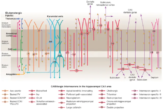
While the principal cells of the hippocampus form relatively homogenous groups regarding their morphological and physiological features, the GABAergic cell population comprises a large variety of cell types. They influence temporal dynamics of synapses, organize network oscillations, participate in selecting cell assemblies and implementation of brain states (Klausberger & Somogyi, 2008). Most of the GABAergic cells in the hippocampus take part in local control of the circuit, so as termed interneurons; however some of them project out from the hippocampus, innervating subcortical and/or cortical areas (Figure 2).
Here the GABAergic cells will be classified based on their axonal and dendritic morphology. Generally, the dendritic location of neurons defines the inputs they receive, and the region targeted by their axons defines the specific domain in their target cells they can influence. We can dissect four major groups of interneurons in the hippocampus based on their axonal arborization. The perisomatic region-targeting interneurons innervate the perisomatic region of principal cells including the soma, proximal dendrites or axon initial segments. These synapses are in a strategic position to control the output of the pyramidal cells. The dendritic layer-targeting interneurons contact on several dendritic segments, often in a pathway-selective manner, thus they are able to influence a specific type of input. Most of the inhibitory cells (except axo-axonic neurons) innervate both pyramidal cells and interneurons; however, interneuron-selective interneurons target predominantly other
14
interneurons. The long-range projecting GABAergic neurons– besides giving rise to local collaterals– project out of the hippocampus with myelinated axons. They are supposed to regulate information flow between different areas (Freund & Buzsáki, 1996).
I.1.4.1. Perisomatic region-targeting interneurons
The soma and axons of perisomatic region-targeting interneurons can be found within or near the pyramidal cell layer. In most of the cases, their dendrites span all layers of the hippocampus.
The axo-axonic cells (AAC) provide the exclusive innervation of the axon initial segments of pyramidal cells (Somogyi, 1977). They release GABA and express the Ca2+- binding protein parvalbumin (PV). The peculiarity of this cell type is that under certain circumstances it may depolarize its target cell due to the higher reversal potential of Cl- present in the axon initial segments (Szabadics et al., 2006; Khirug et al., 2008). But generally, discharges of AACs seem to provide hyperpolarizing current (Glickfeld et al., 2009). Additionally, modeling in vivo-like membrane potential fluctuations, the dominant postsynaptic effect of axo-axonic cells was shown to be inhibition (Woodruff et al., 2011).
Some of the axo-axonic cells have a dendritic tree confined to the stratum oriens (Ganter et al., 2004).
The basket cells are named after their axons forming perisomatic “baskets” around the target cell bodies. There are two types of basket cells in the hippocampus that are markedly different despite their similar dendritic- and axonal morphology. They can be distinguished by neurochemical markers and firing pattern. One of the basket cells expresses a neuropeptide cholecystokinin (CCK) and has a regular-spiking firing pattern. The other basket cell expresses parvalbumin and has a fast-spiking firing property (FS BC). Their different synaptic input-output properties and in vivo behavior propose a distinguished function in the network (Freund, 2003; Klausberger et al., 2003). For instance, the output of regular-spiking basket cells can be modulated by subcortical pathways (e.g. serotonin, acetylcholine), by retrograde signaling molecules (e.g. endocannabinoids) and by autoreceptors (such as GABAB
receptors), thus, they are suggested to play a role in fine-tuning the network operation. The FS BCs are innervated extensively by glutamatergic inputs, and modulated by opioids. This type
15
of cell was proposed to be precisely timed device, which allows to control synchronous activities (Freund & Katona, 2007).
I.1.4.2. Dendritic layer-targeting interneurons
The bistratified cells share many features of PV+ cells. They also express PV and have fast-spiking characteristics (Buhl et al., 1994; Klausberger et al., 2004). Their soma is within or near the pyramidal cell layer; their dendrites arborize in the strata radiatum and oriens, but avoid the stratum lacunosum-moleculare (Halasy et al., 1996). Their axons can be found in the strata oriens and radiatum. The somata and the horizontally-oriented spiny dendrites of oriens lacunosum-moleculare cells (O-LM cells) are located in the stratum oriens. Their axons arborize mainly in the stratum lacunosum-moleculare, suggested to control the entorhinal input of pyramidal cells (McBain et al., 1994). O-LM cells express the neuropeptide somatostatin. The cell bodies of Schaffer collateral-associated cells are located in the stratum radiatum of the CA1, their dendrites project to all layers. Their axons target the oblique dendrites and, to a lesser extent, the basal dendrites of pyramidal cells. These interneurons contain CCK (Cope et al., 2002; Pawelzik et al., 2002). Their corresponding cell type is also present in the CA3 (Lasztóczi et al., 2013). The apical dendrite innervating cells have very similar morphology to Schaffer collateral-associated cells, but instead of oblique dendrites they prefer to innervate the main apical shafts of the pyramidal cells (Klausberger et al., 2005). The somata of the perforant path-associated cells can be found mainly at the border of strata radiatum and lacunosum-moleculare; their dendrites span all layers or remain in the stratum lacunosum-moleculare (Hájos & Mody, 1997; Klausberger et al., 2005). The former three cell types are also immunopositive for CCK (Klausberger, 2009). A large group of subpopulations of hippocampal interneurons (approximately 30% of all GABAergic cells) have a characteristic feature that their local axon arbor is very dense. They are the neurogliaform cells (Price et al., 2005) and ivy cells (Fuentealba et al., 2008). Neurogliaform cells are confined to the stratum lacunosum moleculare, while ivy cells are located mainly close to the pyramidal cell layer. They can contact dendritic spines of pyramidal cells (Tamás et al., 2003), but the majority of their axon endings do not form real synaptic contacts (Oláh et al., 2009), thus, they could provide tonic inhibition of neighboring cells or regulate transmitter release via presynaptic GABAB receptors.
16 I.1.4.3. Interneuron-selective interneurons
Interneuron-selective interneurons have three subtypes (I, II, III), which can be distinguished based on their calretinin and vasoactive intestinal polypeptide content and axonal arborization pattern (Acsády et al., 1996a; Acsády et al., 1996b; Gulyás et al., 1996).
They can effectively control the firing output of other GABAergic cells (Chamberland et al., 2010).
Figure 2. Diversity of interneurons in the CA1 region of the hippocampus. Klausberger and Somogyi distinguished 21 types of interneurons in the CA1. The pyramidal cells are shown in blue, the somato-dendritic region of perisomatic region-targeting interneurons are orange.
The dendritic layer-targeting interneurons are red, while interneuron-selective interneurons are shown in pink. Axons are purple; the main synaptic terminations are yellow. The distribution of the afferent glutamatergic inputs are indicated on the left. VIP, vasoactive intestinal polypeptide; VGluT, vesicular glutamate transporter; O-LM, oriens lacunosum moleculare. Adapted from Klausberger and Somogyi 2008.
17 I.1.4.4. Long-range projecting GABAergic neurons
The GABAergic projection neurons are generally located in the stratum oriens, where they have horizontally oriented dendrites. The axon collaterals of trilaminar cells span three layers of the hippocampus (strata radiatum, pyramidale and oriens) and project toward the subiculum (Sík et al., 1995; Ferraguti et al., 2005). Backprojecting cells send axons backwards to CA3 or DG (Sík et al., 1994), while the double projection neurons project to the medial septum and often also to the subiculum (Gulyás et al., 2003; Jinno et al., 2007). The Oriens or radiatum retrohippocampal projection cells can be found in the stratum oriens or radiatum, they project to the subiculum and retrohippocampal areas (Jinno et al., 2007), as well as to the entorhinal cortex (Melzer et al., 2012).
II. Long-term synaptic changes in hippocampal neurons
Experiences impact the brain function by affecting the activity of neural circuitries, which is manifested partly in changing synaptic weights. Long-lasting modification of synaptic strengths is tightly linked to the storage of new information and considered as the cellular basis of learning (Citri & Malenka, 2008). Long-lasting enhancement of synaptic inputs is termed long-term potentiation (LTP); long-lasting weakening is called long-term depression (LTD). The LTP of excitatory synapses was discovered first in the dentate gyrus (Bliss & Lomo, 1973), followed by the observation of LTD first in the cerebellum (Ito &
Kano, 1982). These types of synaptic changes were then acknowledged as widespread phenomena expressed at excitatory synapses throughout the brain (Malenka & Bear, 2004). In these early years of discovery, the interneurons were considered to be rigid structures lacking any possibilities for potentiation or depression at their input or output synapses (McBain et al., 1999). Indeed, most interneurons lack spines on their dendrites, a structure that provides compartmentalization for signaling cascades, which is necessary for altering synaptic strengths. Moreover the postsynaptic density, a protein matrix which contains the scaffolding proteins and signaling molecules necessary for plastic changes is much weaker at GABAergic synapses compared to excitatory synapses. After improving the recording conditions, it was shown that synapses onto interneurons (Buzsáki & Eidelberg, 1982; Laezza et al., 1999), and also their GABAergic outputs (Chevaleyre & Castillo, 2003; Patenaude et al., 2003) are
18
capable for long-term plasticity. Moreover, it has been clarified that interneurons might have dendritic compartments due to their fast receptor kinetics and activity of Na+/Ca2+ exchanger (Goldberg et al., 2003), which allow local changes in biochemical processes necessary for selective modification of synaptic operations.
Early studies have already established the basic properties of LTP: it exhibits cooperativity, associativity, and input specificity (Nicoll et al., 1988). Cooperativity indicates that LTP can be induced by the simultaneous activation of a critical number of synapses or by coactivation of pre- and postsynaptic neurons. Associativity is the ability to potentiate a weak input when it is activated together with a strong input. Input specificity means that LTP is elicited only at activated synapses and not at adjacent synapses. These classic rules are, however, loosely interpreted in some forms of interneuron plasticity. Both LTP and LTD has early and late phase. In the early phase several second messenger cascades are activated, which are responsible for the induction period, this may last for a few minutes. During the late phase, new protein synthesis occurs (Citri & Malenka, 2008).
II.1. Induction mechanisms
LTP can be induced by several methods, for instance, by high frequency stimulation (HFS, up to 100 Hz) of presynaptic fibers. This approach might model a highly synchronized release of glutamate, e.g. during sharp waves (Buzsáki, 1989). Another way to induce LTP is theta-burst stimulation with or without coincident postsynaptic depolarization (pairing). In contrast, theta-burst stimulation combined with postsynaptic hyperpolarization induces LTD, which might be correlated with plastic changes occurring during theta oscillations (Huerta &
Lisman, 1995; Hölscher et al., 1997). Furthermore, LTD can also be triggered by low frequency (below 10 Hz) stimulation. Various receptor agonists or antagonists are also capable to induce long-term synaptic changes, referred as chemically-induced LTP or LTD.
Another method for induction of long-term synaptic plasticity is the precise timing of single pre- and postsynaptic spikes, called spike-timing-dependent plasticity. LTP occurs, when the presynaptic spikes precede postsynaptic spikes by up to 0-20 ms, whereas LTD is observed when presynaptic spikes follow postsynaptic spikes by 0 to 20-50 ms interval (Feldman, 2009). This induction mechanism operates within a time window allowing coincident spiking of neurons during gamma oscillations (30-100 Hz).
19
II.2. Long-term plasticity of excitatory synapses onto principal cells
The common motif of the induction protocols to trigger long-term synaptic changes in principal cells is the necessity to raise Ca2+ concentration postsynaptically through N-methyl- D-aspartate (NMDA) receptors. This type of ionotropic glutamate receptors is permeable to Ca2+ and requires strong depolarization to become activated. If the increase in Ca2+ levels is fast enough, the cascade of the second messenger Ca2+/calmodulin-dependent protein kinase II is activated, leading to an increase in the conductance of the fast, non-Ca2+-permeable ionotropic receptor of glutamate, the α-amino-3-hydroxy-5-methyl-4-isoxazolepropionic acid (AMPA) receptor, and insertion of new AMPA receptors to the synapse. LTP is usually accompanied by enlargement of dendritic spines and formation of new ones. Conversely, a modest increase in postsynaptic calcium concentration induces LTD, by activating a pathway involving calcineurin phosphatase activation, consequent removal of AMPA receptors from the synapses and shrinkage of dendritic spines (Malenka & Bear, 2004; Citri & Malenka, 2008). The threshold level for generating LTP depends on the history of the synapse. If the synapse has previously undergone LTP, the threshold increases and a calcium influx will result LTD with a higher probability (Bear, 1995).
A special form of LTP occurs at mossy terminals in the hippocampus. The expression of this type of LTP is solely presynaptic (Nicoll & Malenka, 1995). As general mechanisms of presynaptic LTP, it requires the cAMP/PKA signaling pathway and the active zone protein RIM1α.
Another non-conventional type of LTD is mediated by retrograde messenger molecules, the endocannabinoids (eCB, eCB-LTD). It usually requires a coincident rise of Ca2+ and activation of metabotropic glutamate receptors (mGluR) postsynaptically, which triggers the subsequent release of eCBs from the postsynaptic neuron. These eCBs act on presynaptic CB1Rs located at axon endings causing a decrease of transmitter release. This form of LTD is also widely distributed in the brain (Heifets & Castillo, 2009); although in some cases, mGluR-mediated LTD was shown to be independent of CB1Rs (Rouach &
Nicoll, 2003; Nosyreva & Huber, 2005). The role of astrocytes in eCB-LTD induction has also been recently revealed (Min & Nevian, 2012).
20
II.3. Long-term plasticity of excitatory synapses onto interneurons
In the past few years, numerous forms of long-term plasticity have been described in interneurons depending on cell type, brain region or stimulation protocols (Lamsa et al., 2010;
Kullmann & Lamsa, 2011; Laezza & Dingledine, 2011). A great drawback of early studies was the lack of proper identification of interneuron types. As a result of this, data are controversial and often not comparable to each other. Induction of plasticity in interneurons seems to have different rules than in pyramidal cells. In GABAergic cells, HFS often induces LTD instead of LTP (McMahon & Kauer, 1997b; Laezza et al., 1999; Pelkey et al., 2005). In interneurons located in the stratum radiatum, NMDA receptor-dependent LTP could be elicited (Lamsa et al., 2005). However, other interneurons possess NMDA receptor- independent form of long-term plasticity, where Ca2+-influx necessary for synaptic changes provided by Ca2+-permeable AMPA (CP-AMPA) receptors (Nissen et al., 2010), or L-type Ca2+-channels (Galván et al., 2008). A special type of LTP has been observed on interneurons equipped with CP-AMPA receptors: LTP was induced, when HFS was paired with postsynaptic hyperpolarization, named as anti-Hebbian LTP (Lamsa et al., 2007; Oren et al., 2009; Nissen et al., 2010). A group of stratum oriens interneurons show postsynaptic mGluR1/5 -dependent LTP (Lapointe et al., 2004; Topolnik et al., 2006). A series of studies implicated the role of presynaptic mGluR7 receptors in the induction of LTD in CA3 interneurons (Laezza et al., 1999; Pelkey et al., 2005). The subcellular cascades underlying these types of plasticity are divergent and have not been precisely elucidated yet.
II.4. Endocannabinoid-dependent long-term plasticity
Endocannabinoid molecules are the most abundant retrograde messengers in the nervous system. Among eCBs, 2-arachidonoyl glycerol (2-AG) is the most prevalent in the brain (Kano et al., 2009, Katona & Freund, 2012). The CB1Rs are present at excitatory and inhibitory terminals and mediate either short- or long-term depression in the hippocampus (Ohno-Shosaku et al., 2001; Wilson & Nicoll, 2001; Chevaleyre & Castillo, 2003), depending on stimulus type and length of transmitter release (Heifets & Castillo, 2009). The eCB-LTD is the most conventional form of LTD exhibited at GABAergic efferent synapses of CCK- positive interneurons (i-LTD), which was originally discovered in the amygdala (Marsicano et
21
al., 2002). This eCB-iLTD is heterosynaptic, because the presynaptic CB1R-mediated reduction of GABA release requires activation of postsynaptic mGluRs by glutamatergic fibers (Chevaleyre & Castillo, 2003, Figure 3).
Despite the fact that CB1Rs are usually present at glutamatergic terminals at lower levels compared to that found at GABAergic synapses (Kawamura et al., 2006), homosynaptic eCB-LTD occurs in several glutamatergic synapses both in subcortical regions (Gerdeman et al., 2002; Robbe et al., 2002) and -using spike-timing dependent protocol- in the neocortex (Sjostrom et al., 2003; Bender et al., 2006; Lafourcade et al., 2007). Whether this form of LTD is also present at glutamatergic terminals in the hippocampus have remained highly controversial. LTD at excitatory synapses induced by high concentrations of group I mGluR agonist dihydroxyphenylglycine (DHPG) seemed to be independent of CB1Rs in pyramidal cells (Rouach & Nicoll, 2003; Le Duigou et al., 2011) or in interneurons (Gibson et al., 2008; Edwards et al., 2012). In contrast, some studies detected CB1R-dependent LTD in pyramidal cells (Xu et al., 2010; Izumi & Zorumski, 2012).
II.5. Long-term plasticity at excitatory synapses onto PV+ interneurons
Lamsa and colleagues found anti-Hebbian LTP at excitatory synapses onto PV+
interneurons in response to HFS (Lamsa et al., 2007). A recent study showed a persistent increase in intrinsic excitability of PV+ cells (Campanac et al., 2013) in the CA1 region of the hippocampus. This type of potentiation occurred in response to HFS, induced by mGluR5- mediated inactivation of Kv1 potassium channels. Investigations of spike-timing-dependent plasticity in the layer 2/3 of somatosensory cortex showed that fast-spiking (i.e. putative PV+) interneurons express postsynaptic mGluR-dependent LTD regardless of the timing of pre- and postsynaptic spiking (Lu et al., 2007). However, it is still unclear whether eCBs are involved in mGluR-dependent LTD observed in PV+ interneurons.
Figure 3. Summary of the induction mechanism of endocannabinoid-mediated long-term depression (eCB-LTD). Upon excess neuronal activity, group I mGluRs -located at the postsynaptic site, in the perisynaptic annulus- are activated and coupled to phospholipase C (PLC) through Gαq/11 subunit and facilitate diacylglycerol (DAG) formation from
22
phosphatidylinositol (PI). DAG is further converted to 2-AG by diacylglycerol lipase-α (DGL- α). 2-AG is released from the postsynaptic neuron by presumable eCB membrane transporter (EMT) and binds CB1Rs. In some forms of eCB- LTD, eCB mobilization requires postsynaptic Ca2+ rise probably to activate PLC. Postsynaptic action potentials (e.g., during spike timing–dependent protocols) may trigger this Ca2+
rise through voltage-gated Ca2+
channels (VGCC), NMDA receptors, or Ca2+ can be released from the endoplasmic reticulum (ER), by a product of PLC, inositol 1,4,5-trisphosphate (IP3). At presynaptic terminals, CB1R acts via Gαi/o through inhibition of adenylyl cyclase (AC), which reduces the activity of protein kinase A (PKA). This step may also require a presynaptic Ca2+ rise. The reduction of PKA activity leads to activation of the Ca2+-sensitive phosphatase calcineurin (CaN), which shifts the kinase/phosphatase activity balance, thus, induces dephosphorylation of a yet unidentified presynaptic target (T), potentially RIM1α; resulting long-lasting reduction of transmitter release. Adapted from Heifets et al. 2009.
III. Oscillations in the hippocampus
In a cortical network, excitatory principal cells are designated for processing, storing and retrieving information, while inhibitory interneurons are responsible for spatial and temporal control of the firing of these cells, thereby enabling the synchronization of network activity. Oscillations emerge in brain regions where the recurrent excitatory connectivity of principal cells is prominent, including the neocortex, the CA3 region of the hippocampus or
23
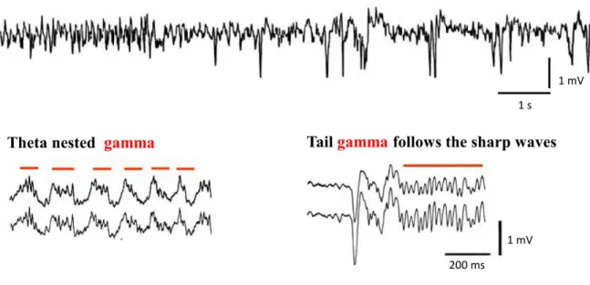
the subiculum. Other brain areas lacking local recurrent collaterals can be entrained by extrinsic rhythmic inputs (Hájos & Paulsen, 2009). Several types of network activities related to memory acquisition have been observed in the hippocampus. For instance, theta (4-8 Hz) and gamma (30-100 Hz) oscillations are contributed to formation of new memory traces, whereas sharp waves and associated ripple (120-200 Hz) oscillations are suggested to play a role in memory consolidation (Buzsáki, 1986, Figure 4). These rhythmic events can be detected as fluctuation of the local field potential, which are proposed to be generated mainly by inhibitory conductances (Oren & Paulsen, 2010). Oscillations in networks can have a role in binding of assemblies coding several modalities of a cue (Singer, 1993) and/or arrange the timing of the discharge of connected cells promoting plasticity. During a network event, at least two features of a given cell activity might carry information: the timing of the cell’s action potential relative to the network event (phase coding) or the frequency of the cell’s spiking (rate coding). Lower frequency oscillations can involve more neurons in a larger brain volume and associated with larger voltage fluctuations, because a broader time window allows recruitment of larger number of cells. In contrast, fast oscillations are contributed to smaller, more localized cell assemblies, and are associated with smaller voltage fluctuations (Axmacher et al., 2006; Buzsáki & Wang, 2012).
Figure 4. Activity patterns of the hippocampus recorded in vivo. Top: Theta oscillation that occurs during exploratory behavior, whereas subsequent large irregular activity dominated by sharp waves which can be observed when the animal is in rest (Modified from Buzsáki,
24
1989). Bottom: Gamma oscillations (indicated by red lines) can be found nested within theta activity (left) or following sharp waves (right). Modified from Buzsáki et al., 2003 (right) and Traub et al., 1996 (left).
III.1. Theta oscillations
Theta oscillations are characteristic activity patterns on the hippocampal EEG during exploratory behavior. The amplitude of theta oscillations is largest in the stratum lacunosum- moleculare of the CA1 region (Bullock et al., 1990), and switches the polarity at stratum pyramidale (Leung & Yim, 1986; Fox, 1989). The MS-DBB and the supramammillary nucleus are the two critical subcortical structures involved in pacing the theta rhythm (Petsche et al., 1962). According to the current theory, the theta rhythm is generated by pacemaker GABAergic neurons located in the MS-DBB (Hangya et al., 2009) that project to the hippocampus and entorhinal cortex, providing a rhythmic inhibition on local inhibitory neurons (Miettinen & Freund, 1992). In addition, the hippocampal perisomatic region- targeting interneurons are tonically excited by acetylcholine released from septal cholinergic afferents. This cholinergic excitation together with the phasic inhibition can generate rhythmic discharges of GABAergic neurons synchronizing a large population of neuronal activities (Freund & Antal, 1988; Stewart & Fox, 1990). The glutamatergic input from the entorhinal cortex can also contribute to the rhythmic excitation, terminating in the stratum lacunosum-moleculare (Winson, 1974). Other non-subcortical mechanisms of theta generation can be also present in the hippocampal networks. For example, after lesioning of the entorhinal cortex, theta rhythm driven by the CA3 recurrent collateral system emerges (Kramis et al., 1975; Lee et al., 1994). Besides perisomatic region-targeting interneurons, at least three other types of GABAergic cells may also contribute to the maintenance of theta.
The O-LM cells receive rhythmic excitation from pyramidal cells, thus they provide a rhythmic inhibition to the stratum lacunosum-moleculare. This rhythmic inhibition can prevent the discharge of weakly activated principal cells by entorhinal fibers, but can allow to spike neurons that code information. The perforant path-associated cells that are capable to discharge with theta frequency in response to cholinergic activation (Chapman & Lacaille, 1999) may also control the impact of the entorhinal input. The hippocampal GABAergic neurons with septal projection can provide rhythmic feedback to the medial septum (Alonso
25
& Kohler, 1982; Tóth et al., 1993). Finally, pyramidal cells display subthreshold resonance in their membrane potential fluctuations, with a preferred frequency in the theta band. This single-cell feature of pyramidal cells can also actively contribute to their somatic and dendritic membrane potential oscillations during theta (Leung & Yim, 1991; Kamondi et al., 1998; Strata, 1998).
III.1.1. Theta rhythm-related place coding
Remarkably, most pyramidal neurons are silent during theta rhythm. The few active cells discharge single spikes at the trough of theta measured in the pyramidal cell layer, when perisomatic inhibition is minimal. A prominent example of hippocampal information processing during theta rhythm is the representation of spatial location of the animal. Those pyramidal cells that are responsible for coding a single part of the territory are called place cells. The area where place cells fire is the cell’s place field which corresponds to the physical environment where the animal is currently present (O'Keefe & Dostrovsky, 1971; O'Keefe &
Recce, 1993). When the animal approaches the place field of a given cell, the cell increases its firing rate and its spikes display phase precession relative to the theta rhythm (Skaggs et al., 1996). This change in the firing phase can reach 180° in the center of the place field (defined by the maximum discharge rate of the neuron). The coding of place fields occurs randomly. If a pyramidal cell is depolarized, e.g. when it receives input from perforant path fibers conveying the actual environmental cues, the synaptic connections between the pyramidal cell and its input can be strengthened, and then the cell can become responsive to a specific environmental input, overriding the rhythmic somatic hyperpolarization (Axmacher et al., 2006).
III.2. Gamma oscillations
Gamma oscillations often occur embedded into theta oscillation (Buzsáki et al., 2003) or following sharp wave activity (Traub et al., 1996). During theta rhythm, perisomatic region-targeting interneurons fire bursts of spikes at the peak of the theta measured extracellularly in the stratum pyramidale, where the spikes in the burst episodes occur at
26
gamma frequency (Buzsáki et al., 1983; Bragin et al., 1995). Indeed, the inhibitory postsynaptic potential provided by these interneurons is known to be reflected in the gamma field (Oren et al., 2010). Two mechanisms could underlie the generation of gamma oscillation: synchronization of interneuronal firing by mutual inhibition (Whittington et al., 1995; Traub et al., 1996; Wang & Buzsáki, 1996) or a reciprocal interaction between excitatory and inhibitory neurons (Csicsvári et al., 2003; Mann et al., 2005) is required (for review see Buzsáki & Wang, 2012). Again, as in the case of theta rhythm, only a small percentage of pyramidal cells are active during gamma oscillation (Csicsvári et al., 2003;
Senior et al., 2008; Colgin et al., 2009). This is in accordance with the theory that in a system linking cell ensembles by close coincident temporal activity, the spiking of most cells should be silenced in order to avoid their near-synchronous firing by chance (von der Malsburg, 1995).
In the hippocampus, at least two types of gamma oscillations can be distinguished.
The slow gamma (30-50 Hz) is generated in the CA3 region, whereas the fast gamma (50-100 Hz) is driven by the rhythmic input from the entorhinal cortex (but see Buzsáki & Wang, 2012). These two types of transient oscillations may occur in different phases of the theta cycles. The slow gamma is preferentially present in the descending phase of theta cycles, while the fast gamma occurs at their trough. These two types of gamma oscillations might have distinguished roles in a learning task. The fast gamma may carry new information from the entorhinal cortex needed to be burnt in the network. For this purpose, the fast gamma provides a 10-15 ms long time window ideally suited to generate LTP. Thus, this type of oscillation may establish the basis for memory encoding. In case of retrieval, the slower gamma generated by the CA3 is suggested to play a role. The slow gamma could readily reactivate the cells representing a recent memory, but as the time window it provides (25-30 ms) is not optimal for potentiation, the retrieval process will avoid re-encoding of previously stored memories. A potential mechanism for a switch between the two types of gamma oscillations might be a strong excitation of the CA3 network or recruitment of distinct subpopulation of interneurons (Colgin & Moser, 2010).
27
III.2.1. Cooperative action of theta and gamma oscillations in the formation of working memory
A theory for the requirement of co-active theta and gamma oscillations was built by Lisman and colleagues. The 7+/-2 short-term memory items that humans can store is represented by the gamma cycles nested in each cycle of theta rhythm. Each item may be stored in one gamma cycle and the 7+/-2 items are always represented in the same order during theta cycles. In this concept, the theta rhythm provides an absolute phase reference to enable the preservation of the sequence. This coupling of theta and gamma oscillations can give rise to a working memory buffer and their frequency relationship is supposed to explain the limit of simultaneously storable memory items (Lisman & Idiart, 1995).
III.3. Sharp wave-ripples
Sharp waves are large amplitude events generated by population discharges. They are generated in the CA3 region during low arousal brain states, when recurrent collaterals of CA3 pyramidal cells are released from subcortical inhibition (Miles & Wong, 1983). They can be detected as a voltage deflection up to 1 mV lasting for 50-100 ms (Buzsáki, 1986).
This field activity reflects the summated excitatory postsynaptic potential (EPSP) of Schaffer collaterals (Csicsvári et al., 2000). The ascending phase and the peak of sharp waves are usually decorated by an embedded, fast oscillatory activity (120-200 Hz) called ripple (Buzsáki et al., 1992; Ylinen et al., 1995). Ripples are generated by the dynamic interaction of excitatory and inhibitory cells. The field potential during ripples may reflect synchronized somatic inhibitory postsynaptic potentials interrupted by synchronous spiking of pyramidal cells in every 5-6 milliseconds (Csicsvári et al., 1999).
III.3.1. Sharp wave-ripples in memory consolidation
Sharp wave-ripples are considered to be the neural correlate of memory consolidation.
During ripples, neuronal assemblies that participated in coding are re-activated in the same or reversed order on a compressed time scale (Skaggs & McNaughton, 1996; Nádasdy et al.,
28
1999; Diba & Buzsáki, 2007). Because sharp waves spread from the CA3 to the CA1, subiculum and then to layers V and VI of the entorhinal cortex (Chrobak & Buzsáki, 1996), they can mediate the transfer of recently acquired memories to their final place of storage in the neocortex. The strong excitation provided by recruitment of large cell assemblies during sharp waves might be able to initiate LTP in neurons which were weakly potentiated during the acquisition phase (Buzsáki, 1986).
III.4. In vitro oscillation models
Due to the lamellar structure of the hippocampus proper, the connectivity of the CA regions is well preserved in horizontal slices, which enables to investigate oscillations in acute slice preparations. These models allow the direct experimental study of the cellular and synaptic mechanisms underlying given network activities. As a corollary advantage, one can selectively target cell types under visual guidance, and pharmacological modifications are also possible. In case of sharp wave-ripples or slow gamma oscillations that emerge intrinsically in CA3 networks, detailed examination of the generation mechanisms becomes also available. While sharp wave-ripples emerge spontaneously in acute slices (Maier et al., 2002), gamma oscillations require induction. They can be induced by activating mGluRs (Whittington et al., 1995; Boddeke et al., 1997), kainate receptors (Hájos et al., 2000; Fisahn et al., 2004; Gloveli et al., 2005), or muscarinic acetylcholine receptors (mAChRs) (Fisahn et al., 1998).
III.4.1. Carbachol-induced in vitro gamma oscillations
Our group studied the properties of in vitro gamma oscillations by activating mAChRs with bath application of an acetylcholine receptor agonist carbachol. This type of gamma oscillation can be maintained for hours, and intended to model in vivo gamma oscillation occurring at high cholinergic tone, that occur during exploratory behavior when gamma oscillation is embedded in theta rhythm (Marrosu et al., 1995; Buzsáki et al., 2003).
Carbachol increases the excitability of CA3 pyramidal cells via M1/3AChRs (Müller &
Misgeld, 1986) and activate M2AChRs on terminals of PV+ cells resulting in a decreased, but
29
sustained GABA release from their axon endings (Szabó et al., 2010). These effects may contribute to the generation of gamma oscillations within the CA3, where the recurrent excitation of CA3 pyramidal cells drives the firing of inhibitory cells.
Carbachol-induced oscillation in the CA3 region shares many features of in vivo gamma oscillations observed in the same area. In accordance with in vivo observations, the phase of the local field potential also reverses in the stratum lucidum. In vivo and in vitro oscillations have identical current source density profiles. Pyramidal cells discharge with the highest probability at the trough of the oscillation both in vivo and in vitro when measured in the pyramidal layer, followed by the firing of interneurons with a monosynaptic delay. These similarities suggest that carbachol-induced gamma oscillations in slices are generated with a similar mechanism as in vivo occurring gamma oscillations and make this model suitable to study the underlying cellular processes (Hájos & Paulsen, 2009).
III.5. Parvalbumin-positive perisomatic region-targeting interneurons in oscillations
Perisomatic inhibition is considered to be the main current generator in multiple network oscillations synchronizing large assemblies of principal cells (Ellender & Paulsen, 2010). Thus, depending on brain state, PV+ cells seem to be the key elements of the rhythm of a wide range of cortical oscillations. Several studies show the crucial role of these cells in the generation of theta rhythm (4-8 Hz, Korotkova et al., 2010) or gamma oscillations (30-100 Hz, Fuchs et al., 2007; Cardin et al., 2009; Sohal et al., 2009), as well as in sharp wave-ripple activities (120-200 Hz, Rácz et al., 2009; Ellender et al., 2010).
III.5.1. Unique properties of PV+ cells that make them suitable to govern oscillations
Many different properties endow the PV+ cells to effectively control the output of principal cells (Bartos et al., 2007). They strategically synapse on the somata and proximal dendrites (basket cells, Somogyi et al., 1983; Kubota et al., 2007) or axon initial segments (axo-axonic cells, Somogyi, 1977) of principal cells, where the action potentials are generated. The release of GABA from their axon terminals is precisely controlled by their action potentials, which is supported by the P/Q-type voltage-gated Ca2+-channels in the
30
boutons and by the presence of parvalbumin. The P/Q-type Ca2+-channels in the axon endings ensure an efficient rise of Ca2+ concentration in the close proximity of vesicles (Neher, 1998).
The source of Ca2+ is tightly coupled to the Ca2+ sensor, estimated to be within 50 nm distances from the release sites (Hefft & Jonas, 2005). This structure can result in a fast, synchronized release of GABA-containing vesicles, precisely timed to the action potential invading the bouton. The fast and synchronous release of vesicles enables GABA receptors to be opened synchronously, producing a rapid rise time of synaptic currents. The parvalbumin- content of the cells (Katsumaru et al., 1988) provides an efficient buffering of residual calcium, enabling a fast decay of GABA currents (Vreugdenhil et al., 2003).
The membrane features of PV+ cells that determine their action potential generation also contribute to their effective inhibitory and synchronizing properties. The resting membrane potential of these interneurons is generally by 10-15 mV more depolarized (Fricker et al., 1999; Verheugen et al., 1999) than that of pyramidal neurons, which set them a ready- to-fire mode (Jonas et al., 2004). Furthermore, the potassium current mediated via Kv3 channels is pivotal to the fast-spiking phenotype, making them well suited to fire action potentials with high frequency during sustained depolarization without distortion in the action potential shape (Doischer et al., 2008; Goldberg et al., 2011). Fast deactivation, high activation threshold and lack of inactivation of the Kv3 potassium channels are crucial for generating fast spiking (Lien & Jonas, 2003). The action potential of the PV+ cells has small half width, which may also participate in constraining fast decay of GABA release. Large afterhyperpolarization with fast kinetics is optimal for a maximal recovery of Na+ channels from inactivation and a minimal delay in the onset of the action potential initiation (Jonas et al., 2004).
The excitatory synaptic input onto PV+ cells is also unique. They receive EPSPs with short latency and rapid kinetics (Miles, 1990). This is in part due to the fast synaptic currents mediated by AMPA receptors containing the GluA4 subunit (Jonas et al., 1994; Geiger et al., 1995) and to the fast clearance of glutamate from the synaptic cleft (Geiger et al., 1997). PV+
interneurons receive relatively large excitatory inputs from each of their presynaptic pyramidal cells, which can even drive action potential generation (Galarreta & Hestrin, 2001;
Woodruff et al., 2011). Unique electrical properties of dendrites (Hu et al., 2010; Nörenberg et al., 2010) diminish filtering of EPSPs preserving the fast kinetics. The lack of active conductances on the dendrites endows them for precise monitoring of network excitation and generation of an inhibitory output appropriate for controlling the overall system.
31
PV+ cells form a highly interconnected network; they are synaptically (Bartos et al., 2001) and electrically coupled with each other (Galarreta & Hestrin, 1999; Fukuda & Kosaka, 2000). Therefore, they can excite each other through fast electrical synapses (Tamás et al., 2000). The kinetics of the mutual inhibition of these cells is faster than the inhibition received by the pyramidal cells (Bartos et al., 2002), thus PV+ cells are capable to perfectly phase their spiking before synchronizing pyramidal cells throughout large areas.
Collectively, these features together provide PV+ interneurons a narrow time window for synaptic integration, ensuring fast and precisely timed spike generation (Glickfeld &
Scanziani, 2006) and a resulting fast synchronous inhibition. These properties are pivotal for oscillogenesis at different frequencies (Freund & Katona, 2007; Fuchs et al., 2007; Sohal et al., 2009).
III.5.2. Emerging evidences for the role of PV+ cells in oscillations
Several findings indicate the role of PV+ cells in synchronous network operation. For instance, impairing PV+ cell function resulted in changes in the oscillations. In parvalbumin knockout mice, the power of in vitro gamma oscillation is increased, presumably due to an enhanced inhibition in the lack of the high affinity Ca2+-binding protein (Vreugdenhil et al., 2003). Similar increase in the gamma power was observed when Kv3.1 potassium channels responsible for the fast-spiking phenotype were ablated (Joho et al., 1999). The loss of GABAA receptor-mediated synaptic inhibition onto PV+ cells reduced theta rhythm and coupling of theta and gamma oscillations, but left gamma oscillations intact (Wulff et al., 2009). Manipulation of excitatory synaptic inputs onto PV+ cells also caused severe changes in synchrony and memory-related behavior. When either of their two major subunit types of AMPARs (GluA1 or GluA4) was deleted from PV+ cells, in vitro hippocampal gamma oscillations exhibited reduced power. In parallel, the animals showed impairments in hippocampus-dependent memory tasks (Fuchs et al., 2007). In vivo recordings from the GluA1 knockout mice found increase of ripple amplitude, whereas theta and gamma oscillations remained intact (Rácz et al., 2009). Removing the NR1 subunit of NMDA receptors from PV+ cells resulted in altered theta oscillations and damaged spatial navigation.
Elevated power of gamma oscillations was also observed in NR1PVCre-/- mice, which was less modulated by theta (Korotkova et al., 2010). Optogenetic stimulation in the neocortex showed