THE MOLECULAR NEUROANATOMY OF THE SYNAPSES ESTABLISHED BY
THE ASCENDING SEROTONERGIC SYSTEM
ANDRÁS SZŐNYI
Workgroup for Quantitative Functional Anatomy, Laboratory of Cerebral Cortex Research, Department of Cellular and Network Neurobiology
Institute of Experimental Medicine, Hungarian Academy of Sciences
Group leader and consulent: DR. GÁBOR NYIRI
The ascending serotonergic system of the brain plays an important role in many brain functions including attention, motivation, emotions and sleep-wake cycles. The malfunction of this system has been implicated in many severe neuropsychiatric disorders, like major depression, obsessive-compulsive disorders and schizophrenia.
Median raphe is one of the serotonergic nuclei innervating the forebrain. It was shown in the hippocampus, that the median raphe establishes multiple synaptic contacts on a specific subpopulation of local interneurons, which in turn innervate hippocampal pyramidal cells.
Our laboratory described that these median raphe fibres have an excitatory effect on these interneurons, with a strong glutamatergic component. However, the exact molecular machinery of this glutamatergic transmission is still not known.
NMDA-type glutamate receptors are important mediators of glutamatergic transmission, and they take part in long-term synaptic plasticity mechanisms. Therefore, we asked whether NMDA-receptors are present in the raphe-hippocampal synapses. Using immunohistochemistry, we found that most raphe-hippocampal synapses contain postsynaptic NMDA-receptors.
We investigated whether this is a general feature of the synapses established by median raphe.
Using anterograde tracing combined with NMDA-receptor immunohistochemistry, we found that synapses established by the median raphe in hippocampus, medial septum and medial prefrontal cortex do contain NMDA-receptors.
These results suggest that the median raphe can act via glutamatergic transmission not only in the hippocampus, but in the medial septum and prefrontal cortex as well. The NMDA receptor content of these synapses in the different brain areas suggest that the fast glutamatergic neurotransmission is a general feature of these synapses, which fact completely changes our view of the function of median raphe.
TABLE OF CONTENTS
INTRODUCTION
The functions of the hippocampus The neuroanatomy of the hippocampus
1. The hippocampal formation of the mouse; an anatomical overview 2. Macroscopic anatomy of the hippocampal formation
3. Pyramidal cells and their intrinsic connections 4. Interneurons
The functions of the medial septum The neuroanatomy of the medial septum
1. Medial septum; an anatomical overview 2. Macroanatomy of the medial septum
3. Cytoarchitectonic organization of the medial septum The functions of the medial prefrontal cortex
The neuroanatomy of the medial prefrontal cortex
1. The macroscopic anatomy of the medial prefrontal cortex 2. The laminar organization of the neocortex
Interconnections of the hippocampus, medial septum and medial prefrontal cortex 1. Connections between the medial septum and the hippocampus
2. Connections between the medial septum and the medial prefrontal cortex 3. Connections between the medial prefrontal cortex and the hippocampus The functional anatomy of the median raphe
1. The raphe nuclei: an overview
2. Differences between the dorsal and median raphe pathways 3. The ascending median raphe pathway
4. An emerging new concept of the functions of the median raphe Glutamatergic and serotonergic neurotransmission
1. Glutamatergic transmission 2. Serotonergic transmission Aims of the study
MATERIALS AND METHODS Animals and perfusion Stereotaxic surgery Specificity of antibodies
Double labelling immunoelectron microscopy
Anterograde tracing combined with immunoelectron microscopy
RESULTS
Result I.: vGluT3 containing terminals establishing synapses on interneurons located in the distal dendritic regions of the hippocampus originate from the median raphe Result II: NMDA-receptors are associated with the postsynaptic active zones of raphe-hippocampal synapses
Result III: NMDA-receptors are present in the synapses of the median raphe in the forebrain
DISCUSSION
INTRODUCTION
In my thesis, first I will briefly review the functions and anatomical properties of the hippocampus, the medial septum and the medial prefrontal cortex, followed by a description of the interconnections of these brain areas in the mouse brain. After this, I will review the functional anatomy of the median raphe, which is one of the main serotonergic nuclei of the brain. This will be followed by the brief description of the glutamatergic and serotonergic neurotransmission. These descriptions are mainly based on murine brain, because this is where experiments were carried out, however, the most important facts are also discussed in relation to human brain as well, the latter of which show obvious similarities to murine brain.
Finally, the methods and our results will be described, with a final discussion of these results.
Functions of the hippocampus
The hippocampal formation is a brain structure that is a key participant in certain learning and memory functions. Since the famous case of H.M., who lost his ability to memorize new events of his life after his bilateral medial temporal lobectomy, the hippocampus has been a subject of many studies that were aimed to find out the role of the hippocampal formation in these functions. Two main theories emerged in the last decades about hippocampal function;
the first of them claims the hippocampal formation to be involved in the so-called declarative memory, the other claims it to be involved in spatial memory and formation of cognitive maps (Andersen 2007).
The declarative memory can be consciously recalled in humans: it contains our memories about facts (semantic memory) or events (episodic memory). The hippocampus does not have a role in other, so-called non-declarative memory processes like priming or classical conditioning that cannot be recalled consciously (Szirmai 2011). The hippocampus works together with other structures of the medial temporal lobe, including the other parts of the hippocampal formation, the perirhinal and parahippocampal cortices, and other neocortical associational areas to form these memories (Andersen 2007). Its role is important in memory consolidation: the initial memory traces that enter the hippocampus are stabilized later in the neocortex, through the interactions between the two structures. After this consolidation, the hippocampus is not needed to recall these memory traces; even H.M. remembered the facts and events that happened to him before his surgery (Andersen 2007).
The role of hippocampus in spatial memory became clearer with the discovery of the place cells; these cells fire at a higher frequency when the animal or human is in a well-defined point of the space in the environment (O’Keefe & Dostrovsky 1971). As different place cells are responsive in different locations, and they are distributed along the septotemporal axis of the hippocampus (see below), it was supposed that the collective firing of different place-cell ensembles in different environments might be the neural substrate of distinct maps of space (Andersen 2007). These maps form a spatial framework, where the memories of the world can be encoded. This is connected to our declarative memory: we usually start retrieving our memories with remembering to where we were at the moment when the memory trace was created (Andersen 2007). These maps, of course, can be used for spatial navigation.
The hippocampus plays an important role in mood-related processes as well (for instance anxiety and depression). These processes are strongly interconnected with the other hippocampal functions, which become highly relevant in pathological conditions. These
processes are heavily influenced by raphe-hippocampal inputs, the function of which is still not entirely understood.
The neuroanatomy of the hippocampus
1. The hippocampal formation of the mouse; an anatomical overview
The hippocampal formation is a complex archicortical structure of the so-called limbic system. The data of the following chapters are based on the newest edition of The Hippocampus Book (Andersen 2007), without further citations.
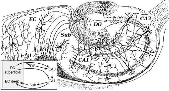
The hippocampal formation (HF) has the following subregions: the entorhinal cortex with the subicular complex, the cornu ammonis 1-3 (CA1-3) and the dentate gyrus. The subregions of the HF are connected to each other in a unique, mainly unidirectional way. All of these main pathways are excitatory; the first step in this intrinsic circuit is the layer II/III of the entorhinal cortex. This pathway has two directions: the one connected with the granule cells of the dentate gyrus is called the perforant path, because it perforates the hippocampal fissure; the other is connected to the CA1-3, and it is called the alvear (or temporoammonic) pathway. In the dentate gyrus, the granule cells are the principal cells; these cells give rise to the mossy fiber projection into the CA3 of the hippocampus. The CA3 pyramidal cells are connected to the CA1 pyramidal cells, via Schaffer collaterals. The CA1 pyramidal cells close the circuit with a direct projection to the layer V of the entorhinal cortex and with a projection to the subiculum, which itself projects to the deep layers of entorhinal cortex as well (Figure 1.).
Figure 1: The hippocampal formation: This is one of the first images representing the different parts of the hippocampal formation (HF). It was drawn by Santiago Ramón y Cajal, the famous Spanish neuroanatomist, based on his experiments made with Golgi-staining of the hippocampus. The Golgi-stained cells and their dendrites are shown in black in the different parts of the HF. The axons originating from the principal cells of the different subregions of the HF can be observed as well; these show the main intrinsic connections of the HF (see text for details). The insert shows a schematic drawing of the information flow; the arrows mark the synaptic contacts between the different areas of the HF. Used with permission from: Histologie du Systeme Nerveux de l'Homme et des Vertebretes, Cajal, 1911.
The intrinsic and extrinsic connections of the HF are carried by three major fiber bundles. The angular bundle carries information between the entorhinal cortex and other parts of HF. The
fimbria-fornix pathway carries the information between the HF and the subcortical and brainstem areas. Finally, the commissural pathways connect the two HF of the hemispheres.
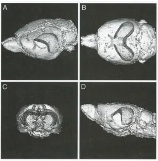
These structures – with the before mentioned other parts of the HF – have an elongated, banana-like shape (Figure 2.).
Figure 2: the location of the hippocampal formation in the brain: The images show a rat brain and inside the hippocampal formation with the fimbria-fornix, reconstructed with MRI from an oblique (A), horizontal (B), coronal (C) and sagittal (D) plane. Used with permission from: The Hippocampus Book, Andersen, 2007
The long axis of this formation in the mouse extends in a C-shaped manner from the midline of the brain (near the septal nuclei, rostrodorsally) to the temporal lobe (caudoventrally). This axis is referred to the septotemporal axis, and the orthogonal is the transverse axis. The subicular complex appears at one-third along the way of the septotemporal axis; the entorhinal cortex is located even more ventrally and caudally. The angular bundle is located between the entorhinal cortex and subicular complex. The fimbria is collected from afferent and efferent fibers of the alveus: it is located laterally from the hippocampus, and it becomes thicker from the temporal to the septal levels. At the most septal level, rostrally from the hippocampus, it continues in the fornix, right below the corpus callosum, near the midline. The commissural pathway comprises of dorsal and ventral commissures; the dorsal is located rostral to the splenium of corpus callosum; the ventral is located caudal to the septal area and dorsocaudal to the anterior commissure.
2. Macroscopic anatomy of the hippocampal formation
The parts of the HF have different subdivisions themselves (Figure 3.). The entorhinal cortex is a neocortical structure, so it has a multilaminate appearance, showing the classical six- layered organization of cells. In contrast, principal cell bodies of the DG are located in one layer: this is the stratum granulosum that is divided to suprapyramidal and infrapyramidal blade. The suprapyramidal blade is between the CA1 and CA3; the infrapyramidal blade is opposite to it, and the two blades are connected in a V-shape. The principal cells of the CA1-3 are also located to one layer, which is called the stratum pyramidale. This can be noticed in the CA3, CA2 and CA1 as well. The CA subregions are divided based on the pyramidal cells
and their connections: CA3 and CA2 pyramidal cells are larger than the CA1 pyramidal cells, but only CA3 pyramidal cells receive mossy fiber input from the dentate gyrus. The subicular complex has a broader pyramidal cell layer than CA1.
Figure 3: The layers of the hippocampal formation: A: Nissl-stained horizontal section of the hippocampal formation of the rat. B: A schematic drawing of the hippocampus shown on A. Abbreviations: ab: angular bundle, al: alveus, CA1-3: cornu ammonis 1-3 of the hippocampus DG: dentate gyrus, EC: entorhinal cortex, fi:
fimbria, gcl: stratum granulosum, hf: hippocampal fissure, ml: stratum moleculare, Para: parasubiculum, pcl:
stratum pyramidale, Pre: presubiculum, pl: hilus/polymorphic cell layer, sl: stratum lucidum, sl-m: stratum lacunosum-moleculare, so: stratum oriens sr: stratum radiatum, Sub: subiculum. The roman numbers show the cell layers of the EC, Para and Pre, respectively. Scale bar: 500μm. Used with permission from: The Hippocampus Book, Andersen, 2007
I define here the superficial-deep axis in the hippocampus. The hippocampal fissure is the point of reference: if a structure is closer to the hippocampal fissure within the dentate gyrus or hippocampus, it is considered to be more superficial. The alveus, a layer comprising of afferent and efferent fibers is located most deeply in the hippocampus. The dense cellular layer superficial to the alveus is the stratum pyramidale. The stratum oriens is located between the alveus and stratum pyramidale; it contains the basal dendrites of the pyramidal cells.
Stratum radiatum is located even more superficially; this thick layer contains the proximal apical dendrites of the pyramidal cells. The distal dendrites are located to the stratum lacunosum-moleculare, right at the border of the hippocampal fissure. In the CA3 in stratum lucidum the thorny excrescences of the dendrites of the CA3 pyramidal cells receive synapses from the large thorny axon terminals of the mossy fibers. All layers receive terminals from extrinsic sources. On the other side of the fissure begins the stratum moleculare of the dentate gyrus, comprising of the dendrites of the granule cells. The stratum granulosum contains the cell bodies of the granule cells. Most deeply in the dentate gyrus is the polymorphic cell layer or hilus that contains the mossy fibers, together with many different kinds of interneurons.
The stratum pyramidale of the CA3 also extends to the hilus.
3. Pyramidal cells and their intrinsic connections
Because the experiments in this study were carried out in the CA regions, I will focus only on this subregion of the HF. CA1-3 differ from each other in many substantial ways in spite of their very similar organization.
The main excitatory input of the CA3 pyramidal cells originates from the mossy fibers of the dentate granule cells; the CA2 and CA1 pyramidal cells do not receive input from the mossy fibers. The CA3 pyramidal cells are connected to each other through CA3-CA3 associational (ipsilateral) and commissural (contralateral) connections; these terminals are present in the strata oriens and radiatum as well. Furthermore, they give rise to the CA3-CA1 Schaffer collateral system; this is the main excitatory input onto CA1 pyramidal cells, and it can be ipsi- or contralateral as well. In contrast with the CA3 pyramidal cells, CA1 pyramidal cells do not give rise to associational connections onto each other; they innervate the subiculum or deep layers of the entorhinal cortex as their main output. However, these CA1 pyramidal cells give rise to recurrent collaterals into the stratum oriens (but not radiatum), where they innervate a subpopulation of interneurons (see below).
Entorhinal cortical afferents may innervate the CA1 and CA3 pyramidal cells directly through the temporoammonic pathway. These entorhinal afferents travel in the alveus, then turn into a radial direction, heading to the stratum lacunosum-moleculare, where they innervate the apical dendrites of the pyramidal cells.
4. Interneurons
Cortical interneurons are GABAergic cells with axons non-projecting to other brain regions (DeFelipe et al., 2013). These cells usually establish inhibitory synapses with their targets;
however, in some cases a depolarizing effect was found as well (Szabadics et al. 2006, but see Glickfeld et al. 2009). The interneurons constitute only about 10% of the total number of cells in the cortex; however they show an extreme heterogeneity (Freund & Buzsáki, 1996). Based on recent studies, there are at least 21 different kinds of interneurons in the hippocampus (Klausberger & Somogyi, 2008), divided by the localization of their soma, their main input and output, neurochemical markers and firing properties.
4.a.: Cells targeting the perisomatic region. The basket cells innervate the somata or proximal dendrites of the pyramidal cells of the hippocampus. Their somata are usually located close to the pyramidal cell layer, and their dendrites span across all layers; their axons ramify in the stratum pyramidale. A basket cell have about 10000 synaptic varicosities, and it makes 2-10 inhibitory synapses on a pyramidal cell, so it can innervate more than 1000 pyramidal cells (Miles et al. 1996). Based on their neurochemical identity, two main subgroups of basket cells are distinguished: one of them contains parvalbumin (PV), and the other contains cholecystokinin (CCK), type 3 vesicular glutamate transporter (vGluT3) or vasoactive intestinal peptide (VIP) and type 1 cannabinoid receptor (Freund 2003, Somogyi et al. 2004).
These two cell populations have different roles in modulating the activity of hippocampal pyramidal cells (Freund 2003). The other type of the hippocampal interneurons targeting the perisomatic region of the pyramidal cells are the axo-axonic or chandelier cells. The somata of axo-axonic cells are located close to the stratum pyramidale, and their dendrites extend to all layers as well. Their axons may innervate up to 1200 pyramidal cells (Li et al., 1992), making 2-30 synaptic contacts on the axonal initial segments of the pyramidal cells (Somogyi et al. 1983).
Figure 4: Interneurons of the hippocampus: The figure shows a schematic drawing of the hippocampal interneurons that target pyramidal cells. The classification is based on morphological properties. The black filled circles show the somata, the black bars indicate the dendrites, and the dark grey rectangles mark the axon arborisation of the different interneurons, respectively. On the background, pyramidal cells are indicated. Arrows on the left show the main intrinsic inputs of the hippocampal formation. For more detail, see text. Abbreviations:
s.o.: stratum oriens, s.p.: statum pyramidale, s.r.: stratum radiatum, s.l.-m.: stratum lacunosum-moleculare. Used with permission from: Interneurons of the Hippocampus, Freund & Buzsáki, 1996.
4.b.: Cells targeting the dendritic regions. This is a very heterogeneous population of interneurons. One subpopulation of them is the oriens-lacunosum-moleculare (O-LM) cells;
the somata and dendrites of these cells are located to the stratum oriens, and they innervate the distal dendrites of the pyramidal cells in the stratum lacunosum-moleculare. The most important excitatory input of these cells is the recurrent collaterals of the pyramidal cells.
Innervating the distal apical dendrites of pyramidal cells, the O-LM neurons provide an effective feedback inhibition on the pyramidal cells (Klausberger et al. 2003).
The somata of the bistratified cells are located usually to the close to the pyramidal cell layer or in stratum radiatum, and their dendrites are present in every layer. Their axons innervate the dendrites of the pyramidal cells in the stratum oriens and radiatum, but not in lacunosum moleculare.
Interneurons located to the stratum radiatum, lacunosum-moleculare and the border of the two layers are the most heterogeneous population of the hippocampal interneurons (Lacaille &
Schwartzkroin 1988). Their dendrites are usually horizontal, but sometimes they extend radially to stratum pyramidale, or show a multipolar organization (Vida et al., 1998). These cells innervate the superficial dendrites of the pyramidal cells, establishing GABAergic
inhibitory synapses onto them (Buhl et al., 1994). Many different neurochemical markers were shown to be present in these cells, like PV, CCK, calbindin (CB), neuronal nitric oxide synthase (nNOS), somatostatin (SOM) and neuropeptide Y (for a detailed review see Freund and Buzsaki, 1996).
4.c.: Interneuron-selective (IS) cells. These cells are located in all layers, and they establish GABAergic contacts specifically with the somata and dendrites of other interneurons. The dendrites of these cells span across several layers as well (Gulyás et al., 1996). Freund and Buzsaki (1996) differentiated three different subpopulations of IS interneurons. These cells contain the calcium-binding protein calretinin (CR) or VIP. IS cells may have an important role in synchronously timing the activity of large populations of pyramidal cells.
Functions of the medial septum
The medial septum (MS) plays an important role in many different cognitive and attention- related processes, for example in memory formation. MS takes part in the generation of hippocampal theta rhythm that has a very important role in learning and memory processes.
Medial septum lesions were often associated with cognitive and attention deficits (Vertes and Kocsis 1997, Colom 2006). Moreover, the cells of the MSDB are among the neurons that suffer the earliest and most severe degeneration in Alzheimer’s dementia, and the severity of the disease correlates with the degeneration of this system (Paxinos 2004).
Medial septal cells receive a strong innervation from the oral part of the pontine reticular formation and the supramamillary hypothalamic nucleus. The tonic activity of these brain areas is transformed in the MS to a rhythmic firing pattern; this rhythmic activity has a powerful effect on the network activity of the hippocampal neurons through the septohippocampal projection (Vertes & Kocsis 1997).
The neuroanatomy of the medial septum
1. Medial septum; an anatomical overview
The septum means the wall between the lateral ventricles of the telencephalon. This structure is the part of the basal forebrain limbic system; besides this, it is part of the basal forebrain cholinergic corticopetal system that innervates every part of the cortex. The septum is differentiated from the many other limbic system-related nuclei located close to it (for example the nucleus basalis, substantia innominata and nucleus accumbens) based on its reciprocal connections with the hippocampus (Paxinos 2004).
There are four groups of nuclei described based on their anatomical location in the septum (Swanson and Cowan 1979). The lateral group involves the lateral septum, septofimbrial and septohippocampal nuclei. The medial group is formed by the medial septum (MS) and diagonal band of Broca (DBB). The posterior group is formed by the triangular nucleus and bed nuclei of the anterior commissure and stria medullaris. Finally, the ventral group contains only the bed nucleus of stria terminalis. This latter part is used to be considered recently to be the part of the extended amygdala (Paxinos 2004).
2. Macroanatomy of the medial septum
The corpus callosum bounds the septum dorsally and rostrally. The lateral borders are the ventricles and the nucleus accumbens. The caudal border of the structure is the fornix. The ventral hippocampal commissure and the anterior commissure pass through the structure.
Afferents and efferents pass through four main routes: fornix-fimbria connects the medial and lateral parts with the hippocampal formation; stria terminalis connects the ventral group with the amygdala; the caudal group projects through stria medullaris. Every group of septal nuclei receives information from the brainstem through the medial forebrain bundle, and sends descending projections as well (Paxinos 2004).
Because the experiments of this study were carried out in the medial group of septal nuclei, I will focus on MS and DBB.
3. Cytoarchitectonic organization of the medial septum
There are three main populations of principal cells present in the medial septum: the cholinergic, GABAergic, and glutamatergic neurons.
a. The MS contains cholinergic neurons in a great number that were shown to be positive for choline-acetyltransferase and cholinesterase (Mesulam et al. 1983). These neurons were shown to express other neuropeptides as well (Paxinos 2004).
b. The second group of MS and DBB neurons that project to other brain areas are the GABAergic neurons. These neurons can be clearly distinguished from the former population based on electrophysiological and neurochemical features (Sotty et al.
2003, Freund 1989). The most important feature of these GABAergic neurons is that they express parvalbumin, while the cholinergic cells do not (Freund 1989).
c. A recently described third group of cells in the MS and DBB are the glutamatergic cells that contain the type 2 vesicular glutamate transporter (vGluT2). Based on in vitro experiments, these cells have different electrophysiological properties from the above-mentioned two cell populations, and they modulate hippocampal network activity as well (Sotty et al. 2003, Colom et al. 2005, Huh et al. 2010).
The cholinergic, GABAergic and glutamatergic cell groups of the medial septum are interconnected, so these neurons have a role in the build-up of the local circuits of the MS and DBB (Leranth & Frotscher 1989, Colom et al. 2005, Colom 2006).
Finally, there are GABAergic cells in the MS that are local inhibitory interneurons, and another population of neuroendocrine cells containing gonadotropine-releasing hormone (Paxinos 2004).
The functions of the medial prefrontal cortex
The neocortex is the phylogenetically newest part of the brain. The volume of the brain in proportion of the body weight shows a tendency of growing in the evolution of the vertebrates, but this growing is uneven. The expansion is the greatest in the neocortex, and there are differences in the relative increase of the neocortex areas as well: associative cortices and the prefrontal cortex show the largest expansion relative to the sensory areas (Shepherd 2004).
Many cognitive functions depend on the right operation of the medial prefrontal cortex (mPFC), like decision-making, error detection, motivation and attention (Szirmai 2011).
Many studies deal with the role of the mPFC in both long-term and short-term memory processes, together with memory consolidation (Euston et al. 2012). mPFC is an associational area with a broad range of connections with the other parts of the neocortex, subcortical and brainstem areas (Heidbreder & Groenewegen 2003). It is important from the functional point of view that the mPFC has an access to information about motivational stimuli like reward and pain, and this feature seems to be important in decision making: based on the experiences of the past together with the momentary inner state (motivation and emotions), the animal will behave in the most appropriate way in the given situation (Euston et al. 2012). Some aspects of the above-mentioned functions are very human-like and hard to be examined in rodents;
but still, many mPFC-related behavioural patterns seem to be very similar in the two species.
Many diseases related to these functions are described, like obsessive-compulsive disorders and anxiety; some of them can be interpreted completely only in humans, like schizophrenia (Szirmai 2011). The malfunction of the mPFC is widely described in these neuropsychiatric disorders (Szirmai 2011).
The neuroanatomy of the medial prefrontal cortex
1. The macroscopic anatomy of the medial prefrontal cortex
The mPFC is divided to at least four cytoarchitectonically different areas: medial precentral area (or Fr2), anterior cingulate cortex (Brodmann’s area of the human brain 24), prelimbic area (area 32) and infralimbic area (area 25). However, it has been suggested, that there exists a main subdivision of the mPFC to a dorsal component containing the medial precentral, anterior cingulate and dorsal prelimbic areas and to a ventral component containing the ventral prelimbic and infralimbic areas. This latter differentiation is based on anatomical and functional-behavioural findings (Heidbreder & Groenewegen 2003).
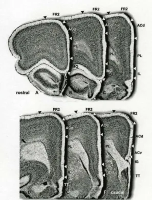
Figure 5: The anatomy of the medial prefrontal cortex.
A-F: Adjacent Nissl-stained coronal sections show the anatomical composition and subregions of the mPFC of the rat. Image A is the most rostral, image F is the most caudal section. The white and black arrowheads indicate the borders of the subregions. Abbreviations: ACd: anterior cingulate cortex dorsal part, Acv: anterior cingulate cortex ventral part, Fr2: medial precentral area, IL: infralimbic area, IG:
induseum griseum, MO: medial orbital cortex, PL: prelimbic cortex, TT: taenia tecta. Used with permission from: The medial prefrontal cortex in the rat: evidence for a dorso- ventral distinction based upon functional and anatomical characteristics, Heidbreder & Groenewegen, 2003.
2. The laminar organization of the prefrontal cortex
There are six cell layers present in the prefrontal cortex, based on the size, density and shape of the cells. Some layers might be thinner or even absent in some prefrontal areas.
I. stratum moleculare: it comprises of a dense plexus of fibers
II. stratum granulosum externum: it contains small pyramidal and stellate cell somata III. stratum pyramidale externum: it contains mainly pyramidal cells
IV. stratum granulosum internum: a dense layer of spiny stellate cells
V. stratum pyramidale internum: it consists of medium-sized and large pyramidal cells VI. stratum multiforme: it contains many variously shaped cells, together with afferent
and efferent fibers
Although the cortex has a laminar organization, the functional units seem to be organized in a columnar way. János Szentágothai made the first, pioneer studies in the examination of the anatomical grounds of the functional organization of the neocortex (Szentágothai 1975).
Somata of different interneurons – that have similarly diverse features like in the hippocampus – can be found in every layer. These cells are usually not spiny – unlike the pyramidal and stellate cells – and they establish GABAergic synapses with their targets (Shepherd 2004).
Interconnections between the hippocampus, medial septum and medial prefrontal cortex
1. Connections between the medial septum and the hippocampus
Both the cholinergic, GABAergic and glutamatergic cells of the MS project to the hippocampus, through anatomically different fiber tracts:
- the type I fibers of the septum, which are thick, with large en-passant boutons or baskets around cell somata are GABAergic. These cells innervate the different interneuron populations of the hippocampus, involving the PV, CB, CCK, VIP and SOM-immunoreactive cells. The function of these cells is to disinhibit the principal cells of the hippocampus by inhibiting large populations of interneurons (Freund 1989, Freund & Antal 1988, Gulyas et al. 1991).
- the type II fibers of the septum, which are thin and never form baskets around somata, but their varicosities are much smaller, are cholinergic. They establish synaptic contacts with pyramidal cells and interneurons as well (Gulyas et al. 1991). These inputs have a depolarizing effect on their targets through muscarinic cholinergic receptors (for a review see Vertes & Kocsis 1997).
- the recently described glutamatergic fibers of the MSDB that have an excitatory effect on CA3 pyramidal cells (Huh et al. 2010).
MS receives input from the hippocampus through the hippocampo-septal system. The hippocampal innervation of the MS and DBB is more limited than the septo-hippocampal innervation. Only GABAergic cells innervate the MS from the hippocampus; these cells contain calbindin or somatostatin, they innervate mainly the GABAergic cells of the MS (with a more limited innervation of the cholinergic cells as well), and their somata are located to the stratum oriens in the CA1. Similar cells were described in the CA3, containing somatostatin
or calretinin, but not CB (Toth & Freund 1992, Toth et al. 1993, Gulyas et al. 2003).
Moreover, the hippocampo-septal cells innervate septo-hippocampal cells and vice versa, so there is a reciprocal connection present at the cellular level (Toth et al. 1993, Takacs et al.
2008).
2. Connections between the medial septum and the medial prefrontal cortex
The medial septum has prominent connections with different cortical areas including the entorhinal, cingulate and medial prefrontal cortices (Semba et al. 2000, Paxinos 2004). The connection between the MS and mPFC is bidirectional, and it is topographically organized (Heidbreder & Groenewegen 2003). The more ventral parts of the mPFC project more medially (eg. to the MS and vertical limb of DBB), while the more dorsal parts of the mPFC project more laterally (to the horizontal limb of DBB). Both cholinergic and non-cholinergic cells of the MS and DBB are reached by these fibers. In turn, more lateral parts of the medial group of the septal nuclei project more laterally in the PFC, and medial parts project more medially (Gaykema et al. 1990, Heidbreder & Groenewegen 2003, Paxinos 2004).
3. Connections between the medial prefrontal cortex and the hippocampus
There is a huge amount of associational connections present in the mPFC that seem to be topographically organized. The infralimbic (IL) area projects to the medial orbital and prelimbic (PrL) areas and to a lesser extent to the entorhinal and anterior cingulate cortices (ACC). Prelimbic area projects to more dorsal cortical areas, but it reaches the IL and medial precentral area, ACC, and to a lesser extent sensorimotor cortices. Moreover, there is an intrinsic association system present in the PrL. The ACC and medial precentral areas project more significantly to sensorimotor and visual-related cortices. Afferent connections to the mPFC arrive from different associational areas like perirhinal cortex, ventral agranular insular area and secondary visual and sensorimotor cortices in the parietal and temporal lobe.
Interestingly, the mPFC does not seem to have a prominent projection to the hippocampus, but the hippocampus (especially ventral CA1) seem to project to the ventral areas of the mPFC (Heidbreder & Groenewegen 2003).
The functional anatomy of the median raphe
1. The raphe nuclei: an overview
The raphe nuclei are the parts of the nine cell groups of the rodent brain that are considered to use serotonin as primary transmitter. This classification is based on the study of Dahlström and Fuxe (1964), in which the Falck-Hillarp technique of immunofluorescence was used to describe the monoaminergic cell groups in the central nervous system.
Classically, we differentiate between the descending and ascending serotonergic systems. The descending system consists of the B1-4 nuclei; these nuclei innervate the brainstem and the spinal cord, and have an important role in pain perception (Zhao et al. 2007). The ascending serotonergic system consists of the B5-9 nuclei that give the serotonergic innervation of the forebrain. This system plays an important physiological role in sleep-wake cycles, affective and motivational states and emotional association with memory. The damage or disruption of serotonergic system and/or transmission is found in many pathological states, like major depression, obsessive-compulsive diseases, schizophrenia and drug abuse (Hensler 2006). The
B5 and B8 nuclei compose the pontine and midbrain part of the median raphe (MR) and the B6 and B7 nuclei compose the pontine and midbrain part of the dorsal raphe (DR), respectively (Figure 6.; Paxinos 2004). The serotonergic pathways emerging in the dorsal and median raphe nuclei differ from each other in many anatomical aspects, and some fundamental functional differences were found by recent studies.
Figure 6: The midbrain raphe nuclei: Immunohistochemical staining for serotonin clearly shows the dorsal and median raphe nuclei of the mouse. The decussation of the median cerebellar peduncles can be observed between the two nuclei. The shiny dots indicate serotonin-immunoreactive cell bodies. Abbre- viations: DR: dorsal raphe, MR: median raphe, PMR:
paramedian raphe. Source: own fluorescent immunoreaction with a rabbit-anti-serotonin antibody (see Table 1. for antibody specifications).
2. Differences between the dorsal and median raphe pathways
The most obvious difference between the dorsal and median raphe pathways is the anatomical build-up of their fibers. The dorsal raphe gives rise to thin fibers with little pleomorphic en- passant varicosities that can be hardly distinguished from the axons. The fibers of the median raphe, on the contrary, are thick, and they end in large boutons forming recognizable baskets around the somata and proximal dendrites of the cells they innervate (Kosofsky & Molliver, 1987). This morphological difference was obvious using anterograde tracing and serotonin- immunostaining as well.
Ultrastructural studies by Descarries and colleagues showed that only about one-third of the serotonergic terminals in the forebrain establish synaptic junctions; these junctions are asymmetric, and target the dendritic spines and shafts (Séguéla et al. 1989, Oleskevich et al.
1991, Descarries and Mechawar 2000). These studies, however, did not differentiate between the origins of the serotonergic varicosities they examined. Freund et al. (1990) found that the anterogradely traced axons of the MR and also serotonergic axons establish large boutons around cell somata and dendrites in the hippocampus, suggesting a strong coupling between the boutons and targets. This is supported by recent ultrastructural and physiological evidence (Varga et al. 2009). Ultrastructural evidence is shown in the medial septum about the synaptic junctions established by the majority of MR fibers on the somata and dendrites of target cells (Leranth & Vertes 1999). These findings suggest that the DR and MR fibers differ in the type of their neurotransmission. DR fibers are responsible for a non-synaptic, “volume”
transmission of serotonin in the brain areas they target, while MR fibers establish synaptic contacts in a target-selective manner in the forebrain (Freund et al. 1990).
The fibers of the two raphe nuclei usually innervate different brain areas. Moreover, MR cells seem to innervate specific subpopulations of neurons in the innervated brain areas, while DR fibers do not seem to show this target selectivity.
Finally, the molecular anatomical composition of these fibers is different as well. Studies using toxic amphetamine derivatives showed that DR fibers are vulnerable to these compounds, while MR fibers are not, because the serotonin-transporter is not present on the plasma membrane of the axons of the MR fibers (Mamounas & Molliver 1988, Mamounas et al. 1991, Brown & Molliver 2000). This fact suggests further differences in the dynamics of the neurotransmission of the DR and MR pathways.
3. The ascending median raphe pathway
MR was considered for a long time to be a serotonergic nucleus, and many studies used the terms “serotonergic” and “originating from MR” as synonyms. Köhler and Steinbusch in 1982 found 5-HT negative cells in the median raphe in a study using retrograde tracers and 5-HT immunostaining, but the presence of a strong non-serotonergic pathway arising in the median raphe was considered only recently.
The support for a new concept of the function of the MR was given by the discovery of a new, third type of vesicular glutamate transporters (vGluT3); this protein was shown to be active by amino acid uptake assay, and strongly present in different cholinergic and serotonergic cell groups (Gras et al. 2002); moreover, the most dense immunoreactivity for vGluT3 was observed in the raphe nuclei (Gras et al. 2002). The protein was colocalized with different monoaminergic markers all over the brain, like vesicular monoamine transporter type 2, serotonin-transporter and 5-HT (Gras et al. 2002, Somogyi et al. 2004, Shutoh et al. 2008, Jackson et al. 2009, Amilhon et al. 2010). However, these studies, together with others found that there is a significant non-serotonergic pathway present in the forebrain ascending from the MR (Aznar et al. 2004). So taking these data together, three subpopulations of the forebrain-projecting cells of the MR can be distinguished based on their 5-HT and vGluT3- content: one of them is only-serotonergic (5-HT-positive), one of them is only-glutamatergic (vGluT3-positive), and one of them is serotonergic and glutamatergic (5-HT/vGluT3-positive) at the same time (Shutoh et al. 2008, Jackson et al. 2009).
Median raphe innervates every brain area I examined before. The topography of these connections is the following in the different brain areas:
a. Hippocampal formation: the MR fibers innervate every subfield of the dorsal and ventral HF. The fibers of the MR travel to the hippocampus through two main routes: the fimbria- fornix pathway and the cingulum bundle (Moore et al. 1978). The DG receives a strong innervation: the MR fibers are confined to the granule cell layer, right below the cell bodies of the granule cells. In the CA3-CA2, the MR fiber labelling is strong in the stratum lacunosum- moleculare, and it avoids the stratum pyramidale and lucidum. This pattern is continued in the CA1 region, with a strong innervation of the border of strata radiatum and lacunosum- moleculare. There is a moderate amount of MR fibers present in the str. oriens as well.
b. Neocortex: basically the neocortex receives a much stronger DR innervation. However, some cells in the superficial layers I-III, and sometimes in the deep layer VI receive MR innervation. This can be observed in the prefrontal, somatomotor, somatosensory and visual cortices. The more rostral areas of the neocortex, especially the prefrontal areas receive much stronger innervation from the MR. The entorhinal cortex receives a similar innervation by both DR and MR fibers, with one exception: MR fibers form a dense band in the layer III of the lateral entorhinal cortex (Köhler et al. 1980).
c. Medial septum: the MS and DBB receive a very strong MR innervation through the medial forebrain bundle. MR fibers establish baskets around GABAergic cells of the MS, including PV- and CB-immunoreactive neurons (Leranth & Vertes 1999, Aznar et al. 2004).
Median raphe innervates many other brain areas as well (Vertes et al. 1999). Many of them (for example the mediodorsal and reuniens nucleus of the thalamus) are connected to the above-mentioned forebrain areas that can have functional significance in the regulation of the network activity of these areas.
The MR pathway shows target-selectivity in each of these target regions. The targets of the MR seem to be GABAergic cells in these brain areas (Freund et al. 1990, Leranth & Vertes 1999, Aznar et al 2004). In the hippocampus, the main targets of MR-fibers are the CB- positive cells in the distal third of the stratum radiatum and in the stratum lacunosum- moleculare (Freund et al. 1990). These CB-positive GABAergic interneurons innervate the dendrites of the hippocampal pyramidal cells (Gulyás & Freund 1996). Moreover, CCK and VIP-positive cells were also shown to be the targets of MR fibers (Papp et al. 1999, Somogyi et al. 2004). These cells innervate the dendrites and the somata of pyramidal cells. Another interesting feature of these CCK and/or CB-containing cells, that they express the 5HT3- receptor that is the only known ionotropic receptor in the serotonin-receptor family (Morales
& Bloom 1997, Morales et al. 1998). Similar cells were described in the neocortex, where CCK-and CR-containing cells showed 5HT3-receptor positivity. These cells were located to the superficial layers II-III of the cortex, which layers are reached by MR fibers (Morales &
Bloom 1997, Morales et al. 1998). However, there is still no direct evidence showing that MR fibers innervate 5HT3-receptor-containing interneurons in the neocortex. In the medial septum, the MR innervates PV- or CB-positive cells, as mentioned before.
4. An emerging new concept of the functions of the median raphe
The majority of the literature considers the MR pathway to be a serotonergic, modulatory system. However, some very interesting features of the MR have been described recently that suggest a more accurate function. Activation of the MR in urethane-anaesthetized rats resulted in a desychronization of the hippocampal EEG, while inhibition of the MR resulted in a long lasting theta-activity in the hippocampus (for a review of these studies, see Vertes & Kocsis 1997). Moreover, blockade of 5HT3-receptors resulted in the increase of theta frequency, in increased amplitude of the elevated long-term potentiation in the hippocampus, and better performance in the radial maze in freely-moving rats (Stäubli & Xu 1995). MR is in an ideal position to change the activity of the hippocampal network, because it innervates also the MS and the hippocampus heavily.
The presence of vGluT3 in the MR supported further the view that the MR might have a more precise effect on the forebrain than previously thought. vGluT3 was shown to cooperate with VMAT2 in the vesicular transport of serotonin, because vGluT3 knock-out animals showed a disrupted serotonergic transmission in the hippocampus, together with an anxious phenotype (Amilhon et al. 2010).
Our laboratory showed in 2009 using in vivo and in vitro experiments that the MR can have a very fast and strongly coupled effect on a subpopulation of interneurons in the hippocampus.
The light-sensitive cation channel channelrodopsin (ChR) was expressed in MR neurons using an adeno-associated virus, and after appropriate survival time, ChR-containing fibers were illuminated by light in the hippocampus, in vitro. The interneurons at the distal apical dendritic regions of the hippocampus responded reliably with EPSPs to this illumination. The amplitude of these EPSPs was decreased by 25% after bath application of a selective 5HT3-R
antagonist, but it was abolished only when the ionotropic glutamate receptors were blocked as well. This was further supported by in vivo experiments of electrical stimulation of the MR and recording units in the hippocampus. Moreover, the activation of these interneurons resulted in a disynaptic IPSP in the hippocampal pyramidal cells. The synapses established by MR on these hippocampal interneurons were reconstructed by correlated light- and electron- microscopy. Furthermore, anterogradely traced and vGluT3-immunoreactive terminals were serially reconstructed in the CA1, and the presence of the ionotropic AMPA-type glutamate receptor was shown in the postsynaptic active zones of these synapses. This was the first direct evidence of a strong, fast and precise glutamatergic input from the MR to the forebrain (Varga et al. 2009), that questioned the slow and modulatory role of the MR.
Glutamatergic and serotonergic neurotransmission
1. Glutamatergic transmission
Glutamate is the most abundant neurotransmitter of the brain, used by the majority of synapses. Its excitatory effect was recognized in the 1950s (Hayashi 1954), and since then a great effort was made to describe the presence, localization and function of its molecular machinery in the different brain areas.
Glutamate is an amino acid with many metabolic functions, so it is easily available in the cells. Glutamate cannot pass through the blood-brain barrier; it is synthesized in the brain.
Glutamate can be synthesized from alpha-ketoglutarate by glutamate-dehydrogenase, through transamination or by glutaminase. The latter reaction needs the help of glial cells: glutamate in the glia is transformed to glutamine by glutamine-sythetase, then glutamine is transported to the neurons, where it is transformed back to glutamate by glutaminase, on the inner membrane of mitochondria (Ádám 2006). Glutamate is transported into synaptic vesicles by the vesicular glutamate transporters (vGluTs) that are localized to different brain areas and cell types. vGlut1 is present primarily in cortical principal cells; vGluT2 is located mainly to the subcortical areas; and vGluT3 is present in different kinds of interneurons in the brain and in several monoaminergic nuclei (Fremeau et al. 2004). After release to the synaptic cleft, glutamate acts on its different receptors, and finally it is removed from the synaptic cleft by excitatory amino acid transporters (Figure 7.).
Figure 7: The glutamatergic transmission. For details see text. Abbreviations: AMPA: AMPA-receptor, EAAT: excitatory amino acid transporter, Gln: glutamine, Glu: glutamate, mGluR: metabotropic glutamate receptor, NMDA: NMDA-receptor, VGLUT: vesicular glutamate transporter. Used with permission from:
Targeting the glutamatergic system to develop novel, improved therapeutics for mood disorders, Sanacora et al., 2008.
Glutamate acts on metabotropic (mGluRs) and ionotropic (iGluRs) receptors. The eight described types of mGluRs can be divided into three families (mGluRI-III) based on the G- proteins they are coupled with, and the different chemical compounds they bind (Conn & Pin 1997). iGluRs are divided into three subtypes: these are the AMPA-, NMDA- and kainate- receptors; every receptors comprise of different subunits (Dingledine et al. 1999). AMPA receptors comprise of the subunits GluA1-4; kainite receptors contain subunits GluK5-7 and KA1-2; and NMDA receptors comprise of GluN1, GluN2A-D and GluN3A-B subunits that have different physiological properties and anatomical distributions (Dingledine et al. 1999).
After the release of glutamate from the vesicles, the first participants of the signal transduction are the AMPA-Rs. After the opening of these channels, Na+-ions start to flow across the channel, thus depolarizing the postsynaptic membrane. The glutamate binds to the NMDA-receptors as well, but ion-flux cannot start in these channels, because the pore of the receptor is closed by Mg2+-ions. After the depolarization of the membrane, the Mg2+-ions move out from the pore, and Ca2+-ions flow across the channel, resulting in a strong excitatory postsynaptic potential (EPSP).
Ca2+-ions can trigger a lot of secondary messenger pathways. One result of the fast influx of Ca2+-ions to the postsynaptic side of the membrane together with the depolarization of the postsynaptic cell may be the upregulation of the number of AMPA-Rs in the membrane. This results in a more prominent activation of that synapse later, with a rise in the EPSP amplitude.
This phenomenon is called long-term potentiation (LTP) that is associated with many learning and memory processes in different cortical areas of the brain (Malenka & Bear 2004).
2. Serotonergic transmission
Serotonin (5-HT) is an amino acid derivative. 5-hydroxy-tryptophan is synthesized from tryptophan by the enzyme tryptophan-hydroxylase (TpH), and this compound is transformed by aromatic amino-acid-decarboxylase to 5-hydroxytryptamine (5-HT). In the central nervous system, serotonin is packed into vesicles by vesicular monoamine transporter type 2 (VMAT2). After release from the vesicles, 5-HT is removed from the synaptic cleft (or the extracellular space) by the high-affinity serotonin-transporter (SERT) back to the cytoplasm.
5-HT is catabolised by monoamino-oxidase A (MAO-A) that transforms it into 5-hydroxy- indol-acetic acid (Fürst 2004, Ádám 2006). Serotonin acts through many different receptors all over the brain. The serotonin-receptor family consists of receptors named 5HT1-7; these receptors have the most diverse effects in the different brain areas and cell types (Fürst 2004).
Except the ionotropic nonselective monovalent cation channel 5HT3-R, every serotonin receptor is metabotropic: 5HT1-Rs and 5HT5-Rs are coupled to Gi/o-proteins, 5HT2-Rs are coupled to Gq/11-proteins, and 5HT4-Rs, 5HT6-Rs and 5HT7-Rs are coupled to Gs-proteins.
5HT3-Rs were shown to have a fast excitatory effect on hippocampal interneurons (Ropert &
Guy 1991), and they show different kinetics in different intracellular Ca2+-ion concentrations (McMahon & Kauer 1997). The presence of these receptors on the cells targeted by MR fibers can have a great importance in understanding the exact dynamics of the MR-innervation on the different forebrain areas.
Aims of the study
In the former chapters I overviewed three forebrain areas: the hippocampus, the medial septum-diagonal band, and the prefrontal cortex from an anatomical point of view, together with some functional considerations. I presented a short insight to the glutamatergic and serotonergic neurotransmission, and their co-operation in the ascending pathway from the median raphe. The important role of the NMDA-receptors in the glutamatergic transmission, involving long-term synaptic plasticity mechanisms was shown as well (Malenka & Bear 2004).
MR has a fast and strongly coupled effect on a subpopulation of hippocampal interneurons, but whether NMDA-receptors play a role in this effect was unknown. Our first question was whether the NMDA-receptors are also present in the raphe-hippocampal synapses.
There is a strong serotonergic innervation present in the MS and the mPFC as well. These brain areas were described to have an important role in attention, motivation-related behaviour and learning and memory processes; their malfunctions lead to several neuro- psychiatric disorders like major depression, Alzheimer’s disease and obsessive-compulsive disorders (Hensler 2006). Moreover, the median raphe seems to target GABAergic cells in these brain areas as well. The features of the synapses established by the MR in the MS or in the mPFC have not been investigated before. It would be very interesting to know, whether the molecular machinery of these synapses is similar to that in the hippocampus, because the similar molecular composition of these synapses would suggest similar physiological functions in these brain areas. Therefore we asked whether NMDA-receptors are present in the synapses of the MR established in other brain areas as well.
To answer these questions, we performed anterograde tracing combined with double immunogold-immunoperoxidase reactions, together with correlated light- and electron microscopy.
MATERIALS AND METHODS
Animals and perfusion
All experiments have been approved by the Animal Care and Experimentation Committee of the Institute of Experimental Medicine of Hungarian Academy of Sciences and the Animal Health and Food Control Station, Budapest and performed in accordance with the ethical standards laid down in the 1964 Declaration of Helsinki and its later amendments.
Seven 29-118 days old male C57BL/6J mice were sacrificed. For perfusion, mice were anaesthetized with isoflurane followed by an intraperitoneal injection of an anesthetic mixture (containing 0.83% ketamine, 0.17% xylazin hydrochloride, 0.083% promethazinium chloride, 0.00083% benzethonium chloride, and 0.00067% hydrochinonum) to achieve deep anesthesia.
After that the mice were perfused transcardially first with 0.9% NaCl solution for 2 min followed by 4% paraformaldehyde for 40 min and finally with 0.1M phosphate buffer (PB) for 10 min.
Stereotaxic surgery
Three 62-118 days old male C57BL/6J mice were anaesthetized with isoflurane followed by an intraperitoneal injection of an anaesthetic mixture (containing 0.83% ketamine, 0.17%
xylazin hydrochloride in 0.9% saline, 10ml/kg body weight); then they were mounted on a stereotaxic frame. For anterograde tracing experiments we injected 5-10 nl 5% solution of the well-known (Lanciego & Wouterlood 2011) anterograde tracer biotinylated dextrane amine (BDA; molecular weight: 10 kDa; Molecular Probes) into the median raphe region of three mice. We used borosilicate micropipettes (Drummond, Broomall, PA) for the injections with tips broken to 28-36 μm. The coordinates for the injection were 4.4 mm posterior from the bregma, in the midline, and 4.5 mm below the level of the horizontal plane defined by the bregma and the lambda (zero level). For the injections we used a Recording Nanoject-R precision microinjector pump (Drummond, Broomall, PA). The coordinates were defined according to the atlas (Paxinos & Franklin, 4th edition). After the surgeries, the animals received 0.5-0.7 ml physiological saline and 0.03-0.05 mg/kg of the anti-inflammatory drug meloxicam (Metacam, Böhringer Ingelheim, Germany) intraperitoneally, and were placed into separate cages to survive for 10-14 days before perfusions.
Specificity of antibodies
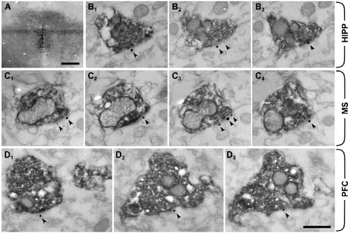
For a summary of the primary antibodies used, see Table 1. The specificity of the antibodies for vesicular glutamate transporter type 3 (vGluT3), neuroligin 2 (NLGN2) and C-terminus of the NMDAR subunit GluN2A was tested extensively before using wild-type and vGluT3-/- , NLGN2-/- or GluN2A-/- null-mutant mice, respectively (Watanabe et al. 1998, Szabadits et al.
2011, Takács et al. 2013). To further test the anti-GluN2A antibody under our experimental conditions, we measured the linear density of immunogold particles on the presynaptic plasma membrane of identified type I synapses established on spines in every reaction using the ImageJ image analyzer software (NIH, USA), and this was considered to be the background labelling. We compared this to the linear density of the immunogold particles on the postsynaptic membrane of the very same type I synapses and on the postsynaptic membrane of the immunoperoxidase-labelled synapses on the very same reactions. We found that the
linear density of the immunogold particles on the postsynaptic side was 30-270 times higher compared to the background, demonstrating that the background labelling is negligible (Figure 10). The anti-serotonin antibody is characterized by Shutoh et al. (2008), and we also detected a typical staining pattern for serotonin, which further confirms its specificity.
Secondary antibodies were also extensively tested for possible cross-reactivity with the other secondary or primary antibodies, and possible tissue labelling without primary antibodies was also tested to exclude specific background labelling by the secondary antibodies. No specific- like staining was observed under these control conditions.
Table 1: Antibody specifications
Raised in
Raised
against Dilution Source
Catalog number
Lot
number Characterized Rabbit Neuroligin 2 1:600
Synaptic
Systems 129 203 10
Takács et al.
2013 Rabbit GluN2A
1:150- 1:250
gift of M.
Watanabe - -
Watanabe et al. 1998 Guinea-
pig vGluT3
1:3000-
1:4000 Millipore AB5421 LV1453193
Szabadits et al.
2011 Rabbit Serotonin 1:10000
ImmunoStar
Inc. 20080 1131001
Shutoh et al.
2008
Double labelling immunoelectron microscopy
For NLGN2 and GluN2A immunolabelling, after perfusion of 4 mice and removal of their brains from the skull, coronal hippocampal sections were cut on a Leica VT1200S vibratome at 50 μm. This was followed by washing out of fixative in 0.1 M phosphate buffer (PB) for 1h. Then sections were cryoprotected sequentially in 10% (overnight) and 30% (2h) sucrose in PB and freeze-thawed three times over liquid nitrogen. For synaptic detection of NLGN2, sections were washed subsequently in 0.1 M PB following tris buffered saline (TBS) and blocked in 1% HSA (Sigma-Aldrich) in TBS. Then they were incubated in a mixture of primary antibodies for NLGN2 (rabbit, 1:600) and vGluT3 (guinea pig 1:3000) diluted in TBS. For synaptic detection of NMDARs, pretreatment with pepsin was essential (Watanabe et al., 1998). Sections were incubated in 0.2 M HCl solution containing 1 mg/ml pepsin (Dako) at 37C° for 10 min. They were then incubated in 50mM glycine (Sigma-Aldrich) and blocked in 1% HSA in TBS, followed by incubation in a mixture of primary antibodies for GluN2A subunit (rabbit;1:250) and vGluT3 (guinea pig; 1:4000) diluted in TBS containing 0.1% BSA-c (BSA-c/TBS; Aurion) for 72 h. After repeated washes in TBS or BSA-c/TBS, respectively, sections labeled for NLGN2 were incubated in blocking solution (Gel-BS) containing 0.2% cold water fish skin gelatine and 0.5% HSA in TBS for 1h. Sections labeled for GluN2A were washed intensively in BSA-c/TBS. After this, both series of sections were incubated in mixtures of secondary antibody solutions containing 1.4 nm gold-conjugated goat anti-rabbit (Nanogold Fab’conjugated antibody; NanoProbes; in a concentration of 1:100 in NLGN2 experiments and 1:300 with GluN2A stainings) and biotinylated goat anti guinea- pig (Vector, 1:200) diluted in Gel-BS or BSA-c/TBS, respectively, for 24 h. After intensive washes in TBS or BSA-c, the sections were treated with 2% glutaraldehyde in 0.1 M PB for 15 min to fix the gold particles into the tissue. This was followed by incubation in avidin–
biotinylated horseradish peroxidase complex (Elite ABC; 1:300; Vector Laboratories) diluted in TBS for 3 hours. The immunoperoxidase reaction was developed using DAB (3,3- diaminobenzidine; Sigma-Aldrich) as chromogen. This was followed by incubation in silver
enhancement solution (SE-EM; Aurion) for 40 min at room temperature. The sections were treated with 0.5% osmium tetroxide in 0.1 M PB on ice and were dehydrated in ascending alcohol series and in acetonitrile and embedded in Durcupan (ACM; Fluka). During dehydration, the sections were treated with 1% uranyl acetate in 70% ethanol for 20 min.
After this, 100-nm-thick serial sections were prepared using ultramicrotome (Leica EM UC6) and picked up on single-slot copper grids. The sections were examined using a Hitachi H- 7100 electron microscope and a Veleta CCD camera.
Anterograde tracing combined with immunoelectron microscopy
The stereotaxic injection of 10kDa BDA, perfusion and pretreatment of the sections before incubating in primary antibody solutions was performed as described above. This was followed by incubation of the sections in a primary antibody solution containing the rabbit anti-GluN2A antibody (1:250) diluted in BSA-c/TBS for 72h. After subsequent washes in BSA-c/TBS, sections were incubated in a secondary antibody solution containing 1.4 nm gold-conjugated goat anti-rabbit antibody (1:300) for 24h. The following immunoperoxidase reaction and silver-intensification was performed as described above.
RESULTS
Result I.: vGluT3 containing terminals establishing synapses on interneurons located in the distal dendritic regions of the hippocampus originate from the median raphe
There are two sources of vGluT3-containing terminals in the hippocampus: a) the first originates from basket cells and establishes synaptic contacts in the stratum pyramidale on the perisomatic region of pyramidal cells; b) the second originates from MR and establishes synapses on the somata and proximal dendrites of interneurons located in the outer third of the stratum radiatum and in stratum lacunosum-moleculare (Somogyi et al. 2004). However, it might happen, that some terminals of the vGluT3-positive basket cells climb to the distal dendritic regions; to determine the exact distribution of these latter terminals in the distal dendritic layers, we performed double immunogold-immunoperoxidase labelling for neuroligin 2 (NLGN2) and vGluT3. NLGN2 is a postsynaptic transmembrane protein present in the GABAergic and cholinergic synapses (Takács et al., 2013), and it was never shown to be present in serotonergic or glutamatergic synapses (Varoqueaux et al. 2004). At the light microscopic level, we always found the pattern of vGluT3-labelling, as described before (Somogyi et al. 2004); briefly, two dense band of terminals in the strata pyramidale and lacunosum-moleculare could be observed, and in every section there were several strongly immunopositive interneurons. At the electron microscopic level, NLGN2 labelling was associated to postsynaptic membranes. We found in the CA1 region that only the 6.1% and 12.8% of the examined vGluT3-positive synapses were NLGN2-positive (49 and 39 serially reconstructed synapses in two mice, respectively; Figure 10. A). In contrast, the adjacent vGluT3-negative (presumably GABAergic) terminals were always NLGN2-positive (Figure 8. D-E). These results show that the majority of the vGluT3-positive terminals located to the distal dendritic regions of the CA1 originate from the median raphe.