Cholinergic modulation of distinct types of perisomatic region targeting interneurons and their involvement in carbachol-
induced fast network oscillation in the CA3 region of the hippocampus
Ph.D. Dissertation Gergely Szabó Semmelweis University
János Szentágothai Doctoral School of Neurosciences
Supervisor: Norbert Hájos Ph.D., D.Sc.
Institute of Experimental Medicine Hungarian Academy of Sciences Laboratory of Network Neurophysiology
Official Reviewers of the Ph.D. Dissertation:
Gábor Czéh Ph.D., D.Sc.
Zita Puskár Ph.D.
Members of the Theoretical Examination Board:
Béla Halász Ph.D., D.Sc. - Chairman József Kiss MD., Ph.D., D.Sc.
György Karmos MD., Ph.D.
Budapest
2012
TABLE OF CONTENTS
TABLE OF CONTENTS __________________________________________________ 2 ABBREVIATIONS_______________________________________________________ 4 I. INTRODUCTION______________________________________________________ 7 Reviewing the literature___________________________________________________ 8 I/1. Role of hippocampus in memory functions and spatial navigation _________________ 8 I/2. Hippocampal anatomy: structure, connections, cell types _______________________ 12 I/3. Network activity patterns correlate with hippocampal function __________________ 27 II. AIMS OF THESIS____________________________________________________ 41 III. MATERIALS AND METHODS _______________________________________ 42 III/1. Experimental animals and ethical approval _________________________________ 42 III/2. Slice preparation for in vitro physiology ____________________________________ 42 III/3. Paired recordings _______________________________________________________ 43 III/4. Recording oscillations in slices ____________________________________________ 44 III/5. Measurements of evoked and miniature events ______________________________ 45 III/6. Post hoc anatomical identification of interneurons____________________________ 45 III/7. Identification of FSBCs and AACs using double immunofluorescent labelling _____ 45 III/8. Data analysis and materials_______________________________________________ 46 IV. RESULTS __________________________________________________________ 49
IV/1. Identification of different types of perisomatic region targeting interneurons______ 49 IV/2. Characterization of basic properties of synaptic connections between perisomatic region targeting inhibitory cells and postsynaptic pyramidal neurons_________________ 54 IV/3. The ACh receptor agonist carbachol reduces the amplitudes of uIPSCs in a
presynaptic cell type dependent manner _________________________________________ 56 IV/4. Carbachol changes the short-term depression of FSBC– and AAC–pyramidal cell synapses in a frequency-dependent manner ______________________________________ 59 IV/5. Asynchronous GABA release from RSBC terminals shows frequency dependence _ 61 IV/6. Synaptic cross-talk between terminals of AACs may elongate the decay of synaptic currents____________________________________________________________________ 63 IV/7. Perisomatic region targeting inhibitory cell types have distinct firing characteristics during CCh-induced network oscillations________________________________________ 67 IV/8. DAMGO, an opioid receptor agonist reduces CCh-induced fast network oscillations via µ-opioid receptors ________________________________________________________ 74 IV/9. µ-opioid receptor activation suppresses synaptic inhibition causing desynchronization of pyramidal cell activity______________________________________________________ 77
IV/10. Activation of µ-opioid receptors reduces inhibitory transmission, but leaves
excitatory transmission and pyramidal cell properties intact ________________________ 80 IV/11. GABA release from FSBC and AAC terminals is differently affected by DAMGO application in the presence of carbachol _________________________________________ 83 V. DISCUSSION________________________________________________________ 86
V/1. Special properties of perisomatic inhibition (Study I.)__________________________ 86 V/2. Involvement of perisomatic inhibition in generating fast network oscillations ______ 91 (Study II)___________________________________________________________________ 91 V/3. Functional implications ___________________________________________________ 95 VI. SUMMARY ________________________________________________________ 98 VII. ÖSSZEFOGLALÁS _________________________________________________ 99 VIII. LIST OF REFERENCES ___________________________________________ 100 IX. ACKNOWLEDGEMENTS___________________________________________ 125 X. LIST OF PUBLICATIONS ___________________________________________ 126
ABBREVIATIONS
5HT-3 receptor: 5-hydroxy-triptamine receptor type 3 AAC: axo-axonic cell
ACh: acetyl-choline
ACSF: artificial cerebrospinal fluid
AF/DX116: M2 receptor preferring antagonist AIS: axon initial segment
BP: band pass CA: cornu Ammonis
CB1R: cannabinoid receptor type I CCh: carbachol
CCK: cholecystokinin CR: calretinin
CTAP: D -Phe-Cys-Tyr- D -Trp-Arg-Thr-Pen-Thr-NH2; MOR antagonist DAMGO: [D-Ala2,N-Me-Phe4,Gly5-ol]enkephalin acetate, MOR agonist DG: dentate gyrus
DSI: depolarization induced suppression of inhibition EC: entorhinal cortex
ECoG: electro-corticogram EEG: electro-encephalogram
eGFP: enhanced green fluorescent protein EPSP: excitatory postsynaptic potential FSBC: fast spiking basket cell
GABA: γ-aminobutyric acid
GAD65: glutamate decarboxylase-65
GIRK: G protein-activated inwardly rectifying K+ current HIPP cell: hilar perforant path-associated cell
IPSC: inhibitory postsynaptic current ISI: interneuron-selective interneuron KCC2: K+-Cl-cotransporter type 2
KO: knockout
LFP: local field potential LIA: large irregular activity LTP: long term potentiation
M1-5 receptor: muscarinic acetylcholine receptor type 1-5 mAChR: muscarinic acetylcholine receptor
mEPSC: miniature excitatory postsynaptic current MFA cell: mossy fiber associated cell
mGluR: metabotropic glutamatergic receptor mIPSC: miniature inhibitory postsynaptic current MOR: µ-opioid receptor
MS-DBB: medial septum-diagonal band of Broca nAChR: nicotinic acetylcholine receptor
NBQX: 2,3-dihydroxy-6-nitro-7-sulfamoyl-benzo[f]quinoxaline-2,3-dione; an AMPA receptor antagonist
NGS: normal goat serum
O-LM cell: oriens-lacunosum-moleculare cell PB: phosphate buffer
PET: positron emission tomography PV: parvalbumin
QX-314: [2(triethylamino)-N-(2,6-dimethylphenyl) acetamine; a voltage dependent Na+ channel blocker
RCA neuron: recurrent collateral-associated neuron REM: rapid eye movement
RLM cell: radiatum-lacunosum moleculare cell RSA: rhythmic slow activity
RSBC: regular spiking basket cell
SCA cell: Schaffer collateral-associated cell SD: standard deviation
SEM: standard error of mean SOM: somatostatin
Str: stratum
SWR: sharp wave-associated high-frequency ripples
T50 value: the width of currents at the half of the peak amplitude TTX: tetrodotoxin
uIPSC: unitary inhibitory postsynaptic current VGLUT: vesicular glutamate transporter VIP: vasoactive intestinal polypeptide WT: wild type
I. INTRODUCTION
In cortical structures only every fifth neuron is GABAergic (Somogyi et al., 1998), yet these cells significantly influence information processing in neuronal networks (Miles et al., 1996; Pouille and Scanziani, 2004). Based on the target preference the cortical GABAergic cells can be divided to cells innervating the perisomatic region of principal neurons and to cells targeting their dendrites. Perisomatic region targeting inhibitory cells can effectively modulate the generation of sodium-dependent action potentials, being able to determine the output of principal cells. On the other hand, dendrite targeting neurons are believed to be responsible for the control of the efficacy and plasticity of glutamatergic inputs received by dendritic domains (Cobb et al., 1995; Miles et al., 1996; Lovett-Barron et al., 2012). The investigations discussed in this thesis focused on the former neurons.
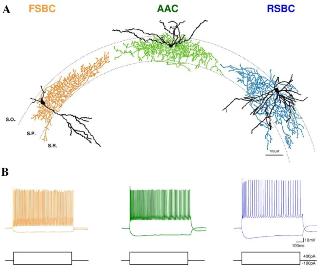
Perisomatic region targeting interneurons can be classified into two neurochemically distinct categories, namely to the group of the calcium binding protein parvalbumin (PV)-expressing cells and to the group of neurons that do not contain parvalbumin, but express the type I cannabinoid receptors (CB1R) on their axon terminals (Freund and Katona, 2007). The cell group containing parvalbumin consists of basket cells with fast spiking phenotype (FSBCs) that innervate the somata and the proximal dendrites of pyramidal cells as well as axo-axonic or chandelier cells (AACs) targeting the axon initial segments (AIS) of pyramidal neurons. GABAergic cells expressing CB1Rs involve basket cells with regular spiking phenotype (RSBCs) innervating the soma-near membrane surface of pyramidal cells.
Cholinergic neuromodulation is known to exert potent effects on different cognitive functions like attention (Deco and Thiele, 2009), learning (Hasselmo, 2006) and REM sleep (Heister et al., 2009). Since GABAergic interneurons are reported to be the target of cholinergic modulation in many ways (Hájos et al., 1998; Fukudome et al., 2004; Neu et al., 2007; Lawrence, 2008), it might be possible that acetylcholine released from the fibers with basal forebrain origin to distinct cortical areas exerts its effect, at least partly, through the influence of GABAergic interneurons. Although several investigations have been carried out regarding the cholinergic receptor composition in interneurons and their sensitivity to cholinergic receptor agonists, the effect of cholinergic receptor activation and its mechanisms have not been tested specifically on distinct types of perisomatic region targeting interneurons.
Since perisomatic region targeting interneurons are capable of synchronizing the firing of large cell assemblies (Cobb et al., 1995), these GABAergic cells may have a pivotal role in generating oscillatory activities in cortical networks. Indeed, recent studies have shown the participation of parvalbumin containing interneurons in the generation of oscillations at gamma (30-80 Hz) frequencies (Cardin et al., 2009; Fuchs et al., 2007), which oscillatory activities often coincide with sensory encoding, neuronal assembly formation, or memory storage and retrieval (Sederberg et al., 2003; Tiitinen et al., 1993;
Montgomery and Buzsáki, 2007). Previous studies, however, have not separated the function of FSBCs and AACs in the generation of gamma oscillations and have not clarified the potential involvement of RSBCs in the oscillogenesis either. The main goal of this thesis was to investigate the effects of cholinergic receptor activation on the properties of perisomatic region targeting interneurons and to reveal their contribution to the generation of cholinergically induced fast network oscillations. These investigations were carried out in a cortical network, in the hippocampus. In the following chapters I will outline the main findings and experiences that paved the road for the experimental work that will be presented in the thesis.
Reviewing the literature
I/1.
Role of hippocampus in memory functions and spatial navigationThe hippocampus and its related structures are one of the most extensively studied areas of the nervous system. This is partly due to its well defined three laminar structure, which shows complex organization together with relatively simple feature compared to other cortical brain regions. Furthermore, the structure of this archicortical brain region is similar in all mammals, in addition, it has several common features with the medial cortex of the reptiles (Northcutt, 1981; Rodriguez et al., 2002; López et al., 2003). These properties together with the unambiguous relation to such basic cognitive functions like learning and memory or spatial navigation rendered the hippocampus to be a popular experimental model of neurobiologists.
The potential role of the hippocampus in certain memory functions has emerged in the middle of the past century, when it was clarified that the damage of the hippocampus and other temporal lobe structures leads to memory loss. The most well known precedent is
related to Henry Gustav Molaison (1926–2008), famously known as H.M., an American memory disorder patient, who was widely studied from late 1957 until his death. In 1953 H.M. had a radical surgery to stop his uncontrollable epileptic seizures. He had an almost complete removal of hippocampi as well as the surrounding cortical structures involving a part of the amygdalar nuclei on both sides (Scoville and Milner, 1957; Corkin et al., 1997).
After his surgery the seizures almost ceased but the operation also caused an unexpected result: he lost the capability to remember new facts about people, places or things after a few seconds. His severe anterograde amnesia accompanied him along his life (Corkin, 2002).
Besides the case of H.M. several studies have come out investigating amnesic patients. These studies reported similar memory deficits with more localized lesions in the hippocampus itself, or in its surrounding areas, such as the parahippocampal cortices, the entorhinal region or in the perirhinal cortical areas (Rempel-Clower et al., 1996; Aggleton and Saunders, 1997; Buffalo et al., 1998). Such human neuropsychiatric observations served as a basis for memory storing theories, suggesting that the function of the hippocampus is to create and store specific memories, which later can be recalled with appropriate (modified or partial) recalling stimuli. Furthermore, the hippocampus may participate in a so called memory consolidation process, during which the repeated reactivation of the memories stored in the hippocampus leads to the reorganization of memory-representations in the neocortex (Káli and Acsády, 2003). As a result, those memories that could originally be recalled only by the hippocampus, progressively become independent on it, and finally may build into the already existing unit of neocortical knowledge stored in the neocortex (Goshen et al., 2011).
Not all kinds of memories are influenced by the hippocampus. Those types of memories that do not require the long-term storage and retrieval of factual events are usually not damaged in amnesia. Typically the short-term or working memory as well as the so called implicit or non-declarative memories including sensory or motor skills, classical operant conditioning and the so called priming remain intact in patients with hippocampal dysfunctions. On the other hand, those memories that are related to specific facts, persons or happenings, mostly intentionally recollected information of previous experiences, are believed to require the participation of hippocampal formation and are typically damaged in amnesia. We refer to this type of memory as explicit or declarative memory (since the encoded information can be explicitly declared) and it consists of two
subtypes, episodic and semantic memory. Episodic memory consists of the recollection of singular events of one’s life, events that can be recalled as personal experiences. Semantic memory can be defined as factual information from one’s past that cannot be necessarily bound to a given event; a knowledge of historical events and figures, knowledge of handling objects, meaning of words and concepts. To date it is a debated question whether episodic and semantic memories are similarly involved in amnesia. According to certain theories the temporal lobe areas including the hippocampus contribute equally to the processing of all kind of declarative memories (Squire, 1992; Squire and Zola, 1998). An alternative view is that the hippocampus is necessary for remembering ongoing life's experiences (episodic memory), but not necessary for the acquisition of factual knowledge (semantic memory; Tulving and Markowitsch, 1998). This idea was proposed by Vargha- Khadem and her colleagues on the basis of their study of three young people suffering from anterograde amnesia caused by hippocampal damage soon after their birth. Although these patients had severely impaired episodic memory, the acquisition of factual knowledge typical of their age was normal (Vargha-Khadem et al., 1997). The finding that in anterograde amnesia it is quite possible for episodic memory to be more severely impaired than semantic memory suggests that semantic memory can occur independently of episodic memory. Taken together, the hippocampus (together with anatomically related structures) is principally essential for episodic memory (Káli and Acsády, 2003).
Ability of navigation in space is another special feature that is attached to hippocampal function. As it is known for some decades, the firing frequency of certain hippocampal pyramidal cells markedly increases, when the animal enters to a given area of its environment. We refer to this area as a place field and to the corresponding cell as a place cell (O'Keefe and Dostrovsky, 1971). The discovery of place cells led to the
“cognitive mapping” theory (O'Keefe and Nadel, 1978), which claims that the hippocampus functions as a cognitive mapping system by creating the spatial representation of the animal’s location relative to its environment. Such a cognitive map would serve to plan the most appropriate track to the place, where the animal wants to go. The most frequently used tool to test spatial navigational skills of experimental animals is the Morris water maze, where the animal has to find a hidden platform to climb out, while swimming in opaque water. The time spent in the water negatively correlates to the extent of spatial representation. Rats learn quickly the location of the platform, even if it is under the water.
However, in an experiment, when the hippocampi were partly isolated from their
connections by cutting the fornices, the rats were unable to find the platform (Morris et al., 1982). In another experiment the hippocampi were transiently inactivated in rats before the learning phase or during the retrieval phase in the maze. The researchers found that the hippocampus is necessary either to learn or retrieve information regarding the location of the platform (Riedel et al., 1999).
Spatial navigation skills have been demonstrated to be related to the hippocampus also in humans. It has long been known that spatial orientation presents difficulty to people with hippocampal damage. When amnesic patients with bilateral hippocampal lesions were studied, they were found to have no information about the spatial environments they contacted after the injury, however, they had precise information about the environment, where they had grown up (Teng and Squire, 1999; Rosenbaum et al., 2000). The involvement of the hippocampus in spatial navigation has also been shown in a line of studies of taxi drivers in London. When structural MRIs of taxi drivers’ brain were compared to control subjects, they were found to have significantly larger posterior hippocampi (Maguire et al., 2000). Interestingly, when taxi drivers were compared to bus drivers, who have to follow a constrained set of routes all day, the authors found that taxi drivers had greater gray matter volume in mid-posterior hippocampi, suggesting that higher spatial knowledge requires higher computational power of the hippocampus (Maguire et al., 2006). In another study of the same group positron emission tomography (PET) was used to examine the brain function in London taxi drivers with many years of experience, while they recalled complex routes around the city. Compared with baseline this resulted in activation of a network of brain regions, including the right hippocampus (Maguire et al., 1997).
Contextual learning is also a typical feature that can be linked to the hippocampus.
For example, it is known for several learning tasks that during the recall the response is much stronger, if the experiment is achieved at the same environment (same context), where the learning has taken place. With lesioned hippocampus this effect could not be observed (Good and Honey, 1991). Fear learning, a phenomenon that is closely linked to the function of amygdalar nuclei, also has a contextual component, which was shown to require intact hippocampus (contextual fear conditioning, Kim and Fanselow, 1992).
In this section I outlined the basic hippocampal functions. Before I would review the literature of the possible cellular mechanisms behind these functions and the network oscillations it is necessary to delineate the structure and anatomy of the hippocampus.
I/2.
Hippocampal anatomy: structure, connections, cell typesAs I mentioned above, the hippocampus is a quite conservative formation, the basic hippocampal architecture is common to all mammals, yet, it nonetheless demonstrates substantial species differences. Since our experiments have been carried out in mice, I disregard to review these differences and restrict to discuss only the anatomy of the rodent hippocampus.
Hippocampal connectivity, principal cells
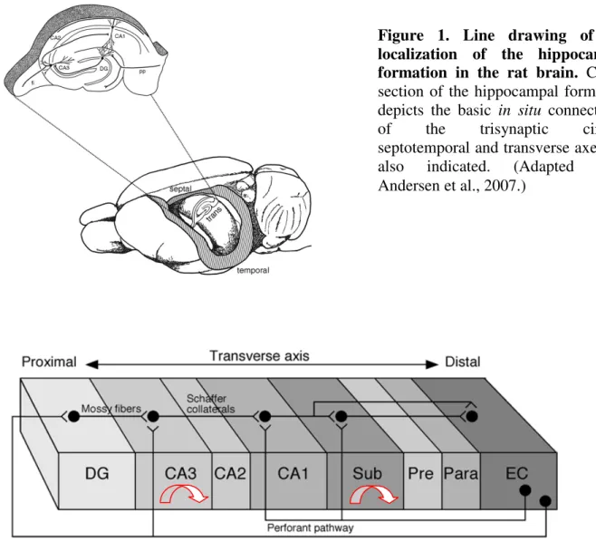
The hippocampus is a part of the middle arc of the limbic system. The crescent- shaped, bilateral structure is localized between the neocortex and the diencephalon (Fig. 1).
Through the fimbria it is reciprocally connected to subcortical areas and to the contralateral hippocampus by the commissural fibers. The only neocortical input originates from the entorhinal cortex, we refer to this as perforant path; the neocortical output mostly consists of indirect or direct projections back to the entorhinal cortex through the subiculum or straight to the entorhinal cortex, respectively (Fig. 2).
Besides its reciprocal connections with the entorhinal cortex, the hippocampus (CA1 region, see later) gives rise to projections to the perirhinal and postrhinal cortices as well as to prefrontal cortex and also to the amygdaloid complex. With the exception of the prefrontal cortex, these areas all project back to the hippocampus (CA1) (Andersen et al., 2007). The hippocampus proper consists of three subdivisions, named after the Cornu Ammonis by Lorente de Nó: CA3, CA2, and CA1. The connected areas together compose the hippocampal formation constituting a functional unit, which includes the dentate gyrus, the subiculum, the presubiculum, the parasubiculum, and the entorhinal cortex.
Hippocampal connectivity has a characteristic feature, which differs markedly from the connections organized in the neocortex. Whereas commonly the neocortical regions are reciprocally connected (Felleman and Van Essen, 1991), this is not true for the connectivity of the hippocampal formation, since as first described by Ramón y Cajal (1893), these connections are predominantly unidirectional (Fig. 2).
The afferent and efferent connections of the hippocampal formation mostly consist of glutamatergic fibers originating from the principal cells, which neurons compose the Figure 2. Scheme of the connections of the different subregions of the hippocampal formation. The information flow through the hippocampus. The hippocampus possesses bidirectional communication with the EC. Layer II cells of the entorhinal cortex (EC) project to the dentate gyrus (DG) and to the CA3 region of the hippocampus via the perforant path. Layer III neurons of the EC project to the CA1 region of the hippocampus via the temporoammonic pathway. The axons of the granule cells in the DG forming mossy fibers innervate the CA3 cells. Besides their dense local recurrent collaterals (indicated by arrows), CA3 pyramidal cells innervate the CA1 neurons via the Schaffer collaterals. Pyramidal cells of the CA1 field of the hippocampus and the subiculum project back to the layer V-VI of the EC. (Adapted from Andersen et al., 2007, modified.)
Figure 1. Line drawing of the localization of the hippocampal formation in the rat brain. Cross- section of the hippocampal formation depicts the basic in situ connectivity of the trisynaptic circuit, septotemporal and transverse axes are also indicated. (Adapted from Andersen et al., 2007.)
85% of the neuronal population in the brain region. The first step in the intrinsic hippocampal circuit is traditionally considered to be the cells in layer II of the entorhinal cortex, due to the fact that much of the neocortical input reaching the hippocampal formation does so through the entorhinal cortex (Andersen et al., 2007). The pyramidal cells of layer II of the entorhinal cortex that project to the dentate gyrus give rise to the major hippocampal input pathway called the perforant path. The dentate gyrus does not project back to the entorhinal cortex.
The principal cells of the dentate gyrus, called the granule cells, give rise to axons called mossy fibers synapsing on pyramidal cell dendrites in the CA3 field of the hippocampus. Mossy fibers do not innervate granule cells but make synapses on the dendrites of the so called mossy cells, which cells are localized in the polymorphic layer of the dentate gyrus (hilus). Mossy cells are glutamatergic and project back to the molecular layer of dentate gyrus and innervate granule cell dendrites both ipsi- and contralaterally.
These cells thus seem to be the major source of the glutamatergic associational/commissural projection to the dentate gyrus (Andersen et al., 2007).
Similarly to the dentate gyrus, the principal cells of the CA3 region do not project back to the granule cells. The output of CA3 pyramidal cells composes the major input of CA1 pyramidal cells, these fibers are called Schaffer collaterals and are also nonreciprocal.
Likewise, CA1 pyramidal cells project unidirectionally to the subiculum or back to the entorhinal cortex. The entorhinal cortex also project to the CA1, but this projection originates from layer III cells and not from layer II, the origin of perforant path. This projection (so called temporo-ammonic pathway) follows a topographical organization: the lateral entorhinal area projects to the distal portion of the CA1 (close to the subiculum), while those fibers that originate in the medial entorhinal area innervate the proximal portion (close to CA2). It has been recently demonstrated that layer III entorhinal cortical cells also project to the CA2 region, thus layer II and layer III entorhinal cortical inputs converge on CA2 pyramidal neurons (Chevaleyre and Siegelbaum, 2010).
The sequential information flow in the hippocampus introduced above is also known as the trisynaptic circuit, involving the entorhinal cortex-dentate-gyrus; dentate gyrus-CA3; CA3-CA1 connections (Fig. 1). The specific features of these connections and the participating hippocampal fields will be discussed in details in the following section together with introducing the here localized interneurons.
Interneurons
Besides principal cells the remaining 15 % of hippocampal neurons are mainly GABAergic neurons (i.e. releasing γ-aminobutyric acid as the main neurotransmitter).
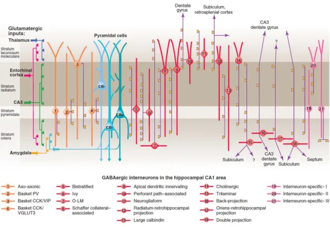
These inhibitory cells are usually defined as interneurons, since the majority of them are local-circuit neurons, although some types with commissural or even extrahippocampal projections are also known. Whereas the glutamatergic cells in the hippocampus show relative homogeneity, interneurons have been found to be much more diverse. By way of example, in a study of CA1 interneurons 21 different types of GABAergic neuron were collected (Klausberger and Somogyi, 2008; Fig. 3).
The categories of interneurons based on morphological characteristics and certain neurochemical marker expressions are fairly similar in all hippocampal regions. Thus, the interneuron repertoire of the CA1 region shown by the review of Klausberger and Somogyi (2008) might be more or less valid to all the hippocampal fields; the homologous cell types of CA1 interneurons often can be found in the CA3, or in the dentate gyrus, even if they have different nomenclature.
The morphological classification of interneurons considers mainly their dendritic and axonal arborization. The dendritic localization provide information about the inputs, whereas the location of axonal terminals shows the outputs of interneurons, allowing us to predict their potential role played in neuronal networks.
Figure 3. Interneuron diversity in the CA1 region of the hippocampus. In 2008 at least 21 different interneuron types were identified by Klausberger and Somogyi. Three types of pyramidal cells were also distinguished based on localization and immunopositivity for the calcium binding protein calbindin. The main targets of glutamatergic inputs are shown on the left. The somata and dendrites of interneurons synapsing on pyramidal cells (blue) are orange, and those innervating mainly other interneurons are pink. Axons are purple; the main synaptic terminations are yellow. Axon terminals originating from different interneuron types innervate the perisomatic region (left) and either the Schaffer collateral/commissural or the entorhinal pathway termination zones (right), respectively.
VIP, vasoactive intestinal polypeptide; VGLUT, vesicular glutamate transporter; O-LM, oriens lacunosum moleculare. Source: Klausberger and Somogyi, 2008.
The comparison of morphological categories based on neurochemical markers produces further distinctions that may reveal differences in neuronal function. For instance, as we will see, the presence or absence of the Ca2+ binding protein parvalbumin assigns basically different functions to morphologically similar cell types.
Based on their axonal arborization hippocampal inhibitory cells can be classified into four main groups:
1) perisomatic region targeting cells, with axons terminating on the perisomatic region of the target neurons;
2) dendrite targeting neurons that innervate the dendritic regions of their target cells;
3) interneuron-selective interneurons that synapse on other inhibitory cells;
4) long-range projecting GABAergic cells that project their axons to other brain regions.
Perisomatic region targeting interneurons, which are the main subject of interest of the thesis, play critical role in regulating the action potential generation in principal cells.
Only single inhibitory postsynaptic potentials (IPSPs) produced by perisomatic region targeting interneurons were shown to be able to suppress repetitive firing of pyramidal cells (Miles et al., 1996).
Perisomatic region targeting interneurons are largely similar in all cortical areas including the hippocampus. All types, that can be distinguished based on either morphology or neurochemical markers, occur in the dentate gyrus and also in the cornu Ammonis. Perisomatic region of pyramidal cells is defined as a domain of plasma membrane, which includes the cell body, the axon initial segment, and the proximal apical and basal dendrites (Freund and Katona 2007). This region is targeted by at least three types of inhibitory neurons, the fast spiking basket cells (FSBC), the regular spiking basket cells (RSBC) and the axo-axonic cells (AAC).
Basket cells by forming a “basket” around the somata of pyramidal neurons give rise to multiple contacts on their membrane (Colonnier, 1968; Freund and Buzsáki, 1996).
In contrast, AACs specifically target the axon initial segments (AIS) of pyramidal cells (Somogyi, 1977). Whereas basket cells innervate not only principal cells but also other interneurons, AACs exclusively target principal cells (Somogyi et al., 1982).
The two basket cell types have characteristically different expression patterns of certain types of receptors, transmitters or modulators (Freund, 2003). For instance, RSBCs are predominantly immunopositive for cholecystokinin and express CB1Rs, whereas FSBCs are known to express the Ca2+ buffering protein parvalbumin (PV) (Pawelzik et al., 2002). RSBCs show regular spiking phenotype and are able to integrate consecutive independent excitatory inputs, which make them fire. In contrast, FSBCs show fast spiking phenotype and respond reliably to subtle and repetitive excitation with precisely timed action potentials (Glickfeld and Scanziani, 2006).
A special perisomatic region targeting neuron type of the CA3 region is called mossy fiber associated (MFA) neuron. Typically, these cells have axons coaligned with the mossy fibers. Their dendrites are located in the strata radiatum and oriens, the axons are terminated on the proximal apical dendritic shafts, and to a lesser degree, on the somata of pyramidal cells being in a position to selectively suppress the dentate gyrus input (Gulyás et al., 1993; Vida and Frotscher, 2000). The boutons of MFA cells are sensitive to CB1R activation (Losonczy et al., 2004). MFA cells show quite similar neurochemical and physiological properties to RSBCs (Szabó GG, Papp O, Hájos N, unpublished observations).
AACs also tend to show fast spiking phenotype and express PV. These cells are particularly in a position to effectively control pyramidal cell firing by targeting the site of action potential generation, i.e. on the AIS (Somogyi, 1977; Somogyi et al., 1982). In spite of their GABAergic nature, in certain conditions their postsynaptic effect were reported to be excitatory, because of the low levels of K+ - Cl - cotransporter (KCC2) in the AIS (Szabadics et al., 2006). Since then, the hyperpolarizing or depolarizing nature of AACs is still a debated question waiting to be resolved (Glickfeld et al., 2009; Woodruff et al., 2010).
Dendrite targeting interneurons compose the majority of inhibitory input in the hippocampus. For instance, in the CA1 region 92% of GABAergic synapses terminates the dendritic region of pyramidal cells (Megiás et al., 2001). The axonal endings of the dendrite targeting interneurons usually terminate on those dendritic membrane surfaces of principal cells, where the specific glutamatergic inputs are received. Therefore they are believed to be responsible for the control of the efficacy and plasticity of excitatory inputs targeting the same dendritic region (Miles et al., 1996).
As mentioned above, the layer II entorhinal cortical input through the perforant path arrives to the dentate gyrus. These fibers synapse on the dendritic spines of the granule cells in the so called molecular layer. The granule cells themselves compose a compact layer called the granule cell layer. The third layer of the dentate gyrus is the hilus or polymorphic layer, which localized next to the CA3 region (Fig. 1). A variety of dendrite targeting interneurons can be found in all the three layers. Similarly to the CA regions, all the dendritic surfaces of granule cells are covered by the axon terminals of different types of dendrite targeting cells (Freund and Buzsáki, 1996). This arrangement probably allows to control all the excitatory inputs received by granule cells (entorhinal afferents, commissural/associational afferents, mossy fiber collaterals). Since the thesis focuses on the CA3 region these interneurons will not be characterized in details, but their organization is well noticable on Figure 4.
The granule cell axons leaving the dentate gyrus enter the CA3 region forming a bunch of fibers, called mossy fibers because of their special synaptic terminals innervating the CA3 pyramidal neurons. The single large boutons of mossy fibers synapsing on the complex branched spines together give rise to the so-called thorny excrescences (Hamlyn, 1962). Because of the lack of myelin the layer formed by this special contact can be visualized in fresh tissue, thus it was named as stratum lucidum. This layer is unique to the CA3, the other two CA regions do not have stratum lucidum, since they do not receive mossy fiber input. The somata of the principal cells constitute the pyramidal cell layer.
CA3 pyramidal cells have typically larger somata compared to the CA2 or CA1 region. The apical dendrites of CA3 pyramidal cells span three strata: the stratum lucidum, the stratum radiatum and the stratum lacunosum moleculare. The basal dendrites extend into stratum oriens towards the alveus. CA3 region consists three subregions: CA3c, b and a (from the hilus towards the CA1, respectively).
20
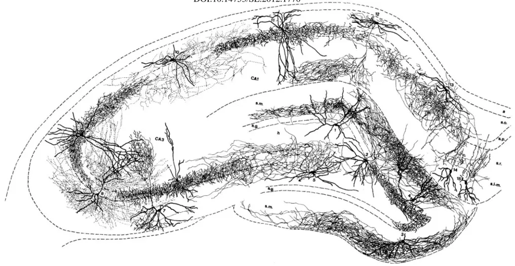
Figure 4. Drawing of different types of interneurons of the hippocampal formation based on camera lucida reconstructions in rodents.
Thick lines represent the dendritic trees, thin ones indicate axons. Note that the different interneurons innervate all the dendritic and perisomatic regions of both granule and pyramidal cells. Most interneurons of the dentate gyrus shown here are not mentioned in the text. 1:
HICAP (hilar commissural-associational pathway related) cell. 2: HIPP (hilar perforant path-associated) cell. 3: MOPP (molecular layer perforant path-associated) cell. 4: VIP-containing basket cell. 5: Trilaminar cell of CA3c. 6: Axo-axonic (or chandelier) cell. 7: 0-LM cell of CA3. 8: Bistratified cell of CA3. 9 Basket cell with axon in CA3 and CAl. 10: Bistratified cell of CA1. 11: Basket cell of CA1. 12: OLM cell of CA1. 13: Horizontal trilaminar cell of CA1. 14,15: IS-2 (interneuron selective) VIP-containing cells. a, alveus; s.o., s.p., s.r., s.1-m., strata oriens, pyramidale, radiatum, and lacunosum-moleculare of the hippocampus; s.m., s.g., h, strata moleculare, granulosum, and hilus of the dentate gyrus. Adapted from Freund and Buzsáki, 1996.
The dendrite targeting interneurons localized here show marked heterogeneity similarly to other parts of the hippocampus. Here I briefly summarize these interneurons with a particular interest to the differences compared to the well documented CA1 region (Klausberger and Somogyi, 2008; Fig. 3.).
The somata and dendrites of Oriens-lacunosum-moleculare cells (OLM cells) are localized in the stratum oriens and their axonal arbor in the stratum lacunosum moleculare (McBain et al., 1994; Sík et al., 1995; Maccaferri and McBain, 1996). They innervate the apical tufts of the pyramidal cell dendrites, being able to influence the excitation caused by entorhinal cortical inputs on pyramidal cells. OLM cells express the neuropeptide somatostatin, a metabotropic glutamate receptor type mGluR1α (Baude et al., 1993) and parvalbumin (PV) (Klausberger et al., 2003). Bistratified cells are mostly located with their cell bodies within or close to the pyramidal layer and synapse onto oblique and basal dendrites of pyramidal cells (Buhl et al., 1994; Klausberger et al., 2004). Similarly to the previously introduced fast spiking basket cells, it expresses PV and shows fast spiking phenotype (Klausberger et al., 2004). OLM and bistratified cells occur both in CA3 and CA1 region.
CA3 (and also CA2) region characteristically possesses extended recurrent collaterals originating from the pyramidal cell axons. These collaterals form dense associational connections in the stratum radiatum and stratum oriens (Li et al., 1994).
Probably the GABAergic regulation of these excitatory inputs is partly achieved by the recurrent collateral associated (RCA) neurons, which cells are located in the stratum radiatum and project its axons to the dendritic regions of pyramidal cells in the stratum radiatum. Similarly, the radiatum-lacunosum moleculare (RLM) cells, with their somata located in the stratum radiatum and their axonal arbor in the stratum lacunosum moleculare, probably participate in the control of incoming information through the perforant path.
Both cell types are CB1R immunopositive (Szabó GG, Papp O and Hájos N, unpublished observations).
The CA2 subfield is a narrow zone of cells interposed between CA3 and CA1 regions. Similarly to CA3, CA2 has pyramidal cells with large cell bodies but is not innervated by mossy fibers like CA1. Its role in hippocampal computation remained elusive, and its presence in the hippocampus is often neglected, however recent studies suggest that the participation of CA2 in hippocampal circuitry has to be reconsidered (Chevalyre and Siegelbaum, 2010; for a review see Jones and McHugh, 2011). The
interneuronal composition of CA2 area seems to be also quite unique (Mercer et al., 2010;
Mercer et al., 2012), but far less data are available regarding inhibitory cells compared to the CA3 or the CA1 region.
Following the route of information flow through the trisynaptic circuit the next station is the CA1 region. Pyramidal cells of the CA3 and CA2 project to the CA1. The fibers of CA3 pyramidal cell axons reaching CA1 compose the Schaffer collaterals and organized in a three-dimensional, topographical fashion.
Similarly to CA3, the input-specified interneurons occur also in CA1. The so called perforant path-associated cells innervate the apical dendritic tufts of the pyramidal cells.
The cell bodies of these cells can mostly be found at the border of the strata radiatum and lacunosum-moleculare, their dendrites can reach all layers or remain in the stratum lacunosum-moleculare (Hájos and Mody, 1997; Klausberger et al., 2005). To the target area of CA3 input the so called Schaffer collateral-associated (SCA) cells are partnered. These interneurons synapse on the oblique dendrites and to a lesser extent also on the basal dendrites of pyramidal cells. The somata of this cell group are mainly located in the stratum radiatum, their dendrites reach all layers (Hájos and Mody, 1997; Cope et al., 2002;
Pawelzik et al., 2002). Additionally, Klausberger also defines a category of the so called apical dendrite innervating cells having quite similar morphology to SCA cells, but innervating mostly the main apical shaft instead of the oblique dendrites (Klausberger et al., 2005).
Specific interneurons, such as neurogliaform cells and ivy cells can also be found both in the CA3 and CA1 region (Price et al., 2005; Szabadics and Soltész, 2009;
Price et al., 2005). Neurogliaform cells uniquely produce slow GABAA and GABAB
receptor-mediated responses on their target cell, release GABA by volume transmission and show gap junctional coupling to each other (Price et al., 2005; Simon et al., 2005; Zsiros and Maccaferri, 2005; Szabadics et al., 2007; Zsiros et al., 2007; Oláh et al., 2009).
Neurogliaform cells can usually be found in the stratum lacunosum moleculare and target the apical tufts of the principal cells (Price et al., 2008). Ivy cells are similar to neurogliaform cells in many aspects but are located in the strata pyramidale or radiatum and innervate the oblique and basal dendrites of pyramidal cells (Fuentealba et al., 2008).
In contrast to the different inhibitory cells introduced so far, in the hippocampus such interneurons also exist that specifically innervate other interneurons. These cells are called interneuron-selective interneurons. Their group may be divided to at least three
different subclasses and a typical common feature of them is their immunopositivity for the Ca2+ binding protein calretinin and/or the vasoactive intestinal polypeptide (VIP)(Acsády et al., 1996a; Acsády et al., 1996b; Freund and Buzsáki, 1996; Gulyás et al., 1996).
In the hippocampus not all the GABAergic cells are local-circuit neurons. Several types of inhibitory neurons have projections to other brain areas, these cells are refered to as long-range projecting GABAergic cells. Long-range GABAergic connections are supposed to be well suited to coordinate the timing of activity between brain regions (Buzsáki and Chrobak, 1995). The most studied remote target of the hippocampal GABAergic projecting neurons is the medial septum. These hippocampo-septal cells can be found in all parts of the hippocampus: in the CA1 region they are mainly located in the stratum oriens; in the CA3 region, they are scattered throughout all the layers; and in the dentate gyrus (DG), they are exclusively located in the hilar area (Totterdell and Hayes, 1987; Jinno and Kosaka, 2002; Gulyás et al., 2003). It has to be mentioned that the hippocampus also receives GABAergic inputs from the medial septum, thus these two areas are reciprocally connected (Raisman, 1966; Takács et al., 2008). Furthermore, septal GABAergic neurons specifically innervate hippocampal GABAergic neurons (Freund and Antal, 1988; Gulyás et al., 1991). Another group of projecting neurons projects to the subiculum. These cells were described mostly in the CA1 stratum oriens (Losonczy et al., 2002; Ferraguti et al., 2005). Moreover, a GABAergic projection from the CA1 region to the retrosplenial cortices were also reported (Jinno and Kosaka, 2002; Van Groen and Wyss, 2003; Jinno et al., 2007). And finally, Attila Sík and his colleagues also described a long-range, cross-regional inhibitiory cell projecting from the CA1 region back to the CA3 and hilar regions (Sík et al., 1994). More recently such somatostatin (SOM) expressing interneurons have also been identified in the CA1 region and also in the dentate gyrus which project back to the medial entorhinal cortex. Additionally, PV expressing GABAergic neurons in the medial entorhinal cortex have been shown to project back to the hippocampus and target on other interneurons, indicating bidirectional entorhinal- hippocampal connectivity (Melzer et al., 2012).
Subcortical connectivity of the hippocampus
Similarly to all cortical areas the hippocampus receives extended subcortical afferents. From the brain stem all the ascending monoaminergic pathways reach the
hippocampal regions. The hippocampus receives noradrenergic input from the locus coeruleus (Jones and Moore, 1977; Moore and Bloom, 1979), serotoninergic fibers from the medial and dorsal raphe nuclei (Freund et al., 1990a; Freund, 1992) and dopaminergic input from the ventral tegmental area (Scatton et al., 1980). All types of monoaminergic molecules released from these fibers were demonstrated to act on cortical principal neurons and influence glutamate release at the presynaptic sites (Scanziani et al., 1993; Aghajanian and Marek, 1999; Chu et al., 2010). Furthermore, neuromodulatory signals from the supramammillary and tuberomammillary nuclei of the hypothalamus reach the CA2 area and also the dentate gyrus (Panula et al., 1989; Maglóczky et al., 1994).
Often the effects of monoamines are not directly exerted on principal cells but transmitted by hippocampal interneurons. For instance, RSBCs that express 5-hydroxy- triptamine-3 (5HT-3) receptors (Morales and Bloom, 1997; Ferezou et al., 2002) are innervated by serotonergic afferents (Freund et al., 1990b), thus they might be able to convey the information carried by the raphe nucleus to all the neurons that are controlled by the basket cells (Papp et al., 1999). Moreover, Varga and his colleagues have recently demonstrated that fibers originating from the median raphe nucleus produce not only a slow modulatory effect but a rapid activation of hippocampal interneurons by glutamate/serotonin co-transmission (Varga et al., 2009).
The most dominant and most studied subcortical input to the hippocampus originates from the medial septum-diagonal band of Broca (MS-DBB), which is a mixed fiber system containing both cholinergic afferents and also reciprocal GABAergic projections (Mosko et al., 1973; Baisden et al., 1984; Amaral and Kurz, 1985; Freund and Antal, 1988; Tóth and Freund, 1992; Tóth et al., 1993). Since the main subject of the current thesis is the cholinergic modulation of interneurons and fast network oscillations, I will introduce this subcortical input and its effect on the hippocampus more detailed. The GABAergic component of MS-DBB input will be presented in the section introducing the generation of the theta (4-8 Hz) rhythm.
Fifty to 75% of the cells in the MS-DBB that project to the hippocampal formation are cholinergic (Andersen et al., 2007). Most of these cholinergic fibers are diffuse and rough in topographical organization (Záborszky et al., 1999). The nature of neurotransmission between these fibers and hippocampal cells is not fully explored or remained controversial (reviewed by Sarter et al., 2009). Several studies concluded that the great majority of cholinergic terminals make synaptic specializations only rarely and rather
influence their targets by volume transmission (Chedotal et al., 1994; Umbriaco et al., 1994; Descarries and Mechawar, 2000; Mechawar et al., 2000). In some studies, septal cholinergic fibers were shown to establish contacts with both principal neurons and interneurons on their cell bodies, dendritic shafts and spines (Frotscher and Léránth, 1985, 1986). Additonally, a few intrahippocampal cholinergic cells were also described (Frotscher et al., 1986). These cholinergic cells also express CR and/or VIP, thus may belong to the interneuron-selective category (Freund and Buzsáki, 1996). In a recent study the authors using molecular-anatomical approaches have demonstrated that the subcellular distribution of the muscarinic receptor type 1 (M1 receptor), which is the most predominant subtype of muscarinic cholinergic receptors (mAChRs) in the hippocampus, suggest that these receptors are likely to be activated by volume transmission (Yamasaki et al., 2010).
Cholinergic neuromodulation has complex effects on both glutamatergic and GABAergic neurons in the hippocampus exerted by the binding of acetylcholine (ACh) either to ionotropic nicotinic receptors (nAChRs) or mAChRs at pre- and postsynaptic locations. Hippocampal principal neurons do not express functional nAChRs at anatomically detectable levels (Frazier et al., 1998b; Frazier et al., 2003), however, physiological studies suggested the existence of the α7 subunit containing nAChRs on glutamatergic terminals in the hippocampus (Gray et al., 1996; Albuquerque et al., 2009).
Regarding the mAChRs, with the exception of M5, all subtypes are expressed in the hippocampus (Levey et al., 1995). While the M1 and M3 types are widely expressed in pyramidal cells, the M2 and M4 receptors are expressed mostly in interneurons (Levey et al., 1995).
Acetylcholine on pyramidal cells usually induces membrane depolarization via the activation of M1 and M3 receptors, both are Gq -linked mAChRs (Young et al., 2005;
Gulledge and Kawaguchi, 2007), although mAChR activation might also cause hyperpolarizing effects on the membranes of pyramidal cells (Gulledge and Kawaguchi, 2007). Gq activation leads to closure of various types of potassium channels (Krnjevic et al., 1971; Nakajima et al., 1986; Guerineau et al., 1994), as a result, depolarizes the membrane of pyramidal neurons and increases membrane resistance (Cole and Nicoll, 1984). In the neocortex this effect has been demonstrated to be clearly due to the activation of M1 receptors (Gulledge et al., 2009). Beside the effect on excitability mAChRs have also been shown to influence the glutamate release from Schaffer collaterals in the CA1
region suggesting a presynaptic locus of action, which is mediated by M4 receptors (Dasari and Gulledge, 2011).
Moving to the interneuron populations the number of potential targets of cholinergic action is increasing. Hippocampal interneurons can be influenced by activation of nAChRs.
Both pharmacological (Jones and Yakel, 1997) and synaptic activation (Frazier et al., 1998a) of nAChRs produce a brief and fast depolarization or inward current due to their ionotropic nature. Different interneuron types show great variability regarding their responsiveness to nicotinic receptor agonists. According to a study carried out in the CA1 region, interneurons with axons ramifying throughout dendritic layers respond with fast depolarization mediated by α7-subunit containing nACh receptors. Interneurons located in the stratum oriens with axons ramifying in the stratum lacunosum moleculare (OLM cells) display a dual-component response with an initial fast phase mediated by α7-subunit containing nAChRs followed by a non- α7 nAChR-subunit dependent depolarizing phase.
Perisomatic region targeting interneurons were found to be insensitive to nAChR agonists (McQuiston and Madison, 1999b).
Activation of mAChRs influences inhibitory transmission in a similarly complex manner. For instance, as it has been demonstrated, pharmacological activation of mAChRs increases the frequency and amplitude of spontaneous IPSCs (Pitler and Alger, 1992a), whilst at the same time depresses monosynaptically evoked IPSCs and the frequency of miniature IPSCs (Behrends and Ten Bruggencate, 1993). This means that mAChRs may affect the function of interneurons at least at two different loci: influence the firing rate by shifting the membrane potential (postsynaptic effect); and control the release of GABA by acting on presynaptically localized receptors.
Regarding the postsynaptic effects of mAChRs, CA1 interneurons have been shown to exhibit a wide range of responses to muscarinic receptor agonists; while some interneurons hyperpolarize, others exhibit a biphasic response, or do not respond.
Muscarinic receptor activation increases the excitability of the majority of interneurons in all layers of the CA1 region by shifting the membrane potential to more positive values (Parra et al., 1998; McQuiston and Madison, 1999a). In the interneurons localized in the stratum oriens, these depolarizing effects have been shown to be due to the action of M1 and M3 receptors (Lawrence et al., 2006). Regarding other layers of the CA1, membrane potential of regular spiking basket cells (RSBCs) and Schaffer collateral associated neurons has been demonstrated to be controlled via M3 mAChRs, whereas the parvalbumin (PV)
expressing fast spiking basket cells (FSBCs) solely via M1 mAChRs (Cea-del Rio et al., 2010; Cea-del Rio et al., 2011).
Also the transmitter release from GABAergic terminals is controlled by mAChRs.
In these processes usually the M2 and M4 receptors participate. In perisomatic region targeting interneurons M2 receptors have been shown to be expressed at the parvalbumin positive axon endings (Hájos et al., 1998), where they can effectively reduce GABA release (Fukudome et al., 2004) via triggering G protein-activated inwardly rectifying K+ (GIRK) currents (Ben-Chaim et al., 2003).
An indirect action of muscarinic receptor agonists that affect presynaptic GABA release is also known. Activation of postsynaptically localized M1/M3 receptors on pyramidal cell membrane may trigger the release of endogenous cannabinoid molecules (endocannabinoids), which binding to CB1Rs effectively inhibits the release of GABA from cholecystokinin (CCK) expressing terminals (Fukudome et al., 2004; Lawrence, 2007;
Neu et al., 2007).
Taken together, the massive variety of targets where acetylcholine might exert its effect suggests that if we want to make predictions about the functions of such a complex phenomenon like cholinergically-induced network oscillations, it is crucial to know how the different elements of the network behave during cholinergic receptor activation.
I/3.
Network activity patterns correlate with hippocampal functionOscillatory activities in the hippocampus and their potential roles in higher order cognitive functions
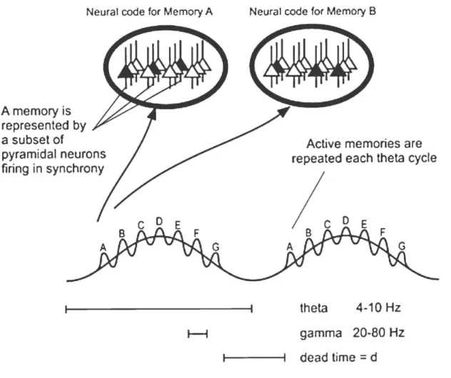
The encoding capacity of cortical regions becomes several fold higher, if the information to be stored is based not only on the firing frequency (rate coding), but also on the relative temporal position of action potentials (phase coding). Therefore, precise timing of action potentials at the subsecond range is likely to be essential for information processing in neuronal networks (Káli and Acsády, 2003). But what could serve as a time signal for the neuronal activity to be organized around? To arrange neuronal activities in a proper order, the nervous system generates sub-threshold membrane potential fluctuations by which many cells will be close to the firing threshold at the same time, thus enabling the
synchronization of large cell assembles. Hence, not only the firing frequency of a given cell, but also the time relative to the fluctuating activity of cell populations might carry specific information. The alternating firing and non-firing periods of cell populations produce synchronized synaptic potentials through the surface membranes of targeted neurons, which are manifested as oscillatory pattern on the electroencephalogram (EEG) or electrocorticogram (ECoG) (Buzsáki, 2006).
The functions of the hippocampus have been thoroughly examined in living animals, either in anesthetized or in freely-moving conditions. These in vivo experiments revealed that the oscillatory activities observed in the hippocampal field potential are correlated to the animal’s behavior. In rodents, theta (4–10 Hz) oscillations typically occur during exploratory behavior and rapid eye movement sleep (Grastyán et al., 1959;
Vanderwolf, 1969; Soltész and Deschénes, 1993). Sharp wave-associated high-frequency ripples (SWRs, 120–200 Hz) of 100ms duration occur during slow-wave sleep, awake inactivity, and consummatory behaviors (Buzsáki et al., 1983). Additionally, in awake brain, oscillations at gamma frequencies (30–100 Hz) can also be observed embedded in theta activity, following sharp-waves as well as gamma rhythm may follow or precede epileptic activity (Traub et al., 1996; Bragin et al., 1997; Traub et al., 2001; Buzsáki et al., 2003) (Fig. 5).
These hippocampus related activity patterns can also be recorded in humans. For instance, hippocampal rhythmic slow activity (RSA) rhythm during rapid eye movement (REM) sleep has been reported to occur in humans (Bódizs et al., 2001). Although human RSA activity shows lower frequency (1.5-3 Hz) compared to the theta recorded in rodents, it is considered to be the counterpart of the mammalian theta (Bódizs et al., 2001; Buzsáki, 2002; Buzsáki et al., 1986). Nevertheless, it has to be mentioned that such activity has been recorded in the human hippocampus during virtual movement that has fallen into the theta range (4-8 Hz; Caplan et al., 2003; Ekstrom et al., 2005). SWRs may be observed also in humans, particularly during non-REM but also during REM sleep and awake (Staba et al., 2004), together with interictal epileptic spikes (Bragin et al., 1999), or temporally associated to sleep spindles (Clemens et al., 2007). Hippocampal gamma rhythm can be recorded during short-term memory tasks or memory recall in humans (Fell et al., 2001;
Sederberg et al., 2003).
These behavior related network activities reflect the underlying mechanisms of information processing in the hippocampus, so investigating the origin of these network
activities may help understanding hippocampal function. In the following section the current knowledge regarding the mechanisms behind these phenomena and their role in hippocampal computation will be briefly summarized.
Theta rhythm and sharp waves – possible functions
Theta rhythm has been shown to be particularly prominent in association with voluntary movement (Vanderwolf, 1969; Whishaw and Vanderwolf, 1973), including free running and rearing (O'Keefe and Nadel, 1978). These activities are collectively termed as exploratory behavior, during which the animal gains new information about the environment. Considerable research has also focused on the correlation of theta rhythm with learning and memory (Berry and Thompson, 1978; Winson, 1978). Lesions of the medial septum and fornix reduce hippocampal theta power (Rawlins et al., 1979) and cause impairments in a number of memory-guided tasks, for instance in spatial reversal in a T- maze. These impairments appear specific to recent, episodic memory, since fornix lesions do not impair the initial learning of a goal location, but impair the learning of reversal (M'Harzi et al., 1987).
These and similar observations have contributed to the widely accepted hypothesis that theta activity represents the first phase of storing episodic memories: during theta the environment to be learned is represented in the hippocampus, which may induce a weak and transient potentiation of connectivity in a subgroup of CA3 pyramidal cells. This would be the memory acquisition stage (Buzsáki, 1989). The next step is the memory consolidation process, when the weakly changed synaptic weights are consolidated. This process takes place during sharp wave ripple oscillations (SWRs) (Buzsáki, 1986) (Fig. 5).
SWRs having 50-150 msec duration usually occur embedded in the non-rhythmic irregular pattern of the hippocampal field potential, which is called large irregular activity or LIA.
LIA interrupted by SWR activity can be observed during eating, grooming and slow wave sleep. SWRs are the result of the synchronous discharges of numerous pyramidal cells, which, according to Buzsáki’s theory, might be sufficient to induce long term synaptic modifications in the neurons of the CA3 region and in their targets in CA1. Hence, the memory trace initiated during theta oscillation will be imprinted into the hippocampal network by SWRs.
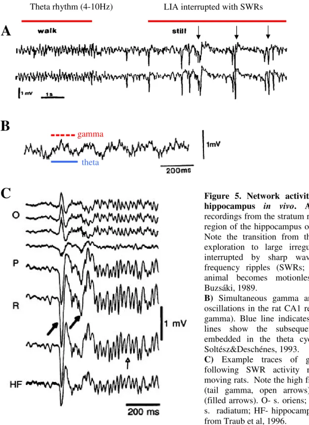
Figure 5. Network activity patterns in the hippocampus in vivo. A) Field potential recordings from the stratum radiatum in the CA1 region of the hippocampus of both hemispheres.
Note the transition from theta rhythm during exploration to large irregular activity (LIA) interrupted by sharp wave-associated high- frequency ripples (SWRs; arrows) when the animal becomes motionless. Adopted from Buzsáki, 1989.
B) Simultaneous gamma and theta frequency oscillations in the rat CA1 region (theta –nested gamma). Blue line indicates a theta cycle, red lines show the subsequent gamma cycles embedded in the theta cycle. Adopted from Soltész&Deschénes, 1993.
C) Example traces of gamma oscillations following SWR activity recorded in freely moving rats. Note the high frequency oscillation (tail gamma, open arrows) following SWRs (filled arrows). O- s. oriens; P-s. pyramidale; R- s. radiatum; HF- hippocampal fissure. Adopted from Traub et al, 1996.
C
B
gammatheta
Theta rhythm (4-10Hz) LIA interrupted with SWRs
A
The idea that SWRs could be capable of inducing long term modifications of synaptic contacts is largely based on studies on long term synaptic plasticity including the widely known phenomenon called long term potentiation or LTP. LTP has been first described in the dentate gyrus of the rabbit (Bliss and Lomo, 1973). Since this study, it has been reported in many brain areas and by using many experimental protocols. LTP is a long-lasting enhancement in signal transmission between two neurons that results from their coinciding activity (Bliss and Gardner-Medwin, 1973). Several methods exist to evoke long-lasting changes in glutamatergic synapses, but the easiest way to induce LTP is to stimulate many incoming fibers with high frequency impulses at the same time to ensure sufficient synaptic input to evoke action potentials postsynaptically (Cooke and Bliss, 2006). Since SWRs reflect synchronous discharge of bulk of pyramidal cells, during SWRs similar processes may take place as during LTP induction protocols. Supporting this idea, during SWRs Ca2+ mediated action potentials have been reported to be generated in the dendrites of CA1 pyramidal cells, which may contribute to the changes of synaptic weights (Kamondi et al., 1998). Besides its role in memory consolidation, SWRs may participate in the information transfer to the neocortex, during which the memory traces produced by hippocampal networks are transported to the neocortex in order to be permanently stored (Chrobak and Buzsáki, 1996; Skaggs and McNaughton, 1996; Nádasdy et al., 1999;
Goshen et al., 2011).
As mentioned above, the encoding capacity might be much higher, if the timing of action potentials also carries specific information. A particularly striking example for the importance of the timing of pyramidal cell firing relatively to theta rhythm is the phenomenon called phase precession (O'Keefe and Recce, 1993). Place cells respond to particular regions of the environment termed as place field. O’Keefe’s observation was that as the rat traversed the place field of a place cell, the phase of firing of that cell relative to the ongoing theta oscillation systematically changed. A place cell, while the animal moving through its place field, fires systematically at earlier theta phase during the subsequent theta cycles till the animal leaves the place field. Consecutive experiments have led to the idea that phase precession reflects the cued recall of upcoming places in a known environment (Jensen and Lisman, 1996; Tsodyks et al., 1996). Thus, the rate code, which expresses the location of the animal in space will be completed with a phase code, which serves as an additional temporal dimension expressing whether the encoded place field belongs to the
left or to the forthcoming places that the animal will reach. Such a mechanism might be involved in the storage of episodic memories, which often involve complex sequences of events (Lisman and Buzsáki, 2008).
Theta rhythm and sharp waves –mechanisms
In general, when considering the mechanisms behind oscillations, it is fundamental to distinguish current generation and rhythm generation. Current generator refers to the transmembrane currents giving rise to the deflections of field potential with a given magnitude, whereas rhythm generator refers to the mechanism that determines the pattern and frequency of oscillatory activities. In the case of theta, extracellular field deflections are assumed to be generated by the summed activity of IPSPs and EPSPs on the somata and dendrites of principal cells, respectively. The interplay between coherent excitatory (from perforant path) and inhibitory (septal connections and local feed-forward inhibition) inputs reaching hippocampal pyramidal cells, functioning as current generators (dipoles), might produce the unique properties of theta pattern appearing on field potential (Buzsáki, 2002).
The origin of theta rhythm genesis is still a question of debate. Several brain regions have been identified to be responsible for, or at least contribute, to the pacemaking activity involved theta oscillation. The rhythm generating properties are usually attributed to interneuron-principal cell networks. For instance, at least three hippocampal interneuron types have riveted particular attention regarding the hippocampal theta rhythm generation.
Perisomatic region targeting interneurons are thought to be important in the timing of action potential generation of pyramidal cells during theta oscillation. Basket and axo- axonic cells discharging rhythmically in the gamma range on the descending phase of pyramidal layer theta provide rhythmic hyperpolarization on the perisomatic region of pyramidal cells. As a consequence, dendritic region will be periodically isolated from somatic regions due to the periodic activation of these interneurons. Another group of interneurons that have been suggested to be involved in hippocampal theta rhythm targets the dendritic region of pyramidal cells. Specifically, OLM and HIPP (hilar perforant path- associated) cells innervate the zones where the entorhinal inputs are terminated on pyramidal cells and granule cells, respectively. Since current flow through the distal dendrites of principal cells is a determining event in generating theta waves, inhibitory control of these inputs might play a crucial role in the regulation of theta rhythm. The third