Distinct behavior and synaptic properties of perisomatic and dendritic-region targeting interneurons during physiological
and pathological network states in the CA3 region of the mouse hippocampus in vitro
Ph.D. Dissertation
Zsolt Kohus Semmelweis University
János Szentágothai Doctoral School of Neuroscience
Supervisor: Attila I Gulyás, Ph.D Official Reviewers: Lucia Wittner, Ph.D
Zoltán Somogyvári, Ph.D
Head of the Final Examination Committee: Pál Czobor, MD, Ph.D Members of the Final Examination Committee: Tibor Zelles, MD, Ph.D
György Karmos, MD, Ph.D
Budapest
2017
2 I. TABLE OF CONTENTS
I. TABLE OF CONTENTS ... 2
II. LIST OF ABBREVIATIONS ... 5
III. INTRODUCTION ... 7
III.1. The hippocamus ... 9
III.1.1. The cytoarchitecture of the hippocampus... 9
III.I.2. Principal cells ... 11
III.1.3. Interneurons of the CA3 region ... 15
III.1.3.1. Perisomatic region-targeting interneurons ... 15
III.1.3.2. Dendritic region-targeting interneurons ... 17
III.1.3.3. Interneuron-selective interneuron ... 20
III.1.3.4. Long-range projecting GABAergic cells ... 21
III.2. Behavior related activity patterns of the hippocampus ... 22
III.2.1. Theta oscillation in the hippocampus ... 24
III.2.2. Gamma oscillation in the hippocampus ... 26
III.2.3. Sharp wave-ripples in the hippocampus ... 27
III.2.4. Pathological oscillation in the hippocampus ... 28
III.3. Forms and mechanisms of short-term synaptic plasticity ... 29
IV. AIMS OF THE THESIS ... 33
V. MATERIALS AND METHODS ... 35
V.1. Animals... 35
V.2. Slice perparation ... 35
V.3. Electrophysiological recordings ... 36
V.3.1. Local field potential recording ... 36
V.3.2. Multichannel local field potential recording ... 37
V.3.3. Optical stimulation ... 37
V.3.4. Paired recordings ... 37
V.3.5. Investigation of short-term plasticity and unitary synaptic properties 38 V.3.6. Stimulation-evoked postsynaptic currents ... 39
V.4. Electrophysiological characterization and clustering of cell types ... 40
V.5. Post hoc anatomical identification of interneurons ... 40
V.6. Modeling the short-term plasticity of inhibitory synapses ... 41
V.7. Asynchronous charge calculation ... 43
3
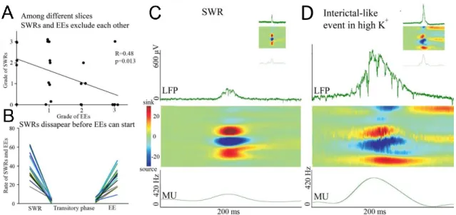
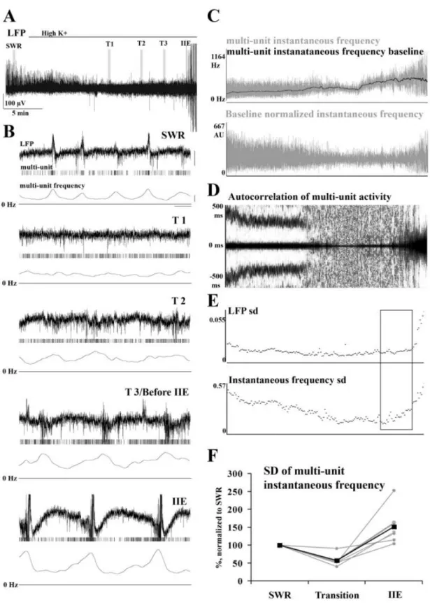
V.8. Statistical analysis ... 44 VI. RESULTS ... 45 VI.1. Physiological sharp wave-ripples and pathological interictal-like events are different forms of transient high activity events in the CA3 region of
hippocampus ... 45 VI.1.1. Induction of transition from sharp wave-ripple to epileptiform events generating state ... 45 VI.1.2. Sharp wave-ripples and high K+ induced interictal-like events are different transient high activities ... 48 VI.1.3. Rearrangement of synchrony during the transitory phase separates sharp wave-ripples from interictal-like events ... 49 VI.1.4. Identification of different classes of CA3 neurons ... 51 VI.1.5. Activity of identified CA3 neurons during sharp wave-ripples and interictal-like events... 53 VI.1.6. Membrane potential changes of hippocampal CA3 neurons during interictal-like events... 57 VI.1.7. Stages of interictal-like events correlate with intracellular potentials of hippocampal neurons ... 60 VI.1.8. High K+ application alters cellular and network parameters ... 61 VI.1.9. The strength and short-term depression of PV+ basket cell –
pyramidal cell synapses is modulated by high K+ application ... 62 VI.2. Properties and dynamics of inhibitory transmission between pyramidal cells and interneurons expressing parvalbumin of cholecystokinin ... 65
VI.2.1. Identification of interneuron-pyramidal cell and interneuron-
interneuron cell pairs in the CA3 region of hippocampus ... 65 VI.2.2. Hierarchical clustering based on electrophysiological properties identifies interneuron types ... 67 VI.2.3. Frequency of interactions among homogeneous and heterogeneous types of inhibitory neurons ... 71 VI.2.4. The type of IN determines the properties of unitary IPSCs onto PCs74 VI.2.5. Cell type-specific short-term plasticity characterizes the synaptic transmission of INs onto PCs ... 75 VI.2.6. Properties of unitary IPSCs among INs are also connection type specific ... 80 VI.2.7. Short-term depression among PV+ basket cell pairs is frequency- dependent ... 82
4
VI.2.8. Interactions among CCK+ interneurons show heterogeneous
dynamics regardless of axonal location ... 83
VI.2.9. Simple mathematical models of short-term plasticity effectively capture the temporal properties of transmission of different types of hippocampal interneurons ... 86
VI.2.10. Possible physiological significance of short-term plasticity in sharp wave-ripple generation ... 91
VII. DISCUSSION ... 94
VII.1. Part I ... 94
VII.1.1. In vitro sharp wave-ripples and high extracellular potassium induced interictal-like events are different network phenomena with distinct properties. ... 94
VII.1.2. The inhibitory control especially from parvalbumin-positive basket cells fails during interictal-like events ... 95
VII.1.3. The changes of cellular and network parameters underlie the transition from sharp wave-ripple generating state to high K+ induced interictal-like event generation state ... 97
VII.2. Part II ... 98
VII.2.1. Clustering based on single-cell electrophysiological properties can reliably separate PV + cells from CCK + cells and PV + BCs from AACs. ... 98
VII.2.2. Interneurons of the same class (except AACs) tend to form reciprocally connected subnetworks. ... 98
VII.2.3. The identity of both the pre- and postsynaptic cells influences inhibitory transmission and its temporal properties ... 99
VII.2.4. Heterogeneities in the release properties of CCK+ interneurons .. 100
VII.2.5. Physiological relevance of inhibitory dynamics among interneurons and their target cells in different network states ... 101
VIII. CONCLUSIONS ... 103
IX. SUMMARY ... 105
X. ÖSSZEFOGLALÁS ... 106
XI. REFERENCES ... 107
XII. LIST OF PUBLICATIONS ... 123
XIII. ACKNOWLEDGEMENTS ... 124
5 II. LIST OF ABBREVIATIONS
4-AP: 4-aminopyridine AAC: axo-axonic cell
ACSF: artificial cerebrospinal fluid AIS: axon initial segment
AM251: N-(Piperidin-1-yl)-5-(4-iodophenyl)-1-(2,4-dichlorophenyl)-4-methyl-1H- pyrazole-3-carboxamide
AMPA: α-amino-3-hydroxy-5-methylisoxazole-4-propionic acid AP: action potential
BC: basket cell
CA1-CA3: Cornu Ammonis fields Calocal: local calcium concentration Cares: residual calcium
CB1R: cannabinoid receptor type I CCK: cholecystokinin
CCK+ BC: cholecystokinin positive basket cell
CCK+ DTI: cholecystokinin positive dendritic region-targeting interneuron CCK+: cholecystokinin positive
DG: dentate gyrus
DsRED: red fluorescent protein
DTI: dendritic region-targeting interneuron EC: entorhinal cortex
EE: epileptiform event EEG: electro-encephalogram
eGFP: enhanced green fluorescent protein
EPI: epilepsy protocol, 30 action potentials at 160 Hz followed by 4 action potentials at 320 Hz
EPSC: excitatory postsynaptic current GABA: γ-aminobutyric acid
HFO: high frequency oscillation IIE: interictal-like event
6 IN: interneuron
IPSC: inhibitory postsynaptic current IS: interneuron-selective interneuron KW: Kruskal-Wallis ANOVA LFP: local field potential
mACSF: modified artificial cerebrospinal fluid MS: medial septum
MW: Bonferroni-corrected Mann-Whitney U test
NBQX: 2,3-dihydroxy-6-nitro-7-sulfamoyl-benzo[f]quinoxaline-2,3-dione; an AMPA receptor antagonist
NMDA: N-methyl-D-aspartate PC: pyramidal cell
PTI: perisomatic region-targeting interneuron PV: parvalbumin
PV+ BC: parvalbumin positive basket cell PV+: parvalbumin positive
PVeGFP mice: transgenic mice expressing enhanced green fluorescent protein controlled by PV promoter
RIP: ripple protocol, 4 action potentials at 160 Hz STD: short-term depression
STF: short-term facilitation SWR: sharp wave-ripple
uIPSC: unitary inhibitory postsynaptic current
7 III. INTRODUCTION
The hippocampal formation is one of the most studied neuronal system in the brain and appears to be crucial for many high order cognitive functions, such as encoding and retrieval of memory and spatial navigation (Buzsáki, 1986; O’Keefe and Recce, 1993).
The hippocampus and related structures show rhythmic oscillatory activities in various frequency ranges in a behavior-dependent manner (Buzsáki and Draguhn, 2004; Singer, 1999), which activity patterns strongly correlate with different cognitive functions. In freely moving rodents, three types of hippocampal oscillations have been observed: theta (5-10 Hz) and embedded gamma rhythms (30-100 Hz) are observed during exploration and rapid eye movement sleep (Bragin et al., 1995; Buzsáki, 1989; Colgin, 2013; Colgin et al., 2009; Csicsvari et al., 2003; Hasselmo et al., 1996). In addition, sharp-wave associated ripples (100-300 Hz) appear during awake immobility, consummatory behavior and slow-wave sleep (Csicsvari et al., 2000; Ylinen et al., 1995).
The alternation of these behavior-associated hippocampal activity patterns is an inherent and general property of the healthy hippocampal network and plays a critical role in information processing and memory consolidation (Girardeau et al., 2009; Jadhav et al., 2012). A key requirement for the generation of network oscillations is the regular and synchronized network activity: the regular and synchronous firing of several neurons generates a rhythmic activation of output synapses providing a periodic fluctuation in the membrane potential of postsynaptic target cells, resulting in a fluctuating field potential signal. All of these network activities require a temporally precise and effective inhibition to control the discharge of pyramidal cells (Aradi et al., 2002; Buzsáki and Draguhn, 2004; Földy et al., 2004; Somogyi and Klausberger, 2005). Several lines of studies show that perisomatic region-targeting interneurons have a crucial role in generating physiological oscillatory activities in the cortical network by providing effective and precisely timed inhibition of large cell assemblies (Gulyás et al., 2010; Schlingloff et al., 2014). However, changes in the balance between excitation and inhibition towards excitation can result in a network hyperexcitability and recurrent pathological seizures – epileptiform events (Florence et al., 2009). Therefore, it is essential to understand the role of individual neurons in the generation of physiological and pathological activity patterns,
8
and to identify the mechanisms leading to transition from physiological to pathological network states.
One way to get closer to understand the genesis of these complex activity patterns is the measurement of relevant network, cellular and synaptic parameters. Hippocampal slices have been proven as a powerful experimental model for investigating the functional properties of hippocampal oscillations and underlying mechanisms. In an improved slice holding chamber, thick (450 μm) hippocampal slices were shown to produce in vivo-like activity levels and patterns including spontaneous sharp wave-ripples and pharmacologically induced gamma oscillation (Hájos and Paulsen, 2009; Hájos et al., 2009). Moreover, they allow rapid pharmacological interventions, optogenetic manipulation, local drug application, and the parallel measurement of network and cellular activity during spontaneous and triggered network oscillations, as well as the interactions among neuronal elements of the circuit (Szabó et al., 2010; Gulyás et al., 2010; Hájos et al., 2013; Schlingloff et al., 2014;).
First we address the questions how the same network can generate physiological and pathological high activity levels and what is the contribution of principal cells and different types of interneurons to the generation of interictal like-events. To answer these questions we induced transition from in vitro sharp wave-ripples to interictal like-events, and analyzed the network, cellular and synaptic parameters leading to the reorganization of synchrony and firing patterns.
The functional connectivity between neurons can give rise to oscillations at different frequencies (Schnitzler and Gross, 2005). To understand the connection trends and interaction among CA3 interneurons, we performed paired recordings between perisomatic and dendritic region-targeting interneurons and their interneuron and pyramidal cell targets, and physiologically and pathologically relevant action potential trains were used to stimulate presynaptic cells. We also fitted dynamic models of synaptic transmission to the data to provide formalized description of inhibitory transmission among cells. Finally, we show how inhibitory dynamics between PV+ basket cells and pyramidal cells could contribute to sharp wave-ripple generation.
Earlier in vitro studies investigating the transmission parameters between interneurons and their target cells were carried out in ‘classical’ artificial cerebrospinal fluid (ACSF) producing reduced neuronal activity in slices (Gulyás et al., 2010; Szabó et
9
al., 2010). Because the activity level in a network can tune the excitability and transmission parameters (Reig and Sanchez-Vives, 2007), in order to mimic in vivo environment, we used thicker hippocampal slices (350-450 μm), and during experiments the ‘classical’ ACSF was switched to modified ‘in-vivo like’ ACSF, that resulted in high, in vivo-like activity patterns.
III.1. The hippocamus
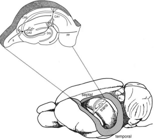
The hippocampal formation, including the dentate gyrus (DG), the hippocampus proper (consisting of the Cornu Ammonis (CA) fields CA1-C3) and the subiculum, is a double C-shaped structure located in the temporal lobe of the brain (Figure 1). The hippocampus and DG are also referred as archicortex composed only of three layers (in contrast to the neocortex which is generally thought to comprise of five or six layers), and has a direct connection to the parahippocampal formation consisting of presubiculum, the parasubiculum, the medial (MEA) and lateral (LEA) entorhinal cortical areas and the perirhinal cortex.
The first layer of the hippocamus is the polymorph deep layer, called stratum oriens.
Superficial to the deep layer lies the CA1-CA3 principal cell layer. The most superficial layer is the the stratum lucidum (CA3), stratum radiatum and stratum lacunosum moleculare (van Strien et al., 2009).
Although there are substantial differences in the cytoarchitecture and connectivity patterns of the hippocampus between different species, the layout of cells as well as afferent and efferent fibers are phylogenetically conserved and are similar in humans and rodents. Since experiments described in this thesis were carried out in mice, the chapters describing the hippocampus (cytoarchitecture, its connectivity, activity patterns, short- term plasticity) are limited to the rodent hippocampus.
III.1.1. The cytoarchitecture of the hippocampus
To understand how the hippocampus maintains its function, a detailed knowledge of the structure-function relationship of microcircuits and individual cell types is required.
Hippocampal neurons can be divided into two main categories based on their main
10
neurotransmitter. The majority of the neurons (~ 85%) are glutamatergic excitatory principal cells (pyramidal cells). On the other hand, the diverse minority of hippocampal cells (~ 15%) are formed by GABAergic cells (releasing inhibitory γ-aminobutyric acid, GABA). They represent a highly heterogeneous cell population with cell bodies scattered through all layers (Freund and Buzsáki, 1996; Somogyi and Klausberger, 2005).
Figure 1. The anatomy of the hippocampus. The drawing depicts a rat brain after the neocortex overlaying the hippocampus was removed, revealing the position and the shape of hippocampus.; TRANS: transverse axis, orthogonal to the septotemporal axis. The inset shows an enlarged section along the transverse axis, with the trisynaptic circuit. CA1, CA2, CA3: cornu ammonis 1, 2, 3 region of the hippocampus; DG: dentate gyrus; PP: perforant path. Adapted from Andersen et al., 2007
11 III.I.2. Principal cells
The principal cells of the CA field are the pyramidal cells located in the stratum pyramidale (or pyramidal cell layer). However, the CA has a heterogeneous structure due to different features of pyramidal cells. Thus, the CA has been described by Lorente de Nó as having three subfields named CA1, CA2 and CA3.
The CA3 region lies proximal to the DG and is subdivided into CA3c, CA3b and CA3a (from the hilus towards the CA1). CA3 pyramidal cells have relatively large cell body (20-30 µm of soma diameter, with two to four time larger surface than CA1 pyramidal cells). The dendritic tree has two distinct domains: the basal dendrites arise from the base of the soma and extend into stratum oriens towards the alveus, whilst apical dendrites descend from the apex of the soma and traverse three strata: the stratum lucidum, the stratum radiatum and the stratum lacunosum moleculare (Spruston, 2008).
The dendrites of CA3 pyramidal cells are densely covered by dendritic spines, serving as postsynaptic targets primarily for glutamatergic terminals. In addition to dendritic spines, CA3 pyramidal cells also have large complex spines called thorny excrescences, which are the synaptic target of mossy fiber boutons. The thorny excrescences are mainly concentrated on the proximal part of the apical dendrites in stratum lucidum (Gonzales et al., 2001). The CA2 and CA1 regions of CA does not receive mossy fiber inputs, therefore they lack the stratum lucidum and CA2 and CA1 pyramidal cells are devoid of thorny excrescences (Dudek et al., 2016; Ishizuka et al., 1995).
CA3 pyramidal cell axons project ispi- and contralateraly to all CA subfields and to the lateral septal nuclei (Ishizuka et al., 1990; Li et al., 1994). They give rise to the Schaffer collateral inputs to CA1 pyramidal neurons, and they form an extensive associational connection between CA3 pyramidal neurons (called recurrent collateral system). Their postsynaptic targets include both pyramidal cells and interneurons (Gulyás et al., 1993a; Li et al., 1994; Miles, 1990; Sik et al., 1993).
The CA2 subfield is a narrow zone positioned between CA3 and CA1. CA2 pyramidal cells have similar morphology to those in CA3, however, they have larger cell bodies than those in CA3, and do not receive inputs from the DG (they lack thorny excrescences). Axons of CA2 pyramidal cells, similar to CA3 pyramidal cells, project to the ipsi- ad contralateral CA1-CA3 areas and contribute to the commissural/associational
12
system (Li et al., 1994; Mercer et al., 2007). CA2 pyramidal cells have also been reported to project back to the medial entorhinal cortex (Hitti and Siegelbaum, 2014; Kohara et al., 2014).
CA1 pyramidal cells tend to be smaller than in CA3. The cell bodies have a diameter of ~15 µm, located in the CA1 stratum pyramidale, however, CA1 pyramidal cells have also been found in the stratum radiatum (Cajal, 1968, Gulyas, 1998) and stratum oriens (Bannister and Larkman, 1995a). CA1 pyramidal cells are not homogeneous but can be grouped by molecular (Baimbridge et al., 1991), morphological (Slomianka et al., 2011) and functional properties (Valero et al., 2015). Another source of variability of CA1 pyramidal cells is the position or depth relative to the cell layer (Bannister and Larkman, 1995a). Early studies have demonstrated segregation of calbindin immunoreactivity in the deep and superficial substrata of CA1 (Baimbridge et al., 1991). It has been also shown, that PV+ basket cells differentiate among deep and superficial CA1 pyramidal cells and provide distinct perisomatic inhibitory interactions (Lee et al., 2014), and they show different intracellular dynamics during sharp wave-ripples (Valero et al., 2015).
The basal dendrites (two to eight) of CA1 pyramidal cells emerge from the soma in the sratum oriens, and the apical dendrites extend into the stratum radiatum and form a dendritic tuft in stratum lacunosum-moleculare (Megías et al., 2001). The dendrites of CA1 pyramidal cells are densely covered with dendritic spines, however, proximal basal dendrites are sparsely spinous (Bannister and Larkman, 1995b; Megías et al., 2001).
CA1 pyramidal cells represent the main output of the hippocampal formation. The local axon collaterals of CA1 pyramidal cells provide a major excitatory input to interneurons, and form synapses onto neighboring CA1 pyramidal cells. However, the recurrent connectivity in the CA1 area is very low (Deuchars and Thomson, 1996). On the other hand, CA1 pyramidal cells heavily project to the subiculum and the deeper layers of entorhinal cortex (Amaral and Witter, 1989). They also send long projections to other brain areas including amygdala, hypothalamus, prefrontal cortex, nucleus accumbens, olfactory regions, auditory cortex, visual cortex, as well as to the lateral septal nucleus and the diagonal band of Broca (Cenquizca and Swanson, 2007).
Within the hippocampal formation, the flow of information is mostly unidirectional, and the special chain of excitatory synaptic connections is also known as the classical trisynaptic hippocampal circuitry (Figure 2). The trisynaptic circuit or trisynaptic loop is
13
a relay of synaptic transmission in the hippocampus, and was initially described by Santiago Ramon y Cajal.
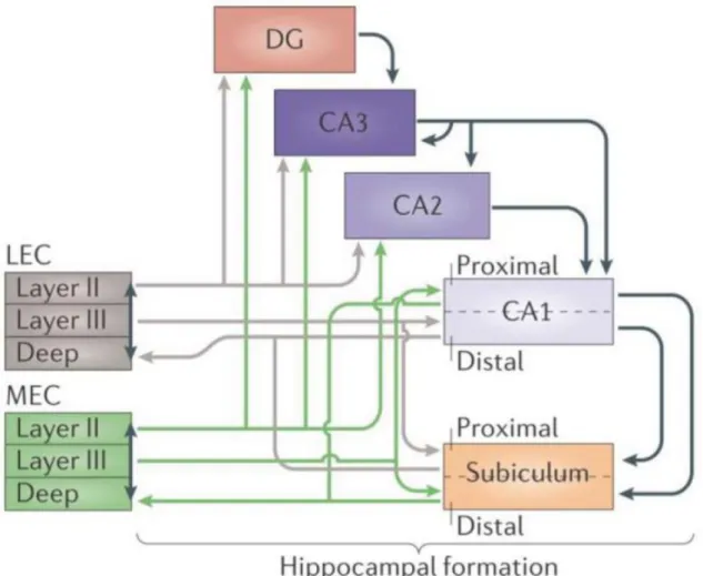
Figure 2. The information flow through the hippocampus. The major cortical input to the hippocampus and the dentate gyrus is the entorhinal cortex (EC) through the perforant path, arising mainly from layers II and III. Medial entorhinal cortex (MEC) layer II provides input to the dentate gyrus (DG), CA3 and CA2, and layer III project to CA1 and subiculum. CA1 and subiculum give rise to output to EC layer V. This connection route (green lines) is paralleled by the route starting in the lateral entorhinal cortex (LEC, gray lines). LEC layer II projects to the outer third of molecular layer of the DG and the outer half of the stratum lacunosum moleculare of CA3, whereas layer II cells of MEC establish connections with the middle third of the molecular layer of DG and inner half of the stratum lacunosum moleculare of CA3. LEC layer III form synapses with CA1 pyramidal cells on the most distal portion of apical dendrites in stratum lacunosum moleculare. The two pathways converge onto single neurons in the DG, CA3 and CA2, but target different neurons in CA1 and subiculum. The axons of granule cells in the DG give rise to mossy fibers and form synapses with CA3 neurons. In turn, CA3 pyramidal cells project to CA1 via Schaffer collaterals and form synapses on the apical and basal dendrites of CA1 pyramidal cells. Note tha the DG and layer II of MEC and LEC also project to CA2, which in turn connects to CA1 pyramidal cells. This suggests that CA2 is also involved in the trisynaptic circuitry). Adapted from Moser et al., 2014.
14
The major input is carried by axons of the perforant path (1st synapse), which convey polymodal sensory information from the neurons of the entorhinal cortex (EC) (layer II). Axons of the medial and lateral EC form excitatory synapses with the outer and middle third of dendrites of granule cells in DG (in stratum moleculare). The layer III of EC also project topographically to the CA1; the lateral EC project to the distal part of CA1, bordering subiculum, while medial EC give rise to fibers terminating in the proximal part of CA1 (bordering CA2). The axons of the granule cells in DG (also known as mossy fibers) innervate the complex spines (thorny excrescences) of proximal and apical dendrites of CA3 pyramidal cells (2nd synapse). The CA3 pyramidal cells, in turn, project to ipsilateral CA1 pyramidal cells through Schaffer collaterals (3rd synapse), and contralateral to CA3 and CA1 pyramidal cell through commissural connections. The CA3 pyramidal cells have an extensive network of recurrent collaterals forming excitatory connections with other pyramidal cells in strata oriens and radiatum. The axons of CA3 pyramidal cells (called Schaffer collaterals) are organized in a three-dimensional, topoghraphic manner. CA3 pyramidal cells located in CA3c region are more likely to innervate CA1 pyramidal cells near the subiculum, whilst CA3 pyramidal cells near the CA2 border (CA3a) are more likely to connect proximal CA1 pyramidal cells, located closer to the CA2 border (Andersen et al., 1971). However, it has recently been shown, that mossy fibers enter the CA2 region and innervate by small boutons the shaft and simple spines of CA2 pyramidal cells. Moreover, the layer II (but not layer III) medial and lateral EC also project to the CA2 region. This suggests an existence of an alternative trisynaptic circuit: DG-CA2-CA1 (Kohara et al., 2014) (Figure 2).
15 III.1.3. Interneurons of the CA3 region
Whereas the majority of principal cells of the hippocampus proper have their cell bodies organized into the pyramidal cell layer, the cell bodies of GABAergic inhibitory cells (interneurons) are scattered throughout all layers. Interneurons represent only ~15%
of the total hippocampal neuronal population, but they show extreme heterogeneity with respect to their morphological and physiological characteristics, as well as with their expression of neurochemical markers and transcription factor (Somogyi and Klausberger, 2005). As the experiments in our studies were performed in the CA3, in this part I will focus on interneurons described within this region.
As the most distinct anatomical feature of interneurons is the layer-specific distribution of their axons and dendrites, the majority of classification schemes have considered mainly their dendritic and axonal arborization. The localization of dendrites provides information about the inputs, while the axonal tree determines the output of interneurons, and predicts their potential role in neuronal networks.
Based on their axonal arborization hippocampal inhibitory cells can be classified into four major groups (Klausberger and Somogyi, 2008):
1. perisomatic region-targeting interneurons, innervating the perisomatic region of pyramidal cells
2. dendritic region-targeting interneurons, innervating the dendritic region of pyramidal cells
3. interneuron-selective interneurons targeting predominantly, if not exclusively other interneurons
4. long-range projecting GABAergic cells, sending long-range projections to other brain areas.
III.1.3.1. Perisomatic region-targeting interneurons
The perisomatic region of pyramidal cells is a defined domain of plasma membrane, including the soma, the axon initial segment, and the proximal and basal dendrites up to 100 μm (Freund and Katona, 2007; Megías et al., 2001). This region is targeted by three types of perisomatic region-targeting interneurons: the parvalbumin positive (PV+) axo- axonic cells (AACs), PV+ basket cells (PV+ BCs) and the cholecystokinin expressing
16
(CCK+) basket cells (CCK+ BCs) (Figure 3). All three types of interneurons often have their soma close to the pyramidal cell layer, but some CCK+ BCs can be found in st.
oriens and str.radiatum . The location of output synapses of perisomatic region-targeting interneurons is well suited to influence the timing and synchronization of action potential firing in a large population of principal cells (Cobb et al., 1995; Freund and Katona, 2007).
Axo-axonic cells are known to innervate almost exclusively the axon initial segment (AIS) of pyramidal cells (Freund and Buzsáki, 1996). Their axonal tree terminates mainly in the pyramidal cell layer, and form radial rows of boutons around the str. pyramidale – str.oriens border. Their radial dendritic tree spans all hippocampal layer with a prominent tuft in str. lacunosum moleculare (Viney et al., 2013). They show fast spiking phenotype (Szabó et al., 2010), and through GABAA receptor-mediated inhibition on the AIS can effectively control the action potential generation of pyramidal cells (Buhl et al., 1994).
The basket cells are named after their axons forming a perisomatic basket around the cell bodies and proximal dendrites of pyramidal cells. They have dense axon collaterals in the principal cell layer, and based on their neurochemical content can be classified as PV+ BCs and CCK+BCs (Freund and Buzsáki, 1996).
Besides anatomical similarities the input and out properties of the two basket cell types are highly dissimilar (Glickfeld and Scanziani, 2006). PV+BCs have fast spiking character and respond reliably to repetitive excitation with short and precisely timed action potentials. Thus, PV+BCs act as rapid signaling interneurons translating precisely the rapid excitatory inputs into fast and short-latency inhibitory outputs (Jonas et al., 2004). In contrast, CCK+BCs show regular spiking phenotype, require extensive integration of excitatory inputs to generate spike trains, and respond less readily to excitatory signals (Glickfeld and Scanziani, 2006). Differences have also been shown at axon terminals of the two basket cell types: P/Q type calcium channels located presynaptically on PV+BC terminals are tightly coupled to vesicle fusion, providing quick, highly synchronized GABA release with low failure rate. In contrast, CCK+ cells express N-type calcium channels located more remotely from the release site. The GABA release from CCK+BC terminals shows strong fluctuation in latency and amplitude, as well as higher failure rate, resulting in weak inhibition with low temporal precision (Hefft
17
and Jonas, 2005). In addition, PV+ BCs and CCK+ BCs are sensitive to different neuromodulators. For example, PV+ BCs express μ opioid receptors that can reduce inhibitory transmission (Gulyás et al., 2010). On the other hand, cannabinoid receptor type 1 (CB1R), are present on presynaptic terminals of CCK+ BCs but not PV+ BCs (Földy et al., 2006; Szabó et al., 2014), and can mediate suppression of GABA release from CCK+ terminals (Földy et al., 2006).
A special type of GABAergic cells in the CA3 region is the mossy-fiber associated (MFA) interneuron. MFA interneurons have extensive axonal arbors in the str. lucidum, co-alignated with the mossy fibers, and often give rise to additional axon collaterals in the hilus. Their dendrites are located in the str.radiatum and oriens. Their axons are terminated on the proximal apical dendritic shafts, and to a lesser degree on the somata of principal cells (Szabó et al., 2014; Vida and Frotscher, 2000). MFA interneurons, similar to CCK+ BCs, are sensitive to CB1R activation (Losonczy et al., 2004; Szabó et al., 2014)
III.1.3.2. Dendritic region-targeting interneurons
The inhibition provided by perisomatic region-targeting interneurons plays a crucial role in the control of action potential discharges. However, the majority of inhibitory inputs are located in the dendritic region, where they often match the spatial distribution of the afferent excitatory pathways (Klausberger, 2009). The layer-specific distribution of dendritic region-targeting interneurons provides spatially defined inhibition and controls the local dendritic electrogenesis of principal cells (Miles et al., 1996a).
The peculiar anatomical arrangement of oriens lacunosum-moleculare (O-LM) cells suggests their unique functional role in the hippocampal network. The cell body and horizontal dendrites bearing elongated filopodia-like spines are found in str. oriens, while their axons project to the str. lacunosum moleculare, where they extensively branch (Hájos et al., 2013) (Figure 3). O-LM cells are positioned to provide inhibition to the excitatory input arriving at the distal dendrites of CA3 pyramidal cells via excitatory entorhinal perforant path afferents (Hájos and Mody, 1997).
The cell bodies of perforant path-associated cells are located in the str. radiatum or str. lacunosum moleculare, often at the border of these two layers (Figure 3). Similar to CCK+ basket cells they express CB1R on their axon terminals (Szabó et al., 2014). Their
18
dendrites can reach all layers or remain in the str. lacunosum moleculare and adjacent str.
radiatum. Their axons terminate in the str. lacunosum moleculare and overlap with the excitatory input from the entorhinal cortex, the perforant path (Hájos and Mody, 1997;
Pawelzik et al., 2002; Vida et al., 1998), where they innervate the apical tuft of pyramidal cells (Klausberger et al., 2005). However, Szabó et al. (2014) found examples of perforant path-associated cells where axon collaterals left str. lacunosum moleculare and formed a meshwork in the str. radiatum close to the border of str. lacunosum moleculare.
Stratum radiatum (R-interneurons) (Figure 3) and stratum lacunosum-moleculare interneurons (LM-interneurons) in the CA3 region were first described by Gulyás et al.
(1993b). The somata of R- and LM-interneurons are positioned approximately 147 and 257 μm from the boundary between str.pyramidale and lucidum, respectively, and approximately 100 μm from the medial extent of the suprapyramidal blade of the DG (Calixto et al., 2008). They are characterized by one to six typically aspiny dendritic tree stemming from the soma, and give rise to elaborated axonal arbors, which often extend beyond their layer of somatic residence: into the str. radiatum (for LM-interneurons) and str. lacunosum-moleculare (for R-interneurons) (Ascoli et al., 2009). It has been shown that R- and LM-interneurons possess voltage-dependent conductances playing role in shaping the temporal integration of converging excitation. Moreover, they can act as a coincident detector for subtreshold input from the perforant path and mossy fibers (Calixto et al., 2008)
Oriens-oriens and oriens-radiatum cell somas are located in str.oriens (Figure 3).
Oriens-oriens cells have smooth dendrites and axons restricted to str.oriens. On the other hand, the rarely ramifying axons of CA3 oriens-radiatum cells could be observed in strata oriens and radiatum, and occasionally penetrating into the CA1 region (Hájos et al., 2013)
19
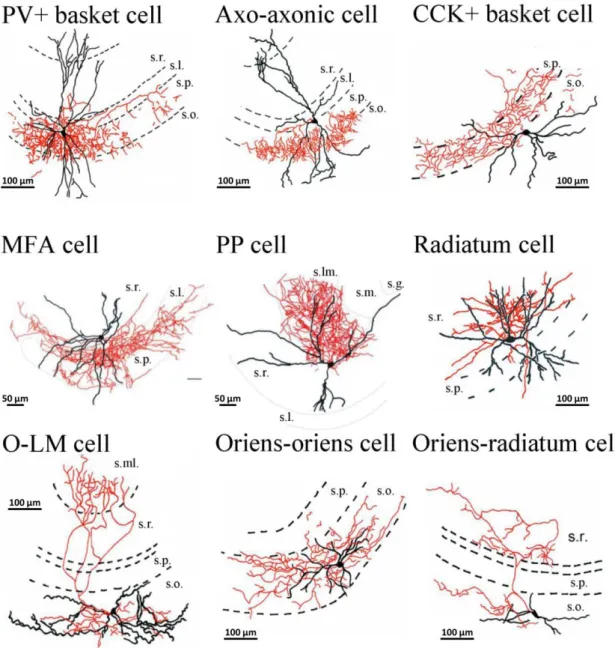
Figure 3. Camera lucida reconstruction of intracellularly labeled CA3 perisomatic and dendritic region- targeting interneurons. In the camera lucida reconstruction, the cell bodies and dendrites are shown in black, while the axon collaterals in red. Note that the majority of axon arbor of perisomatic region- targeting interneurons (PV+ basket cell, axo-axonic cell, CCK+ basket cell) and MFA cell, PP cell, radiatum cell, oriens-oriens cell (dendritic region-targeting interneurons) can be restricted to a given layer. In case of O-LM and oriens-radiatum cells (dendritic-region targeting interneurons) axons could cover a substantially wider range, often penetrating into neighboring strata. s.r., stratum radiatum; s.l., stratum lucidum; s.p., stratum pyramidale, s.o., stratum oriens; s.ml., stratum moleculare; s.lm., stratum lacunosum moleculare, s.g., stratum granulosm; MFA cell, mossy fiber-associated cell; PP cell, perforant path-associated cell; O-LM cell, oriens-lacunosum moleculare cell. Adopted from Hájos et al., (2013) (PV+ basket cell, axo-axonic cell, CCK+ basket cell, O-LM cell, radiatum cell, oriens-oriens cell, oriens- radiatum cell) and Szabó et al. (2014) (MFA cell, PP cell), with permission.
20
Neurogliaform cells are probably the most compact interneurons in the stratum lacunosum moleculare (Capogna, 2011). The soma and dendrites are located within the same layer (Price et al., 2005), with dense axonal arbor in the str. lacunosum molecular and the border of str. lacunosum moleculare and str. radiatum. As far as we know, neurogliaform cells has been described only in the CA1 and DG within the hippocampus (Armstrong et al., 2011; Capogna, 2011; Vida et al., 1998). However, it does not necessarily mean that neurogliaform cells are not present in the CA3 str.lacunosum- moleculare. Ivy cells are closely related to neurogliaform cells, but their soma is located in the strata pyramidale or radiatum. They have dense and fine local axonal plexus innervating predominantly the basal and oblique dendrites of pyramidal cells. Ivy cells and neurogliaform cells express neuropeptide Y and often express neuronal nitric oxide synthase (nNOS) (Fuentealba et al., 2008; Price et al., 2005). They represent the most abundant interneuron class in the hippocampus, comprising 37% of GABAergic cells (Capogna, 2011; Fuentealba et al., 2008).
III.1.3.3. Interneuron-selective interneuron
Interneurons belonging to the same class are often connected by GABAergic synapses and tend to form reciprocally connected networks (Chamberland and Topolnik, 2012). In addition, GABAergic cells receive synapses from a population of inhibitory cells, which specifically innervate other interneurons, the so-called interneuron-selective (IS) interneurons. Gulyás et al. (1996) distinguished two types of calretinin-expressing IS interneurons in the CA3 subfield based on the dendritic morphology and somatic distribution. The cell body and the entire dendritic tree of the first type is restricted to the hilus and str.lucidum of the CA3 subfield, with always myelinated axons, and because of the presence of often branching spines on their dendrites they are named as spiny calretinin-interneurons. The second type is referred to as spine-free calretinin- interneuron. A remarkable feature of calretinin-interneurons is the formation of dendro- dendritic and axo-dendritic contacts with other calretinin-interneurons. Moreover, they also form synaptic contacts with other calbindin, calretinin and vasoactive intestinal peptide-immunoreactive interneurons (Gulyás et al., 1996). Besides calretinin-containing IS interneurons, vasoactive intestinal peptide-positive IS interneurons have also been
21
described. However, these cells were identified in the CA1 region (Acsády et al., 1996;
Chamberland et al., 2010; Tyan et al., 2014).
III.1.3.4. Long-range projecting GABAergic cells
In addition to the local circuit of GABAergic cells, a specific subpopulation of inhibitory neurons sends long-range inhibitory projections to subcortical and other cortical areas. These cells are called long-range projecting GABAergic cells.
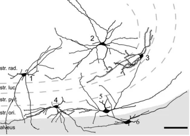
Figure 4. Camera lucida reconstruction of retrogradely labeled CA3 spine-free and spiny hippocampo- septal cells. The soma of spine-free hippocampo-septal (HS) cells are found in strata radiatum (1,2), oriens (4, 5) and alveus (6). HS cells in str.oriens and alveus have horizontally oriented spine-free dendrites located mainly in str.oriens and alveus. The dendrites of spine-free HS cells in the str.radiatum run in all directions and penetrate all layers. Densely-spiny HS cells (3) in the CA3 area located always in the str.lucidum. str.rad., stratum radiatum; sr.luc., stratum lucidum; str.pyr., stratum pyramidale; str.ori., stratum oriens. Scale bar, 100 μm. Adopted from Takács et al., (2008), with permission.
22
The hippocampo-septal cells innervate the most studied remote target of hippocampal GABAergic cells, the medial septum (MS). The sparsely spined hippocampo-septal cell somata are located in str. oriens and str. radiatum, where recurrent axons of pyramidal cells arborize. On the other hand, the soma of densely spined hippocampo-septal cells (characterized by profusely covered dendrites and elongated soma with spines) is observed in str. lucidum (Figure 4) (Takács et al., 2008). They give rise to myelinated axons projecting back to the medial septum-diagonal band of Broca, as well as to local collaterals, where they target local pyramidal cells and interneurons in strata oriens and radiatum (Gulyás et al., 2003; Jinno et al., 2007; Takács et al., 2008).
Hippocampo-septal cells receive inhibitory inputs from the MS, thus these two regions are reciprocally connected (Raisman et al., 1966; Takács et al., 2008). The main postsynaptic targets of hippocampo-septal cells in the MS are PV-expressing GABAergic and cholinergic neurons (Tóth et al., 1993).
CA3 Trilaminar cells show several similarities to O-LM cells: they have cell bodies in str. oriens and their dendritic tree is restricted to this region. However, their axons are distributed to three adjacent layers: strata radiatum, pyramidale and oriens, and also send axon collaterals into neighboring areas (i.e. to CA1 and subiculum) (Gloveli et al., 2005;
Hájos et al., 2004). The postsynaptic targets of trilaminar cells include both pyramidal cells and interneurons (Ferraguti et al., 2005).
III.2. Behavior related activity patterns of the hippocampus
The hippocampus plays prominent role in spatial and episodic memory formation, which processes require a precisely synchronized and coordinated activity of distributed neurons. The alternating firing and non-firing periods of cell populations (including both interneurons and principal cells) are responsible for the synchronized synaptic potentials of targeted neurons, which are manifested as oscillatory patterns on the EEG (Colgin, 2016). In rodents, hippocampal rhythms (EEG patterns) include theta rhythms (4-12 Hz) (Bullock et al., 1990), sharp wave associated ripple oscillation (SWRs, 100-250 Hz ripples superimposed on 0.5-3Hz sharp waves) (Buzsáki, 1986) and gamma rhythms (30- 100 Hz) (Csicsvari et al., 2003) (Figure 5). Each rhythm is observed during particular
23
behavioral pattern, is associated with characteristic neuronal firing properties, and is thought to carry out distinct functions (Colgin, 2016).
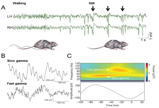
Figure 5. Network activity patterns in the hippocampus in vivo. A: Local field potential recording from symmetrical locations of the left (LH) and right (RH) dorsal CA1 stratum radiatum during locomotion (walking) and awake immobility (still). Note the transition from theta rhythm during exploration to large irregular activity interrupted by sharp wave-ripples (black arrows). Adopted from Buzsáki, 2015. B:
Example of slow gamma (25-50 Hz) and fast gamma (65-140 Hz) oscillation from CA1 stratum pyramidale.
C: Time frequency representation of power for representative recording in B (top), averaged across all theta cycles (bottom). Frequency is plotted on the y axis, time point t = 0 correspond to the theta trough.
Note that slow and fast gamma are associated with different portions of the underlying theta cycle. Adapted from Colgin et al., 2009.
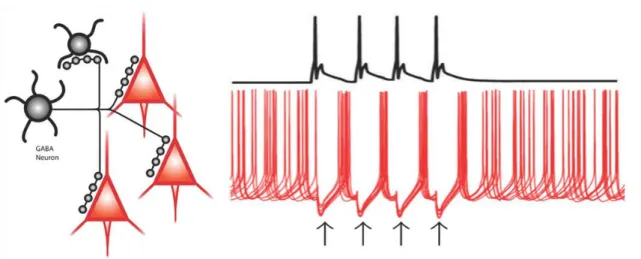
The EEG rhythms originate from the synchronized activity of pyramidal cells reflecting the electrical changes in the extracellular space. During oscillations, the firing of pyramidal cells is synchronized, and this synchrony is mediated by the fast synaptic inhibition originating from GABAergic interneurons (Colgin, 2016) (Figure 5). As single hippocampal interneurons can target multiple postsynaptic cells (including pyramidal cells and interneurons of the same type), the output of many GABAergic neurons could entrain the activity of large number of cells within the network (Figure 6). Thus,
24
GABAergic interneurons contribute to the alteration of firing and non-firing periods of cell populations, resulting in different oscillatory patterns on EEG (Gonzalez-Burgos and Lewis, 2008).
Figure 6. The GABAAR-mediated inhibition provided by GABAergic interneurons is efficient in synchronizing neuronal activity. The diagram on the left indicate that the axons of individual perisomatic region-targeting GABAergic cells (black) form multiple contacts with postsynaptic pyramidal cells (red) and other GABAergic cells. The right panel shows the membrane potential of the GABAergic cell (black trace) and superimposed traces of membrane potential of three pyramidal cells (red) illustrating how the asynchronous firing of pyramidal cells becomes transiently synchronized by the phasic inhibition provided by the GABAergic cell. Shortly after the presynaptic action potential, an inhibitory postsynaptic potential is produced (black arrows) preventing postsynaptic pyramidal cells from firing. When inhibition decays, the pyramidal cells fire nearly in synchrony. Adapted from Gonzalez-Burgos & Lewis, 2008 and Gonzalez- Burgos et al., 2011.
III.2.1. Theta oscillation in the hippocampus
A prominent network pattern in the hippocampus is a slow oscillation in the theta frequency range (4-12 Hz). During wakefulness theta is associated with voluntary movement and exploratory behavior, during which the animal gains new information about the environment (Berry and Thompson, 1978) (Figure 5). Theta also occurs during rapid eye movement sleep (Jouvet, 1969) and occasionally during immobile attention or arousal (Buzsàki and Eidelberg, 1983).
Theta oscillation is the most regular in frequency and has a largest amplitude in the stratum lacunosum-moleculare of CA1 (Kamondi et al., 1998). Besides hippocampus
25
theta has been observed in other brain regions including the subiculum (Paré et al., 2002), striatum (DeCoteau et al., 2007), neocortex (Kahana et al., 2001), hypothalamus (Kocsis and Vertes, 1997) and medial septum (Vinogradova et al., 1995).
The hippocampal theta activity depends critically on the integrity of the afferent inputs from the medial septum (MS). MS contains two major types of neurons that are thought to play distinct roles in the theta rhythm generation: cholinergic cells modulate the activity of hippocampal pyramidal cells (Cole and Nicoll, 1984), whereas GABAergic cells are targeting hippocampal interneurons (Freund and Antal, 1988). It has also been reported, that MS GABAergic cells expressing parvalbumin can drive hippocampal theta activity by rhythmic disinhibition of hippocampal pyramidal cells (Hangya et al., 2009).
Because the largest amplitude theta oscillations are observed in CA1 stratum lacunosum- moleculare (Kamondi et al., 1998), rhythmic excitation of the distal apical dendrites of pyramidal cells by entorhinal afferents is also assumed to play an important role in the theta oscillation generation (Buzsáki, 2002).
Combining these data proposed a model for hippocampal theta generation: the cholinergic neurons of MS provide slow depolarization of their pyramidal cell targets;
meanwhile MS GABAergic neurons mediate rhythmic disinhibition of pyramidal cells.
The rhythmic excitatory postsynaptic potentials provided by the input from the entorhinal cortex are responsible for the current sink in the stratum lacunosum-moleculare (Buzsáki, 2002).
Hippocampal theta oscillation has a well-established role in memory encoding (Buzsáki and Moser, 2013). The impairment in a spatial memory task caused by the lesion of the MS correlates with the reduction of hippocampal theta rhythm (Rawlins et al., 1979; Winson, 1978) and causes impairment in tasks including delayed spatial alternation (Givens and Olton, 1990), delayed non-match to position (Markowska et al., 1989) and spatial reversal (M’Harzi et al., 1987). Moreover, theta may also integrate various types of sensory information received by the hippocampus by linking neurons coding different aspects of the same experience. As theta correlate with movements to acquire sensory stimuli, they are well suited to coordinate multimodal sensory information (such as olfactory and visual inputs) (Colgin, 2016).
26 III.2.2. Gamma oscillation in the hippocampus
Hippocampal gamma rhythms (30-100 Hz) are thought to provide a temporal structure for information processing in the brain, and contribute to cognitive functions, such as memory formation and sensory processing (Bartos et al., 2007) through coordinating cell assemblies (Colgin and Moser, 2010). They are commonly seen superposed onto the theta rhythm during exploration (theta-nested gamma) (Figure 5), but can also be observed in the absence of theta activity in both awake and anesthetized rodents (Csicsvari et al., 2003; Penttonen et al., 1998).
Hippocampal gamma oscillations are thought to be generated in two independent ways (Colgin and Moser, 2010). In behaving rats, the largest estimated excitatory currents during gamma oscillation are observed in the middle third of the DG molecular layer, which is the termination zone for the medial EC projection. In the EC, gamma oscillation arises at high frequency (~ 90 Hz, “fast gamma”), and can spread to the DG and to the CA regions (Bragin et al., 1995). The activity of the lower gamma frequency range (30- 50 Hz) is termed as “slow gamma” and is driven by the CA3 region itself (Csicsvari et al., 2003). The two gamma oscillators have been shown to be independent but still able to couple in time, allowing the alternating processing of information originating from the EC or CA3 region (Colgin et al., 2009).
Current source density analysis of intrinsically generated “slow gamma” oscillation revealed alternating pairs of sinks and sources in the str. pyramidale and radiatum of both the CA3 and CA1 region. The sink-source pairs of CA3 precedes those observed in CA1 suggesting, that the “slow gamma” generated in CA3 entrains CA1 circuitry (Csicsvari et al., 2003). The capability of CA3 to generate gamma oscillation is likely due to the recurrent excitatory collateral system among pyramidal cells (Hájos and Paulsen, 2009).
The rhythmic discharge of perisomatic region-targeting interneurons is also pivotal for the emergence of gamma oscillation within the CA3 network, especially PV-containing inhibitory neurons (Mann et al., 2005; Cardin et al., 2009; Szabó et al., 2010; Gulyás et al., 2010).
27 III.2.3. Sharp wave-ripples in the hippocampus
Sharp wave-ripples (SWRs) are large amplitude, irregularly occurring hippocampal activity pattern observed in animals during waking immobility, consummatory behaviors and slow-wave sleep (Buzsáki, 1986; O’Neill et al., 2006) (Figure 5). The possible role of SWRs originates from theories suggesting their specific role in hippocampus- dependent memory consolidation during sleep (Buzsáki, 1989; Csicsvari and Dupret, 2014). As during SWRs large number of neurons fire synchronous action potentials, it has been suggested that during SWRs the previous network activity is reactivated, which represents memory traces that might undergo memory consolidation and could enable the transfer of memory traces to extra-hippocampal regions (Girardeau et al., 2009; Nádasdy et al., 1999; Wilson and McNaughton, 1994). On the other hand, awake SWRs in particular can reactivate sets of place fields encoding forward and reverse paths associated with both current and past locations. This reactivation during awake SWRs can contribute to learning, memory retrieval and consolidation, as well as trajectory planning (Carr et al., 2011; Dupret et al., 2010; Jadhav et al., 2012; O’Neill et al., 2010).
SWRs in CA1 can be reorganized based on two particular local field potential patterns. While the strong negative deflection with 50-100 ms duration in the CA1 str.
radiatum reflects the synchronous discharge of CA3 pyramidal cells (giving rise to excitatory inputs to CA1 pyramidal cell dendrites), the high frequency ripple oscillation (120-200 Hz) represents the rhythmic activation of interneurons and pyramidal cells to the strong depolarization and synchronous drive (Buzsáki, 2015).
The CA3 region is known to be implicated in the generation of SWRs. CA3 pyramidal cells (especially CA3a adjacent to CA2 and distal CA3b) give rise to extensive and strongly recurrent collateral system, which enables the generation of population bursts (Traub and Wong, 1982). Interestingly, the CA2 region mirrors several aspects of CA3a (Tamamaki et al., 1988). It has been recently shown, that superficial CA2 pyramidal cells fire prior to synchronized activity of CA3 neurons, and the population activity from CA2 spreads to CA3a-b and c and finally to CA1 subregion of the hippocampus. These results suggest that CA2 region could play a leading role in the initiation of SWRs (Oliva et al., 2016). These rhythmic burst from CA2 (CA3) finally
28
propagate to CA1 region, where they reach both pyramidal cells and inhibitory neurons via feed-forward manner (Csicsvari et al., 1999b, 1999a).
During SWRs only a subset of pyramidal cells is active at a low firing rate, while the majority of them are silent (Ellender et al., 2010; Sullivan et al., 2011; Wu et al., 2005). However, the co-activation of these subsets of pyramidal cells (often called
“ensembles”) strongly excites GABAergic interneurons, especially PV+ basket cells (Buzsáki, 2015; Stark et al., 2014). The tonic excitatory envelop drives reciprocally connected PV+ basket cells, which, in turn, give rise to ripple-frequency spiking that is phase-locked through reciprocal inhibition. These results support the hypothesis that the sufficiently excited network of reciprocally connected PV+ basket cells is sufficient to generate SWRs (Schlingloff et al., 2014).
III.2.4. Pathological oscillation in the hippocampus
The hippocampus shows oscillatory activities with different frequencies ranging from 4 Hz to up to >100 Hz. The irregularly occurring, transient, high activity events carry distinct forms of superimposed high frequency oscillation (HFO). Transient HFOs are field potentials that reflect short-term synchronization of neuronal activity, and are believed to play important role in both physiological and pathological brain states (Engel et al., 2009).
The frequency spectrum covered by HFOs has been subdivided into so called ripples (80-200 Hz) (Buzsáki et al., 1992) and fast ripples (200-500 Hz) (Bragin et al., 1999a). It is assumed by several research groups that ripples reflect physiological and fast ripples reflect pathological HFO. However, the distinction cannot be based on the differences in their respective frequency range alone: fast ripples can be recorded from different areas of normal neocortex, whilst ripple frequency range can be observed in epileptic DG where normal ripples never occur (Engel et al., 2009; Menendez de la Prida et al., 2015).
Pathological HFOs are historically linked to “fast ripples” described in the hippocampus and entorhinal cortex of rats with recurrent spontaneous seizures (250-500 Hz) (Bragin et al., 1999a). These oscillations were termed fast ripples as they were
“faster” than normal, sharp wave-associated ripple oscillation (100-200 Hz) (Buzsáki et
29
al., 1992). In animal models of chronic limbic epilepsy, HFO with frequency range of 250-500 Hz occur in the DG, CA1, CA3, subiculum and entorhinal cortex, but only in rats exhibiting spontaneous seizures (Bragin et al., 1999b, 1999a). This interictal HFOs, or fast ripples, are considered to be pathological, as they are associated with sites of seizure onset (Bragin et al., 1999b, 2000). Moreover, they sometimes precede seizure onset, (Jirsch et al., 2006; Khosravani et al., 2009), but exclusively in epileptogenic regions (Bragin et al., 2000) and do not appear in animals subjected to epileptogenic insult without exhibiting epileptic seizure (Bragin et al., 1999a).
The neuronal mechanism underlying physiological and pathological HFOs are thought to involve different degrees of interneuron-mediated inhibition. During physiological ripples, neuronal firing occurs around the ripple, and reflects the synchronous firing of approximately 20% of pyramidal cells, coordinated by the synchronous interneuron-mediated inhibitory postsynaptic potentials (Jefferys et al., 2012; Menendez de la Prida et al., 2015; Schomburg et al., 2012; Ylinen et al., 1995). In contrast, pathological HFOs reflect brief bursts of population spikes arising from groups of abnormally synchronized principal cells within local areas (Bragin et al., 2000).
However, the exact mechanism of pathological HFOs is not clear, and one of the main goals of this thesis is to reveal possible underlying mechanisms of pathological HFO generation.
III.3. Forms and mechanisms of short-term synaptic plasticity
During physiological activity patterns, the dynamic modification of synaptic connectivity enables the brain to adequately respond to environmental changes. Although synapses throughout the brain share many features, they also have distinct properties. In the majority of cases, the same general sequence of events leads to neurotransmitter release: an action potential generated in the presynaptic neuron is propagated down the axon, where Ca2+ ions entering the presynaptic terminal through voltage-gated calcium channels trigger vesicle fusion. The liberated neurotransmitter binds to the receptors on the postsynaptic cell and triggers postsynaptic response (reviewed in Blitz et al., 2004).
The synaptic transmission is highly dependent on recent neuronal activity, and the synaptic efficacy can increase (synaptic facilitation) or decrease (synaptic depression) on
30
timescales from a few milliseconds to several tens of seconds after the onset of specific temporal activity pattern. This temporal increase or decrease of synaptic strength in response to use is called short-term plasticity, and can directly affect the timing and integration of presynaptic inputs to postsynaptic neurons (Fortune and Rose, 2001). The short-term variations of synaptic efficacy (short-term facilitation – STF, short-term depression - STD) may serve critical roles in the processing of complex and natural streams of activity (Kandaswamy et al., 2010; Klyachko and Stevens, 2006), and can directly affect the synaptic and circuit function during computation (Abbott and Regehr, 2004).
The strength of connection is fundamentally dependent on three main factors: the number of synaptic contacts, the probability of neurotransmitter release at each synapse and the amplitude of the postsynaptic response elicited by the release of neurotransmitters from a single vesicle (quantal size). Short-term plasticity is a synapse-specific phenomenon that depends on the identity of both pre- and postsynaptic neurons (Losonczy et al., 2002; Markram et al., 1998; Reyes et al., 1998; Zucker and Regehr, 2002). It is generally agreed that factors contributing to short-term plasticity are located presynaptically, however, the properties of postsynaptic receptors can also contribute to short-term plasticity (Blitz et al., 2004; Hennig, 2013).
The extent of short-term plasticity depends on the initial probability of release.
Synapses with a high initial probability of neurotransmitter release tend to depress, whilst synapses with low release probability usually facilitate (Regehr, 2012).
For most synapses with low initial release probability, the repeated stimulation at short intervals leads to short-term facilitation. According to the hypothesis introduced by Katz & Miledi (1968), the initial presynaptic action potential evokes local increase of calcium concentration (Calocal), which triggers neurotransmitter release. After the action potential and neurotransmitter release, the calcium persists at a lower level (called residual calcium, Cares) in the presynaptic bouton. This means that the consequent action potential would result in facilitation if the Cares concentration becomes a significant fraction of the local calcium signal. However, Cares represents only 1% of Calocal
suggesting that this mechanism results only in a moderate increase of Cares and a very little facilitation (Regehr, 2012). It has also been hypothesized that Cares increases release probability by acting on a calcium sensor distinct from synaptogamin (Atluri and Regehr,
31
1996). Synaptogamin binds Ca2+ rapidly, but with low affinity, and can respond quickly to a brief Ca2+ signal and can evoke rapid vesicle fusion. A calcium sensor with high- affinity calcium-binding site with slow kinetics might respond slowly to calcium and Cares. As a consequence of repeated presynaptic action potentials the increased Cares
fraction of Calocal can be responsible for facilitation (Fioravante and Regehr, 2011). Such a good candidate for calcium sensor located presynaptically, binding Ca2+ with high affinity and slow kinetics is synaptotagmin7. It has been shown by Jackman et al. (2016) that the deletion of synaptotagmin 7 eliminates short-term facilitation without altering the initial release probability at facilitating Schaffer collateral synapses between hippocampal CA3 and CA1 pyramidal cells, thalamocortical synapses between layer VI cortical pyramidal cells and thalamic relay cells, mossy fiber synapses between DG granule cells and CA3 cells, and perforant path synapses between layer II and II cells of the entorhinal cortex and granule cells. These results show that synaptotagmin 7 plays a crucial role in mediating short-term facilitation, as it may act as the proposed specialized Ca2+ sensor to increase release probability during facilitation (Jackman et al., 2016).
Short-term depression, which is represented by a transient decrease of synaptic strength during trains of presynaptic stimulation have predominantly presynaptic origin.
Many aspects of STD are explained by the depletion of the readily releasable pool (Fioravante and Regehr, 2011). According to this model, the number of vesicles released by an action potential depends on the size of the readily releasable pool (RRP), as well as on the release probability of vesicles. If an action potential releases a large fraction of the RRP, the subsequent stimulation (action potential) will release fewer vesicles, as the released vesicles are not immediately replenished (Zucker and Regehr, 2002). The sustained high-frequency stimulation of presynaptic neurons results in profound depression, however, the recovery from this depression in the order of several second and can be significantly accelerated by elevating of presynaptic calcium (Dittman and Regehr, 1998; Dittman et al., 2000). Thus the extent of depression crucially depends on the number of vesicles in the RRP at each active zone, the number of vesicles released by an action potential and the replenishment of vesicles in the RRP (recovery from depression) (Hennig, 2013; Zucker and Regehr, 2002).
Short-term plasticity can also be mediated by postsynaptic mechanisms.
Desensitization of postsynaptic receptors can reduce synaptic responses during repeated
32
activation of presynaptic neurons (Jones and Westbrook, 1996; Sun et al., 2002). In addition, this repetitive activity can also lead to receptor saturation and subsequent decrease of synaptic response. However, there is a marked difference between receptors with slow (NMDA receptors) and fast (AMPA receptors) kinetics. It has been shown by Chen et al. (2002), that the presynaptic bursts pronounced AMPA receptor desensitization and reduced the efficacy of AMPA component, whereas the NMDA component was accentuated during the same burst.
33 IV. AIMS OF THE THESIS
The main goal of this thesis is to understand how the same network (CA3 region of hippocampus) can produce different forms of activity patterns, i.e. physiological sharp wave-ripples and pathological interictal-like events; and how pyramidal cells and interneurons contribute to the dynamics of these transient high activity states. Therefore, two main objectives were determined:
The first objective was to uncover the differences between physiological sharp wave-ripples and interictal-like events, and reveal the underlying mechanisms resulting in the transition from the physiological to the pathological network state. For this purpose, we asked the following questions:
What is the phenomenological difference between physiological sharp wave-ripples and pathological interictal-like events?
How the same identified principal cells and interneurons behave during sharp wave-ripples and interictal-like events?
What network and cellular mechanisms underlie the transition from sharp wave-ripples to interictal-like events?
The transition from sharp wave-ripples to interictal-like events, the activity of identified CA3 neurons as well as the membrane potential changes of principal cells and interneurons were recorded together with Dr. Rita Karlócai (RESULTS part VI.1.1.- VI.1.7.). Dr. Rita Karlócai conducted electrical stimulation experiments (RESULTS part VI.1.8.). The paired recordings between monosynaptically coupled interneurons and pyramidal cells were performed by me (RESULTS part I.9)
The second objective was to quantitatively and qualitatively describe the connectivity of perisomatic and dendritic region-targeting interneurons in the hippocampal CA3 area, with attention on the following points:
Can different classes of CA3 interneurons be distinguished based on their passive and active membrane properties? (The electrophysiological characterization and clustering analysis of CA3 interneurons was performed by Dr. Szabolcs Káli)
34
What is the connection trend between different classes of CA3 interneurons?
Are there differences in the properties of unitary transmission and short- term dynamics between interneurons and their pyramidal cell and interneuron targets?
Can mathematical models of short-term plasticity effectively capture the transmission properties of different types of hippocampal interneurons?
(Mathematical models were designed and fitted to the data by Dr.Szabolcs Káli)
What is the possible physiological relevance of short-term plasticity in the sharp wave-ripple generating state? (Optogenetic experiments were performed by Dániel Schlingloff)