fncir-12-00106 December 21, 2018 Time: 15:28 # 1
REVIEW published: 21 December 2018 doi: 10.3389/fncir.2018.00106
Edited by:
Miguel Angel Morales, Universidad Nacional Autónoma de México, Mexico
Reviewed by:
Istvan Jozsef Merchenthaler, University of Maryland, Baltimore, United States Andrew L. Gundlach, The Florey Institute of Neuroscience and Mental Health, Australia Jacki Crawley, University of California, Davis, United States
*Correspondence:
Tomas Hökfelt Tomas.Hokfelt@ki.se
†Present address:
Eugenia Kuteeva, Atlas Antibodies AB, Bromma, Sweden Erwan Le Maitre, Unit of Rheumatology, Center for Molecular Medicine, Department of Medicine, Karolinska Institutet, Karolinska University Hospital, Stockholm, Sweden
Received:28 June 2018 Accepted:05 November 2018 Published:21 December 2018
Citation:
Hökfelt T, Barde S, Xu Z-QD, Kuteeva E, Rüegg J, Le Maitre E, Risling M, Kehr J, Ihnatko R, Theodorsson E, Palkovits M, Deakin W, Bagdy G, Juhasz G, Prud’homme HJ, Mechawar N, Diaz-Heijtz R and Ögren SO (2018) Neuropeptide and Small Transmitter Coexistence: Fundamental Studies and Relevance to Mental Illness.
Front. Neural Circuits 12:106.
doi: 10.3389/fncir.2018.00106
Neuropeptide and Small Transmitter Coexistence: Fundamental Studies and Relevance to Mental Illness
Tomas Hökfelt1* , Swapnali Barde1, Zhi-Qing David Xu1,2, Eugenia Kuteeva1†, Joelle Rüegg3,4,5, Erwan Le Maitre1†, Mårten Risling1, Jan Kehr6,7, Robert Ihnatko8,9, Elvar Theodorsson8,9, Miklos Palkovits10, William Deakin11, Gyorgy Bagdy12,13,14, Gabriella Juhasz11,12,15, H. Josée Prud’homme16, Naguib Mechawar16,17, Rochellys Diaz-Heijtz1and Sven Ove Ögren1
1Department of Neuroscience, Karolinska Institutet, Stockholm, Sweden,2Department of Neurobiology, Beijing Key Laboratory of Neural Regeneration and Repair, Beijing Laboratory of Brain Disorders (Ministry of Science and Technology), Beijing Institute for Brain Disorders, Capital Medical University, Beijing, China,3Department of Clinical Neuroscience, Karolinska Institutet, Stockholm, Sweden,4The Center for Molecular Medicine, Stockholm, Sweden,5Swedish Toxicology Sciences Research Center, Swetox, Södertälje, Sweden,6Pronexus Analytical AB, Solna, Sweden,7Department of Physiology and Pharmacology, Karolinska Institutet, Stockholm, Sweden,8Department of Clinical Chemistry, Linköping University, Linköping, Sweden,9Department of Clinical and Experimental Medicine, Linköping University, Linköping, Sweden,
10Department of Anatomy, Histology and Embryology, Semmelweis University, Budapest, Hungary,11Neuroscience and Psychiatry Unit, University of Manchester, Manchester, United Kingdom,12Department of Pharmacodynamics, Semmelweis University, Budapest, Hungary,13MTA-SE Neuropsychopharmacology and Neurochemistry Research Group, Hungarian Academy of Sciences, Semmelweis University, Budapest, Hungary,14NAP 2-SE New Antidepressant Target Research Group, Hungarian Brain Research Program, Semmelweis University, Budapest, Hungary,15SE-NAP2 Genetic Brain Imaging Migraine Research Group, Hungarian Brain Research Program, Semmelweis University, Budapest, Hungary,
16Douglas Hospital Research Centre, Verdun, QC, Canada,17Department of Psychiatry, McGill University, Montreal, QC, Canada
Neuropeptides are auxiliary messenger molecules that always co-exist in nerve cells with one or more small molecule (classic) neurotransmitters. Neuropeptides act both as transmitters and trophic factors, and play a role particularly when the nervous system is challenged, as by injury, pain or stress. Here neuropeptides and coexistence in mammals are reviewed, but with special focus on the 29/30 amino acid galanin and its three receptors GalR1, -R2 and -R3. In particular, galanin’s role as a co-transmitter in both rodent and human noradrenergic locus coeruleus (LC) neurons is addressed.
Extensive experimental animal data strongly suggest a role for the galanin system in
depression–like behavior. The translational potential of these results was tested by
studying the galanin system in postmortem human brains, first in normal brains, and
then in a comparison of five regions of brains obtained from depressed people who
committed suicide, and from matched controls. The distribution of galanin and the four
galanin system transcripts in the normal human brain was determined, and selective and
parallel changes in levels of transcripts and DNA methylation for galanin and its three
receptors were assessed in depressed patients who committed suicide: upregulation
of transcripts, e.g., for galanin and GalR3 in LC, paralleled by a decrease in DNA
methylation, suggesting involvement of epigenetic mechanisms. It is hypothesized that,
when exposed to severe stress, the noradrenergic LC neurons fire in bursts and release
galanin from their soma/dendrites. Galanin then acts on somato-dendritic, inhibitory galanin autoreceptors, opening potassium channels and inhibiting firing. The purpose of these autoreceptors is to act as a ‘brake’ to prevent overexcitation, a brake that is also part of resilience to stress that protects against depression. Depression then arises when the inhibition is too strong and long lasting – a maladaption, allostatic load, leading to depletion of NA levels in the forebrain. It is suggested that disinhibition by a galanin antagonist may have antidepressant activity by restoring forebrain NA levels.
A role of galanin in depression is also supported by a recent candidate gene study, showing that variants in genes for galanin and its three receptors confer increased risk of depression and anxiety in people who experienced childhood adversity or recent negative life events. In summary, galanin, a neuropeptide coexisting in LC neurons, may participate in the mechanism underlying resilience against a serious and common disorder, MDD. Existing and further results may lead to an increased understanding of how this illness develops, which in turn could provide a basis for its treatment.
Keywords: allostatic load, epigenetics, galanin, locus coeruleus, major depression disorder, neuropeptides, resilience
INTRODUCTION
The first evidence for chemical signaling in the central nervous system was reported by Eccles et al. (1954), when they demonstrated that acetylcholine is the transmitter released from motor neuron collaterals onto Renshaw cells in the spinal cord. Some 10 years later the Canadian electrophysiologist Hugh McLennan in his monograph “Synaptic transmission”
(McLennan, 1963) reviewed in some detail the evidence for a number of molecules being transmitters: “Acetylcholine,”
“Catecholamines,” “5-Hydroxytryptamine,” “Substance P,” “Factor I and the Inhibitory Transmitter,” “GABA and Glutamic Acid,”
and “Cerebellar Excitatory Factor” were the chapter sub-headings.
Some further compounds were mentioned, like other amino acids.
A detailed table of the regional distribution of these molecules was included. In the “Conclusions” McLennan stated “With the exception of a number of cholinergic and rather fewer adrenergic systems, the data supporting a certain type of chemical mediation in any given situation are quite inadequate, and in spite of the inherent difficulties the number of problems to be solved are of great interest.” Indeed, many efforts in the following years rapidly expanded the number of candidates and ‘certified’ their transmitter status – work still ongoing. However, to identify a molecule as a transmitter was at that time often a difficult process with strong pro and contra arguments. More recently completely different molecules have appeared on the scene, not stored in vesicles and thus not exocytosed, like nitric oxide (NO) and hydrogen sulfide (H
2S), sometimes called “gasotransmitters”
(Paul and Snyder, 2015). Subsequently, substance P, mentioned already by McLennan, was identified as a member of the by far most diverse group of signaling molecules ( > 100) in the nervous system, the neuropeptides (Burbach, 2010).
The purpose of the present article is to review data on one of these peptides, galanin, which was discovered by Tatemoto et al. (1983) at Karolinska Institutet, a peptide that is a co- transmitter in many systems. In particular, focus is on recent
results describing the distribution of galanin and it three receptors GalR1-3 in the ‘normal’ human brain by studying post mortem tissue samples (Le Maitre et al., 2013). More importantly, results are discussed showing significant changes in expression of the galanin family ‘members’ in post mortem brains from depressed patients having committed suicide, as compared to controls (Barde et al., 2016). A hypothesis is presented on a possible role of galanin, coexisting in noradrenergic neurons in the locus coeruleus (LC), in the development of depression and in resilience. This hypothesis is based on results from extensive animal experiments, so discussion of the human studies is preceded by an overview of “neuropeptides” with some comments on “methodological approaches,” of “neuropeptide – small transmitter molecule coexistence,” of the neuropeptide
“galanin,” followed by a summary of the critical and relevant animal experiments.
NEUROPEPTIDES
The concept of neuropeptide transmitters was introduced by the late Dutch scientist David de Wied and colls. (see De Wied and De Kloet, 1987). Neuropeptides are different from classic transmitters in several ways (Strand, 1991). In brief, neuropeptides are ribosomally synthesized as large precursor molecules in cell soma and dendrites (Noda et al., 1982; Mains et al., 1987), and the bioactive peptide(s) is excised from prepropeptide precursors by convertase enzymes (Seidah and Chretien, 1999). Packed in storage vesicles the peptides are axonally transported and released by exocytosis from nerve terminals, and also from dendrites and soma.
Neuropeptides in the nervous system encompass > 100
members (Burbach, 2010), almost always acting via one or
more of a correspondingly large number of 7-transmembrane,
G protein-coupled receptors (GPCRs) ( > 200). Much research is
ongoing in the neuropeptide field. A search on PubMed with the
fncir-12-00106 December 21, 2018 Time: 15:28 # 3
Hökfelt et al. Galanin-Noradrenaline Coexistence and Major Depression
terms “neuropeptides, review” (August 1, 2018) generated 35.579 hits. However, work on neuropeptides has not been without controversies. Already in the 1990’ies doubts were expressed with regard to functional significance [see for example the article entitled “Superfluous neurotransmitters” (i.e., neuropeptides) by Bowers (1994)]. The recent statement by Sudhof (2017) still reflects a cautious attitude: “At the forefront of early molecular neuroscience was the identification of neuropeptide precursors and neuropeptide receptors (Noda et al., 1982), but since then the question of neuropeptide signaling has largely faded from view with a few exceptions.”
However, peptides have an important and well accepted physiological function, when they are expressed in neurosecretory systems (Scharrer and Scharrer, 1937; Bargmann, 1949; Bargmann and Scharrer, 1951; Swaab et al., 1975;
Vandesande and Dierickx, 1975; Brownstein and Mezey, 1986; Swanson et al., 1986; Ceccatelli et al., 1989; Meister, 1993; Morris et al., 1998; Gainer et al., 2002; Landgraf and Neumann, 2004; Jurek and Neumann, 2018), releasing their peptides into the general circulation (e.g., vasopressin, oxytocin) (Acher and Chauvet, 1954; Du Vigneaud, 1954), or into the hypothalamic portal circulation [thyrotropin releasing hormone (TRH), luteinizing releasing hormone (LHRH), somatostatin (a.k.a. growth hormone release-inhibiting hormone, GHR-IH), corticotropin releasing factor/hormone (CRF/CRH), and growth hormone releasing hormone (GHRH)] (Guillemin, 1978; Schally et al., 1978; Spiess et al., 1981, 1983; Vale et al., 1981; Brazeau et al., 1982; Rivier et al., 1982).
It is fair to say that many of the initial, high expectations of neuropeptides were not met. Examples are: (i) the discovery of the first endogenous ligands met- and leu-enkephalin for the morphine receptor (Hughes et al., 1975), present in dorsal horn interneurons (Hokfelt et al., 1977b), was expected to lead to new efficacious medicines for fighting pain, without the serious side effects of morphine; and (ii) antagonists to substance P, present in sensory neurons and the spinal dorsal horn (Lembeck, 1957;
Hokfelt et al., 1975b; Takahashi and Otsuka, 1975) and acting as a transmitter (Otsuka et al., 1975; Henry, 1976) via NK1 receptors (Mantyh et al., 1995), were anticipated to represent a new type of painkiller.
These ‘failures’ have occurred in spite of considerable efforts from academia and pharmaceutical companies. For example, a substance P (neurokinin 1, NK1) antagonist was tested some 25 years later in the clinic but did not induce analgesia (Hill, 2000; Herbert and Holzer, 2002). However, and interestingly, it was also reported in a placebo-controlled trial in patients with moderate to severe major depression that the substance P (NK1) antagonist MK-869 (Aprepitant, EMEND), has robust antidepressant activity (Kramer et al., 1998). Moreover, the improvement was similar to that observed (in the same study) with the widely used antidepressant serotonin reuptake inhibitor (SSRI) paroxetine (Paxil, Seroxat) and essentially without (the common sexual) side effects seen with SSRIs (Kramer et al., 1998).
However, a phase 3 trial failed to reproduce the antidepressant effects of MK-869 (Keller et al., 2006). Reasons for the failure in the treatment of depression have recently been analyzed (Rupniak and Kramer, 2017), and psychiatric studies of NK1
are still ongoing (e.g., Frick et al., 2016; Schank and Heilig, 2017). Neuropeptides and pharmacotherapy for depression will be discussed further below.
There is, however, one ‘sphere’ where neuropeptides have achieved a significant ‘status,’ and that is as markers for specific neuron populations, in particular in cortex and hippocampus
1, without defining their functional role. This said, there are interesting examples, where a neuropeptide is essential for particular mouse behaviors. For example, in the lateral amygdaloid nucleus gastrin releasing peptide (GRP) regulates fear via the GRP receptor (Shumyatsky et al., 2002), and the same peptide and receptor modulate sighing in the preBötzinger complex in the ventrolateral medulla oblongata (Li et al., 2016). Arcuate AgRP neurons projecting to i.a. the parabrachial nucleus (Broberger et al., 1998) represent another example. These neurons are GABAergic and also express and release NPY, thus a good example of peptide and small molecule co-transmission.
Alhadeff et al. (2018) have now shown that, of these three molecules, NPY via its NPY Y1 receptor is selectively responsible for a pain-inhibiting effect. Finally, based on a Drosophila study (Asahina et al., 2014), Zelikowsky et al. (2018) use a battery of the most recent methodologies to conduct a landmark study that demonstrates a key role for the neuropeptide tachykinin 2/neurokinin B and its receptor NK3 in chronic isolation stress, opening up for a new treatment strategy of this serious mood disorder.
The therapeutic potential of neuropeptide signaling has been extensively discussed based on animal experiments. These experiments also consider a possible role of neuropeptides in behaviors related to stress and mood regulation, and explore their receptors as possible targets for antidepressant drug development, a main theme of this review (Herbert, 1993;
Maubach et al., 1999; Hokfelt et al., 2003; Holmes et al., 2003;
Sajdyk et al., 2004; Nemeroff and Vale, 2005; Millan, 2006;
Steckler, 2008; Wu et al., 2011; Griebel and Holsboer, 2012;
Griebel and Holmes, 2013).
LOCALIZATION AND FUNCTION OF NEUROPEPTIDES: METHODS
Four methods are of crucial importance for the exploration of neuropeptides and their coexistence with small molecule transmitters: Immunohistochemistry (IHC), radioimmunoassay (RIA), in situ hybridization (ISH) and real-time (quantitative)
1There are many examples: interneurons in neocortex are partly defined by (five) neuropeptides (Somogyi and Klausberger, 2005). For example, somatostatin- positive cortical interneurons are associated with gamma-rhythms (Veit et al., 2017), with the development of neuropathic pain (Cichon et al., 2017) and possibly with mental illness (Hamm and Yuste, 2016); and galanin-immunoreactive neurons in the medial preoptic area govern parental behavior (Wu et al., 2014), and in the ventrolateral preoptic nucleus they are sleep active (Gaus et al., 2002).
However, in none of these studies is a functional role assigned to the peptide.
Neuropeptides as phenotype marker are thus similar to calcium-binding proteins (such as parvalbumin) (Baimbridge et al., 1992;Andressen et al., 1993), which e.g., in neocortex label subpopulations of interneurons, often in combination with neuropeptides (e.g.,Somogyi and Klausberger, 2005).
polymerase chain reaction (qPCR).
2These methods allow not only studies of the localization and levels of various neuropeptides but also give a hint toward functionality.
Neuropeptides released from nerve endings have to be replaced by ribosomal synthesis in cell soma followed by axonal transport. Thus, replacement can take a considerable time, of course especially in neurons with long projections, and especially in large brains like the human brain. However, here dendritic release is special as the distance between site of release and site of synthesis is short and allows for rapid replacement. In fact, dendritic release is associated with distinct features: peptide release (see below) via exocytosis is stimulated by depolarization- induced Ca2 + entry through voltage-gated calcium channels, whereby the SNARE proteins in the dendrites may partly differ from those in nerve endings (Ludwig and Leng, 2006; Kennedy and Ehlers, 2011; Ovsepian and Dolly, 2011; van den Pol, 2012;
Ludwig et al., 2016).
Neuropeptide dynamics distinctly contrast those of classic transmitters: the latter are enzymatically produced also at release sites (in the nerve endings), and they have a membrane reuptake mechanism (transporters) at both the cell and storage vesicle membrane (Kanner, 1994; Liu and Edwards, 1997; Chen et al., 2004; Eiden et al., 2004; Hahn and Blakely, 2007; Torres and Amara, 2007). These transporters allow rapid replacement at the site of release, i.e., no axonal transport is needed. Such transporters have not been demonstrated for neuropeptides. This said, there is evidence that galanin after intraventricular injection can accumulate in a small number of neurons, e.g., in the hippocampus (Jansson et al., 2000).
Monitoring peptide mRNA levels with ISH provides a measure of activity of specific neurons. If analyzed in an experimental paradigm, one may even associate involvement of a peptide with a certain function. For example, an increase in galanin transcripts in dorsal root ganglion (DRG) neurons, after peripheral nerve injury, has been interpreted as a defense against pain (Xu et al., 2008) and as a signal for repair (Hobson et al., 2010).
However, reporting of mRNA levels alone always raises the issue of translation: Can the presence of transcript really equal
2IHC is based on antibodies and allows demonstration of the cellular and ultrastructural localization of peptide/proteins in the microscope. The method was introduced already in the early 1940s byCoons et al. (1942)but was not applied to the nervous system until almost 30 years later (Geffen et al., 1969). Since peptides are rapidly transported out from the cell body after synthesis, the mitosis inhibitor and axonal transport-blocker colchicine is often needed to visualize cell bodies in the brain with this method (Barry et al., 1973;Ljungdahl et al., 1978). Using RIA, also based on (actually often the same) antibodies, developed byYalow and Berson (1959)almost 60 years ago, concentrations/levels of peptides/proteins can be quantified in tissues and fluids. ISH, also a histochemical technique, detects nucleic acid sequences in tissue sections (Brahic and Haase, 1978;Gee et al., 1983).
Since transcripts (mRNAs) are detected, the signal labels cell soma (and to some extent dendrites). The PCR method was invented byMullis et al. (1987). A note of concern: In addition to specificity problems, especially associated with IHC and GPCRs, histochemical techniques often lack sensitivity to detect low-abundance molecules. Evidence for this view is provided by single cell analysis (Eberwine and Bartfai, 2011). This is particularly true for receptor transcripts, since these proteins have a low turnover (in any case compared to releasable molecules like neuropeptides). And only few receptor molecules are needed for signaling. The present review may ‘underestimate’ the number of molecules that coexist in a neuron and its signaling.
the presence of protein (peptide)? Many studies suggest this to be the case in DRGs, for example. Also, the experiments on human postmortem brains, where transcript (qPCR) and peptide (RIA) were analyzed in the same samples (Barde et al., 2016) support this view (see below). Ideally this issue can be solved by double-labeling of individual cells: ISH for transcript and IHC for neuropeptide (Grabinski et al., 2015). Contrasting ISH it is, however, difficult to quantify peptide levels at the microscopic level with IHC. Also, IHC requires fixed tissues, whereas snap-frozen fresh tissue is used for ISH. Nevertheless, these histochemical/biochemical approaches have been applied in countless animal experimental studies to explore a possible functional role of neuropeptides in specific neuronal populations.
NEUROPEPTIDE AND SMALL TRANSMITTER COEXISTENCE
In the 1970’s several groups reported that a neuron may release more than one transmitter. These findings were often considered to violate “Dale’s principle,” a rule generally thought to state that a neuron only produces and releases one neurotransmitter. This was subsequently clarified as a misunderstanding (e.g., Eccles, 1986). Several of the early studies on transmitter co-existence focused on invertebrates, and only on classic transmitters and not neuropeptides (Kerkut et al., 1967; Brownstein et al., 1974;
Hanley et al., 1974; Cottrell, 1976). Since then the analysis of co-transmission in this class of animals has been extremely informative. Thanks to in-depth analyses of the comparatively easily accessible and well-characterized systems in invertebrates using front-line methods, detailed knowledge of the mechanisms underlying co-transmission, and of its functional consequences has been generated (as reviewed in, e.g., Kupfermann, 1991;
Bargmann, 1993; Nusbaum et al., 2017; Nassel, 2018). In the present article, the focus is on transmitter coexistence in mammalian systems.
In mammals, co-existence of noradrenaline (NA) and serotonin (5-hydroxytryptamine, 5-HT) in the same synaptic vesicle of sympathetic nerves in the pineal gland was reported (Jaim-Etcheverry and Zieher, 1973); but, serotonin presumably originated from pinealocytes and had been translocated into the storage sites with the help of cell and vesicular membrane transporter molecules. At that time, evidence was also presented for a developmental transmitter “switch” from a cholinergic to a noradrenergic transmitter phenotype in sympathetic neurons in vitro, with some neurons temporarily expressing both acetylcholine and noradrenaline (Furshpan et al., 1976); later work revealed that this also occurred in vivo (Landis and Keefe, 1983). Furthermore, several groups, in particular Burnstock and coworkers, provided evidence that ATP is a transmitter and co- transmitter (Burnstock, 1972), at that time a controversial view (Burnstock, 2012).
This was also the period when attention started to focus on peptides/neuropeptides in the brain. David de Wied and colleagues in the Netherlands studied the effects of pituitary hormones on behavior (de Wied and Bohus, 1966).
Guillemin and Schally’s groups discovered that the hypothalamic
fncir-12-00106 December 21, 2018 Time: 15:28 # 5
Hökfelt et al. Galanin-Noradrenaline Coexistence and Major Depression
thyrotropin-releasing hormone is a tripeptide (Boler et al., 1969;
Burgus et al., 1970), and several new peptides were isolated from the intestine and brain (Tatemoto and Mutt, 1980; Mutt, 1989). Also substance P was isolated from the intestine (von Euler and Gaddum, 1931), but only after 40 years (!) was it chemically identified as an undecapeptide (Chang and Leeman, 1970; Chang et al., 1971). Last but not least, a very large number of important peptides were isolated from the skin of various frog species by Erspamer et al. (1978). In a visionary review, Burnstock raised the question “Do some nerve cells release more than one transmitter?” with focus on ATP and also mentioning neuropeptides (Burnstock, 1976).
At that time the neuropeptide somatostatin was, surprisingly, localized to peripheral sympathetic neurons (Hokfelt et al., 1977a) already known to signal via NA, the transmitter of sympathetic neurons (von Euler, 1948; Hamberger and Norberg, 1963) (Figures 1A,B). Somatostatin had been discovered as an inhibitor of growth hormone release from the anterior pituitary (Brazeau et al., 1973; Vale et al., 1975; Guillemin, 2008). However, it turned out that somatostatin was not only present, as expected, in neurosecretory nerve endings in the hypothalamic median eminence (Dubois et al., 1974; Hokfelt et al., 1974a; Pelletier et al., 1975), but also in many other brain nuclei (Hokfelt et al., 1974a, 1975a; Brownstein et al., 1975; Dubé et al., 1975; Elde and Parsons, 1975). This indicated roles far beyond that of a hypothalamic hormone controlling pituitary growth hormone release. Then somatostatin was shown to have a depressant action on cortical neurons (Renaud et al., 1975). So somatostatin in noradrenergic neurons was the first example of coexistence of a neuropeptide transmitter with a classic neurotransmitter in mammals (Hokfelt et al., 1977a).
Other early examples of this type of coexistence were vasoactive intestinal polypeptide with acetylcholine (Lundberg et al., 1979), and the neuropeptide Y (NPY) with NA (Lundberg et al., 1982). In the brain substance P was found in 5-HT (serotonin) neurons (Chan-Palay et al., 1978; Hokfelt et al., 1978), and cholecystokinin (CCK) in dopamine neurons (Hokfelt et al., 1980b), followed by many more combinations.
Regarding function, it could be shown, for example, that VIP contributes to the atropine-resistant vasodilation in exocrine glands (Lundberg et al., 1980), that NPY interacts with NA in sympathetic functions (Allen et al., 1982; Lundberg et al., 1982;
Ekblad et al., 1984), and that CCK affects dopamine release (Kovacs et al., 1981; Starr, 1982), binding (Fuxe et al., 1981;
Murphy and Schuster, 1982) and behavior (Crawley et al., 1984).
In an elegant landmark study on a frog sympathetic ganglion Jan and Jan demonstrated that cholinergic presynaptic fibers express and release an LHRH-like peptide that is responsible for the late, slow excitatory post-synaptic potential via ‘volume transmission’
(Jan and Jan, 1982).
Taken together, these findings suggested a new principle:
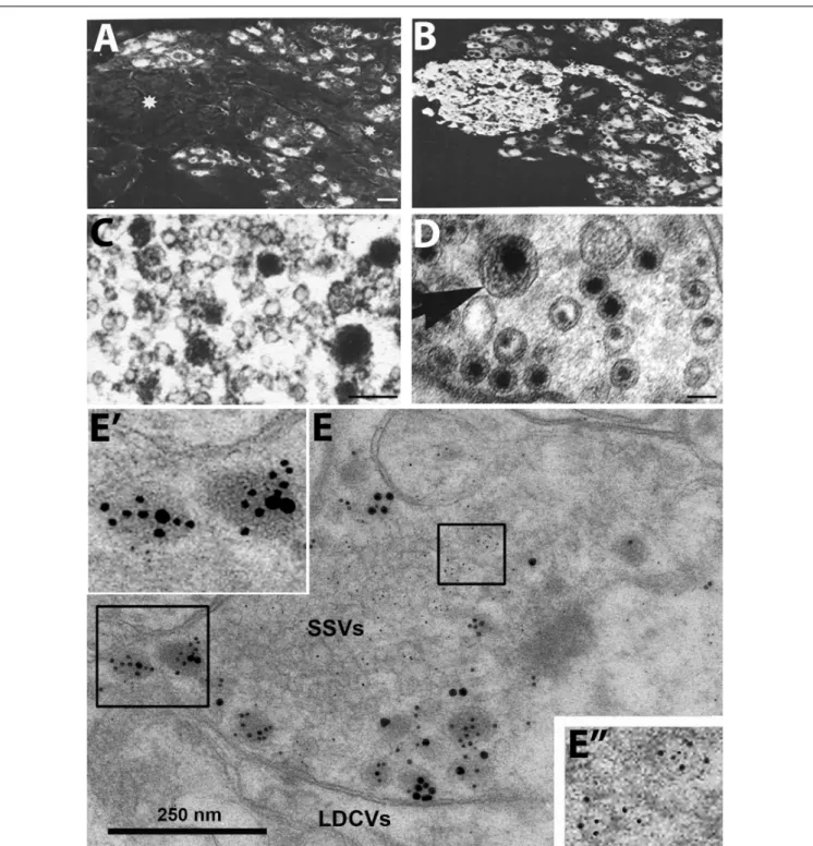
co-transmission - the release of a neuropeptide and a classic (small molecule) transmitter from the same neuron. In fact, the view emerged that neuropeptides always ‘co-exist’ with small molecule transmitters. Moreover, many groups, using IHC at the ultrastructural level, found that peptides are stored in large dense core vesicles (LDCVs) (diameter ∼ 1,000 Å) (Goldsmith
and Ganong, 1975; Swaab et al., 1975; Vandesande and Dierickx, 1975; Castel and Hochman, 1976; Dube et al., 1976; Krisch, 1976; Pelletier et al., 1981; Merighi, 2002) (Figures 1C,E), whereas monoamines like NA are present both in synaptic vesicles (diameter ∼ 500 Å) and LDCVs as shown with potassium permanganate fixation (KMnO4) (Figure 1D) (Richardson, 1966;
Hokfelt and Jonsson, 1968). The number of LDCVs in a nerve ending is mostly low compared to synaptic vesicles, indicating a lower content of peptide molecules vs. classic transmitters.
However, the affinity at peptide receptors is in the low nanomolar range, allowing efficacious signaling even by low numbers of peptide molecules in the extracellular space.
It was not clear, whether IHC could exclude that peptides are stored in synaptic vesicles. Pelletier et al. (1981) incubated adjacent, ultrathin sections with antibodies against substance P and 5-HT, respectively, but in both cases only LDCVs were stained, not synaptic vesicles. This in spite of the fact that monoamines are (mainly) stored in synaptic vesicles (Figure 1D).
Thus, it did not seem possible to visualize the main transmitter (5-HT) in the synaptic vesicles with IHC, contrasting, e.g., the KMnO4 method for NA (Figure 1D). So perhaps IHC also failed to demonstrate neuropeptides in synaptic vesicles?
Therefore, subcellular fractionation studies were carried out, strongly suggesting lack of peptide in the synaptic vesicle pool but presence of NPY in the fraction with many LDCVs (Figures 2A–E) (Lundberg et al., 1981; Fried et al., 1985)
3. In contrast to monoaminergic neurons, in sensory glutamatergic neurons the amino acid appears to be exclusively stored in synaptic vesicles (Merighi, 2002) (Figures 1C,E).
Furthermore, peptides are in general released when neurons fire at high frequency or in bursts (Lundberg et al., 1980;
Andersson et al., 1982; Bondy et al., 1987; Bartfai et al., 1988;
De Camilli and Jahn, 1990; Verhage et al., 1991; Consolo et al., 1994; Xia et al., 2009), and often extrasynaptically (Zhu et al., 1986) (Figure 3). The latter was already indicated in a pioneering study on the presynaptic structure of the synapse, showing docking sites for the synaptic vesicles which, however, are not spacious enough to leave room for LDCVs which are twice-the- size (1,000 Å) (Pfenninger et al., 1969) (Figure 3)
4. This is of course not valid for somato-dendritic release and where true synapses do not exist, nor for the peripheral autonomic nervous system, where there is a considerable distance between the nerve ground plexus (Hillarp, 1949; Falck, 1962) and the smooth
3The preparation used inFried et al. (1985)was very suitable for the purpose: The muscle layer of rat vas deferens contains a dense network of noradrenergic nerve terminals storing NPY (Figures 2A–C). However, the very thick, compact smooth muscle layer makes isolation of nerves/storage vesicles difficult. This obstacle was circumvented by castrating rats which leads to muscle atrophy. Thus, fairly pure fractions containing, respectively, synaptic and large vesicles, many of the latter with a visible dense core (Figure 2E), could be obtained (Fried et al., 1985).
4Of note, Figure 24–4A in Chapter 24 by A. I. Basbaum and T. M. Jessell shows an electron micrograph of an afferent C fiber nerve ending making a type 1 synapse with a dendrite in themonkeysuperficial dorsal horn. Here a string of LDCVs are seen close to the presynaptic membrane opposite to the postsynaptic density. It is not possible to definitely decide, if the LDCVs reach the presynaptic membrane.
Nevertheless, the ‘rule’ of extrasynaptic release of LDCVs may not be without exceptions. The micrograph is by courtesy of H. J. Ralston, III. [from the Fourth Edition of the Textbook “Principles of Neural Science” (2000), edited by E. R Kandel, J. H. Schwartz and T. M. Jessell.]
FIGURE 1 |Immunofluorescence micrographs of the guinea-pig inferior mesenteric ganglion(A,B)and electron micrographs from different types of nerve endings (C–E).(A,B)Two adjacent sections incubated with antibodies to somatostatin(A)and the noradrenaline (NA) synthesizing enzyme dopamine ß-hydroxylase (DBH) (B). The majority of the principal ganglion cells are somatostatin-positive, whereas the small intensely fluorescent (SIF) cells (asterisk) lack the peptide. Virtually all ganglion cells and the SIF cells are DBH-positive, i.e., are noradrenergic.(C–E)Examples of transmitter storage in nerve endings based on or immunohistochemistry (C,E)or potassium permanganate fixation(D).(D)In sympathetic nerve endings NA (black precipitate) is stored in both (small) synaptic vesicles and large dense core vesicle (LDCVs) (arrow). Note that content varies between vesicles, both in the synaptic and LDCVs.(C)Substance P, a neuropeptide (black precipitate), in a sensory nerve ending in the monkey dorsal horn, is stored exclusively in LDCVs, all synaptic vesicles are empty.(E)Peptide and glutamate co-storage and coexistence in the dorsal horn of the rat spinal cord based on immunogold immunohistochemistry. Substance P/CGRP is detected with 10/20 nm gold particles and glutamate with 5 nm gold particles. Note that substance P and CGRP can be stored within the same LDCV (left box, magnified inE’). Staining for glutamate is restricted to synaptic vesicles (right box, magnified inE”). The results suggest that glutamate, a small molecule transmitter, isnotstored in LDCVs in sensory nerve endings, and release of peptide and amino acid may be separate events. This contrasts NA (seeD). Bars: 40µm, for(A,B); 100 nm for(C,D); 250 nm for(E).(A,B) FromHokfelt et al. (1977a).(C)FromDiFiglia et al. (1982), with permission.(D,E)Courtesy of Dr. A. Merighi (cf.,Merighi, 2002).
fncir-12-00106 December 21, 2018 Time: 15:28 # 7
Hökfelt et al. Galanin-Noradrenaline Coexistence and Major Depression
FIGURE 2 |Coexistence and subcellular distribution of neuropeptide Y (NPY) and noradrenaline (NA) in the rat vas deferens.(A,B)Immunohistochemical visualization of NPY-(A)and tyrosine hydroxylase (TH)-(B)positive nerve terminals in adjacent sections. Overlapping, dense NPY and noradrenergic networks are seen in the muscle layer. Note sparse NPY-only positive nerves (arrow) in the subepithelial region, possibly cholinergic nerves.(C)Electron microscopic micrograph of several nerve terminal profiles in the muscle layer after potassium permanganate (KMnO4) fixation, showing small synaptic vesicles with a dense core and LDCVs. The dense core indicates presence of NA both in the synaptic and LDCVs (cf.Figure 1D). No profiles without small vesicle with a dense core are seen, suggesting a pure adrenergic innervation of the muscle layer.(D,E)Subcellular distribution of NA (x) and NPY (o) in a density gradient of rat vas deferens. There is only one peak for NPY (fraction 7;E), whereas there are two peaks for NA (fraction 5 and 7), tentatively representing synaptic vesicles and LDCVs, respectively. Note many LDCVs (arrows), as well as many vesicles of the same size but without dense core (double-headed arrow). The peptide is only present in the heavy fraction (in agreement withFigures 1C,E), whereas NA is present also in the light one (in agreement withFigure 1D). On the abscissa, totally recovered sedimentable substance is given as picomoles per milliliter after centrifugation at 145,000×gmaxfor 45 min. On the ordinate, density gradient fractions 1–10 are given, corresponding to the following sucrose molarities: 1 (0.26 M), 2 (0.32 M), 3 (0.47 M), 4 (0.56 M), 5 (0.69 M), 6 (0.74 M), 7 (0.84 M), 8 (0.91 M), 9 (0.98 M), 10 (1.2 M). Recoveries of NA = 70%, of NPY = 65%, and of protein = 87%. Reprinted fromFried et al. (1985), with permission.
muscle cells, as shown with electron microscopy combined with electrophysiology (Merrillees et al., 1963). Furthermore, in the brain, extrasynaptically released neuropeptides may diffuse over long distances, so called volume transmission (Fuxe et al., 2010).
The exocytotic machinery underlying neurotransmitter release has been thoroughly characterized with regard to release of small molecule transmitters stored in synaptic vesicles (De Camilli and Jahn, 1990; Sudhof, 2014). However, the exocytotic neuropeptide release from LDCVs is less well defined. In early studies on synaptosomes it was shown that CCK release from LDCVs is triggered by small elevation of Ca
2+concentration in the bulk cytoplasm, whereas glutamate release from the synaptic vesicles requires the higher concentrations produced close to Ca
2+channels in the active zone (Verhage et al., 1991). This is in agreement with the localization of the two types of vesicles consistently observed in electron microscopic micrographs of the nerve endings: many synaptic vesicles with some close to the presynaptic membrane, versus a few LDCVs virtually always distant from the synapse (Figure 3).
There is evidence for involvement of SNAREs [soluble N-ethyl maleimide (NEM)-sensitive factor attachment protein receptor protein family] (Sudhof, 2014) also in dendritic release from magnocellular dendrites (Schwab et al., 2001; de Kock et al., 2003;
Ovsepian and Dolly, 2011). The calcium-dependent activator protein for secretion (CAPS) (Walent et al., 1992) has been identified as a priming factor for exocytosis of LDCVs (Stevens and Rettig, 2009; James and Martin, 2013). Thus CAPS2, but not CAPS1, is required for LDCV exocytosis as shown in cerebellar granule cells and hippocampal interneurons (Sadakata et al., 2004; Shinoda et al., 2011).
Taken together, these early findings suggested that neuropeptides were not the main neuronal messengers.
Moreover, when neuropeptides are released, the fast small molecule transmitters are already active in the synaptic cleft – i.e., no peptide release without release of classic transmitters. The discovery of coexistence and co-transmission was summarized in several books/reviews (Burnstock, 1978; Hokfelt et al., 1980a, 1986, 1987a; Cuello, 1982; Chan-Palay and Palay, 1984;
Jaim-Etcheverry, 1994; Merighi, 2002), and since then further efforts have been made to understand co-signaling involving neuropeptides, including co-release of both an excitatory and an inhibitory neuropeptide. For an up-to-date overview of many aspects on neuropeptide signaling (see e.g., Salio et al., 2006; van den Pol, 2012; Ludwig et al., 2016).
More recently it has become clear that coexistence of small
molecule transmitters, encompassing various combinations of
GABA, glycine, glutamate, dopamine and acetylcholine (e.g.,
Guiterrez, 2009; Hnasko and Edwards, 2012; Trudeau et al., 2014)
(Figure 3). For example, coexistence of GABA and glycine was
first reported in the cerebellum (Ottersen et al., 1988), and then
in the spinal cord (Todd and Sullivan, 1990; Ornung et al., 1994),
where evidence for GABA-glycine co-transmission was obtained
in the dorsal horn, and possible co-release from the same synaptic
vesicles (Jonas et al., 1998) (Figure 3). Moreover, mesencephalic
dopamine neurons can also release glutamate (Hnasko et al.,
2010) and GABA (Tritsch et al., 2012), whereby GABA is
FIGURE 3 |Cartoon showing coexistence of a neuropeptide with classic and ‘unconventional’ neurotransmitters in a nerve ending synapsing on a dendrite. Two types of storage vesicles are shown: synaptic vesicles (diameter 500 Å) storing classic transmitters (e.g., 5-HT, NA, GABA or glutamate), mainly released at synapses; large dense core vesicles (LDCVs) storing neuropeptides and, in amine neurons NA or 5-HT. The peptides are in general released extrasynaptically (“volume transmission”), when neurons fire with high frequency or in bursts. Peptide receptors are essentially extrasynaptic or presynaptic, whereas ligand-gated receptors are mostly localized in the postsynaptic membrane. ‘Gaseous’ (e.g., nitric oxide, NO) and other non-conventional transmitters are not stored in vesicles, but are generated upon demand (Snyder and Ferris, 2000). The presynaptic grid, an egg basket-like structure, originally described byPfenninger et al. (1969), is indicated in the nerve ending and high-lighted to the right. Note that the LDCV does not fit into the grid and thus cannot attach to the presynaptic membrane for release. In contrast, there is room for the synaptic vesicle. This supports the concept that peptides are mostly not released into the synaptic cleft. Drawing by Mattias Karlen. Modified fromPfenninger et al. (1969),Lundberg and Hokfelt (1983), andLang et al. (2015).
not synthesized via the classic enzyme glutamate decarboxylase (GAD) but via aldehyde dehydrogenase 1a1 (Kim et al., 2015).
Thus, the number and combinations of transmitters present in a nerve ending (and/or dendrites) virtually seem endless, and it is difficult to define rules according to which neurotransmitters co-exist and are involved in co- transmission, as is discussed further in this Frontiers special topic. Furthermore, neurotransmitter switching, the gain of one and loss of another transmitter in the same, mammalian neuron, can occur not only during development but also in adult animals (Spitzer, 2017).
There is an increasing interest in neuropeptide/neurotrans- mitter coexistence and a need to explore transcriptional changes in defined healthy and diseased brain circuitries (Akil et al., 2010). In fact, there are many interesting results from animal disease models, suggesting involvement of neuropeptides and neuropeptide coexistence in patho-physiological processes with potential therapeutic implications. However, information on the significance of transmitter and neuropeptide coexistence in the normal and diseased human nervous system is limited. In this article, the focus is on galanin co-existing in noradrenergic neurons in the LC, and on galanin receptor expression in postmortem brains from normal subjects and depressed patients who committed suicide (Le Maitre et al., 2013; Barde et al.,
2016). This is in line with previous extensive work carried out on postmortem brains from depressed humans, showing changes in transcripts related to neurotransmitters/neuropeptides and their receptors and to transporters, growth factors in nerve cells, and in glia, in cortical, limbic, hypothalamic and lower brain stem regions (Evans et al., 2004; Iwamoto et al., 2004; Aston et al., 2005; Choudary et al., 2005; Kang et al., 2007; Anisman et al., 2008; Kozicz et al., 2008; Tochigi et al., 2008; Klempan et al., 2009; Sequeira et al., 2009, 2012; Sibille et al., 2009; Poulter et al., 2010; Bernard et al., 2011; Bloem et al., 2012; Kerman et al., 2012;
Zhurov et al., 2012; Labonte et al., 2013, 2017; Li et al., 2013; Du et al., 2014; Lopez et al., 2014a,b; Hayley et al., 2015; Maheu et al., 2015; Torres-Platas et al., 2016; Roy et al., 2017).
GALANIN
Galanin was originally isolated from porcine intestine as a 29- amino acid (30 in humans) neuropeptide (Tatemoto et al., 1983;
Schmidt et al., 1991) (Figure 4A) with a wide distribution in the rat brain as shown with RIA (Skofitsch and Jacobowitz, 1986), IHC (Rokaeus et al., 1984; Melander et al., 1985, 1986b,c,d;
Skofitsch and Jacobowitz, 1985; Merchenthaler et al., 1993), and
ISH (Gundlach et al., 1990b; Jacobowitz and Skofitsch, 1991;
fncir-12-00106 December 21, 2018 Time: 15:28 # 9
Hökfelt et al. Galanin-Noradrenaline Coexistence and Major Depression
FIGURE 4 | (A)Structure of galanin in three species. Galanin is composed of 29 amino acids in most species, except humans (30 amino acids). Note conservation of N-terminal portion.(B)Signaling pathways of galanin receptor subtypes. Galanin, via GalR1 and GalR3, opens potassium channels leading to membrane hyperpolarization. Galanin can via GalR2 activate PLC resulting in generation of IP3, release of Ca2+from the smooth endoplasmic reticulum, opening of Ca2+ channels and eventually transmitter release. AC, adenylate cyclase; cAMP, 30, 50-cyclic adenosine monophosphate; DAG, diacylglycerol; K+, G-protein-regulated inwardly rectifying potassium channel; sER, smooth endoplasmic reticulum; IP3, inositol triphosphate; PIP2, phosphatidylinositol bisphosphate; PKC, protein kinase C; PLC, phospholipase C. Modified fromIismaa and Shine (1999)andLang et al. (2015). Drawing by Mattias Karlén.
Jacobowitz et al., 2004). The distribution of galanin in the mouse brain is similar to that in rat, both with regard to galanin peptide (Perez et al., 2001) and to its mRNA (Cheung et al., 2001;
Lein et al., 2007). The galanin system has also been characterized in the monkey brain (Melander and Staines, 1986; Kordower and Mufson, 1990; Walker et al., 1991) (for human brain, see below).
For many years galanin was considered as the sole endogenous ligand for GalR1-3 but more recently additional ligands were described (Lang et al., 2015)
5. Currently, three galanin receptors, GalR1-3, have been cloned, all three belonging to the family of seven transmembrane-spanning GPCRs, with different transduction mechanisms, with GalR1 and -R3 having distinct similarities (Habert-Ortoli et al., 1994; Fathi et al., 1997; Howard
5First to be identified was the galanin message-associated peptide (GMAP), a product generated from the same precursor as galanin (Rokaeus and Brownstein, 1986). In brain it was also recognized that the N-terminal fragment galanin (1–
16), conserved throughout species, is recognized by high affinity receptor sites in the forebrain (Fisone et al., 1989), and subsequently other fragments have been identified in the brain (Sillard et al., 1992;Ihnatko and Theodorsson, 2017).
Almost 20 years ago the galanin-like peptide (GALP) was discovered in the porcine hypothalamus and shown to be an endogenous ligand of GalR2 (Ohtaki et al., 1999). GALP (9–21) is identical to galanin (1–13) with a high sequence homology among species. In the analysis of ganglioneuroma tissues Santic and colleagues discovered a splice variant of GALP mRNA, a 25 amino acid peptide and named it Alarin (Santic et al., 2006). This peptide, however, does not bind to any of the three galanin receptors, but still is considered a member of the galanin family (Lang et al., 2015). More recently it was found that spexin, a 14-amino acid peptide, is a ligand at the GalR2 and -R3 receptors (Kim et al., 2014).
et al., 1997; Wang et al., 1997; Ahmad et al., 1998; Smith et al., 1998; Iismaa and Shine, 1999; Branchek et al., 2000; Lang et al., 2007, 2015) (Figure 4B). The three galanin receptors are present in most parts of the rat brain, but could not be detected e.g., in dorsal cortical areas and the hippocampal formation (HiFo) in early autoradiographic ligand binding studies (Skofitsch et al., 1986; Melander et al., 1986a, 1988).
Galanin receptors have also been mapped in the mouse brain using 125I-galanin binding autoradiography (Jungnickel and Gundlach, 2005). A direct comparison with results in rat in the study by, e.g., O’Donnell et al. (2003) reveals an overall similar distribution but with some remarkable, apparently qualitative species differences. Thus, mouse shows, i.a., a strong signal in two important regions, the striatum and the cerebellum (Jungnickel and Gundlach, 2005) which both lack binding in the rat (Skofitsch et al., 1986; Melander et al., 1988; O’Donnell et al., 2003). To our knowledge, no attempts have been made to identify the cellular localization and origin of, e.g., the structures binding galanin in the mouse striatum.
The cloning of the receptors allowed localization with ISH and qPCR, which revealed that the transcripts for GalR1 and GalR2 are widely distributed in the rat brain, primarily in the brain stem and in ventral cortical areas (Landry et al., 1998; Mitchell et al., 1999; O’Donnell et al., 1999, 2003; Burazin et al., 2000;
Waters and Krause, 2000; Mennicken et al., 2002). However,
the GalR2 transcript is transiently highly expressed in neocortex
during the first week after birth (Burazin et al., 2000). The distribution of GalR3 is limited (Mennicken et al., 2002). Only the GalR1 transcript has been mapped with ISH in the mouse brain (Hohmann et al., 2003; Lein et al., 2007). Thus, The Allen Brain Atlas (Lein et al., 2007) lacks results on GalR2 or GalR3, suggesting that they are expressed at low levels. This is also supported by the demonstration that the 125I-galanin binding sites are absent in a GalR1 knock-out mouse (Jungnickel and Gundlach, 2005). Taken together, these results suggest that GalR1 is the predominant receptor in the mouse brain, and that distinct species differences exist between mouse and rat.
GalR3 has emerged as a complex receptor (Lang et al., 2015), not present in all mammals (Liu et al., 2010). Its signaling properties are still not well defined, even though GalR3- transfected cell lines have now been generated (Lu et al., 2005b;
Robinson et al., 2013). However, these cells could so far not be used for stable signaling experiments (see Lang et al., 2015).
Still, GalR3 presumably acts via a PTX sensitive G
i/o-type G protein which in turn regulates inwardly rectifying K
+channels (Smith et al., 1998), as do GalR1 receptors (Smith et al., 1998) (Figure 4B). This lack of knowledge contrasts the substantial information about GalR1 and GalR2 (see Lang et al., 2015). The cloning of the receptors was useful, also because it has been difficult to raise specific antibodies to GalR1-3 (Lu and Bartfai, 2009; Brunner et al., 2018). A similar situation exists for other GPCRs (Michel et al., 2009). Detailed tables on the distribution of galanin and GalR1-3 in rodent brain are found in O’Donnell et al. (1999, 2003), Burazin et al. (2000), Hohmann et al. (2003), and Jungnickel and Gundlach (2005).
Early research on galanin was initiated because of its strong reaction to nerve injury. Transection of the sciatic nerve in rat causes an > 100-fold increase in galanin synthesis (mRNA and peptide levels) in the corresponding somata of DRG somata (Hokfelt et al., 1987b). Upregulation could also be detected in the brain after various types of injury/manipulations (Cortes et al., 1990a,b; Villar et al., 1990; Agoston et al., 1994; Palkovits, 1995). In fact, galanin meets the criteria of a neurotransmitter/-modulator, but also has trophic functions, as shown both in brain and the peripheral nervous system (Hobson et al., 2010). Galanin has, in fact, many characteristics similar to the brain-derived neurotrophic factor (BDNF), including storage in, and exocytotic release from LDCVs and both transmitter and trophic functions (Barde, 1994). For example, galanin affects spine density (Sciolino et al., 2015), and it is well-known that BDNF influences dendritic morphology (Bennett and Lagopoulos, 2014). Thus, trophic functions of galanin are potentially interesting but will not be discussed here.
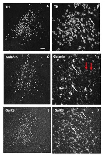
A further early finding in the rat was the coexistence (Figures 5A,B”) of galanin (Figure 5B) in both noradrenergic neurons in the LC (Figure 5B’) (Rokaeus et al., 1984;
Skofitsch and Jacobowitz, 1985; Melander et al., 1986b,c;
Holets et al., 1988; Moore and Gustafson, 1989) and in serotonergic neurons in the dorsal raphe nucleus (DRN) (Melander et al., 1986c; Fuxe et al., 1990; Priestley et al., 1993;
Xu and Hokfelt, 1997), two systems associated with mood- related behavior. The LC neurons also express transcripts for
both GalR1 and -R2 (O’Donnell et al., 1999; Burazin et al., 2000).
Thereafter galanin biology has since the early 1990’s been regularly summarized in books/journal from meetings (Hökfelt et al., 1991, 1998; Hökfelt and Crawley, 2005; Hokfelt, 2010;
Hokfelt and Tatemoto, 2010); and in peer-reviewed articles focusing on the nervous system (only such published after 2004, and not included in the books/journals cited above, are listed here) (Lundstrom et al., 2005; Holmes and Picciotto, 2006;
Karlsson and Holmes, 2006; Ogren et al., 2006, 2007, 2010;
Robinson et al., 2006; Walton et al., 2006; Wrenn and Holmes, 2006; Lu et al., 2007; Tortorella et al., 2007; Picciotto, 2008;
Robinson and Brewer, 2008; Butzkueven and Gundlach, 2010;
Picciotto et al., 2010; Webling et al., 2012; Diaz-Cabiale et al., 2014; Freimann et al., 2015; Weinshenker and Holmes, 2016;
Millon et al., 2017a; Genders et al., 2018a); and in some major comprehensive reviews (Lang et al., 2007, 2015).
GALANIN INHIBITS RAT LOCUS COERULEUS NEURONS
Locus coeruleus is a small, compact bilateral nucleus in the pons located in the gray matter close to the lateral aspect of the 4
thventricle (Maeda, 2000). Dahlstrom and Fuxe first reported that NA is a transmitter in the rat LC, a.k.a. the A6 group (Dahlstrom and Fuxe, 1964). They used the formaldehyde, or Falck-Hillarp, fluorescence method that allows microscopic visualization of catecholamines and serotonin in tissue sections (Carlsson et al., 1962; Falck, 1962; Falck et al., 1962).
In the rat, the LC contains 2,800–3,600 neurons (both sides) (with an additional 260 neurons in the subcoeruleus area, the vast majority of which are noradrenergic with wide projections to virtually all parts of the central nervous system (Ungerstedt, 1971; Descarries and Saucier, 1972; Swanson and Hartman, 1975;
Swanson, 1976; Morrison et al., 1978; Moore and Bloom, 1979;
Goldman and Coleman, 1981; Foote et al., 1983; Aston-Jones et al., 1995). NA nerve terminals are also extensively present in primate cortex (Lewis et al., 1986).
When explored with electrophysiological methods galanin has
effects on the membrane potential of several neuron systems
(see Xu et al., 2005). Galanin hyperpolarizes noradrenergic LC
neurons in a slice preparation (Seutin et al., 1989; Sevcik et al.,
1993; Pieribone et al., 1995), mediated via GalR1 (Ma et al.,
2001) (Figure 5C). However, the GalR2 (R3) agonist ARM-1986
(Liu et al., 2001; Lu et al., 2005b) does not cause any effect on
the membrane potential (Ma et al., 2001) (Figure 5C). GalR2
may instead have a presynaptic role in the projection areas
of LC neurons, perhaps mainly acting as an autoreceptor (Ma
et al., 2001). In agreement, galanin is present in noradrenergic
[dopamine ß-hydroxylase (DBH)]-positive nerve terminals in
cortex and the hippocampus (Melander et al., 1986d; Xu et al.,
1998). Galanin activation of GalR1, but not -R2 or R3, has
been shown also in other studies on the rat and mouse LC
(Hawes et al., 2005; Mitsukawa et al., 2009). In addition to this
direct effect, galanin at low concentrations (10
−9M) enhances the
autoinhibitory effect of NA on LC neurons via alpha-2A receptors
fncir-12-00106 December 21, 2018 Time: 15:28 # 11
Hökfelt et al. Galanin-Noradrenaline Coexistence and Major Depression
FIGURE 5 | (A–B”)Immunofluorescence micrographs of the dorsal pontine periventricular region of mouse after double-staining of a section with antibodies to galanin (green) and tyrosine hydroxylase (TH) (red), the rate-limiting enzyme for catecholamine synthesis and thus a marker for NA neurons. Note that both antibodies stain neurons in the locus coerulus (LC)(B,B’), whereby many (yellow,B”), but not all TH-positive neurons express galanin [arrowheads point to TH-only neurons (red), apparently lacking galanin](B’). Galanin is also present in many structures outside the LC. Colchicine treated animal. Courtesy Joanne Bakker and Mingdong Zhang. Bar for(A)200µm, for(B–B”)20µm.(C)Effect of galanin and the GalR2 agonist AR-M1896 on LC neurons (upper two traces), and the dose–response curves of galanin (red), the AR-M1896 (green) and the mixed GalR1-GalR2 M961 agonist (magenta) (lower trace). Note strong hyperpolarization of galanin and a less strong effect of M961, whereas that AR-M1896 hardly causes any effect at all. FromMa et al. (2001). (D, left panel) Effect of galanin on the response of LC neurons to NA. NA (applied from a pipette at the arrowhead) induces a persistent outward current (upper trace). When galanin (0.1 nM) is present, the NA-induced outward current is enhanced, and the duration is prolonged (middle trace). After wash out of galanin, the amplitude and duration of the NA response was similar to that seen before galanin administration (lower trace). (D, right panel) Effect of galanin on dose-response (upper figure) and duration (lower figure) of NA. The NA dose-response curve is shifted to the left, when galanin (0.1 nM) is present (upper figure). The duration of the NA-induced current is increased in the presence of galanin (lower figure).∗∗P<0.01. FromXu et al. (2001)with permission.
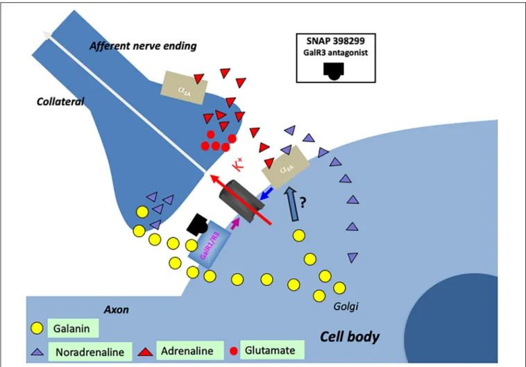
(Xu et al., 2001) (Figure 5D). This may in fact be the primary action of galanin in controlling the firing of LC neurons. Thus, galanin can via different autoinhibitory mechanisms exert a two- step inhibition on LC neurons, at low concentrations enhancing the inhibitory alpha-2A receptor effect.
Autoinhibition of LC neurons, mediated by NA via alpha- 2A receptors, was early discovered by Svensson et al. (1975) and Aghajanian et al. (1977). It is assumed that autoinhibition, both at NA and serotonin neurons, at least in part, is responsible for the delayed onset of the clinical effect of monoamine reuptake inhibitors (Artigas et al., 1996; Mongeau et al., 1997;
Millan, 2006). Autoinhibition via NA in LC was originally suggested to be a consequence of the release from collaterals (Aghajanian et al., 1977). There is, however, evidence that NA can be released from soma/dendrites (Pudovkina et al., 2001; Pudovkina and Westerink, 2005), and more recently release was shown to occur from individual vesicles by combined measurements using amperometry and patch clamp methodologies (Huang et al., 2007). This is in agreement with electron microscopic analysis, showing synaptic vesicles with a dense core in LC dendrites (Shimizu et al., 1979). Thus, collaterals are not necessarily the only structure involved in the autoinhibition.
There is another source of catecholamine input to the LC neurons originating from one of the three C neuron groups in the medulla oblongata: adrenaline (epinephrine) containing afferents (Figure 6) (Hokfelt et al., 1974b, 1984; Howe et al., 1980;
Armstrong et al., 1982), which synapse on LC dendrites (Milner et al., 1989). This was supported by early tracing experiments, although at that time no transmitter histochemical identification was performed (Cedarbaum and Aghajanian, 1978). One likely origin is C1 neurons, since they display a high degree of collateralization, including inputs to the LC (Figure 6) (Haselton and Guyenet, 1990).
Early studies suggested that the adrenaline (Cedarbaum and Aghajanian, 1976) and the C1 neurons (Aston-Jones et al., 1991) inhibit LC neurons. However, the more recent discovery that the C1 neurons are glutamatergic together with optogentic analysis demonstrated excitation as the primary effect (Figure 6) (Abbott et al., 2012). Released adrenaline may act as a modulator not only on postsynaptic but also presynaptic (Li et al., 1995) alpha- 2A receptors, which will, respectively, directly and indirectly, dampen LC neuron activity (Figure 6) (Guyenet et al., 2013).
Taken together, galanin prevents overexcitation of LC, but is only one of several molecules performing this task (Aston-Jones et al., 1991; Singewald and Philippu, 1998; Van Bockstaele, 1998;
Berridge and Waterhouse, 2003; Van Bockstaele and Valentino, 2013). This comprehensive network is perhaps a sign of how important it is to balance the activity of the noradrenergic LC neurons, which are involved in the control of many bodily functions (see below).
Kehr and colleagues have analyzed the effect of intracerebroventricularly administered galanin in freely moving rats and mice, monitoring several neurotransmitters using in vivo microdialysis (Ungerstedt, 1984) and a sensitive HPLC method. Their studies indicate that galanin reduces basal and desipramine-induced extracellular NA levels (Yoshitake et al.,
2003, 2004). This effect is assumed to be exerted via GalR1 at the noradrenergic cell bodies/dendrites in the LC.
Galanin and Dendritic Release
Studies on the hypothalamic magnocellular hormones vasopressin and oxytocin have provided compelling evidence that these two peptides are not only released from nerve endings in the posterior pituitary but also, independently, from dendrites in the paraventricular and supraoptic nuclei (Morris et al., 1998;
Landgraf and Neumann, 2004; Ludwig and Leng, 2006; Kennedy and Ehlers, 2011; Ovsepian and Dolly, 2011; Ludwig et al., 2016).
There is evidence for involvement of SNAREs [soluble N-ethyl maleimide (NEM)-sensitive factor attachment protein receptor protein family] (Sudhof, 2014) in release from magnocellular dendrites (Schwab et al., 2001; de Kock et al., 2003; Ovsepian and Dolly, 2011). Results from studies on CAPS2-dependant neuropeptide release from soma of dorsal root ganglion neurons (Bost et al., 2017; Shaib et al., 2018) may also be relevant for dendritic/somatic release in the brain. Galanin may be released from soma and dendrites in the LC (Pieribone et al., 1995;
Vila-Porcile et al., 2009) (Figure 6). Therefore, it has been hypothesized that stress-induced firing increases galanin release from nerve terminals in the forebrain and dendrites-soma of LC neurons. This could lead to activation of GalR1 autoreceptors and inhibition of firing of LC neurons, a possible mechanism involved in resilience and development of depression-like behavior in animals (Sciolino et al., 2015) (see below).
Other Co-transmitters in the LC
Neuropeptide Y is expressed in LC neurons in rat (Everitt et al., 1984; Chronwall et al., 1985; Yamazoe et al., 1985; Holets et al., 1988) and human (Chan-Palay et al., 1990). Recently it has been shown in mice that dopamine is co-released with NA in the hippocampus (Kempadoo et al., 2016; Takeuchi et al., 2016) and the paraventriculer thalamic nucleus (Beas et al., 2018) and is involved in memory consolidation and control of stress responsitivity, respectively.
GALANIN AND DEPRESSION-LIKE BEHAVIOR IN RODENTS
Galanin influences mood-related behavior in a region-specific way (Bing et al., 1993; Moller et al., 1999). Moreover, results from a number of rat experimental models suggest that galanin can be both prodepressive/anxiogenic and antidepressive (Fuxe et al., 1990, 1991, 1998; Weiss et al., 1998, 2005; Bellido et al., 2002;
Khoshbouei et al., 2002; Barrera et al., 2005; Sergeyev et al., 2005;
Lu et al., 2005a, 2007, 2008; Holmes and Picciotto, 2006; Karlsson and Holmes, 2006; Ogren et al., 2006; Kuteeva et al., 2008, 2010;
Kozlovsky et al., 2009; Picciotto et al., 2010; Le Maitre et al., 2011;
Sciolino et al., 2012, 2015; Weinshenker and Holmes, 2016).
In many of the early studies listed above on depressive-
like behavior the receptor involved was not identified, or the
site of action was not defined experimentally, but there was a
general consensus that it is GalR1 that mediates the depressive
behavior and that GalR2 may be prodepressive (summarized in
fncir-12-00106 December 21, 2018 Time: 15:28 # 13
Hökfelt et al. Galanin-Noradrenaline Coexistence and Major Depression
FIGURE 6 |Cartoon showing several transmitters and signaling pathways in the locus coeruleus (LC) (part of a cell body with initial axon and an afferent nerve ending and a possible axon collateral). A noradrenergic LC neuron co-expresses galanin (yellow LDCVs) originating in the Golgi complex. The peptide in the LDCVs is, after transport to the somatic and dendritic cell membrane, released by exocytosis. Galanin then acts on inhibitory autoreceptors (GalR1/R3), opening potassium channels, in this way attenuating noradrenaline (NA) release in the forebrain. Galanin at low concentrations enhance the alpha2A mediated inhibition of the LC neuron (by an unknown mechanism). Galanin could also be released from collaterals. The GalR3 antagonist (SNAP 398299) may exert an antidepressive action by disinhibiting the LC neuron and restituting forebrain NA levels. With regard to small transmitters, NA (purple triangles) can be released from soma-dendrites and collaterals, acting on somato-dendritic, postsynaptic and presynaptic alpha2A receptors. The afferent nerve ending originates from C1 neurons which are
glutamatergic (red dots) and co-release adrenaline (red triangles). Also adrenaline can act on the alpha2A receptors. The basis for this cartoon is animal experiments, and in the case of the galanin system, results from human postmortem brains are also incorporated.