Exploration of unique physiological features of dentate gyrus granule cells
Ph.D. dissertation
Máté Neubrandt
Semmelweis University
János Szentágothai Doctoral School of Neurosciences
Supervisor: János Szabadics, Ph.D.
Official Reviewers: Márk Kozsurek, Ph.D.
Gábor Molnár, Ph.D.
Members of the Final Examination Board:
Anita Kamondi, DSc.
Gábor Gerber, CSc.
Bence Rácz, Ph.D.
Budapest
2017
2
Table of contents
Table of contents ... 2
Abbreviations ... 5
1. Introduction ... 8
1.1. General introduction of the hippocampus ... 8
1.1.1. Functions of the hippocampus ... 8
1.1.2. Anatomy and connectivity of the hippocampus ... 13
1.2. The dentate gyrus -CA3 interface ... 17
1.2.1. Computation requirements from the DG and the CA3 circuit ... 17
1.2.2. Granule cells ... 19
1.2.3. Adult born granule cells... 23
1.2.4. The feedforward inhibitory circuit in the CA3 region ... 25
1.2.5. Synaptic transmission and plasticity mechanisms ... 26
2. Objectives ... 30
3. Materials and methods ... 31
3.1. Virus mediated birth-dating of granule cells ... 31
3.2. Slice preparation ... 31
3.3. Electrophysiological recordings ... 32
3.4. Calculation of the integrative and biophysical parameters of ABGCs ... 33
3.5. Monosynaptic connections from mossy fibers ... 35
3.6. Disynaptic connections from mossy fibers ... 36
3.7. Anatomical analysis ... 37
3.8. Classification of the postsynaptic cells ... 38
3.9. Statistical analyses ... 39
3
4. Results ... 40
4.1. Functional maturation of adult born granule cells ... 40
4.1.1. Maturation of the biophysical and integrative properties of ABGCs ... 41
4.1.2. Independence of the output properties of individual ABGCs of age and input resistance ... 44
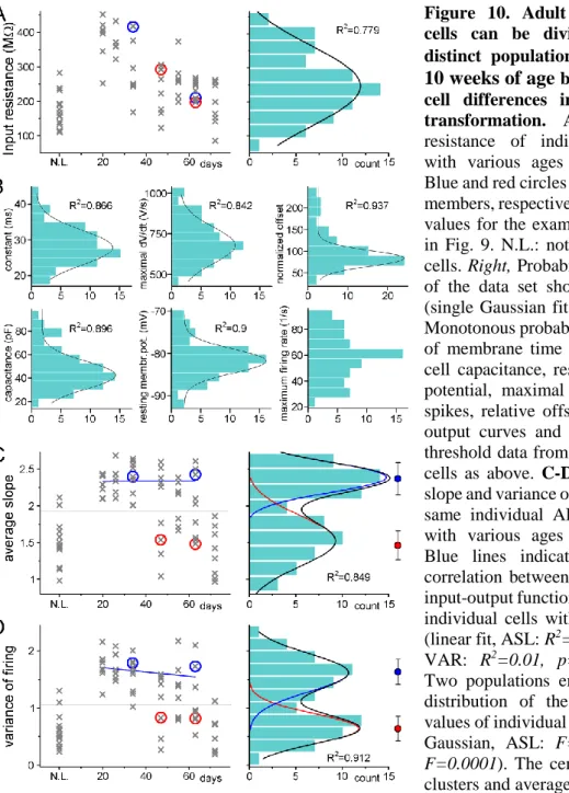
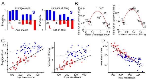
4.1.3. Two functionally distinct populations among non-labeled GCs in adult rats ... 46
4.1.4. Complex mechanisms maintain the two stable states of the input–output transformation of individual cells ... 48
4.2. Effect of single physiological granule cell bursts on the recruitment of postsynaptic CA3 neurons ... 51
4.2.1. Effect of single presynaptic MF bursts in FF-INs ... 52
4.2.2. Postsynaptic cell type-specificity of the single burst-effects ... 56
4.2.3. Burst length dependence of the burst induced potentiation ... 58
4.2.4. Presynaptic origin of the burst-induced amplification in FF-INs ... 60
4.2.5. The amplification does not require large Ca2+ influx ... 61
4.2.6. Complete utilization of release capacity after single bursts ... 63
4.2.7. Occlusion of the amplification by phorbol esters suggests that bursts promote vesicle priming ... 64
4.2.8. The amplification is accompanied by accelerated release ... 66
4.2.9 The amplification does not involve molecular pathways that are known to affect vesicle priming ... 67
4.2.10. Single mossy fiber evoked disynaptic IPSCs reflect the strengthening of feedforward inhibition of a non-specific PC population ... 69
4.2.11. Pyramidal cell firing reflects only ongoing bursts ...71
4
5. Discussion ... 73
5.1. Adult-born granule cells mature through two functionally distinct states ... 73
5.2. Single bursts of single mossy fibers functionally reorganize feedforward inhibition in the CA3 area ... 76
6. Conclusions ... 82
7. Summary ... 84
8. Összefoglalás ... 85
9. References ... 86
10. List of publications ... 103
11. Acknowledgements ... 104
5
Abbreviations
AAC Axo-axonic cell
ABGC Adult-born granule cell AMN082 mGluR7 selective agonist
AMPA α-amino-3-hydroxy-5-methyl-4-isoxazolepropionic acid AP Action potential
ASL Average slope of the input-output funftions
BC Basket cell
BINA Selective positive allosteric modulator of mGluR2 CA1-3 Hippocampal regions, stands for cornu ammonis CA3 GC Ectopic granule cell located in the CA3 region Calphostin C Diacylglycerol binding domains inhibitor CCK Cholecystokinin
CNQX AMPA/kainate receptor antagonists DAB 3,3'-Diaminobenzidine
DCG IV Group II mGluRs agonist DG Dentate gyrus; gyrus dentatus
DIC Nomarski's differential interference contrast microscopy diEPSC Disynaptic inhibitory postsynaptic current
diIPSC Disynaptic excitatory postsynaptic current DMSO Dimethyl sulfoxide
EGTA Ethylene glycol-bis(2-aminoethylether)-N,N,N′,N′-tetraacetic acid EPSC Excitatory postsynaptic current
FFI Feedforward inhibition FF-IN Feedforward interneuron
6
GABA Gamma-aminobutyric acid, inhibitory neurotransmitter Gabazine (SR 95531); GABAA antagonist
GC Granule cell
GFP Green fluorescent protein
GIRK G protein gated inwardly-rectifying potassium channel Go 6976 PKC inhibitor
GF 109203X (Bisindolylmaleimide I); PKC inhibitor
IN Interneuron
IPSC Inhibitory postsynaptic current IvyC Ivy cell
KT5720 Protein kinase A inhibitor LEC Lateral entorhinal cortex
L-group ABGCs with linear input-output transformation LTD Long-term depression
LTP Long-term potentiation MEC Medial entorhinal cortex
MF Mossy fiber
MFB Mossy fiber bouton
mGluR Metabotropic glutamate receptor ML297 GIRK1/2 channel selective activator MSOP Group III mGluRs selective inhibor
PC Pyramidal cell
PDBu Phorbol 12,13-dibutyrate, diacylglycerol analoge PKA Protein kinase A
PKC Protein kinase C
7 PKC (19-36) Protein kinase C inhibitory peptide PLC Phospholipase C
PP Perforant pathway
PPR Paired pulse ratio PTP Post-tetanic potentiation
PV Parvalbumin
RFP Red fluorescent protein Rin Input resistance
SLC Spiny lucidum cell Str. Stratum
S-group ABGCs with supra-linear input-output transformation U 73122 Phospholipase C inhibitor
VAR Average variance of the slope of input-output functions
8
1. Introduction
The hippocampus, being a simple cortical structure with many generally applicable properties became one of the most extensively studied brain region. On the other hand, hippocampus also have some unique anatomical and physiological specializations, consistently with its essential role in important cognitive functions. Many of these features, despite the long history of gaining the interest of researchers, remain poorly understood. My Ph.D. studies aimed to address intriguing and unique aspects of the hippocampal physiology on the cellular and circuit level. Accordingly, I am providing a general introduction of the hippocampus first, than I am discussing specifically the dentate gyrus (DG; gyrus dentatus) – CA3 interface which was the main focus of the research presented here. Notably, most of the available experimental data originates from the rodent hippocampus, and we used rats as model organism. Therefore, I will primarily discuss the rodent hippocampus, supplemented with some relevant human aspects.
Due to the coexistence of partly different nomenclatures in the literature I clarify what I use in this thesis hereafter. Although “hippocampus” sometimes only used to refer the CA regions (cornu ammonis or hippocampus proper) I consider dentate gyrus as part of the “hippocampus” because of its essential and inseparable role in some “hippocampus related” functions and phenomena e.g. pattern separation, sparse coding, will also be discussed. The “hippocampal complex” refers the complex of entorhinal cortex, DG, CA regions, subiculum, presubiculum, parasubiculum.
1.1. General introduction of the hippocampus
1.1.1. Functions of the hippocampus Mnemonic functions
Association of the hippocampal complex with certain memory functions largely arose from the extensive studies of human amnesiac patients. Among them, the most famous is H.M. who developed severe memory dysfunctions after his brain surgery, when large part of his medial temporal lobe has been removed in order to treat his epileptic seizures (Scoville and Milner, 1957; Squire, 2009). The removed structures included significant portion of the hippocampus and the associated cortical regions bilaterally. After the
9
operation, H.M. had anterograde and retrograde global amnesia, even though many of his cognitive abilities remained unaffected. For instance, he could actively participate in an ongoing conversation, followed the topic, until it was disrupted. Upon the partner left the room for a short time, H. M. forgot the entire conversation and could not even recognize the person he talked with. He was incapable of storing new memories (anterograde amnesia), and did not remember a certain period preceded the surgery (retrograde amnesia), however he could clearly recall older memories. On the other hand, he was capable of learning motoric tasks that required practice, indicating that his ability to form so called “procedural memory” was remained. H. M. showed day by day improvements in these tasks even though he did not remember the process of the practices. Later, the conclusions of the extensive studies of H.M. were supported by further observations.
Learning of random associations of complex multimodal inputs for a single exposure is impaired in patients with lesion of the hippocampal complex. In contrast, simple associative pairings like classical conditioning, and motor learning remain intact.
Moreover, the largely intact short-term memory of these patients indicate the involvement of different cortical regions in those specific abilities (Squire, 1992; Andersen et al., 2007). Supporting the uncompromised episodic memory functions, many evidence is accumulated so far that hippocampus also has crucial role in learning the order of sequential events (Eichenbaum, 2013). In summary, the consensus is that hippocampus is essential for processing complex, multi-sensory experiences and establishing new memories. Such memories however transiently deposited in the hippocampus, for long- term storage they might be transferred to other cortical regions.
Spatial navigation
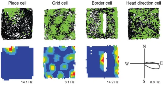
Another major and well described function of the hippocampal complex is its essential role in spatial navigation. The discovery of this feature was recognized by the Nobel Prize in 2014 received by John O'Keefe, May-Britt Moser and Edvard I. Moser “for their discoveries of cells that constitute a positioning system in the brain”. While recording hippocampal cells in freely moving rats O'Keefe and Dostrovsky recognized that the activity of certain cells remarkably increase when the animal enters a specific restricted area of the environment, what they denominated as place fields (Fig. 1). When an animal explores a new environment it progressively recruits assemblies of place cells, with place fields covering the elapsed route, thereby forming a cognitive map of the entire
10
environment. Later, when the animal returns to the familiar environment, the firing fields of the previously established place cells remain the same. A hippocampal cell with an existing place field, however, still can contribute to mapping of a distinct novel environment, by forming a new place field (O’Keefe and Dostrovsky, 1971; O’Keefe and Nadel, 1978). It was believed for a long time that a place cell has a single firing field, because in general experimental conditions animals explore only small spaces. However, it was found recently by using much larger environments, that hippocampal neurons have their own individual propensity for forming place fields in random manner during exploration. Therefore, some cells express many firing fields and many form few or none (Rich et al., 2014). Place cells were initially described in the CA1 and these cells are showing the sharpest edges by their place field and the highest firing rate, the existence of place cells was reported in all hippocampal sub-regions. After long standing controversy regarding the spatial behavior of dentate gyrus granule cells, it was recently clarified that even though they are sparsely active during exploration they can form single stable place fields. When the animal traverses through their place fields, granule cells can fire remarkably high frequency bursts of action potentials (Jung and McNaughton, 1993;
Leutgeb et al., 2007; Neunuebel and Knierim, 2012; Danielson et al., 2017; GoodSmith et al., 2017; Senzai and Buzsáki, 2017).
The other milestone linking the hippocampal formation with spatial navigation was the discovery of grid cells in the medial entorhinal cortex by the group of May-Britt Moser and Edvard I. Moser. Whereas place cells mostly have single firing fields in a given environment in the case of grid cells the entire space is tiled by multiple firing fields of cells forming a grid (Fig. 1). Such a grid constituted of equilateral triangles and therefore showing a characteristic hexagonal pattern with 60° rotation symmetry when illustrated by a spatial autocorrelogram. Grid cells retain their grid-spacing and also the orientation of the grid relative to the borders in any environment, thereby provide a reference metric for the neural representation of the spatial environment (Hafting et al., 2005; Moser et al., 2014).
The way rodents explore the environment by sniffing and whisking during locomotion is markedly different from how humans or primates approach the same task, predominantly by visual observation. Accordingly, the neural equivalents of place and grid cells were also identified in primates when visually exploring a virtual environment.
11
Similar spatial representations were found during visual observation in human epileptic patients, when recordings were performed during surgery, in order to identify the locus of epileptic activity in the temporal lobe (Killian et al., 2012; Jacobs et al., 2013; Miller et al., 2013).
Figure 1. Examples of four fundamental spatial cells in the hippocampal complex. The upper row shows the animals trajectory (black line) in square boxes (1 x 1 m with 50 cm high walls, except in the case of the place cell, where the box was 62 x 62 cm) with indication of the neurons firing activity (green dots). In the case of border cells an additional 50 cm long barrier is inserted.
Lower row: heat maps show the locational firing rate or polar plot shows the directional firing rate of the cells. The peak firing rates are shown below the rate maps. The figure is adopted with modifications from (Hartley et al., 2013).
It is generally accepted that the brain needs to maintain two cooperative mechanisms to efficiently accomplish the various challenges related to spatial navigation. First, it is necessary to build an accurate so called “allocentric” map, the static positional representation of the spatial environment (O’Keefe and Nadel, 1978). The other component is a self-referenced navigation system that requires information of the locomotion speed, the elapsed time and the starting point as a reference. This is a complex mechanism, required for example to calculate the shortest way home after an exploratory trip without the necessity of repeating the exact same route in the reverse direction (McNaughton et al., 1996). In addition to behavioral evidences linking both mechanisms to hippocampus, further related specific representations were also described in the hippocampal complex (Buzsáki and Moser, 2013; Moser et al., 2014). Such neural elements are head direction cells in the entorhinal cortex which not only show directional tuning but also found to be modulated by the moving speed (Sargolini, 2006), and border
12
cells that respond to environmental boundaries in orientation specific manner (Fig. 1) (Agster et al., 2014).
Current perspectives of the hippocampal functions
As discussed above, the hippocampal complex has long history of being associated with two main, seemingly distinct brain functions, the episodic memory and the spatial navigation. On the other hand, these two functions supposable must have much in common since they obviously share similar computational and circuit requirements (Buffalo, 2015). How these functions relate to each? The reconciliation of mnemonic and spatial functions was addressed from at least two perspectives so far. First, Buzsáki and Moser proposed that these two functions are in direct evolutional relationship, considering their underlying mechanisms. Their hypothesis is memory formation was evolved from spatial navigation, and the neuronal mechanisms of navigation in both physical and mental space is fundamentally the same. The review highlights the dichotomy in both navigation and memory. The numerous similar aspects and cellular activities related, on one hand, to the explicit “allocentric cognitive map” and “semantic memory”, and to the first person experience of “path integration” and “episodic memory”
on the other hand are paralleled in the review (Buzsáki and Moser, 2013).
Eichenbaum proposed a second, relatively simpler perspective that spatial representation together with the representation of time are the organizing basics of the episodic memory. The same hippocampal neuron population that provide place cells can also encode temporal regularities of experience. If the activity of neurons is linked to a specific moment within a temporally structured episode it is called “time cell”. During learning, the dimension of the context determines whether the activity of a cell will be associated with either time or space (Eichenbaum, 2013, 2014).
Some recent results further elaborated the functions of hippocampus. In a clever behavioral paradigm, rats had to identify and find a specific sound frequency by adjusting the tone on a continuous scale by a “joystick” to deserve a reward. Remarkably, during this task certain hippocampal and entorhinal cortical cells were activated, so that they mapped the entire sound spectrum by each being rendered to a specific narrow sound frequency range. The progressive mapping of the changing sound showed many similarities to the well described manner of mapping the spatial environment (Aronov et
13
al., 2017). These results also argue that the hippocampus has a general ability to encode and map various environmental contexts and more abstract dimensions than the space.
Thereby the same neural circuit elements might concurrently support spatial and mnemonic functions. Next, I am going to discuss the structural basis of these functions.
1.1.2. Anatomy and connectivity of the hippocampus Architecture of the hippocampal regions
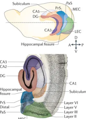
The hippocampus is an archicortical brain region with evolutionary conserved structure that is similar across all mammalian species. The hippocampus has a relatively simple laminar structure that distinguishes it from the other, more complex cortical regions. The simple architecture was also an important factor that made the hippocampus a popular research subject, beyond the broad interest of its implications in memory functions. Some fundamental principle determine the laminar structure at each hippocampal sub-region. The somata of the principal cells are concentrated into a single dense layer, their apical dendrites distributed parallel, and the major afferent- and efferent fiber tracts densely permeate certain cellular domains. In a trans-section, the hippocampus appears as two opposing “horse shoes” these are the major regions, the dentate gyrus (DG) and the hippocampus proper (Fig 2). The latter is divided into further sub-regions CA1, CA2, CA3 denominated according to the abbreviation of its Latin name cornu ammonis (Lorente de Nó, 1934; Andersen et al., 2007).
In the DG, granule cells (GC) are the principal cells. The uniformly polarized morphology of GCs designates the three layers of the DG. The GC dendrites are located in the str. moleculare, the somata in the str. granulosum and the axons in the centrally positioned hilus. The hilus hosts another glutamatergic cell population, the mossy cells that provide an excitatory feedback loop to GCs in both the ipsi- and the contralateral hemispheres (Amaral, 1978). The principal cells of the cornu ammonis are pyramidal cells (PC). The somata of the PCs are organized into a single dense layer called str.
pyramidale. This pyramidal cell layer forms a successive continuum along the CA3-CA2- CA1 sub-regions. The thin basal dendrites of PCs radiate in the str. oriens towards the alveus. The majority of the dendritic tree of PCs, deriving from their much thicker apical dendrites, is largely located in the str. radiatum, only the distal most branches terminate in the str. lacunosum moleculare. Beyond this general scheme, the CA3 region has an
14
additional sublayer the str. lucidum. This layer is formed by the dense mossy fiber (MF) tract, the efferent axons of DG GCs that target the most proximal part of the apical dendrites of PCs (Andersen et al., 2007).
Figure 2. The rodent hippocampal complex.
Position and orientation (upper, drawing) of the hippocampus and its related structures in the intact brain and in a traverse section (bottom).
Abbreviations: MEC and LEC – medial and lateral entorhinal cortex, PrS and PaS – pre- and parasubiculum. The figure is adopted with modifications from (Moser et al., 2014)
Excitatory connectivity – orthogonal information flow
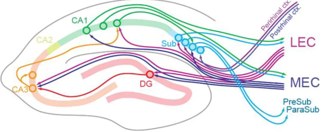
The hippocampal complex is reciprocally connected with practically all sensory and associative cortices consistently with its central role in many cognitive functions. What is the secret of the hippocampus that distinguish it from other brain areas? One major unique feature of the hippocampal circuitry is the orthogonalized information flow that proposed to be a key element in its functions (Fig. 3)(Cajal, 1893). The entorhinal cortex provides the major input to the hippocampus, this afferentation is called the perforant pathway (PP). The entorhinal cortex layer 2 fibers terminate in the DG in the outer 2/3 of the str. moleculare, and the CA3 str. lacunosum moleculare. The layer 3 fibers project to the CA1 str. lacunosum moleculare. The MF pathway originating from the DG GCs provides the intrahippocampal afferentation of the CA3 region. The CA3 PCs are extensively connected with each other and forming a so called associative network that also involves commissural fibers coming from the contralateral CA3. Such an
15
autoassociative network is capable of information storage. It is believed that in the hippocampus, previously acquired information is stored in the associative synapses of the CA3 network (Rolls, 2007). The efferent projection of the CA3 PCs is called Schaffer collaterals that terminate in the str. radiatum of the CA1. The CA1 PCs project to the subiculum and the entorhinal cortex thus closing the hippocampal loop. Notably, the above described fundamental circuit did not consider the CA2 region which is much smaller than the two neighboring regions. The CA2 starts where the MF pathway ends and accordingly the stratum lucidum diminishes. The main afferentation of the CA2 comes from the layer 2 of the entorhinal cortex which distinguishes it from the CA1.
Recently, the identification of some appropriate marker molecules facilitated the research of this region, which predicts novel understanding of its role in the hippocampal circuitry in the near future (Cui et al., 2013; Kohara et al., 2013).
Figure 3. Connectivity of the hippocampus. The main local as well as the afferent and efferent excitatory pathways of the hippocampus. Abbreviations: MEC and LEC - medial and lateral entorhinal cortex, Sub - subiculum. The figure is adopted with modifications from (Hartley et al., 2013).
Inhibitory cells of the hippocampus
Like in other cortical regions, the hippocampal GABAergic cells show substantially larger heterogeneity than that was found in the case of principal cells. Exact classification of GABAergic cells requires consideration of their multiple features, such as the localization of the axonal and dendritic arborization, target cell specificity, neurochemical markers, electrophysiological properties, timing of activity relative to the network oscillations and developmental origin (Freund and Buzsáki, 1998; Buzsáki et al., 2004;
Klausberger and Somogyi, 2008; Kepecs and Fishell, 2014). Instead of going into detailed
16
classification I would like to highlight some key aspects of the diversity that relates to the generalized connectivity schemes and the major network functions.
Key determinant of GABAergic cells function is their target selectivity. Specific IN populations innervate the perisomatic region of principal cells (such as axo-axonic and basket cells), they are optimally positioned to effectively control the spiking output of their postsynaptic partners (Somogyi et al., 1982; Papp et al., 2013; Szabó et al., 2014).
Others, innervating different dendritic domains of the principal cell, are suited to control local signaling and integrative processes of the distal inputs (Klausberger, 2009; Lovett- Barron et al., 2012). However most of the inhibitory cells are indeed local, and rightly referred to as interneurons, projecting GABAergic cells are also exist in the hippocampus (Jinno et al., 2007; Jinno, 2009). Such projecting hippocampo-septal cells inhibit other inhibitory cells of the medial septum that project back to hippocampal GABAergic cells closing a recurrent inhibitory loop. Besides the cholinergic afferents, the septal inhibitory connections have important role in rhythmic network activities of the hippocampus (Freund and Antal, 1988; Bland et al., 1999). A specific inhibitory cell population selectively target other GABAergic cell types within the circuit, similarly to the local collaterals of the hippocampo-septal cells. This circuit motif, the indirect control of the network excitability by the interneuron selective inhibitory cells is referred to as dis- inhibition (Hájos et al., 1996; Tóth et al., 1997; Chamberland and Topolnik, 2012).
Another functionally important property of inhibitory cells is the localization of their soma-dendritic arbor, which determines the main source of their excitatory input. In accordance with this notion, GABAergic cells mostly involved in either one of the two fundamental inhibitory wiring motif, feedforward or feedback inhibition (Glickfeld and Scanziani, 2006; Andersen et al., 2007). Those GABAergic cells whose activity is mostly driven by the afferent projections from extra hippocampal areas or upstream sub-regions contribute the feedforward inhibition. A largely different cell population is recruited by the recurrent collaterals of the local principal cells establishing the feedback inhibition.
The above discussed main categories of inhibitory cells (e.g. target specific and functional types) can be found all over the hippocampus and comprise largely equivalent cell populations across sub-regions.
17
1.2. The dentate gyrus -CA3 interface
As I discussed earlier, several hippocampal functions require the fast storage and subsequent recall of specific sets of multimodal environmental stimuli. What is so special in hippocampus that enables establishing complex memory traces? Why the neocortex which is responsible for the highest order cognitive functions needs the hippocampus?
Neocortex is assumed to be involved in continuously creating and referring to a general internal representation of the observed world. However, it seems that the structure of the neocortex may not be suited for rapid interference free acquisition of new memory traces.
By contrast, the hippocampus appears to be evolved exactly for this task, to complement the learning capacity of the neocortex (Acsády and Káli, 2007). What are the key features, specific roles, and computational tasks that assumed to be accomplished by the DG-CA3 circuit?
1.2.1. Computation requirements from the DG and the CA3 circuit Autoassociative memory networks and pattern completion
At the CA3 region PCs are connected to each other, with local and contralateral collaterals, forming a so-called autoassociative network. Moreover, these associative synapses are plastic that establishes the basis of information storage. Because of these features, the CA3 region has been considered for a long time to have a central role in memory formation. In theoretical models, such an autoassociative memory network is optimal for storing large amount of information and capable of recalling complex memory traces even when only a fraction of the original input is presented to the network. This latter feature is referred to as pattern completion. More specifically, it is believed that when the hippocampus is encoding a complex memory trace, a set of CA3 neurons, so- called ensemble, become active, and the synapses connecting them become potentiated.
Thereafter a certain set of neurons will be allocated to a specific memory trace. During memory recall activation of a subset of the neurons belonging to a certain memory trace will recruit the rest of the neurons coding the memory. To be able to perform these tasks independently, CA3 network needs to maintain two distinct, specialized input system.
The PP input is suggested to be activated during memory recall, and the MF input is responsible for encoding memory traces (Marr, 1971; Treves and Rolls, 1992, 1994;
18
Debanne et al., 1998; Kesner, 2007; Guzman et al., 2016; Mishra et al., 2016). Multiple unique features, such as the “detonator” synapse, the sparse physiological activity, and the target specificity of GCs cooperatively support the MF system to perform this operation. I will discuss these features in more details in the following sections.
Pattern separation
To avoid interference between memories, pattern separation is an essential step in information processing. In general, pattern separation occurs in a network when the output patterns are less similar to each other than the input patterns. In the entorhinal cortex two different experiences likely represented with a certain degree of overlap in the populations of active cells. Particularly, when only subtle differences distinguish two items to be encoded, it is essential to separate them in order to produce independent representations. In the hippocampus, the DG is assumed to perform this key step of memory formation. Multiple features of the DG support this function. First, the anatomical arrangement of this region seems to be optimized for this computation by showing a large divergence. The number of cells in the DG is an order of magnitude larger than that is found in its primary input source, the entorhinal cortex. Second, individual GCs are capable of effectively depolarize, and drive the firing their downstream target neurons, the CA3 PCs. Finally, DG operates with the so-called sparse coding scheme. In this type of neural code each event is encoded by the strong activation of a small set of neurons. (Marr, 1971; McNaughton and Morris, 1987; Deng et al., 2010; Knierim and Neunuebel, 2016).
Sparse coding, the hippocampus specific code
Sparse coding is a coding scheme falls between the two extremes of the so-called localist coding, where a single neuron codes a single memory item, and the fully distributed coding, where each memory is coded by the activity of large neuron populations. In the hippocampus, each memory trace or item is represented by the strong activation of a small set of neurons. Consequently, in the hippocampus the background activity in principal neurons is remarkably low, and only minority of the cells are active at a given time, those who are participating in the ongoing coding processes. These features of the neuronal activity, and the sparse coding scheme is largely different from most of the other cortical regions. Those are usually characterized by higher background
19
activity and relatively weaker modulation by their relevant stimuli, and perform denser coding (Acsády and Káli, 2007; Rolls and Treves, 2011; Wixted et al., 2014).
In the light of the above notions, the DG can be considered as the gate of the hippocampus and its roles are to form pattern separated, sparse representations of the entorhinal cortical activities and effectively transmit it to the CA3 network (Acsády and Káli, 2007). These complex functions require unique solutions, functional specializations from the DG-CA3 circuit, which were the main focus of my Ph.D. studies.
1.2.2. Granule cells
Morphological properties
GCs are the principal cells of the DG and they represent the most numerous neuron type in the hippocampus as their number reaches about one million cells in the rodent brain (Bayer, 1982). The morphology of GCs markedly differs from that of PCs. GCs do not have a pronounced apical dendrite, instead one or a few main dendritic branches originate from their small somata. These dendrites are branching close to the cell body resulting in multiple equivalent thin dendrites which are densely covered by spines (Claiborne et al., 1990; Schneider et al., 2012). Our earlier results revealed a mechanism that is optimal for silencing individual dendrites by an unusual postsynaptic mGluR2 receptor mediated current (Brunner et al., 2013). Another interesting finding of our laboratory is related to GC dendrites: back propagating action potentials retain analog information about the somatic membrane potential which affects local dendritic Ca2+
signaling.
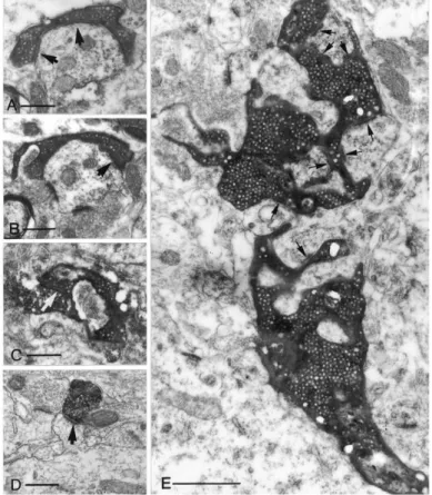
Probably the most unique morphological feature of GCs is their axon, called mossy fiber (MF) that is substantially different from any other cortical axons. Usually a single axon fiber runs through the CA3 str. lucidum and only a few collaterals branch in the hilus. In healthy conditions GCs do not innervate each other, which is ensured by the polarized arrangement of the axons and dendrites (Claiborne et al., 1986; Williams et al., 2007). MF have three different types of axon terminals (Fig. 4). The most special is the large MF bouton (MFB) with its exceptional size reaching 4-10 µm in diameter. This huge presynaptic structure contains on average 25 individual active zones (Rollenhagen et al., 2007). Large MFBs emanate up to 3-4 filopodial extensions which terminate regular
20
sized (0.5-2 µm) axon terminals. The number of these filopodia varies depending on the age or even previous learning experiences (Ruediger et al., 2011; Wilke et al., 2013).
Finally, regular sized (0.5-2 µm) en passant varicosities along the main axon represent the third terminal type (Claiborne et al., 1986; Acsády et al., 1998). The heterogeneity of the terminal types parallels with target specificity, as the large MFBs innervate mossy cells in the hilus and PCs in CA3, while the regular sized terminals specifically target GABAergic cells in both sub-regions (Fig. 5). This target selectivity enables the simple estimation of the ratio of excitatory and inhibitory cells among the postsynaptic partners of GCs, that aspect of the circuit also predicts unusual operation and specific functions.
First, each GC project to as few as 10-18 CA3 PCs that results in an oddly specific information pathway with minimalized divergence. Second, GCs innervate more GABAergic cells in the CA3 (40-50) than PCs, rendering the DG - CA3 interface as a prototypical feedforward inhibitory circuit (Acsády et al., 1998). In comparison, PCs in other cortical circuits usually form thousands of synapses and only 15-20% of them target inhibitory cells, that emphasize the unique organization of the MF pathway (Gulyás et al., 1993; Sik et al., 1993).
Figure 4. Comparison of the different MF terminal types on electron microscopic level. A- B. Small en passant terminals establishes a single asymmetrical synapse on a dendritic shaft with long perforated postsynaptic density (arrows). C. A filopodial extension of a mossy terminal forms a synapse (arrow) with a substance- P receptor labeled interneuron. D. A CA3 PC terminal on a CA1 PC dendritic spine is presented for comparison. E. A large, mossy terminal forms multiple contacts (arrows) with thorny excrescences of a CA3 pyramidal cell. The individual release sites are short.
Note that, all micrographs have the same magnification to enable the direct comparison of the sizes of the structures. Scale bars: A-D: 0.5 µm, E:1µm. This figure is adopted from (Acsády et al., 1998).
21 Figure 5. Filopodial extensions of MF
terminals innervate GABAergic cells.
Schematic illustration of two MF boutons (MFB), each with four filopodial extensions (large arrowheads). All filopodial terminals contacted the dendrites or spines of six GABAergic neurons. Five out of six postsynaptic interneurons have spiny dendrites. All synapses were identified at the electron microscopic level (data not shown).
Arrows indicate the main axons. This figure is adopted from (Acsády et al., 1998).
Activity of granule cells
The GCs receive strong feedforward and feedback inhibition from local GABAergic cells, which is reflected by some unusual features of the organization of the inhibitory circuit in the DG (Acsády and Káli, 2007). Basket cells, as well as other interneurons of the DG refrain from innervating the peri-somatic regions of each other, thus, minimalizing the dis-inhibitory influence (Acsády et al., 2000). Moreover, strong excitation from large number of GCs and mossy cells converge onto GABAergic cells of the DG and the hilus. These GABAergic cells in turn extensively innervate the peri- somatic region and the outer layer of the str. moleculare where the PP input terminates.
(Buckmaster and Schwartzkroin, 1995; Sik, 1997; Acsády et al., 2000).
Beyond the strong synaptic inhibition, the intrinsic physiological properties of GCs also appear to be specifically tuned to support extremely low firing activity. GCs were found to have remarkably hyperpolarized membrane potential (about -80 mV) and no spontaneous firing activity in in vitro slice experiments (Staley et al., 1992; Schmidt- Hieber et al., 2007; Krueppel et al., 2011). Consistently, GCs were found to be one of the most quiescent neurons with their extremely low overall firing activity (below 1 Hz) during in vivo recordings (Munoz et al., 1990; Jung and McNaughton, 1993; Penttonen et al., 1997; Kowalski et al., 2016). When GCs are active, they either fire single action potentials (AP), or short high frequency spike bursts (3-7 APs with 100-200Hz) however their activity is often interrupted with several seconds long inactive periods, which explains the low overall firing rate. The high-frequency bursts are usually generated by non-linear dendritic processes in response to coincident inputs. In hippocampal CA1 PCs,
22
it was shown that dendritic plateau potentials can be evoked by coincident stimulation of the two major excitatory path of the CA1, the Schaffer collaterals and the PP. These plateau potentials reliably converted in the axon to high frequency burst output (Bittner et al., 2015; Apostolides et al., 2016). Much less is known about the mechanism related to the burst firing of GCs (Krueppel et al., 2011). Because of the lack of knowledge on this common physiological activity form of GCs, and also because high frequency bursts are considered to be distinct neuronal signals (Lisman, 1997), this interesting activity feature of GCs was one specific focus of my Ph.D. studies.
CA3 granule cells
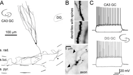
It was reported that a small population of GCs is also present in the CA3 region of the rat hippocampus. CA3 GCs are spread mostly in the str. radiatum and str. lucidum and their abundance is comparable with certain GABAergic cell types. CA3 GCs express GC specific immunohistochemical markers, such as calbindin and Prox1, and share major morphological and physiological properties of the “normal” GCs (Fig 6). They are polarized, their dendrites grow into the str. lacunosum moleculare and receive PP innervation. The axons of the CA3 GCs reside in the str. lucidum and contribute to the MF pathway forming authentic MF terminal types. Most importantly, the synaptic properties are also remarkably similar to those of the DG GCs and this feature qualifies them as an appropriate model for studying MF synaptic functions (Szabadics and Soltesz, 2009; Szabadics et al., 2010).
Figure 6. Morphological and firing characteristics of CA3 GCs. A. Dendritic and axonal arborization of a CA3 GC. The cell was biocytin filled during somatic recording and reconstructed by camera lucida. The inset shows the location of the soma in the hippocampus. B. Light micrographs of the spiny dendrites (top) and the axon with MF terminals (bottom). Arrow heads show the main axon, and asterisks indicate the filopodial extensions. C. The firing pattern of the CA3 GC in A and B compared to a DG GC. Inset indicates the position of the DG GC. The figure is adopted with modifications from (Szabadics et al., 2010).
23 1.2.3. Adult born granule cells
In mammals, neurogenesis almost exclusively restricted to the embryonal development of the brain and discontinues during the postnatal age. However, there are two specific areas in the brain where neurogenesis persists throughout the life. The DG hosts one of these regions at the hilar border of the str. granulosum, named the subgranular zone. The other dedicated region where adult neurogenesis occurs is the subventricular zone of the lateral ventricle (Altman and Das, 1965; Kriegstein and Alvarez-Buylla, 2009; Deng et al., 2010).
In the subgranular zone of the DG, after division of the neural progenitor cells, some of the progeny become glial cells but the majority chooses neuronal lineage and differentiate to DG GCs (Cameron et al., 1993), hereafter referred to as adult born granule cells (ABGCs). After their birth, ABGCs undergo a continuous maturation process, lasting for 8-10 weeks in rodents (Fig. 7). ABGCs became functionally integrated into the hippocampal circuit when they reach 3-4 weeks of age. At this time, ABGCs acquire neuronal properties including synaptic inputs and outputs, and capability of firing action potentials. However, compared to the older GCs they are highly excitable, show enhanced synaptic plasticity and are differently modulated by inhibition compared to ABGCs at the end of their maturation period (Wang et al., 2000; Schmidt-Hieber et al., 2004; Laplagne et al., 2006; Toni et al., 2008; Mongiat et al., 2009; Gu et al., 2012; Marin-Burgin et al., 2012)
Adult neurogenesis provides unique form of plasticity to the neural circuit of the dentate gyrus, which undergoes continuous reconstruction during the postnatal life.
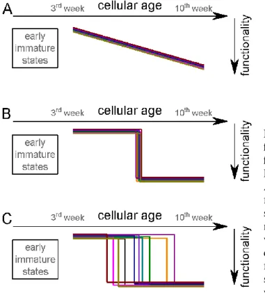
Consequently, the adult DG constitutes of GCs that are largely heterogeneous in age, as well as in intrinsic cellular properties. It is currently believed that ABGCs during their maturation process (between 3-10 weeks of age) are engaged in distinct physiological functions compared to mature GCs. Accumulating evidences support that young ABGCs are necessary for the pattern separation function of the hippocampus (Clelland et al., 2009; Deng et al., 2010; Sahay et al., 2011; Nakashiba et al., 2012). Whereas fully matured cells are potentially involved in rapid pattern completion (Nakashiba et al., 2012), in very sparse and specific encoding (Sahay et al., 2011), in the behavioral state- dependent, precise location representation (Neunuebel and Knierim, 2012), in the
24
preservation of the temporal episodes during later recalls (Aimone et al., 2010a), or that they simply become non-functional, “retired” cells (Aimone et al., 2010b; Alme et al., 2010). However, the transition between young and adult functional states is not completely understood.
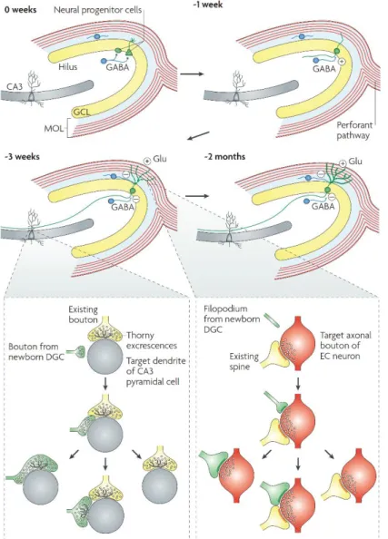
Figure 7. Illustration of the adult hippocampal neurogenesis. The neurogenesis starts with the proliferation of the neural progenitor cells (two different morphologies). They either give rise to adult-born granule cells (ABGCs; green cell) or glial cells (not shown).
ABGCs undergo two months of development, with gradual changes in morphological and physiological characteristics.
1st week of the maturation:
the dendrites of ABGCs extend into the str.
granulosum and str.
moleculare while their axon grows into the hilus, ABGCs receive excitatory (!) GABAergic input from local interneurons (blue cells). 3rd week of the maturation: at this stage ABGCs integrate into the circuit, they receive PP input and start to form synapses with the CA3 cells;
the effect of GABA switches from excitatory to inhibitory.
2 months of age: the maturation is complete, the structural and physiological properties of ABGCs are indistinguishable from those of the elder DG GCs. The bottom inset panels illustrate the competitive nature of synapse formation. Left: a small bouton of the ABGC (green) contacts the dendritic shaft of a CA3 PC (gray) at a site near to an existing MF synapse (yellow). The subsequent development might have three different outcomes, the MFB either conquer the thorny excrescence of the existing synapse, or retracts, or by the growth of new dendritic structures both remains. Right: the filopodium (green) of an ABGC dendrite extends to a PP bouton (red) that is associated with another spine (yellow), that eventually results in a monosynaptic bouton on the new dendritic spine of the ABGC, or a multisynaptic bouton, or leads to retraction of the filopodium. The figure is adopted without any modifications from (Deng et al., 2010).
25
Which physiological parameters support pattern separation function of young ABGCs?
The diverse ion channel content of different types of neurons leads to their different sensitivity and different output in response to similar input patterns (Armstrong and Hille, 1998; Nusser, 2009). The intrinsic excitability determines how neurons integrate their synaptic inputs and convert them to spiking output. The capability of ABGCs to respond with different spiking output to subtle differences in their input strength and frequency is optimal for pattern separation. In my first Ph.D. project, we studied the maturation of multiple membrane parameters of ABGCs to gain insight into the maturation of intrinsic excitability, which potentially underlies changes of the cellular function.
1.2.4. The feedforward inhibitory circuit in the CA3 region
Feedforward inhibition (FFI) is a fundamental network motif in which the afferent input activates a population of the local inhibitory cells, the feedforward interneurons (FF-INs) (Buzsáki, 1984). FFI has been implicated in various circuit functions by controlling the excitability of the principal cells. FF-INs can determine the temporal window in which the excitatory inputs might integrate. Depending on the properties of the local FFI this window can even vary among different subcellular domains (Pouille and Scanziani, 2001). By normalizing input strengths, FFI was shown to expand the dynamic range that a neuronal circuit can represent (Pouille et al., 2009).
As it was discussed earlier, the anatomical features of the MF synapses establish a prototypical FFI circuit in the CA3 region. GCs innervate at least four distinct types of GABAergic interneurons in the CA3: fast-spiking, parvalbumin (PV) expressing basket cells, regular-spiking, cholecystokinin (CCK) expressing basket cells, ivy cells, and septum-projecting spiny lucidum cells (Szabadics and Soltesz, 2009). The axonal arbor of these IN types covers both the perisomatic region of the PCs and a substantial part of their dendritic tree that locates in the str. radiatum and str. oriens. Furthermore, the extensive axon arbor of the INs provide substantial divergence to the their PC targets (Spruston et al., 1997; Vida and Frotscher, 2000). This FFI circuit has been proposed to play key role in the DG – CA3 communication (Lawrence and McBain, 2003; Acsády and Káli, 2007). The strong inhibitory control of the PCs contributes to implementing and maintaining the sparse pattern separated DG input code in the CA3 circuit. The
26
mechanistic understanding of the operation of the DG-CA3 FFI circuit however is not complete. In a FFI network, depending on the wiring specificity of the afferent input and the FF-INs, fundamentally different functional contributions are possible e.g. in the case of lateral inhibition or ensemble-specific FFI. Consideration of the wiring arrangement on single cellular level is particularly important in the case of the DG GCs because their extremely sparse activity decreases the importance of the population level effects.
1.2.5. Synaptic transmission and plasticity mechanisms
Synapses are intercellular junctions of neurons. In the chemical synapses, which are the most fundamental sites for information transfer between neurons, the electrical activity (AP) of the presynaptic cell is converted to chemical signal by releasing neurotransmitters. Binding of neurotransmitter molecules to their receptors results in ion channel openings, and converted back to electrical activity in the postsynaptic neuron.
The release of synaptic vesicles is a remarkably complex process, with multiple successive steps (Südhof, 2013). The AP-evoked membrane depolarization invades the presynaptic terminal and opens voltage gated Ca2+ channels. The inflowing Ca2+, by binding to synaptotagmins, is the key signal mediating the release of neurotransmitters.
To become ready to release, synaptic vesicles must undergo docking and priming steps.
Molecules in the synaptic active zone, such as RIM, Munc13, RIM-BP, α-liprin, and ELKS proteins are identified as key mediators of docking and priming, however these presynaptic processes involve a variety of further molecular components and regulatory mechanisms which are still not completely understood (Südhof, 2012). The presynaptic Ca2+ transient itself also triggers further signaling pathways (Schneggenburger and Rosenmund, 2015; Körber and Kuner, 2016).
According to the quantal hypothesis of del Castillo and Katz, the magnitude of the postsynaptic response is determined by three factors: (1) the number of synaptic contacts, namely the ‘functional release sites’ between the cells, (2) the probability of vesicle release at each release sites, and (3) the quantal size, which is the elementary postsynaptic response for the release of a single neurotransmitter vesicle (Del Castillo and Katz, 1954).
Release probability is controlled by presynaptic regulatory mechanisms, while postsynaptic processes affect the quantal size.
27
Weight of synapses undergoes continuous changes during physiological activity of cells which is necessary for the storage and processing of information. These plasticity phenomena vary on remarkably broad timescale from a few milliseconds to several hours, or even days and weeks (Zucker and Regehr, 2002; Holtmaat and Svoboda, 2009). The scale starts with two fundamental form of short-term plasticity: facilitation and depression. These mechanisms rely on the relationship of the presynaptic Ca2+ transients and its sensors (e.g. the accumulation of residual Ca2+) and the depletion rate of the release-ready vesicles (Fioravante and Regehr, 2011; Jackman and Regehr, 2017).
Prolonged high frequency activity, so-called “tetanus”, in certain synapses can evoke post tetanic potentiation (PTP) or augmentation, forms of plasticity that last for tens of seconds to minutes and usually require the involvement of protein kinase activity, e.g. PKC, PKA beyond the presynaptic Ca2+ transients (Zucker and Regehr, 2002; Fioravante et al., 2014). Notably, the nomenclature and the categorization of these plasticity phenomena is not completely consistent in the literature, thus, certain plasticity forms in different synapses involving substantially different molecular mechanisms might referred similarly if sharing similar temporal profile. Finally, the long-term plasticity, potentiation and depression (LTP and LTD), are permanent changes of the synaptic strength and can be accompanied by structural changes (Yuste and Bonhoeffer, 2001; Holtmaat and Caroni, 2016).
What plasticity mechanisms function in MF synapses? How do they operate during their physiological activity?
The GCs target PCs and inhibitory cells with anatomically different types of presynaptic terminals that suggests functional differences. Indeed, the synaptic transmission from MF to the two major postsynaptic targets operates with substantially different short-term plasticity. At MF-PC synapses the initial release probability is low but in the case of high frequency spiking activity the synapse shows strong short-term facilitation. In contrast, in the case of postsynaptic GABAergic cells the synaptic transmission is relatively stable (e.g. mostly varies between slightly facilitating or slightly depressing) during repetitive activity (Salin et al., 1996; Toth et al., 2000). This functional difference establishes a frequency dependent switch from inhibition to excitation as result of the MF activity in the CA3 circuit. Therefore, the MF terminal often referred to as
“conditional detonator”. Conditional in the sense that high frequency activity, that is a
28
MF burst, is required to overcome the strong FFI network and successfully drive PCs (Henze et al., 2002; Lawrence and McBain, 2003; Mori et al., 2004).
Similarly to the short-term plasticity, dichotomy was found in the long-term synaptic plasticity phenomena when the two major postsynaptic targets of MF synapses, PCs and interneurons were compared. Maccaferri and colleagues applied tetanic stimulation protocol that induced LTP in PCs, however, the same protocol either had no effect or induced depression in postsynaptic interneurons. Similarly, pharmacological activation of the PKA pathway only potentiated synapses on PCs (Maccaferri, 1998).
Another study specifically addressed plasticity of MF synapses on GABAergic cells of the DG and described the presence of LTP and PTP phenomena. In the study of Alle and colleagues the applied stimulus protocol (25 AP at 30Hz repeated 12 times in every third seconds) was either evoked in a presynaptic somatically recorded GC or the MF tract was extracellularly stimulated. The associative form of the protocol, when firing of the postsynaptic fast spiking basket cells followed the presynaptic stimuli, LTP was developed. Whereas, non-associative protocols, when the postsynaptic cell was held in voltage clamp to prevent firing of the cell resulted in PTP. These two plasticity forms involve different molecular pathways as the PTP was found to be sensitive for the blockade of both PKC and PKA while only blocking of PKC reduced the LTP (Alle et al., 2001).
Prolonged stimulation (100AP, 40Hz) of single presynaptic GCs in hippocampal slice culture has been shown to potentiate MF responses for more than 10 minutes in GABAergic neurons as well as in PCs of the CA3. Based on the sustained increase of feed-forward inhibition the authors propose three different state of the MF-CA3 connection: a resting state with low release probability and high failure rate onto PCs; a bursting mode, in which excitation of PCs predominates; and a post-bursting mode, in which the feed-forward inhibition is greatly enhanced (Mori et al., 2007).
In contrast to generally used artificial stimulation paradigms Gundlfinger and colleagues applied natural spike trains to test synaptic dynamics of mossy fibers. They obtained natural spiking activity of GCs by tetrode recording when the animals traversed their place fields thus such natural spike train contained high frequency epochs.
29
Stimulation of multiple GCs with these spike trains resulted in short-term facilitation and LTP in CA3 pyramidal cells (Gundlfinger et al., 2010).
In order to understand the communication between two cells, it is essential to consider all types of the synaptic mechanisms operating on various timescales during the physiological activity of the cells (Abbott and Regehr, 2004). This is particularly important in the case of GCs with such an irregular firing activity that consists of single action potentials, short high frequency bursts and long silent periods. Despite the broad spectrum of studies with various protocols and experimental configurations addressing the MF synapses many unresolved questions remain to be answered. It is not clear whether short, truly physiological high frequency GC bursts activate any specific synaptic mechanisms beyond the short-term plasticity. How does the downstream CA3 network interpret the differences of single action potential and single burst firing in the subsequent period?
30
2. Objectives
My Ph.D. studies addressed the operation of the DG-CA3 circuit and each question aimed to elucidate some yet poorly understood aspects of it. I focused on two main topics with the following specific questions:
My first project, aimed to understand how young and matured adult born granule cells can perform distinct neuronal functions.
• How do the various biophysical properties of ABGCs mature?
• Which specific intrinsic physiological properties enable the emergence of distinct functional populations?
• When and how do ABGCs switch their “young” functionality to “matured”
operation state during their postmitotic development?
In the second project, I addressed the synaptic mechanisms responsible for the interpretation of distinct physiological activities of GCs within the CA3 circuit.
• Do short, truly physiological GC bursts accompanied by distinct synaptic plasticity mechanism than single AP firing?
• How physiologically relevant activity patterns, containing single APs, short high frequency bursts and long quiescent periods are translated by the CA3 neurons?
31
3. Materials and methods
3.1. Virus mediated birth-dating of granule cells
To follow up the development of ABGCs we labeled dividing cells in the DG of 31 to 33 days old rats by Moloney murine leukemia virus vector (Zhao et al., 2006; Jessberger et al., 2007). The surgeries and the virus labeling were done by János Szabadics. Male Wistar rats (95-135 g body weight) were injected with 0.8–1 µl CAG-GFP or CAG-RFP using stereotaxically targeted (5.7-5.8 mm posterior, ±4.4-4.5 mm lateral and 5.6-6 mm ventral from bregma), conventional Hamilton syringe under ketamine/xylazine/pipolphen anesthesia (83/17/7 mg/body kg). By this approach, adult born granule cells were labeled along a broad longitudinal range (2-3 mm) of the hippocampi. Note that in animals 3-10 weeks after virus injection, we never found cells younger than 3-weeks-old based on their cellular properties, indicating the reliability and precision of the birth-dating method.
After the surgical procedure two or three siblings were housed together in large cages (75 cm x 35 cm) equipped with a running wheel, toys and shelters until the electrophysiological experiments were performed, because running is known to increase the number of surviving adult-born neurons (Kempermann et al., 1997; van Praag et al., 1999; Tashiro et al., 2003).
3.2. Slice preparation
All the electrophysiological data during my Ph.D. projects were obtained by in vitro recordings from acute hippocampal slices prepared from Wistar rats. For recording ABGCs adult, postnatal day 51-105 (corresponding to recording of 20-72 days old virus labeled ABGCs) or postnatal day 68-101 (adult non-labeled) male rats were used. GCs born during the development of the brain were recorded in young, 17-18 days old animals.
In the project examining the effect of single GC burst in the CA3 circuit , the acute brain slices were made by using adolescent rats (postnatal day 21–45, both sexes). The animals were deeply anesthetized with isoflurane (in accordance with the ethical guidelines of the Institute of Experimental Medicine Protection of Research Subjects Committee) and 350 μm slices were cut in ice-cold ACSF containing (in mM): 85 NaCl, 75 sucrose, 2.5 KCl,
32
25 glucose, 1.25 NaH2PO4, 4 MgCl2, 0.5 CaCl2, and 24 NaHCO3. The orientation of cutting was close to horizontal, the brains were positioned to obtain multiple slices perpendicular to the axis of the hippocampus at its medial part. This sectioning plane is parallel with the mossy fibers and preserves their connections to target cells in CA3 area (Bischofberger et al., 2006). Slices were incubated at 32°C for 60 minutes after cutting then were kept at room temperature until used for recordings in a solution composed of (in mM): 105,5 NaCl, 37.5 sucrose, 2.5 KCl, 17.5 glucose, 1.25 NaH2PO4, 3 MgCl2, 1.25 CaCl2, and 25 NaHCO3. Under these conditions slices were viable and were used for up to 8-12 hours.
3.3. Electrophysiological recordings
Cells were visualized with an upright microscope (Eclipse FN-1; Nikon) with infrared (900 nm) Nomarksi differential interference contrast optics. The standard recording artificial cerebrospinal solution (ACSF) was composed of (in mM): 126 NaCl, 2.5 KCl, 26 NaHCO3, 2 CaCl2 (unless stated otherwise), 2 MgCl2, 1.25 NaH2PO4, and 10 glucose.
The ACSF was saturated by the mixture of 95% O2 and 5% CO2 during the recordings.
The temperature was held at 35 - 36°C (unless decreased to 28-29°C see Fig. 14) during the experiments. Electrophysiological recordings were acquired with Multiclamp 700B amplifiers (Molecular Devices) and pClamp10 software.
Presynaptic CA3 GCs and MFBs in “whole cell” configuration were recorded in current clamp configuration (digitized at 50 kHz and low-pass filtered at 10-20 kHz) and were held at ~ -75 mV. The pipette capacitance was greatly reduced and optimized by capacitance neutralization in the bridge balance compensated presynaptic current clamp recordings. Action potentials were evoked by current pulses (usually1.5 ms long, 1.8 nA in the case of somatic, and 1 ms long, 300pA in MFB recordings). MFBs however, were preferentially targeted in cell attached configuration. In these recordings MFBs were stimulated in voltage clamp mode, usually 0.5-1 ms long 140-200 mV pulses were required to reach the AP threshold. Action currents were monitored at leak subtracted traces. Postsynaptic cells were recorded in voltage clamp mode (digitized at 50 kHz and low-pass filtered at 4-6 kHz) the membrane potentials were clamped to -70 mV in standard conditions. The series resistance (5-30 MΩ) was monitored by the capacitive
33
artefact in response to a 5 mV step in each trace and controlled during the recordings. Rs
compensation (70-75%) was only applied in some experiments among the disynaptic IPSC (diIPSC) recordings where pyramidal cells were held at -50mV.
Recording pipettes were pulled from either thin or thick wall (1.12-0.86 ID) borosilicate glass capillaries, the pipette resistance was ranging 3–4.5 MΩ for somatic recordings and 10-12 MΩ for MFB recordings. Three different intracellular solutions were used. First, in standard recording conditions ABGCs and monosynaptic pairs (presynaptic CA3 GCs or MFBs to postsynaptic INs), were recorded in an intracellular solution containing (in mM) 90 K-gluconate, 43.5 KCl, 1.8 NaCl, 1.7 MgCl2, 0.05 EGTA, 10 HEPES, 2 Mg-ATP, 0.4 Na2-GTP, 10 phosphocreatine-disodium, and 8 biocytin (pH 7.25). Second, in certain experiments (Fig. 24) presynaptic recordings were done with a modified version of the first intracellular solution where 40mM KCl was substituted for CsCl. Third, for attempting disynaptic connections, postsynaptic pyramidal cells were patched by gluconate based intracellular solution that allows distinguishing between inhibitory and excitatory postsynaptic currents (IPSCs and EPSCs respectively). This solution was composed of (in mM) 133.5 K-gluconate, 1.8 NaCl, 1.7 MgCl2, 0.05 EGTA, 10 HEPES, 2 Mg-ATP, 0.4 Na2-GTP, 10 phosphocreatine-disodium, and 8 biocytin (pH 7.25).
The chemicals for the intracellular and extracellular solutions were purchased from Sigma-Aldrich and pharmacological compounds were purchased from Tocris Bioscience.
DCG IV (1 µM), MSOP (150 µM), SR 95531 (gabazine, 5 µM) were dissolved in ACSF.
Phorbol 12,13-dibutyrate (PDBu, 1 µM), Go 6976 (250 nM), GF 109203X (1 µM), Calphostin C (1 µM), U 73122 (2.5 µM), BINA (5 µM), AMN 082 (1 µM) were dissolved first in DMSO and diluted at least 2000 times in ACSF. EGTA (0.5 - 2.5mM) and PKC (19-36) (100 µM) were added to the intracellular solution.
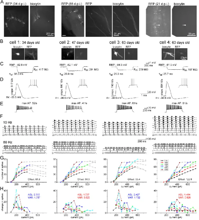
3.4. Calculation of the integrative and biophysical parameters of ABGCs
We characterized the integrative properties of individual ABGCs by two reliable parameters, which measure the gain of the input-output functions: the average slope (ASL) and the variance (VAR) of the slope of input-output curves. The calculated parameters (average slope, variance and offset) were weighted by the different