Egg of Arbacia punctulata
ARTHUR K . PARPART1
Department of Biology, Princeton University, Princeton, New Jersey and Marine Biological Laboratory, Woods Hole, Massachusetts
General
The movement of cellular inclusions independent of Brownian motion and of cytoplasmic streaming has frequently been observed in a large variety of cells. In his recent comprehensive review of protoplasmic streaming, Kamiya (1959) summarized our present knowledge and con- cepts concerning the independent motion of protoplasmic particles. T h e present paper presents some data and ideas about the independent motion of the echinochrome granules in the cytoplasm of the unfertilized egg of Arbacia punctulata (Parpart, 1953).
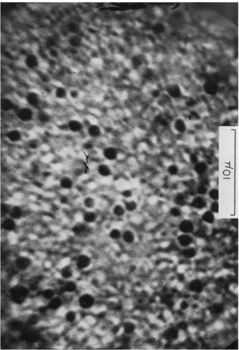
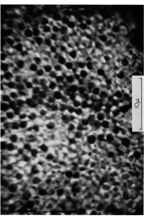
The echinochrome granules averaged 1.3 μ in diameter with a spread of 0.9 to 1.6 μ. Their distribution over an area of ca. 1100 μ2 in the un- fertilized egg is illustrated in Fig. 1, and their distribution over the same area for the same egg, after fertilization, is presented in Fig. 2. In the former case the granules are in active independent motion, whereas in the latter figure the granules have ceased moving and have increased about threefold in number.
The independent motion of the echinochrome granules in unfertil- ized Arbacia eggs was examined by T V microscopy (Parpart, 1951; Par- part and Hoffman, 1956). T h e image on the monitor was recorded on movie film at a rate of 1 frame/sec. T h e eggs were mounted on a micro- scope slide so that their jelly coat became lodged between the slide and a tilted, fixed cover slip. Thus a desired solution could be rapidly flushed around the eggs and the action of a specified agent studied.
Eggs were never compressed between slide and cover slip.
The majority of the echinochrome granules are moving about in a 5-10 μ cytoplasmic layer near the surface. T h e granules are unaffected by the normal cytoplasmic streaming, whether they are at rest or in motion. This motion is entirely random and is of a start-stop nature.
This is illustrated by the typical motion of the granule plotted in Fig. 3.
ι This research was supported in part by the Whitehall Foundation.
471
472 ARTHUR Κ. PARPART
FIG. 1. Photograph of television monitor image of the upper surface, ca. 2 μ below the plasma membrane of the unfertilized egg of Arbacia punctulata. T h e optically dense spherical bodies are the echinochrome granules. The magnification of a 10 μ stage micrometer is included. The sweep lines of the monitor are visible.
FIG. 2. Photograph of the same region of the same egg as in Fig. 1, 10 min after fertilization. All other conditions are the same as for Fig. 1.
474 ARTHUR Κ. PARPART
Studies on several hundred granules of thirty egg cells at 20°C gave an average rate of movement of 0.6 μ/sec with a spread of 0.3-0.8 μ/sec.
These values are from one-third to one-fifth the rate of ameboid move- ment of marine amebae (Pantin, 1924); and from one-quarter to one- two-hundredth of cyclosis in plant cells (Kamiya, 1959). A striking feature of the motion, observable in Fig. 3, is the fact that a granule will vibrate,
ο ι
Displacement (/ι)
FIG. 3. Analysis of the motion of a single echinochrome granule in an unfertilized Arbacia egg. Determined by a motion analyzer from movie recording, at 1 frame/sec, of the monitor of a T V microscope. The numbers at each point represent successive times in seconds. At 25 sec the granule plunged out of the focal plane of the micro- scope. Temperature, 20°C.
Brownian motion, for a period of time (2-3 sec) at one point and then suddenly move 1-5 μ in a given plane in space.
Advantage of this latter characteristic has been taken in analyzing the action of various factors on the motion of these echinochrome granules. Frame-by-frame analysis of the motion of a minimum of one hundred granules was carried out for each factor studied. T h e time required for each granule to go from a state of rest to motion through 3-5 μ was determined. Each such motion was recorded as 1 displacement.
Each displacement required a certain time, and the data were averaged for each 10 displacements as to equivalent time spans. T h e "control"
curve in Figs. 4 and 5 illustrates the spread and mean of these time
10
8h
S 6 Ε
Φ 4
û
Ο Χ Δ
ν- Ο Χ Δ • Λ Δ α
χ ΰ Δ α
Temp. (°C)
• 23
χ 20
δ 18
• χ ο δ • α 15 ο 20 "Return"
» Χ Ο Δ •
Χ Ο Δ •
Χ ΟΔ •
h >· Χ ΟΔ
<· κ 3 Δ •
0 10 20 30 40 50 60 70
Time (seconds)
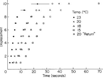
FIG. 4. Arbacia eggs were equilibrated at the temperatures given in the figure.
T h e curve for "20°C Return" was for the same egg cells returned from 15° to 20°C and brought to equilibrium (25 min). Method is outlined in text. Displacement—
100 echinochrome granules at each temperature.
10
8h
Ε 6
δ 4
-i-ox • -|- ox •
-|D Χ 3
ί Spread-—
Control \Mean !
Force Recovery
3000 g |3 mm a l 8 m in
15000g / I min V
x 35 min 10 min o 55 min
0 10 20 30 40 50
Time (seconds) 60 70
FIG. 5. Unfertilized Arbacia eggs were centrifuged in an isosmotic sucrose-sea water density gradient at forces 3000 and 15,000 g for 3 and 1 min respectively, according to the method of Harvey (1932). Then eggs were quickly washed in sea water and echinochrome granule motion studied at the "Recovery" times noted on the graph. Temperature 22°C. Displacement—100 echinochrome granules. Method is outlined in text.
476 ARTHUR K. PARPART
measurements. Thus the behavior of a random population of granules under the influence of various factors was determined.
The action of various factors on the motion of echinochrome gran- ules is recorded in Figs. 4 to 8. These figures record the displacement of 10 time-groups of 100 echinochrome granules against the time required
10 r
ω 6 - · *
§ Temp. 22°C
ö> 41- · χ χ 0.005 M Cysteine 5 min
0 10 20 30 40 50 60 Time (seconds)
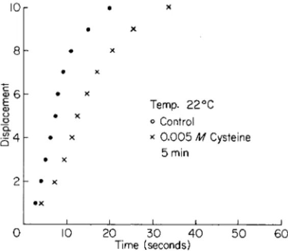
FIG. 6. Unfertilized Arbacia eggs exposed to 0.005 M cysteine dissolved in sea water for the time noted (15 min). Displacement—100 echinochrome granules. Method is outlined in text.
* 10
c 4
£ 2h
% Seawater ο 100 χ 120
A 80
ο 70 ο Return to 100
10 20 30 40 50 60 70 Time (seconds)
FIG. 7. Unfertilized Arbacia eggs exposed to the hypertonic and hypotonic con- centrations of sea water noted on the graph. T h e curve "Return to 100%" was on the cells that had previously been equilibrated in 70% sea water. All equilibrations were for 30 min. Temperature 20°C. Displacement—100 echinochrome granules.
for such displacement. The factors studied were: temperature; centri- fugal force; cysteine; tonicity; sodium iodoacetate and sodium fluoride.
In all cases the action, when properly controlled, was found to be reversible.
£ 6
OK
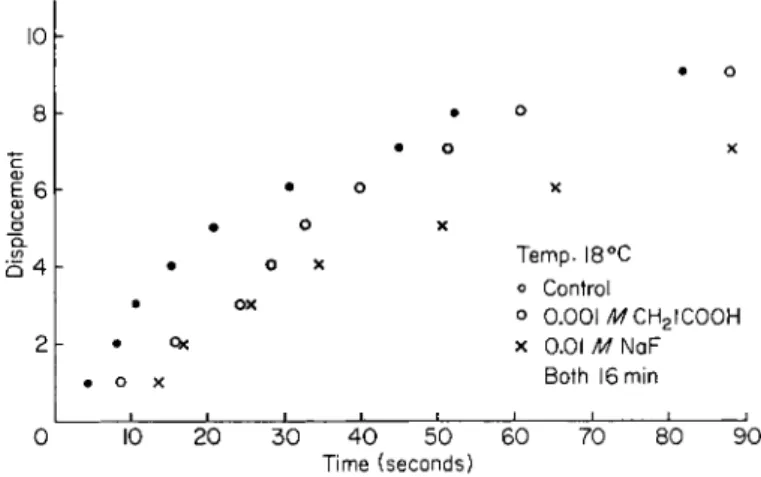
Temp. I8°C ο Control
ο 0.001 /WCH2IC00H
x 0.01 MNQF
Both 16 min
10 20 30 40 50 60
Time (seconds) 70 80 90
FIG. 8. Unfertilized Arbacia eggs exposed for a period of 1 6 min to ( 1 ) 0 . 0 0 1 M N a C H2I C O O H dissolved in sea water and ( 2 ) 0 . 0 1 M NaF dissolved in sea water.
Displacement—100 echinochrome granules. Method is outlined in text.
Discussion of Factor Action
TEMPERATURE
The decrease in the rate of motion of the granules with decrease in temperature is notable in Fig. 4. T h e temperature coefficient (βίο) for the motion of the granules varies between 4.2 and 5.7. This is a surprising action if one examines it from the point of view which Marsland (Marsland and Landau, 1954) has shown, namely, decreasing temperature over the same range leads to an increase in the sol struc- tural character of the layer of cytoplasm ("cortical") in which the major- ity of the echinochrome granules are located. However, if one views the effect of decreasing temperature as a decrease of the fibrous structure of this cytoplasm, then it might appear that these fibers may play some role in the motion of the granules.
However, measurements on the distance that the individual granules will move have shown that it is not affected by temperature; even though its rate is. This is in accord with numerous data in the literature which established that the heat/tension ratios of muscle fibers undergoing isometric contraction are the same over a wide range of temperatures.
478 ARTHUR Κ. PARPART
It would appear that the action of temperature on the motion of the echinochrome granule lends support to the concept that these granules are moved about the cytoplasm by fibers attached to them.
CENTRIFUGAL FORCE
It has long been known that the echinochrome granules have the highest density of any of the granules in the cytoplasm of the Arbacia egg. Thus, in the unfertilized egg, under centrifugal force they go to the centrifugal pole of the egg. Eggs centrifuged by the method of Harvey ( 1 9 3 2 ) were examined for the alteration in motion of these granules. T h e results are given in Fig. 5. Immediately after centrifugation, whether at low ( 3 0 0 0 g) or high ( 1 5 , 0 0 0 g) force, these granules have ceased all motion. Recovery of motion occurs gradually, somewhat faster at lower than higher force, until it is completely reversed. Recovery of the normal rate and distance of motion of the echinochrome granules even at the higher force, occurs long before the reorientation of the yolk and other cytoplasmic granules. Recovery of the normal rate and distance is also evident long before the re-establishment of cytoplasmic streaming.
The data on centrifugal force suggest that during its application the fibers normally attached to the echinochrome granules are torn loose and that one of the first acts of recovery is the re-formation of these fibers. This recovery occurs in the peripheral 5 - 1 0 μ of the cytoplasm.
It is this latter region that is partially gelled, at 2 0 ° C , in the unfertilized egg. It is conceivable that some of the fibers formed in this gel also attach to the echinochrome granules. Such fibers, if contractile, could account for their motion.
CYSTEINE
Unfertilized Arbacia eggs exposed to low concentrations ( 0 . 0 0 1 -
0 . 0 0 5 M) of cysteine dissolved in sea water show a gradual diminution in the rate and distance of motion of their echinochrome granules.
After from 2 0 to 5 0 min exposure all such motion is stopped. It has been demonstrated that cysteine induces a gelation of the egg cytoplasm such that no cytoplasmic granules of any sort can be moved on exposure to centrifugal forces of 15,000 g (Parpart and Ballantine, 1962). It has also been observed that if these eggs are returned to sea water within 1 5 to 2 0 min the effect of cysteine is completely reversible. A typical slowing of echinochrome granule motion after exposure for 5 min is presented in Fig. 6. It was also observed that at this 5-min exposure time all cyto- plasmic streaming had ceased even though echinochrome granule motion had been only partly slowed.
Similar to the data for the effect of temperature, those with exposure
to cysteine and the recovery suggest that there is an optimal degree of gelation of the "cortical" cytoplasm for maximal granule motion. As cysteine continues its action and the fibers of the cytoplasmic gel become more numerous the motion is impeded and stopped. This stoppage is similar to that which occurs on fertilization except for its reversibility if the exposure is not continued too long.
TONICITY
Another factor that will alter the gel-sol relations of the cytoplasm is the amount of water in it. The variables involved in shifting water into or out of the egg cell are numerous. However, an experimental evaluation of such shifts is presented in Fig. 7.
Alteration of the cytoplasmic water content whether toward hyper- tonicity or hypotonicity always produces a decrease in the rate but not the distance of motion of the echinochrome granules. Slight shifts toward the hypotonic (80% sea water) have a more pronounced effect than a larger (70% sea water), but still physiologically reversible, shift.
All these effects are reversible if the egg is returned to sea water within an hour of the change of tonicity. Eggs equilibrated with 6 0 % or 150- 180% sea water have echinochrome granules that have ceased moving.
This reaction is also reversible on return to sea water.
Unfortunately there are no data in the literature, by the present methods of study of in vivo gel-sol transformations, on the influence of the amount of cell water on the state of the gel-sol ratio. T h e data dis- played in Fig. 7 would indicate that the condition of the hypothetical fibers which move the echinochrome granules is optimal in sea water.
Any alteration of the water content of the cytoplasm lowers the con- tractile rate of such fibers. It is also possible that the contractile rate of the fibers may depend on a critical concentration of the Ca++ and/or Mg++ in the cytoplasm, which would be changed by the water shifts.
This line of reasoning has some support from the studies of Bozler (1954). He observed that minute changes in the Ca++ or Mg++ con- centration markedly changes the contractile properties of the isolated and reconstituted muscle fibrils which he studied. Another suggestive bit of evidence is the fact that if unfertilized Arbacia eggs are exposed to 0.005 M ethylenediaminetetraacetate dissolved in sea water, the motion of the echinochrome granules is stopped within 3 to 5 min. This is reversible if the eggs are returned to sea water.
IODOACETATE AND FLUORIDE
The influence of sodium iodoacetate (0.001 M) and of sodium fluo- ride (0.01 M) is recorded in Fig. 8. These metabolic inhibitors act pri-
480 ARTHUR K. PARPART
marily on the "recovery" mechanisms of muscle fibrils. They might, therefore, be expected to slow down gradually the rate of motion of the fibrils that are here hypothesized as responsible for the motion of the granules. This was found to be the case, since not only did it take a long time, ca. 2 hr, for these inhibitors to stop the motion of the echino- chrome granules, but it was observed that the distance of travel of the granules decreased too. Addition of adenosine triphosphate (ATP) to the sea water environment of the egg cell had no effect upon the echinochrome granule motion. This is probably due to the inability of A T P to enter the cytoplasm.
Summary and Discussion
It is obvious from the foregoing that the weight of experimental evidence at present available favors the concept that the random motion of the echinochrome granules of the unfertilized Arbacia egg may be associated with contractile fibrils in the cytoplasm. T h e data strongly suggest that each echinochrome granule has a number of such contrac- tile fibrils attached at many points on its spherical surface. The motion of these granules occurs in unpredictable directions in the three planes of cytoplasmic space. The three-planar motion has not been stressed before since all measurements were made in a two-planar layer of cyto- plasm. However, when one watches the motion of these granules under the microscope or on movie film the echinochrome granules are con- tinuously moving into and out of the focal plane of the microscope field (see legend of Fig. 3). The most dramatic motion of this sort occurs within 1 to 2 min of fertilization. If the upper 2 μ of the cytoplasm are under T V microscope observation this two-dimensional field rapidly fills up with echinochrome granules which quickly travel from deeper in the cytoplasm directly to this surface region (cf. Figs. 1 and 2). T h e majority of these granules, but by no means all, thus come to rest in the outer- most cytoplasm of the egg.
The arrangement of the contractile fibrils is, of course, purely speculative. They may be thought of as attached at many points on the surface of the echinochrome granule. The other ends of these fibrils may be attached 10-20 μ away from the granule or to the extensive villar structures of the plasma membrane of the egg. The latter point of view seems justified from the rapid manner with which an echino- chrome granule 20-30 μ deep in the cytoplasm is "pulled" to the sub- surface layer. The possibility of attachment of these fibrils to fixed gelled regions of the cytoplasm has some confirmation from the fact that as the temperature of the cell is decreased the motion of these granules decreases markedly, and stops completely at about 8°C, even though the cytoplasm
is going over to a more sol state as the temperature decreases. T h e re- ported actions of cysteine (Parpart and Ballantine, 1962), hypertonicity and hypotonicity, and of the metabolic inhibitors also tend to confirm such a picture.
In addition, it would appear that these contractile fibrils, if they exist, must be continuously formed and broken down. T h e action of centrifugal force forces many of these granules from the centripetal region of the egg through 75 μ of cytoplasm to the centrifugal end of the egg. Yet within 5 to 10 min after removal from the centrifuge these granules are again in motion and after 1 hr, even when 15,000 g is used they are very nearly back to the original rate and distance of motion.
This is well before any cytoplasmic streaming can be observed.
The next logical step would be an attempt to demonstrate such fibrils by electron microscopy. All such attempts have failed since we have never been able to find echinochrome granules in electron micro- graphs of sections of Arbacia eggs. This led to an examination of the influence of a great variety of cytological fixing agents on the echino- chrome granules of eggs under observation by T V microscopy. It was observed that none of the standard fixatives would preserve the echino- chrome granules. In fact it was found that the fixatives lead to an explo- sive decomposition of the echinochrome granules (Parpart et al., 1961).
The foregoing concepts are by no means new, except in so far as they apply to the Arbacia egg. T h e recent excellent reviews of Abé (1962) and Kamiya (1959) have stressed somewhat similar ideas for plant cells and amebae. Their articles give the pertinent literature.
There remains one further aspect of these observations on echino- chrome granule motion. It would appear that this method of study of this motion opens up the possibility of study of gel-sol transformations in any cell where such granule motions can be recorded. Such studies could be carried out in all planes of the cytoplasm, and thus make possible not only analysis of region-by-region gel-sol conditions but also of the influence of a variety of agents on such transformations.
REFERENCES Abé, T . H. (1962). Cytologia 27, 111.
Bozler, E. (1954). / . Gen. Physiol. 38, 735.
Harvey, Ε. B. (1932). Biol. Bull. 62, 155.
Kamiya, N. (1959). Protoplasmatologia 8, 1.
Marsland, D., and Landau, J . V. (1954). / . Exptl. Zool. 125, 507.
Pantin, C. F. A. (1924). Brit. J. Exptl. Biol. 1, 519.
Parpart, A. K. (1951). Science 113, 483.
Parpart, A. K. (1953). Biol. Bull. 105, 368.
Parpart, A. K., and Ballantine, T . V. N. (1962). Biol. Bull. 123, 485, and 508.
4 8 2 ARTHUR Κ. PARPART
Parpart, A. K., and Hoffman, J . F. (1956). ./. Cellular Comp. Physiol. 47, 295-303.
Parpart, A. K., VanNorman, E., and Bernhardt, J . C. (1961). Biol. Bull. 1 2 1 , 403.
DISCUSSION
DR. WOLPERT: It is well known that the eggs of various species of sea urchins have different properties. T h e egg of Psammechinus miliaris also shows cytoplasmic movements. Dr. Gustafson and I have made time-lapse movies of the unfertilized egg, and the cytoplasm shows very clear twitching movements and large oscillations that are inhibited by carbon dioxide. In this behavior it is similar to some of the observa- tions made by Dr. Parpart on Arbacia. However, there is an important difference with respect to the presence of a gel-like cortex—a matter that I would like to discuss with Dr. Parpart and the people concerned with hydrostatic pressure. Dr. Mercer and I have examined centrifuged eggs, both fertilized and unfertilized, in the electron microscope and could find no region in which the larger particles, mitochon- dria and yolk granules, were prevented from moving by a gel-like region. W e have not been able to detect, by any means, a typical cortex in Psammechinus.
DR. PARPART: T h e entire cytoplasm of the Arbacia egg is in active streaming motion prior to fertilization. After fertilization all such motion is stopped in the outermost layer of cytoplasm, 5-10 μ thick. As you focus further into the cytoplasm you begin to pick up slower motion, especially as the mitotic apparatus goes into full action. However, the internal cytoplasmic streaming continues, whereas near the surface there is no streaming at all.
DR. WOLPERT: Then the two eggs must be quite different.
DR. ZIMMERMAN: I would be surprised if there were not a cortex in Psamme- chinus just as in Arbacia. In Arbacia, there is a region near the surface of the egg about 5-7 μ thick which has physical characteristics different from those found in the interior of the cell. As Dr. Parpart has said, if you centrifuge a fertilized Arbacia egg, even at very high forces, the echinochrome pigment vacuoles remain in the cortical plasmagel layer. This plasmagel layer becomes very rigid prior to division in the Arbacia egg. I do not know, exactly, what experiments you conducted, but very likely the plasmagel layer in Psammechinus becomes rigid just prior to division, as it does in Arbacia. Your results would depend on the stage of development at which your studies were conducted, since we have reported that there are fluctuations in the strength of the plasmagel in Arbacia during its division cycle. If you tested the strength of the cortex of Psammechinus at a stage when the cortex was relatively weak, you would record a corresponding low value for the gel. Therefore, I would be surprised if there are any major discrepancies between the different eggs.
DR. REBHUN: It appeared to me in your pictures of centrifuged eggs that some of the yolk granules were participating in this motion. I have never seen this in a normal egg and have never seen it in your films before.
DR. PARPART: I avoided mentioning this. It is much harder to detect. One Vidicon tube in the T V system had a high sensitivity. In fact, in the near ultraviolet it permitted exclusive delineation of the echinochrome granules. On the other hand, the Vidicon tubes used for the present studies picked up yolk and other granules.
These yolk and other granules certainly are also in motion of similar type. It is not rapid.
DR. GUSTAFSON: May I take the opportunity to mention an observation I have made on a cytoplasmic change under the influence of cysteine and other SH com- pounds in the egg of Echinocardium. In the same range of concentrations of SH compounds as you used, a marked local contraction of the egg occurs, leading to the
formation of a large concavity, presumably corresponding to the future ventral side of the embryo. T h e effect was irreversible, i.e., persisted after transfer of the egg into pure sea water. T h e same effect can be obtained with histidine. Have you studied the effect of histidine on the cytoplasmic movements?
DR. PARPART: NO; I have not tried histidine, but glutathione has somewhat the same effect as cysteine. Incidentally, if you expose unfertilized eggs to cysteine for certain periods of time and then return them to sea water, there is a reversal of this whole change and actual cleavage occurs. We have gotten up to eight-cell stages with perfectly good cortex around the egg, that is, no breakdown of cortical granules but good cleavage of the egg.
DR. ZIMMERMAN: I have a question to ask Dr. Parpart about the gelating effect.
How do you establish the gelation in Arbacia}
DR. PARPART: I am not certain of this. I only say it is gelation because the yolk and other granules cease to move about in streaming, whereas in the interior similar particles are actively moving about in streaming. This is a normal fertilized egg.
With the cysteine-treated egg, the gelation or cessation of motion of yolk granules goes deeper and deeper with time of exposure.
CHAIRMAN BISHOP: May I ask you if you envisage the fibers as permanent or temporary?
DR. PARPART: They are thought to be broken and re-formed continuously.
CHAIRMAN BISHOP: IS there evidence from electron micrographs of fibrillar con- nections?
DR. PARPART: We cannot get electron micrographs of these granules. If you attempt to observe them under the microscope and put osmium under the cover slip, the granules explode.