1
RESPIRATORY TWIN STUDIES
PhD thesis
Dávid László Tárnoki
Basic Medicine Doctoral School Semmelweis University
Supervisors: Ildikó Horváth MD, D.Sc Viktor Bérczi MD, D.Sc Official reviewers:
Dóra Krikovszky MD, Ph.D Krisztina Bogos MD, Ph.D
Head of the Final Examination Committee:
Zoltán Benyó MD, D.Sc
Members of the Final Examination Committee:
Attila Doros MD, Ph.D
Zsuzsanna Monostori MD, Ph.D
Budapest, 2013
2
Respiratory twin studies Table of Contents
The list of Abbreviations 5
1. Introduction 7
1.1. Twins’ contribution to science 7
1.2. History of Hungarian twin studies 8
1.3. Respiratory system 11
1.3.1. Structure of the respiratory system 11 1.3.2. Assessment of function of the respiratory system:
respiratory function tests 14
1.4. Past studies on the heritability of lung function 18 1.5. Relationship of lung function and cardiovascular diseases 19 1.6. A novel cardiovascular phenotype: arterial stiffness 22 1.7. Secondhand smoke exposure and its health related effects 23 1.7.1. Air pollution and secondhand smoke exposure 23 1.7.2. Cardiovascular effects of secondhand smoke exposure 27 1.7.3. Psychosocial (family) aspects of tobacco smoking 29
2. Objectives 32
3. Methods 33
3.1. Subjects of the twin studies 33
3.2. Study design 33
3.3. Pulmonary function assessment 34
3.4. Hemodynamic measurement 35
3.5. Statistical analysis 37
3.5.1. Data analysis 37
3
3.5.1.1. Descriptive analysis 37
3.5.1.2. Estimating genetic influences on lung function and PWV, AIx 37 3.5.1.3. Estimating the correlation between lung function and PWV, AIx 38 3.5.1.4. Genetic covariance between lung function and PWV, AIx 38
4. Results 39
4.1. Characteristics of the Hungarian Twin Registry 39
4.2. Clinical characteristics and measures 40
4.2.1. Lung function study 40
4.2.2. Smoking habits and secondhand smoke characteristics study 43 4.3. Results of the lung function twin study 43 4.3.1. Heritability analysis of pulmonary function and PWV, AIx 43 4.3.2. Phenotypic correlation between pulmonary function and PWV, AIx 45 4.3.3. Genetic covariance of FEV1, FVC and augmentation indices 47 4.4. Smoking and secondhand smoke characteristics of twins 48
4.4.1. Comparison of smoking habits, smoking characteristics, secondhand smoke exposure, and local home, car and workplace
smoking regulations of monozygotic and dizygotic twins 48 4.4.2. Secondhand smoke exposure in local bars and pubs,
restaurants, cafés and public transportation venues of
monozygotic and dizygotic twin pairs 49
5. Discussion 56
5.1. Research projects of the Hungarian Twin Registry 57 5.2. Consequences of findings of lung function study 58 5.3. Discussion of smoking habits and secondhand
smoke characteristics of twins 62
6. Conclusions 65
7. Summary 66
8. Összefoglaló 68
9. Bibliography 70
10. Bibliography of own publications 98
10.1. Publications related to the current PhD thesis 98
4
10.2. Publications not related to the current PhD thesis 98
11. Acknowledgement 102
5 The list of Abbreviations A = Additive genetic influence
ACE = additive genetic (A), common (C) and unique (E) environmental factors AIx = augmentation index
AQG = air quality guideline AQI = air quality index
ATS = American Thoracic Society BMI = body mass index
BP = blood pressure
BTR = Budapest Twin Registry
C = common familial environmental factor CHD = coronary heart disease
CI = confidence interval
COPD = chronic obstructive pulmonary disease CRP = C-reactive protein
DNA = deoxyribonucleic acid
Dsb = single breath diffusing capacity DZ
=
dizygoticDZO = oppisite sex dizygotic E = unique environmental factor ERS = European Respiratory Society
FEF 25–75 = forced expiratory flow 25–75%
FET = forced expiratory time FEV = forced expiratory volume
FEV1 = forced expiratory volume at 1 second FIF = forced inspiratory flow
FRC = functional residual capacity FVC = forced vital capacity
HCAR = Hungarian Congenital Abnormality Registry HDL = high-density lipoprotein
HRCT = High Resolution Computed Tomography
6 ISP = inflammation-sensitive plasma protein mmHg = millimeter of mercury
MVV = maximal voluntary ventilation MZ = monozygotic
OTKA = Hungarian Scientific Research Fund PAH = polycyclic aromatic hydrocarbons PEF = peak expiratory flow
PEFR = peak expiratory flow rate
PM2.5 = particulate matter less than 2.5 microns in diameter PWV = pulse wave velocity
RR = relative risk
RSP = respirable suspended particle s = secundum
SHS = secondhand smoke
SNP = single nucleotide polymorphism
SPSS = Statistical Package for the Social Sciences t = time
TLC = total lung capacity TV = Tidal volume
USA or US = United States of America
US EPA = United States Enviromental Protection Agency VC = vital capacity
WHO = World Health Organization
7 1. Introduction
1.1. Twins’ contribution to science
Twins have always captured researchers. Twin studies have been a valuable source of information about the genetic basis of complex traits. Comparing monozygotic (MZ) and dizygotic (DZ) twins, genetic background of diseases can be evaluated in susceptibility to a disease. Twins are also an excellent resource for studying the significance of the interaction of a genotype and the environment as well as the contribution of specific polymorphisms to the total genetic variance (Boomsma et al.
2002). There are diseases whose background is only genetic (eg. chromosome defects), or ones which are determinated only by the environment (eg. infections, injuries).
However, most of the diseases possess a genetic and environmental contribution (multifactorial) (eg. hypertension, body composition) (Métneki 2005). Recent statistical models allow simultaneous analysis of many variables in relatives such as MZ and DZ twins in order to calculate the ratio of these contributions to a phenotype (Métneki 2005).
MZ twins share nearly 100% of their genes, because MZ twins derive from a single fertilized egg and inherit identical genetic material (Métneki 2005). In comparison, DZ twins are 50% identical genetically in average (Métneki 2005). MZ twins can be only same-sex, while DZ twins may be of either same or opposite sex (Métneki 2005).
Galton’s classic paper on twins was the first published paper in the nineteenth century using classical twin method (Galton 1875).
The classical twin study model involves MZ and DZ twins by comparing the phenotypic resemblances of the twins (Siemens 1924). In case of a heritable disease, MZ twin pairs are more concordant than DZ twins (Siemens 1924). Additive genetic influence (A) can be calculated from higher correlation in MZ than in DZ twins.
Similarity of correlations suggests a contribution of the common familial environmental factors (C) shared by the twins (e.g. familiar socialization, diet, air pollution). Unique environmental factors (E) which affect only one member of the twin pair, can be estimated using the deviation from perfect MZ co-twin correlation (Métneki 2005).
Univariate quantitative genetic ACE models – which are able to estimate components
8
by capitalizing on several reasonable assumptions - can be fitted to decompose phenotypic variance of the considered parameters into additive genetic effects, or heritability (Neale et al. 2006). Identical twins share their genomes (r=1.0) while this correlates r=0.5 for fraternal twins; both MZ and DZ twins equally share their common environment (C) (r=1 for both MZ and DZ twins). Finally, the unique environment (E) of co-twins remains uncorrelated for both zygosities (Métneki 2005).
Twin studies contribute to discover of the relationship of genes, environment and diseases (Métneki 2005). This field helps researchers to explore the source and genetic determinacy of disorders (such as cancer, heart diseases, arthritis, diabetes) (Métneki 2005). Nowadays twin studies are combined with new gene technologies, provide help in gene localisation, open new ways in prevention and therapy (Métneki 2005).
1.2. History of Hungarian twin studies
Twin studies in Hungary date back to 1970s on the basis of three different databases, all of them through the efforts of Andrew Czeizel. The Budapest Twin Registry (BTR) was launched in 1970 by the Department of Human Genetics and Teratology, National Institute of Hygiene. The notification of all multiple births (including stillbirths) was carried out by physicians of obstetrical institutions in the capital. The registry’s purpose revolved first, around the insurance of twins’
developmental health in the perinatal and postnatal periods. Placentas were collected and analyzed in the “Heim Pál” Children’s Hospital. In addition, twin zygosity was determined in all dichorionic like-sex twins by determining their blood and serum protein groups. Twins’ health was assessed by pediatricians at 6 months and at 1, 3, 6 and 10 years of age (Czeizel et al. 1979). As a byproduct, the BTR offered a unique opportunity for scientific research. For example, a connection between contraceptive pills containing high dose hormones used in the periconceptional period and frequency of dizygotic twin births was demonstrated (Métneki and Czeizel l980).
Other research associated with the BTR focused not only on genetic questions but risk factors in twin births. One such study assessed retinopathia prematorum, which is associated with premature births and is a risk factor of multiple births (Métneki et
9
al. 1991). Also, the duration of gestation and the intrauterine growth of multiple fetuses was compared to singletons (Török et al. 1985; Török et al. 1988). Additional research evaluated the demographic and epidemiological characteristics of multiple births (Métneki 1996; Métneki and Czeizel, 1983, Métneki and Czeizel, 1986).
Unfortunately, due to a lack of funding, the twins’ health program was dissolved in the 1980s and in the 1990s institutional and administrational changes led to the complete discontinuation of the registry. In the early 1980s, after a successful study of lactose intolerance on the population and the need for an adult twin sample in order to estimate the hereditary model of this phenomenon, Júlia Métneki and Andrew Czeizel initiated a second, volunteer adult, twin registry recruiting with newspaper ads and other media presence (Métneki 1996; Métneki et al. 1984; Flatz et al. 1985).
These two twin registries allowed for multiple national research projects assessing child age math aptitude, intelligence, creativity, and musical talent, augmented with neurophysiological assessments (Métneki 1996). Other studies focused on psychological, sociological characteristics; one study assessed premenstrual syndrome symptoms as a mood disorder and a suicide risk factors (Métneki 1996), another the impact of metal load on heart rate variability (Láng et al.
1992).
In a collaboration with dentists, a comparative study was performed on the comsumption of cariogenic food in MZ and same-sex DZ twins (Pados et al. 1989).
After investigating the effect of periconceptional multivitamin supplementation containing folic acid on fertility at the pregnant women taking part in the Hungarian Optimal Family Service, Andrew Czeizel and Julia Métneki first reported their “side- effect”, namely, the higher frequency of twin pregnancies (Czeizel et al. 1994).
Recently, a never before published psychosexual assessment was made available to the international research community (Métneki et al. 2011). Much of the results were reconstructed from notes, summary statistics, presentation slides from the 1980s and 1990s. Attempts to revive the original data involved going through bulk paper storage, without appropriate filing, often matching handwriting for surveys where the paper clips fell off and the pages became shuffled. Despite these efforts we could not reconstruct all of the original data (due to one missing container with almost all DZ
10
male respondents). The female sample was almost entirely reconstructed and is being processed now in hopes of future studies.
The availability of the registries also led to multiple international collaborations including a study with the Hamburg Genetic Institute on alcohol consumption, sensitivity and metabolism (Agarwal et al. 1997), a melanoma prevention study in collaboration of the Hamburg Dermatology Clinic looking at naevi (Roser et al. 1993;
Weichenthal et al. 1994; Roser et al. 1996; Roser et al. 1993), and the already cited study on adult lactose intolerance in collaboration with the World Health Organization and the Hannover Human Genetics Institute (Métneki et al. 1984; Flatz et al. 1985).
The third database, the Hungarian Congenital Abnormality Registry (HCAR), was established in the same year as the BTR (1970) and included personal and medical data of multiple births (Czeizel 1996). This population-based registry offered also a unique opportunity to study the relation of twinning and birth defects in national (Métneki 1978, Métneki and Czeizel 1987; Métneki et al. 1992; Métneki et al. 1996) and international studies (Mastroiacovo et al. 1999). In a Hungarian study of conjoined twins the role of genetic factors was found to be negligible as compared to the environmental (teratogenic and maternal) effect in the etiology (Métneki and Czeizel 1989). Recently, HCAR took part in a multicenter worldwide collaborative epidemiological study of the International Clearinghouse for Birth Defects Surveillance and Research including 21 Clearinghouse Surveillance Programs related to conjoined twins (Mutchinick et al. 2011). The recent international registry-based study in collaboration with 14 European countries 1984-2007 organized by European Surveillance of Congenital Abnormalities outlined the long-term consequences of the increasing prevalence of multiple births observed in the last two decades (Boyle et al.
2013).
11 1.3. Respiratory system
1.3.1. Structure of the respiratory system
The respiratory system is a biological system that introduces respiratory gases to the interior and performs gas exchange, its anatomical features basically include airways, lungs, and the respiratory muscles (Anthea et al. 2010). The human lung contains approximately 300 million alveoli. Nearly 23 generations of branches are present from the trachea to the alveoli (Anthea et al. 2010). Molecules of oxygen and carbon dioxide are passively exchanged (by diffusion) in the alveolar region (Maton et al. 2010). The primary lobule is smallest functional unit of the lung, an adult has approximately 23 millions of it (Müller et al. 2001). The primary lobule comprises all the structures distal to the respiratory bronchiole including 16-40 alveoli (Müller et al.
2001). An acinus contains of approximately 10-20 primary lobules, these structures are located distally to the terminal bronchiole (Müller et al. 2001). The principal physiological functional unit of the lung is the secondary pulmonary lobule, which is the smallest structural unit of lung parenchyma (Miller 1947). The secondary lobule is sorrounded by a connective tissue septum and contains 3-12 acini and measures 1-2.5 cm in diameter (Miller 1947).
Following the segmental, subsegmental brochi, bronchi and bronchioli, the lobular bronchioles enter the core of the secondary pulmonary lobule and divide into a number of terminal bronchioles according to the size of the lobule (Boyden 1971).
(Figure 1.).
12
Figure 1. The structure of tracheobronchial tree.
(Based on: Verschakelen 2006, page 4.)
As the circulation of the human lung is concerned, the arteries accompany the airways, and a corresponding artery belongs to each airway branch (Elliott 1965) (Figure 2.). The dual blood supply of the lung allows a partial communication between the pulmonary and the bronchial arterial systems. The paired bronchial arteries, which arise from the descending aorta, accompany the bronchi and creates an anastomosis between the two vascular networks in the perialveolar capillary system (Lange 2007).
Figure 2. The structure of the blood vessels of the lung.
(Based on: Verschakelen 2006, page 5.)
13
The vessels accompanying the bronchi have elastic characteristics due to their well-developed elastic laminae (Figure 3.). The vessels accompanying the bronchioles down to the level of the terminal bronchioles are muscular arteries (they contain fewer elastic laminae (Verschakelen 2006). More distally, the pulmonary arterioles have only a single elastic lamina after loosing their muscular layer (Verschakelen 2006). The alveoli are sorrounded by small capillary network which is drained by the pulmonary venules which merge at the periphery of the secondary pulmonary lobule (Weibel 1979).
Figure 3. The structure of a secundary lobule (centrilobular artery: blue; vein:
red; lymphatics: yellow).
(Based on: The Radiology Assistant. HRCT part I : Basic Interpretation)
The pulmonary lymphatic system is located in the peribronchiolar and the perivascular spaces, the interlobular septa and the pleural network (Weibel 1979; Fishman and Renkin 1979; Verschakelen 2006).
14
1.3.2. Assessment of function of the respiratory system: respiratory function tests
The function of respiratory system can be variously tested. Pulmonary function tests are the most common in clinical use. Spirometry is an important tool in the assessment of asthma, chronic obstructive pulmonary disease (COPD) and pulmonary fibrosis by giving valuable information about the lung's integrated mechanical function, chest wall and respiratory muscles by measuring the total volume of air exhaled from a full lung (total lung capacity [TLC]) to an empty lung (residual volume) (Miller et al.
2005). Spirometry is a dynamic maneuver of taking in a maximal deep breath and exhaling it as hard, as fast and as long as flow can be maintained, at least 6 seconds (Miller et al. 2005). During the forced vital capacity (FVC) manoeuvre the patient should inhale rapidly and completely from functional residual capacity (FRC). The breathing tube should be inserted into the subject’s mouth, making sure the lips are sealed around the mouthpiece and that the tongue does not occlude it, and then the FVC manoeuvre should be begun with minimal hesitation (Miller et al. 2005).
Preferably patients should be avoided prior to lung function testing from smoking within at least 1 hour, consuming alcohol within 4 hours, performing vigorous exercise within 30 minutes, loosen tight-fitting clothing, eating a large meal within 2 hours (Kreider and Grippi 2007).
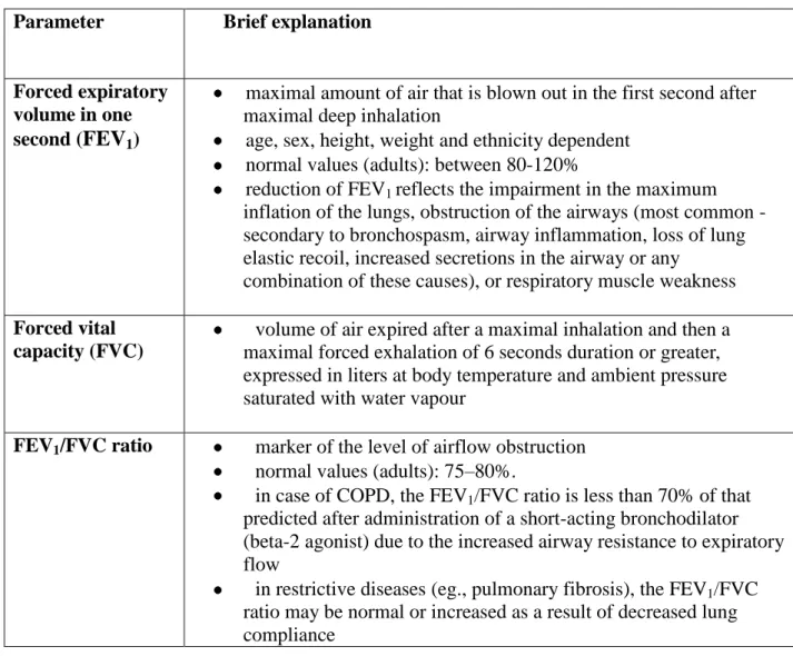
Pulmonary function tests provide several parameters for the examiner. The most common ones are vital capacity (VC), FVC, forced expiratory volume (FEV) at 1 second (FEV1), forced expiratory flow 25–75% (FEF 25–75) and maximal voluntary ventilation (MVV) (Encyclopedia of Surgery). Spirometry results are given in raw data (liters, liters per second) and percent (%) predicted, which means the result as a percent of the predicted values of similar characteristic (age, sex, height, weight) patients (Encyclopedia of Surgery). The most important pulmonary function parameters are summarized in Table 1.
Bronchodilator can be also used in certain cases. By comparing the pre- and post-spirometry-graphs, the effectiveness of the bronchodilator can be assessed (Eigen et al. 2001).
15
Table 1. Most important pulmonary function variables (Source: Lung function — Practice compendium, 1994)
Parameter Brief explanation
Forced expiratory volume in one second (FEV1)
maximal amount of air that is blown out in the first second after maximal deep inhalation
age, sex, height, weight and ethnicity dependent normal values (adults): between 80-120%
reduction of FEV1 reflects the impairment in the maximum inflation of the lungs, obstruction of the airways (most common - secondary to bronchospasm, airway inflammation, loss of lung elastic recoil, increased secretions in the airway or any
combination of these causes), or respiratory muscle weakness Forced vital
capacity (FVC)
volume of air expired after a maximal inhalation and then a maximal forced exhalation of 6 seconds duration or greater, expressed in liters at body temperature and ambient pressure saturated with water vapour
FEV1/FVC ratio marker of the level of airflow obstruction normal values (adults): 75–80%.
in case of COPD, the FEV1/FVC ratio is less than 70% of that predicted after administration of a short-acting bronchodilator (beta-2 agonist) due to the increased airway resistance to expiratory flow
in restrictive diseases (eg., pulmonary fibrosis), the FEV1/FVC ratio may be normal or increased as a result of decreased lung compliance
16 Parameter Brief explanation Vital capacity
(VC)
characterizes the volume change at the mouth between full inspiration and complete expiration
Two forms:
o expiratory vital capacity (EVC) is the maximal volume of air exhaled from the point of maximal inhalation
o inspiratory vital capacity (IVC) shows the maximal volume of air inhaled from maximal exhalation
Total lung capacity (TLC)
the maximum volume of air present in the lungs normally
Diffusing capacity (DLCO)
the carbon monoxide uptake from a single inspiration in a standard time (usually 10 sec)
Maximum voluntary
ventilation (MVV)
the maximum amount of air that can be inhaled and exhaled over a specified period of time (12 sec for normal subjects)
Tidal volume (TV) the amount of air inhaled and exhaled during rest Forced expiratory
flow (FEF) 25- 75%
the mean forced expiratory flow between 25% and 75% of the FVC
the maximum mid-expiratory flow Forced inspiratory
flow (FIF) 25–
75% or 25–50%
the speed of air coming out of the lung during the middle portion of inspiration
Peak expiratory flow (PEF)
the maximal speed reached achieved from the maximally forced expiration initiated at full inspiration
usually obtained from flow–volume curve data Forced Expiratory
Time (FET)
the length of the maximal expiration in seconds from a position of full inspiration
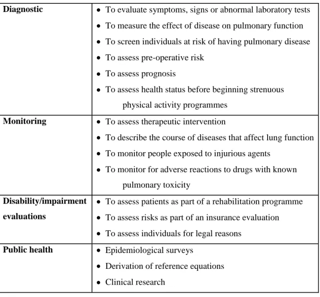
The most common indications of spirometry are the following: asthma, obstructive lung diseases, restrictive lung diseases, dyspnea, assessment of baseline lung function, measurement of bronchial responsiveness, distinction of respiratory from cardiac disease, evaluation of pre-operative risk (for example before anaesthesia or surgery) and monitoring response to treatment (Pierce 2005) (Table 2.).
17
Table 2. Indications for spirometry according to ERS Guidelines 2005 (Source: Miller et al. 2005)
Diagnostic To evaluate symptoms, signs or abnormal laboratory tests To measure the effect of disease on pulmonary function To screen individuals at risk of having pulmonary disease To assess pre-operative risk
To assess prognosis
To assess health status before beginning strenuous physical activity programmes
Monitoring To assess therapeutic intervention
To describe the course of diseases that affect lung function To monitor people exposed to injurious agents
To monitor for adverse reactions to drugs with known pulmonary toxicity
Disability/impairment evaluations
To assess patients as part of a rehabilitation programme To assess risks as part of an insurance evaluation To assess individuals for legal reasons
Public health Epidemiological surveys
Derivation of reference equations Clinical research
The bronchial response can be examined via spirometry using different methods.
Post bronchodilator test: after a “normal” spirometry test, bronchodilator can be administered for another spirometry to compare the results (before and after bronchodilator) (Pierce 2005). By the bronchial challange test, asthma versus COPD can be distinguished and bronchial hyperresponsiveness can be assessed (Pierce 2005).
Inhalation of methacholine, histamine or cold air, and doing rigorous exercise can be applied (Pierce 2005).
18
1.4. Past studies on the heritability of lung function
Heritability of lung function has been investigated in twin and family studies with various results. In one of the first family study, the level of pulmonary function was measured (FEV1 and FEF 25-75) in members of 404 nuclear families living in East Boston, Massachusetts in 1974 (Lewitter et al. 1984). Genetic contribution was found to be consistent through time (41-47%) for parents and their children (Lewitter et al.
1984). Common familial environmental effects on level of pulmonary function explained 1-4% of the variability in children and 11-28% in adults (Lewitter et al.
1984). Another previous twin study estimated genetic control of pulmonary function involving 74 twin pairs (Ghio et al. 1989). Heritability of FEV1 and FVC was found to be significantly heritable ranging between 33-35% (Ghio et al. 1989).
In most studies, the heritability of FEV1 ranged between 10% and 77% and one of the FVC ranged between 26% and 91% (Ingebrigtsen et al. 2011.; Hubert et al. 1982;
McClearn et al. 1994; Hukkinen et al. 2011; Devor and Crawford 1984). Environmental factors could explain a modest part of the variance in most of the studies (Ingebrigtsen et al. 2011.; Hubert et al. 1982; McClearn et al. 1994; Hukkinen et al. 2011; Devor and Crawford 1984). An Indian twin study reported that all lung function variables are highly heritable (23%-99%) except for TV and peak expiratory flow rate (PEFR) (Chatterjee and Das 1995). In contrary, a study of Russian twins certified that shared environmental effects account for the majority (47%) of the variance in FEV instead of genetic factors (Whitfield et al. 1999). Lung function depends also on gender and age (Hallberg et al. 2010; McClearn et al. 1994). A longitudinal study of twins found that the heritability of FEV1 and FVC among never smoking female twins do not change remarkably (32% and 36% for FEV1, 41% and 37% for FVC at baseline and at 3-year follow-up, respectively) (Hukkinen et al. 2011). Genetic factors seem to modestly contribute to lung function variance as demonstrated in a longitudinal study (Gottlieb et al. 2001).
Beyond the “classical” ACE analysis, gene-environment interaction can be also studied in a large sample of twins. A recent Swedish study investigated the genetic and environmental influences on lung function impairment in twins, suggesting that patients with lung diseases such as COPD could benefit from interventions that are sex specific
19
(Hallberg et al. 2010). In this study, the fully adjusted heritability for VC was 59% and did not differ by sex (Hallberg et al. 2010). Heritabilities for FEV1 and diffusing capacity were sex specific: 10% and 15% in men and 46% and 39% in women (Hallberg et al. 2010). Differences between men and women were found in how smoking and symptoms influence the variation in lung function (Hallberg et al. 2010).
Another study tried to find an answer why the effect of cigarette smoking on pulmonary function shows a high inter-individual variancy (some heavy smokers retain normal pulmonary function and others experience profound pulmonary function loss). The authors reported that the heritability of FEV1 was not dependent on smoking status even MZ twins presented little, no or remarkable differences in cigarette use (Tishler et al.
2002). Of note, a recent twin study reported that the A allele of the leptin 19G>A SNP is related to a lower FEV1 and FVC, so the leptin may be important in the determination of maximally attainable lung function (van den Borst et al. 2012).
Twin and family studies lead to genome-wide association studies which can further investigate a genotype if high heritability is present. A such scan of pulmonary function measures (FEV1, FVC, and FEV1/FVC ratio) as part of the National Heart, Lung, and Blood Institute Family Heart Study found an evidence for three chromosomal loci influencing variability in spirometric measures of pulmonary function (Wilk et al.
2003). The FEV1/FVC ratio was linked to chromosome 4 around 28 centimorgans (cM;
D4S1511), FEV1 and FVC were suggestively linked to regions on chromosome 18 (Wilk et al. 2003).
1.5. Relationship of lung function and cardiovascular diseases
The relationship between impaired lung function and atherosclerosis, cardiovascular morbidity and mortality has been poorly investigated (Higgins and Keller 1970; Tockman et al. 1995). An American cohort study determined whether the rate of FEV1 loss independently predicts coronary heart disease (CHD) mortality in apparently healthy men (Tockman et al. 1995). Generally, cardiac mortality increased with increasing quintile of FEV1 decline. CHD mortality follows a large decline in FEV1, independent of the initial FEV1% predicted, cigarette smoking, and other common CHD risk factors (Tockman et al. 1995).
20
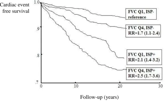
Reduced pulmonary function (mostly FEV1 and FVC) is associated with increased incidences of cardiovascular disease and death (Mendall et al. 2000). Elevated plasma levels of inflammatory markers may partially explain the increased cardiovascular risk among men with low FVC (Mendall et al. 2000). Decreased pulmonary function has been associated with increased levels of C-reactive protein, fibrinogen and white blood cells in some previous papers (Mendall et al. 2000; Dahl et al. 2001; James et al. 1999). Relationship with inflammation-sensitive plasma protein (ISP) levels contributes to the increased risk among men with low FVC (Engström et al.
2002) (Figure 4.).
Figure 4. Cardiac event rates among men with FVC in highest (Q1) and lowest (Q4) quartile and with 0 to 1 (ISP−) or 2 to 5 (ISP+) inflammation-sensitive plasma proteins (ISPs) in top quartile based on the study of Engström and coworkers. The occurrence of
high ISP levels increased the risk significantly among men with low FVC. The differences between groups increased over the entire follow-up period. The results were
similar in smokers and nonsmokers. RR indicates relative risk.
(Source: Engström et al. 2002)
Obviously, smoking contributes to the association between pulmonary function and coronary heart disease as well (Marcus et al. 1989).
Follow-up (years) Cardiac event
free survival
21
Impaired lung function is a major clinical indicator of mortality risk in men and women for a wide range of diseases (Hole et al. 1996). In a prospective general population study assessed the relation between FEV1 and subsequent mortality (Hole et al. 1996). Diminished FEV1 means an increasing risk for both sexes for all the causes of death examined after adjustment for age, cigarette smoking, diastolic blood pressure, cholesterol concentration, body mass index, and social class (Hole et al. 1996).
There are only few previous studies on arterial aging and physical functioning. A British reseach group analyzed associations of arterial stiffness with age, subjective and objective measures of physical functioning, and self-reported functional limitation (Brunner et al. 2011). Their result showed that pulse pressure and mean arterial pressure is linked inversely only with lung function (Brunner et al. 2011). Higher arterial stiffness was associated with poorer lung function adjusted for age, sex, and ethnic group (Brunner et al. 2011). French epidemiological data showed that, even in healthy men, there is a relation between pulmonary function and arterial stiffness. The study reported that reduced pulmonary function was strongly associated with aortic stiffness:
FEV1, FVC and FEV1/FVC ratio were all related to pulse wave velocity (PWV), suggesting that both pulmonary obstructive and restrictive disorders may be implicated in these associations (Zureik et al. 2001). The possible explanations of this finding may be the highly vascular nature of the lung and the anatomic coupling of vascular and parenchymal elements and loss of elasticity of the pulmonary vascular tree, respectively (Enright et al. 1995).
Another possible explanation is that inflammatory mechanisms act as a contributing factor to both vascular stiffness and reduced lung function (Rijcken et al.
1995). Other study underlined the importance of immune complexes and abnormal inflammatory responses (e.g., C-reactive protein) which have been implicated in arterial injury. These factors could lead to vascular changes and modification of stiffness (Selzer et al. 2001). However, no study has ever investigated the genetic influence on the lung function-arterial stiffness relationship.
22
1.6. A novel cardiovascular phenotype: arterial stiffness
Arterial stiffness is a predictor of all-cause and cardiovascular mortality in hypertensive patients and end-stage renal disease (Laurent et al. 2001; Blacher et al.
1999). Arterial stiffness is a dynamic property, which is determined both by vascular function like vascular smooth muscle tone and by the structure of the vessel wall like elastin/collagen content (Van Bortel et al. 2002). Endothelial dysfunction is a marker of increased cardiovascular risk (Kuvin et al. 2001). A large brachial pulse pressure is independently associated with morbidity and mortality from cardiovascular and coronary heart disease (Benetos et al. 1997; Madhavan et al. 1994). Increased arterial stiffness is associated with several risk factors of cardiovascular diseases, such as elevated level of blood glucose, hypertension, obesity, and smoking (Koivistoinen et al.
2007; Liao et al. 1999; Safar et al. 2006).
Arterial stiffness, characterized by PWV and some limited extent: augmentation index augmentation index (AIx), can be estimated non-invasively and has an independent predictive value for cardiovascular events (Laurent et al. 2006). The AIx is given by the ratio of the augmentation pressure and the pulse pressure (PP), being used ever more often in studies as parameters of wave reflection and peripheral vascular resistance (Snieder et al. 2000, Williams et al. 2006; Baulmann et al. 2004). PWV, the most important measure of arterial stiffness, is characterized by the distance traveled (s) by the wave divided by the time (t) for the wave to travel that distance (Snieder et al.
2000).
Carotid-femoral pulse wave velocity reflects the stiffness of both central and peripheral muscular arteries, thus PWV can be used as a simple index for assessing arterial stiffness and atherosclerosis (Safar and O'Rourke 2009; Sugawara et al. 2005).
PWV can be also measured regionally and locally. The Moens-Korteweg equation directly relates PWV and incremental elastic modulus (McDonald et al. 1998).
Bramwell-Hill is an alternate method of measuring PWV, where arterial diameter, compliance, and blood density are taken into account (Bramwell and Hill, 1922). Aortic stiffness has also recently emerged as a strong, independent predictor of cardiovascular mortality and morbidity in patients with essential hypertension (Laurent et al. 2001;
Boutouyrie et al. 2002).
23
1.7. Secondhand smoke exposure and its health related effects 1.7.1. Air pollution and secondhand smoke exposure
Secondhand smoke (SHS) is a complex mixture of the gases andparticles given off by the burning end of a cigarette, pipe or cigar, and the smoke exhaled from the lungs of smokers. Particles emitted from burning cigarettes are in thefine to ultrafine particle size range (0.02 µm–2µm) and have been shown to be inhaled deep into the lungs and to cause an array of adverse health effects (IARC Monographs on the Evaluation of Carcinogenic Risks to Humans 2002, Klepeis et al. 2003; US Department of Health and Human Services 2006) including cancer, heart attacks and asthma (Zhu et al. 2003; Moffatt et al. 2004; Eisner et al. 2005; International Agency for Research on Cancer 2002). PM2.5 particles are air pollutants with a diameter of 2.5 micrometers or less, small enough to invade even the smallest airways and can be measured in micrograms per cubic meter.
SHS is a major public health problem due to its well known adverse health effects (Flouris et al./1 2008; U.S. Department of Health and Human Services 2006).
SHS exposure is known as a cause of asthma exacerbation, otitis media, sudden infant death syndrome, vascular dysfunction, and predisposition toward cardiovascular disease and cancer among children and adults as well (Eisner et al. 2002; Eisner et al. 2005)
To protect the public’s health government health authorities have recommended that indoor smoking be prohibited. Indoor smoking has been found to be a major source of indoor air pollution. The World Health Organization (WHO) has established air quality standards and an air quality guideline (AQG) (World Health Organization 2006). The AQG is a measure for reducing the health impacts of air pollution. An annual average concentration of 10 μg/m3 was chosen as the long-term guideline value for PM2.5. This represents the lower end of the range over which significant effects on survival were observed in the American Cancer Society’s study (Pope et al. 2002).
According to the AQG, an annual mean PM2.5 concentration of 35 μg/m3 or higher is
24
associated with 15% higher long-term mortality risk (World Health Organization 2006).
The WHO’s target air quality guidelines for PM2.5 are much lower, with an average annual mean of 10 μg/m3 and a 24 hour mean of 25 μg/m3. As shown in Table 3, the United States Environmental Protection Agency (EPA) has set limits of 15 μg/m3 as the average annual level of indoor PM2.5 exposure and 35 μg/m3 as an acceptable mean exposure over 24 hours (World Health Organization 2006).
Table 3. US EPA Air Quality Index (AQI) (Source: World Health Organization 2006) Air Quality Air
Quality Index
PM2.5 level (μg/m3)
Health Advisory
Good 0-50 ≤15 None.
Moderate
51-100 16-40 Unusually sensitive people should consider reducing prolonged or heavy exertion.
Unhealthy for sensitive
groups
101-150 41-65 People with heart or lung disease, older adults, and children should reduce prolonged or heavy exertion.
Unhealthy
151-200 66-150
People with heart or lung disease, older adults, and children should avoid prolonged or heavy exertion.
Everyone else should reduce prolonged or heavy exertion.
Very unhealthy
201-300 151-250
People with heart or lung disease, older adults, and children should avoid all physical activity outdoors.
Everyone else should avoid prolonged or heavy exertion.
Hazardous
≥301 ≥251
People with heart or lung disease, older adults, and children should remain indoors and keep activity levels low. Everyone else should avoid all physical
activity outdoors.
25
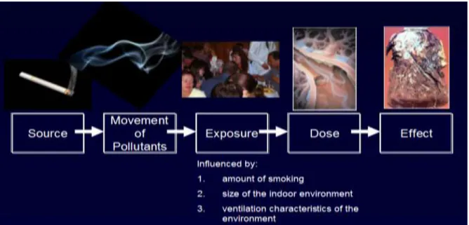
Air pollution can come from many sources, but studies show that indoors the number one source of exposure to small particles comes from tobacco smoke. The exposure from tobacco smoke is also dangerous because the particles themselves are made up of hazardous (know cancer causing chemicals). The dose someone can get from an exposure to tobacco smoke pollution is influenced by many factors including the amount of smoking, size of the indoor environment and ventilation characteristics of the environment (Figures 5-6.). The effect of exposure is influenced by the individuals host characteristics such as pre-existing health conditions, age, and biology (genetics).
Figure 5. Conceptual Model Examining Air Pollution and Public Health.
(Courtesy of K. Michael Cummings, Mark Travers and Andrew Hyland, Roswell Park Cancer Institute, Buffalo, USA)
26
Figure 6. Compution how a pollution source translates to an exposure and eventual harm to human health. Studies exist to provide evidence of how tobacco smoke can
eventually cause harm in humans.
(Courtesy of K. Michael Cummings, Mark Travers and Andrew Hyland, Roswell Park Cancer Institute, Buffalo, USA)
In 2006, the 24-hour PM2.5 standard was lowered (65 to 35 μg/m3) because mounting evidence has established that short-term exposure to PM2.5 can result in numerous health effects including increased mortality (US Environmental Protection Agency 2009). Flash Eurobarometer found that 36% of Hungarians smoke (Hungarian Ministry of Health 2009), a rate similar to that of surrounding countries. A recent WHO survey reported that 84% of Hungarians are being exposed to tobacco smoke in their homes, and 93% report being exposed to smoke outside their homes (World Health Organization 2008).
In 2000, there were around 2.6 million smokers in Hungary, among adults above 18 years of age, 38.3% of men and 23% of women smoked every day. According to the Hungarostudy, the smoking prevalence in Hungary among men was 34.9% in 2002 and 33.9% in 2005 (Susanszky et al. 2007). The costs of harmful effects of smoking and lost income in Hungary in 2004 came to between 315 and 330 thousand million Hungarian
27
Forints (Barta et al. 2006). Non-smokers who are exposed to SHS at home or at work increase their lung cancer risk by 20–30%.
Between 2005 and 2012, cigarette smoking has been prohibited in most of health care facilities in Hungary (World Health Organization 2008) (Health Law 1999, XLII.
and Health Law 2005, CLXXXI. 36§), which was aggravated in 2012 (Health Law 2012). In our air monitoring study, the levels of indoor fine particle air pollution measured in public locations in Hungary where smoking was observed were times higher than the levels in locations where smoking was not observed and in nearly all instances exceeded the levels that the World Health Organization and US Environmental Protection Agency have concluded are harmful to human health (Tárnoki et al. 2009, Tárnoki et al. 2010). Fortunatelly, having taken into account our results as well, a smokefree law was passed in Hungary on April 27, 2011, which made all Hungarian indoor public places smokefree; including closed public places, workplaces and public transport vehicles. The law took effect on January 1, 2012, with a three months grace period before enforcement began. This decision is one of the most effective measures to decrease smoking-related morbidity and mortality. According to the estimations, 1700 deaths will be postponed and 16000 life years will be saved annually in Hungary thanks to the regulation (Adám et al. 2013).
1.7.2. Cardiovascular effects of secondhand smoke exposure
For many years scientists found the link between smoking and heart disease, that active smoking causes heart disease (US Public Health Service 1983).
In this context, smoking was found to kill more people by causing or aggravating heart disease than lung cancer. Later scientists realized the importance of SHS, the exposure to environmental tobacco smoke has been linked to heart disease in nonsmokers few years later (Wells 1988; Kristensen 1989).
Young smokers under 40 years have five times more risk to have a heart attack (Mähönen et al. 2004). The major risk factors for heart disease are smoking, diabetes, total cholesterol concentration, high blood pressure, obesity, left ventricular hypertrophy, increased C-reactive protein and family history of heart disease (Wilson et al. 1998).
28
Studies have shown that several physiological changes involving potential mechanisms of smoking-induced cardiovascular disease can be observed in cigarette smokers compared with nonsmokers who have not been exposed to secondhand smoke (Hatsukami et al. 2006). In the early ninetees, animal studies found that even 5 minute of SHS exposure to the smoke of one cigarette elicits the adhesion of leucocytes to endothelial cells (Lehr et al. 1991). SHS exposure may reduce the distensibility of the aorta (Stefanadis et al. 1998.), has inhibitory effects on endothelium-dependent vasodilatation (Celermajer et al. 1996) and may turn the acetylcholine-induced coronary artery relaxation into a vasoconstriction (Sumida et al. 1998).
SHS causes injury in the vascular endothelium and interfers with the vascular repair system (Heiss et al. 2008), moreover activates blood platelets by increasing the risk for thromboembolic diseases (Elwood et al. 1991; Raupach et al. 2006).
Accordingly, SHS yields to endothelial damage which results atherosclerotic plaque formation and progression, even plaque rupture via decreased vessel dilation, increased vessel contraction, prothrombotic and proinflammatory levels, impaired NO-mediated endothelial function and cell proliferation in the arterial wall (Widlansky et al. 2003).
This mechanism (endothelial dysfunction) leads to impaired arterial stiffness (Mahmud and Feely 2003).
Mahmud et al. reported increasement of aortic arterial stiffness among healthy male persons who breathed SHS from 15 cigarettes in an unventilated room for one hour (Mahmud and Feely 2004). In these subjects, the augmentation index (sign of arterial wave reflection) increased by 15.7% (Mahmud and Feely 2004). A previous study showed an association between SHS and increased carotid intimal thickness as well (Howard et al. 1994).
Passive smoking leads to lower levels of HDL in adults (Mizoue et al. 1999;
Moffatt et al. 1995) and to an increase insulin resistance (Henkin et al. 1999).
SHS exposure changes the systolic blood pressure (Flouris et al./2 2008;
Mahmud and Feely 2004; Sidorkewicz et al. 2006) and cardiac autonomic function (Pope et al. 2001). The risk of coronary heart disease increases significantly by the level of secondhand smoke exposure. For example, nonsmokers who were exposed to 1 to 19 cigarettes per day and to 20 or more cigarettes per day had higher risk of coronary heart disease (He et al. 1999).
29
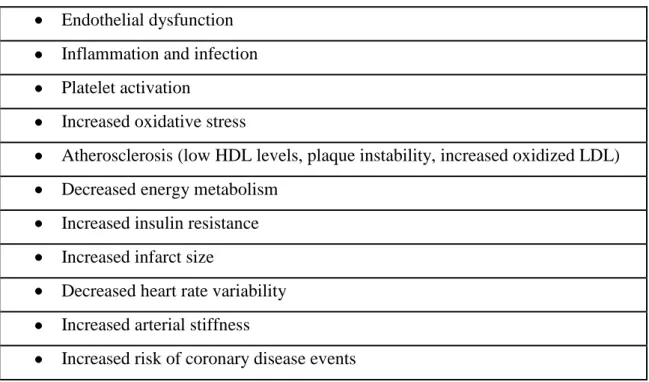
The cardiovascular effects of SHS exposure are summarized in Table 4.
Table 4. Effects of SHS on the cardiovascular system Endothelial dysfunction
Inflammation and infection Platelet activation
Increased oxidative stress
Atherosclerosis (low HDL levels, plaque instability, increased oxidized LDL) Decreased energy metabolism
Increased insulin resistance Increased infarct size
Decreased heart rate variability Increased arterial stiffness
Increased risk of coronary disease events
1.7.3. Psychosocial (family) aspects of tobacco smoking
There is an increasing evidence that socio-economic status of the family effects smoking habits (Tot et al. 2004).
Epidemiological studies found association between cigarette smoking and psychiatric disorders in context with adolescents’ regular smoking, such as conduct disorders, attention-deficit/hyperactivity disorder, internalizing disorders (depression and anxiety) and aggression (Liu 2003; Patton et al. 1998; Kollins et al. 2005; Lerman et al. 1996; Sonntag et al. 2000). Social and commercial tobacco sources play significant role in youth smoking (Johnston et al. 2004).
The most common known psychologic aspect of cigarette smoking is stress, however, the stress levels of adult smokers are slightly higher than nonsmokers.
Smoking has apparent relaxant effect due to the nicotine (Parrott 1999). Psychological stress influences the development of substance dependence, including tobacco dependence (Sinha 2008). The acute effects of the nicotine includes the activation of stress systems and prolongation of physiological stress responses (Fuxe et al. 1989;
30
Pomerleau et al. 1984), moreover stress drives smoking. Its negative effects (smokers experience nicotine deprivation) can be observed between smoking episodes, which motivates smoking through the deprivation reversal (Schachter 1978; Silverstein et al.
1982) and stress induction (Parrott 1999). Interestingly, stress may influence and increase smoking by altering the effects of nicotine, thus smokers smoke more after stress to compensate for attenuated effects (Buchmann et al. 2008).
An Australian study certified that social stream (family and peer networks) play a central role in smoking initiation, progression and youth smoking behaviour (Johnston et al. 2012). A Turkish study found high degree of violent behaviour among smoker school students against friends and family members with a male predominancy (Özge et al. 2006). They published a negative effect of smoking on social relationships, academic performances and suicide attempt or behaviour (Özge et al. 2006). Smoking of the only sibling has an important effect on lifetime smoking, furthermore, both parents and sibling smoking have important effects on current smoking of students (Özge et al.
2006).
Education level of the parents influences the childrens's smoking behaviour rather than ethnicity (Kegler and Malcoe 2005). More permissive parent's children are more likely to smoke, compared with children whose parents have a more
„authoritative” (Jackson et al. 1998; Radziszewska et al. 1996). Previous epidemiological studies confirmed the hypothesis that anti-smoking socialisation is protective against youth smoking (Mahabee-Gittens et al. 2012; Waa et al. 2011).
Parental influences are important for initiation and escalation of smoking (Bricker et al.
2007), during school years peer group behaviours influence smoking initiation and progression as well (West et al. 1999). Gender differences were also shown in the perceptions and reported experiences of smoking in a previously published study.
Female participants were more strongly influenced by peer smoking compared the boys (Simons-Morton and Farhat 2010).
However it is clear that more disorders develop in children who were exposed to environmental tobacco smoke, and postnatal tobacco smoke exposure may cause behavioral problems in children as well (Maytin et al. 1991).
A Greek nation-wide school-based study investigated the relationship between cigarette smoking status and adolescents’ emotional/behavioural problems. An
31
association between smoking and higher levels of emotional/behavioural problems, such as emotional symptoms, conduct problems and hyperactivity/inattention was reported (Giannakopoulos et al. 2010). This study underlined the importance of effective antismoking strategies in school environment and elsewhere with addressing adolescents’ needs regarding their emotional/behavioural health. Smokers have higher chance to divorce comparing nonsmokers (Bachman et al. 1997; Doherty and Doherty 1998).
There is a positive relationship between psychological distress and salivary cotinine levels in smokers and non-smokers, indicating that both firsthand and secondhand smoke exposure may lead to higher levels of mental stress which effects the psychosocial environment (Hamer et al. 2010).
Several twin studies investigated the possible role of genetic factors on nicotine dependence and withdrawal. Nicotine dependence for cigarette smoking or snus use has a moderate genetic determination (30-39%) which is weakly associated with intelligence quotient genetically (Modig et al. 2011; Broms et al. 2007). In addition, nicotine withdrawal symptoms were reported to be moderately heritable (49%) in adult and adolescent smokers (Pergadia et al. 2010), similarly to smoking withdrawal (Carmelli et al. 1992). Heritability of age at first cigarette was 60% for males and 39%
for females in a Danish twin study (Vink et al. 2006). D1A dopamine receptor gene is supposed to be responsible for smoking behavior (Vink et al. 2006).
32 2. Objectives
Since twin studies reveal the proportion of genetic and environmental contribution of a trait, and how the two interact, this model can be applied in a respiratory setting as well. Furthermore, studying twins helps to draw conclusions concerning psychosocial aspects.
Our aims can be summarized in the following points:
1. To establish the Hungarian twin registry and describe the characteristics of the voluntary twin sample whose individuals will be involved in respiratory twin studies.
2. To assess the heritability of lung function, phenotypic correlations between pulmonary function (FEV1, FVC) and hemodynamic variables, furthermore, to determine whether there is a shared genetic relation between lung function and arterial stiffness. We hypothesized that there is a common genetic background between lung function and arterial stiffness.
3. Third, we were specially interested how secondhand smoke exposure effect monozygotic and dizygotic twins in various indoor public places. Even if the heritability of smoking characteristics is well described, to date, there is no information regarding smoking and secondhand smoke characteristics of twins and its psychosocial aspect.
Therefore, the last purpose of the investigation was to assess the smoking habits and sensibility to SHS exposure of monozygotic and dizygotic twins comprehensively in a relatively large twin cohort.
33 3. Methods
3.1. Subjects of the twin studies
In 2006, we began an effort to revive the Hungarian Twin Registry with Levente Littvay. This effort benefited greatly from the help of Júlia Métneki, the person responsible for most of Hungarian twin studies and the management of two twin registries from the 1970s to the 1990s. Levente Littvay could get in touch with Júlia Métneki by the encouragement of Nicholas Martin and continue the work she started in the 1970s. A Hungarian Twin Club was founded in the early 1980s and since then twin meetings are common in the country. Annual meetings are held in Szigethalom (13th annual national meeting in 2012), Ágfalva (6th annual international meeting in 2012) and Kunhegyes (9th biannual meeting in 2012). The old volunteer registry and these meetings are at the foundation of the new volunteer twin registry. Additionally, we are augmenting this list with social media presence, a continuous push in the more traditional media, and via the website (http://www.ikrek.com). In the respiratory twin studies, 151 monozygotic and 62 dizygotic healthy adult twin pairs were involved in Hungary and in the United States.
3.2. Study design
Twin subjects were recruited as part of the International Twin Study 2009 project.
Twins above the age of 18 years were invited to participate. Exclusion criteria included chronic respiratory disease, pregnancy, arrhythmia, acute infection within three weeks of measurement or foreseeable lack of compliance with test procedures and race other than white (to exclude the influence of ethnicity). Zygosity was assigned according to a seven-part self-reported response which is widely used and accepted worldwide with an over 99% accuracy (Heath et al. 2003). Studies were approved by local ethical committees (IRB committee names and project approval numbers: Semmelweis University Regional and Institutional Committee of Science and Research Ethics, TUKEB, 29/2009; Twins Days Festival Ethical Board, 1/2009) and all study subjects gave informed consent prior to entering the study.
34
Hungarian subjects were enrolled from our Hungarian Twin Registry (Littvay et al.
2012). Twins were investigated at Hungarian twin festivals (Ágfalva and Szigethalom) and in two large hospitals in Budapest (Semmelweis University Department of Radiology and Oncotherapy; Military Hospital Department of Cardiology) between July, 2009 and June, 2010.
American twins were tested during two days of the Twins Day Festival in Twinsburg, OH, USA which is the largest twin gathering in the world, in August, 2009.
Lung function and hemodynamic measurements were facilitated by the author and the author’s twin brother (DLT and ADT) at all research sites in order to decrease inter- observer variability and in accordance with guidelines recommended by the European Society of Cardiology (Laurent et al. 2006). Subjects completed a questionnaire separately of each other on the spot concerning smoking and SHS exposure characteristics. Presence of risk factors, medication, past medical history and clinical symptoms were all recorded by the attending physicians on-site.
3.3. Pulmonary function assessment
Lung function was assessed by dynamic spirometry (Minispir Waukesha, WI, USA) (Figure 7.). The spirometer was calibrated daily using a 1 L-syringe. The maneuvers were performed standing while wearing nose clips. The largest FEV1 and the largest FVC from all acceptable maneuvers were used in this analysis. FVC and FEV1
measurements were performed in accordance with guidelines recommended by the American Thoracic Society (Anonymous 2005). Lung function variables were expressed in absolute (measured) values and as percentage of predicted (based on the subject’s age, height, sex, country), using the reference values recommended by the ERS and ECCS93 reference equation values (Pellegrino et al. 2005; Quanjer et al.
1993). Percentage of predicted values are widely used in lung function laboratories to help determine if an individual's lung function is within normal limits.
35
Figure 7. Lung function assessment and questionnaire completion.
3.4. Hemodynamic measurement
Aortic pulse wave velocity (PWV) - a measure of arterial stiffness - and brachial and aortic augmentation indices (AIx) - measure of arterial wave reflection - was assessed by a clinically validated oscillometric device (TensioMed Arteriograph, TensioMed Ltd., Hungary, 1.10.1.1. software). This method enables the calculation of these parameters from oscillometrically recorded pressure waves on the brachial artery (pulse wave analysis) (Baulmann et al. 2008; Horváth et al. 2010) (Figure 8.).
Using inflatable upper arm cuffs with high fidelity sensors, pulsatile volume changes (resulting from pulsatile fluctuations of the brachial artery) are transduced into pressure curves. Pulse waves are recorded at the proximal occlusion site of the cuff whose pressure is 35-40 mmHg above the systolic BP („suprasystolic pressure”).
Computer programs are used to further analyze the recorded pulse waves. Oscillometry method is based on the fact that the forward traveling pulse wave (generated by the ejection of the left ventricle) is reflected in the periphery by creation of a second reflected wave (Qasem and Avolio 2008). Pulse transit time refers to the time it takes a pulse wave to travel between two arterial sites. Accordingly, pulse transit time is determined from the time delay between the forward and the reflected pressure wave.
36
Aortic PWV, a direct marker of arterial stiffness can be automatically calculated from transit time and traveling distance between jugulum (sternal notch) and symphysis pubica according to the following formula: PWVao = distance (m)/transit time (sec) (Williams 2004; O’Rourke et al. 2004). PWV was determined for several (at least 3) cardiac cycles. Stationarity of the subsequent PWV values are accepted when 3 PWV values fall within 1 SD range.
AIx, a measure of pulse wave reflection, can be calculated from brachial pressure curves in combination with automated transfer algorithms. AIx is expressed by the ratio of augmentation pressure and the pulse pressure.
In accordance with guidelines recommended by the European Society of Cardiology (Laurent et al. 2006), all subjects restricted from smoking for three hours, from eating for one hour, and from drinking alcohol or coffee for ten hours prior to measurements.
Subjects were examined in supine position in the dominant arm after at least 10 min of rest. Twins were asked to refrain from speaking or moving during the measurements, and to keep their eyes closed during the test. Adherence to these restrictions was ascertained by querying the subject.
Figure 8. Hemodynamic assessment in American twins (Twinsburg, USA).
37 3.5. Statistical analysis
3.5.1. Data analysis
3.5.1.1. Descriptive analysis
Descriptive analysis (mean ± standard deviation for continuous variables, percentage for categorical variables) for FEV1, FVC values, hemondynamic parameters, smoking and secondhand smoke characteristics of twins was conducted using SPSS (SPSS 17.0 for Windows; SPSS, Chicago, IL). Differences between genders, zygosity and countries were calculated using independent-sample t-tests. p value <0.05 was considered significant.
3.5.1.2. Estimating genetic influences on lung function and PWV, AIx
All analyses were corrected for age, gender, country (significant effect of country, p<0.05) and smoking (the phenotypic correlations were also calculated unadjusted to smoking). Smoking was adjusted according to two groups (never smokers and ex-smokers with a quitting period of at least one year and <5 pack year; and ex- smokers with >5 pack year and active smokers; 1 pack year is defined as smoking 1 pack of cigarettes daily for at least one year). Heritability estimates were determined based on the consideration that greater levels of MZ than DZ within pair similarity indicate a genetic influence on a phenotype, while similarity of co-twin correlations suggests that the variance is due to shared environmental factors. Three general sources of variance were calculated: (i) additive genetic factors or heritability (A) which represents the effects due to genes at multiple loci or multiple alleles at one locus; (ii) common environmental variance (C), which estimates the contribution of the common family environment of both twins (familiar socialization, diet, exposure to high levels of air pollution, parental smoking, shared womb, etc.); and (iii) nonshared environmental variance (E), which represents the effects that apply only to each individual twin and all sources of unique experiences and exposures that cause within-pair differences (e.g., discordance for smoking, differences in illnesses and occupational exposures).
38
Structural equation modeling was performed using the Mplus Version 6.1 weighted least squares estimation due to the categorical variable of interest (Muthén and Muthén 1998- 2010). Empirical confidence intervals were calculated with Bollen-Stine Bootstrap method (Bollen and Stine, 1992). Univariate quantitative genetic modeling was performed to decompose the phenotypic variance of the considered parameters into heritability (A), shared (C), and unshared (E) environmental effects (ACE analysis).
Chi-square model fit p-values are presented where the desired results show insignificant model misfit. Instead of a covariance matrix, the estimation procedure used the raw data matrix. Given the small sample size, no component was fixed to 0 in the model.
3.5.1.3. Estimating the correlation between lung function and PWV, AIx
Correlation coefficients between pulmonary function (FEV1, FVC), aortic PWV and AIx were calculated to measure the strength and the direction of the relationship between variables.
3.5.1.4. Genetic covariance between lung function and PWV, AIx
A bivariate Cholesky decomposition was used to derive the magnitude of covariation between the investigated respiratory function and hemondynamic phenotypes (PWV, AIx) and to estimate what proportion of this correlation is attributable to common underlying genetic and environmental factors. In order to estimate the amount of overlap between genes or environment that influences the two parameters, genetic and environmental correlations between those phenotypes were calculated.