ORIGINAL ARTICLE
Association of drug treatments in pregnant women with the risk of external ear congenital abnormalities in their offspring:
A population-based case-control study
cga_319126..137László Paput1, Ferenc Bánhidy2, and Andrew E. Czeizel3
1Department of Oto-Rhino-Laryngologic and Head/Neck Surgical Department, National Center for Healthcare Audit and Improvement,
2Second Department of Obstetrics and Gynecology, School of Medicine, Semmelweis University, and3Foundation for the Community Control of Hereditary Diseases, Budapest, Hungary
ABSTRACT
The objective of this study was to evaluate the possible association of drug treatments in pregnant women with a higher risk of congenital abnormalities of the external ear, particularly microtia/anotia, in their children. The fre- quency of drug treatments was compared in the mothers of cases with isolated or multiple (syndromic) ear abnormalities and in the mothers of three different controls: controls matched to cases, all controls (these controls had no defects) and mal- formed controls in the population-based large dataset of the Hungarian Case-Control Surveillance of Congenital Abnor- malities. There was no significantly higher use of any drug in the mothers of 354 cases with isolated external ear abnormali- ties than in the mothers of different controls. However, of 156 cases with multiple ear abnormalities, 11 had mothers with hydroxyethylrutosidea treatment and a characteristic pattern of congenital abnormalities was found in these children. Four cases with multiple ear abnormalities were born to epileptic mothers treated with valproate, phenytoin and polytherapy in two cases. Drug treatments are not important in the origin of isolated ear abnormalities. However, a higher risk of multiple ear abnormalities was found in children born to mothers with treatment of hydroxyethylrutosidea or antiepileptic drugs during pregnancy.Key Words: drug treatment, ear abnormality, hydroxye- thylrutosidea, population-based case-control study, pregnancy
INTRODUCTION
The aim of this PhD project is to estimate the possible causal association of structural birth defects (i.e.congenital abnormalities [CAs]) of external ears, mainly microtia and anotia in the offspring of mothers with teratogenic factors during pregnancy because the etiology of these visible CAs is less-known (Aase and Tegtmeier 1979; Mastroiacovoet al. 1995; Suutarlaet al. 2007). The minor proportion of ear CAs can be explained by major mutant genes (Mastroiacovo et al. 1995; Llano-Rivas et al. 1999), but among environmental factors only high altitude (Castillaet al. 1999) had some association with higher risk of microtia and anotia. Prenatal drug exposures were also mentioned as potential causal factors in the origin of microtia/anotia without specification of any drug (Castilla and Orioli 1986).
Thus the objective of the present study was to evaluate different drugs separately in pregnant women who had offspring with exter- nal ear CAs in a population-based case-control study based on the large dataset of the Hungarian Case-Control Surveillance of Con- genital Abnormalities (HCCSCA) (Czeizelet al. 2001a). The birth outcomes of children with external ear CAs has been reported in another paper (Paputet al. 2010).
MATERIALS AND METHODS
The objective of the HCCSCA is to compare the pharmaceutical and other environmental exposures in pregnant mothers of cases and controls.
Cases and controls
Cases with CA were selected from the Hungarian Congenital Abnormality Registry (HCAR) (Czeizel 1997) for the HCCSCA.
The reports of cases with CA is mandatory for physicians from the birth of a child until its first birthday to the HCAR and most CAs are reported by obstetricians (in Hungary practically all deliveries take place in inpatient obstetric clinics and birth attendants are obstetri- cians) or pediatricians (working at neonatal units of inpatient obstetric clinics as well as of various general and special surgical, cardiologic, orthopedic, and oto-rhino-laryngologic inpatient and outpatient pediatric clinics). Autopsy was obligatory for all infant deaths and was usually (about 80%) in stillborn fetuses during the study period. Pathologists sent a copy of the autopsy report to the HCAR if defects were identified in stillborn fetuses or infant deaths.
Fetal defects diagnosed by prenatal diagnostic centers with or without elective termination of pregnancy have also been reported to the HCAR since 1984.
Minor anomalies or morphological variants without serious medical or cosmetic consequences are recorded in the HCAR, but these cases are excluded from the estimation of different CA rates.
CAs were differentiated into two main categories: isolated CAs (only one organ is affected) and multiple CAs (concurrence of two or more CAs in the same person affecting at least two different organ systems) (Czeizelet al. 1988). If minor anomalies were com- ponents of multiple CAs, they were considered at the evaluation of multimalformed cases.
The total (birth+fetal) prevalence of cases with CA diagnosed from the second trimester of pregnancy through the age of 1 year was 35 per 1000informative offspring(liveborn infants, stillborn fetuses and electively terminated malformed fetuses) in the HCAR, 1980–1996 (Czeizel 1997), and about 90% of major CAs were recorded in the HCAR during the 17 years of the study period Correspondence: László Paput, MD, 1062 Budapest, Podmaniczky utca
109-111, Budapest, Hungary. Email: barabas.ev@gmail.com Received November 14, 2010; revised and accepted April 8, 2011.
(Czeizelet al. 1993). The proportion of liveborn infants, stillborn fetuses and electively terminated malformed fetuses was 97.1%, 1.1% and 1.8%, respectively.
The diagnosis of ear CAs was checked in the HCAR and modi- fied if necessary by two steps: (i) If cases with unspecified ear CAs were reported to the HCAR, an extra effort was made by the assistant of the HCAR to contact the medical doctors who reported these cases to specify the diagnosis; and (ii) Parents of cases asked coworkers of the HCAR to organize parental meetings, and there were two parental meetings for the families of cases with ear CAs in the institute of the HCAR in 1988 and 1996. These meetings had three aims: (i) provide information for parents regarding ear CAs and respond to their questions; (ii) experience exchange among parents; and (iii) provide examination of these cases by experts.
Thus we were able to examine personally about one-third of cases with isolated ear CAs.
Both isolated CAs (single, complex, sequence) and multiple CAs (MCAs) (MCA-syndrome, MCA-association and random combi- nation) are further differentiated into three subcategories (Puho et al. 2008). The abbreviation of IECA is used for the category isolated ear CAs. However, at the evaluation of IECA cases, the so-called complex CAs are worth mentioning because severe microtia and anotia (i.e. CAs of the auricle[s]), were frequently associated with narrowing or absence of the external auditory canal/
meatus and sometimes with the CAs of middle ear; thus, there are two or three CAs in the same organ. Most MCAs are not recognized and/or identified as MCA-syndromes or MCA-associations; there- fore, it is not possible to differentiate unrecognized/unidentified MCAs from random combinations of component CAs within the category of MCAs. This diagnostic problem explains that we use the term unclassified multiple CAs in general (Puhoet al. 2008).
Multiple ear CAs had anotia/microtia; therefore, the abbreviation of unclassified multiple anotia/microtiaisUMAM. These cases were described in a previous paper in detail (Paputet al. 2011).
However, cases with IECA and UMAM in the study were selected from the dataset of the HCCSCA.
There were three exclusion criteria in the selection of cases with CA from the HCAR for the HCCSCA: (i) cases reported after three months of birth or elective termination of pregnancy to the HCAR (23% of the total, including mainly mild CAs); (ii) cases with three mild CAs as congenital dysplasia of the hip based on Ortolani click, congenital inguinal hernia, major hemangioma; and (iii) cases with CA-syndromes caused by major gene mutations or chromosomal aberrations (with preconception origin). Thus cases with identified MCA-syndromes, including ear CAs, were excluded from the study.
Controls were identified in the National Birth Registry of the Central Statistical Office for the HCCSCA. These controls were defined as newborn infants without defect and in general two con- trols were matched to every case according to sex, birth week in the year when cases were born and district of parents’ residence.
However, these controls were used in two different approaches: (i) matched controlsof cases with IECA or UMAM; and (ii)all con- trols because their large number improves the statistical power.
However, there was a third control group: (iii)malformed controls selected from the HCAR, that is, liveborn cases with isolated CA (but without cases with IECA) or cases with unclassified MCAs (but without UMAM).
Exposure and confounder data collection
Medically recorded prospective data
Mothers of cases and controls were asked in a mailed explanatory letter to send us the prenatal maternity logbook and other medical
records (mainly discharge summaries of their deliveries) regarding their diseases and related treatments during pregnancy and their child’s CA. Prenatal care was mandatory for pregnant women in Hungary (if a mother did not visit prenatal care, she received no maternity grant and leave), thus nearly 100% of pregnant women visited prenatal care, on average seven times. The first visit was between sixth and twefth gestational weeks. The task of obstetri- cians was to record all pregnancy complications, maternal diseases and related drug prescriptions in the prenatal maternity logbook.
Retrospective self-reported maternal information
A structured questionnaire together with a list of diseases and drugs, and a printed informed consent form were also mailed to the mothers immediately after the selection of cases and controls. To standardize the answers, mothers were asked to read the enclosed list of diseases and medications as a memory aid before replying.
Mothers were also asked to provide a signature for informed consent which authorized us to record the name and address of their children in the HCCSCA.
The period between birth or elective termination of pregnancy and return of the information package (questionnaire, logbook, and informed consent) in our prepaid envelope was 3.5⫾1.2 and 5.2⫾2.9 months for cases and controls, respectively.
Supplementary data collection
Regional nurses were asked to visit all non-respondent mothers of cases (including malformed controls) at home, to help mothers fill in the questionnaire, evaluate available medical documents, and obtain data regarding lifestyle (smoking, drinking, and illicit drug use) and some important symptoms (e.g. high fever) in diseases during pregnancy through a personal interview of mothers and their close relatives living with them. The lifestyle data were collected only in these subsamples due to the unreliability of retrospective maternal information (Czeizelet al. 2004a). Regional nurses visited only 200 non-respondent and 600 respondent control mothers as part of the two validation studies (Czeizel et al. 2003; Czeizel and Vargha 2004), as the ethics committee considered that this follow-up would be disturbing to the parents of healthy children.
Regional nurses used the same method in these control mothers as in non-respondent case and malformed control mothers.
Drug treatment was evaluated according to three different aspects (Czeizel 2009a): (1) The source of information: (a) data only from the prenatal maternity logbooks and/or other medical records; (b) data only from the questionnaire; and (c) concordant data from both medical records and the questionnaire. (2) The timing of exposures (i.e. drug treatments). Thegestational agewas calculated from the first day of the last menstrual period. Three time intervals were considered: (i) The first month of gestation because it is before organogenesis. The first two weeks are before conception while the third and fourth weeks comprise the preimplantation and implanta- tion periods of zygotes and blastocysts, including omnipotent stem cells. Thus CAs cannot be induced by short-term environmental agents in the first month of gestation and explains the ‘all-or- nothing effect’ (i.e. total loss or normal further development). (ii) The second and third months of gestation. The auricle is formed from the first and second branchial arches by a series of auricular hillocks that surround the first pharyngeal groove during the sixth postconceptional week (Poswillo 1973), thus the critical period of external ear CAs is in the eighth gestational week. Thus, mainly the second gestational month as the most sensitive time window of the potential exposures was evaluated, but because of the inaccuracy of this time calculation (both in the gestational age and exposure time), the third gestational month was also considered (Czeizel
2008). (iii) The fourth through ninth months of gestation (i.e. preg- nancy after the organ-forming period). (3) Drug treatments were evaluated according to the chemical structure of medicinal prod- ucts, route of administration, doses, and duration of treatment.
Among potentialconfounding factors, maternal age, birth order, and marital and employment status as indicator of socioeconomic status because it correlated well with the level of education and income (Puhoet al. 2005), and the use of pregnancy supplements, mainly folic acid (Czeizel 2009b) were considered.
Finally, necessary data were collected for 96.3% of cases (84.4%
from reply, 11.9% from visit) and for 83.0% of controls (81.3%
from reply, 1.7% from visit). Informed consent was signed and returned by 98.4% of mothers.
The procedure of data collection was changed in 1997 because regional nurses visit and question all cases and controls, but the recent data had not been validated at the time of this analysis, thus only the dataset of 17 years between 1980 and 1996 is evaluated here.
Classification and evaluation of cases with ear CA
At the evaluation of cases with CAs of the external and middle ear, different classifications were suggested (Marx 1926; Meurman 1957; Tasseet al. 2005); the following classification was used in the HCCSCA:
Type I microtia, as a minor anomaly. The external ear is small and the auricle retains most of its normal structure; therefore, all anatomical structures are distinguishable. The external auditory meatus is present. Each case with type I microtia was excluded from the study of IECA as minor anomaly but type I microtia as compo- nent anomaly was evaluated in UMAM cases.
Type II microtia, as a mild CA. The structure of the smaller external ear is moderately anomalous. The auricle can be hook-, S- or question mark-shaped in appearance with a more or less irregular mass of cartilage, but the external auditory meatus is usually present.
Type III microtia, as a severe CA. The external ear is rudimentary with abnormal structure: the auricle does not include cartilage, only soft tissues and there is no external auditory meatus.
Anotia, as a severe CA. All external ear structures are absent, thus there is no external auditory meatus/canal. The skin of the cheek passes smoothly over the aural area without definite elevation or depression.
Complex ear CA, as a severe CA. These cases had middle ear CAs and anotia/microtia with narrowing or absence of the external auditory canal/meatus.
The diagnosis of ear CAs was checked in the HCCSCA and modified if necessary also by two steps: (i) Results of recent
medical examinations helped to achieve more accurate diagnoses;
and (ii) Frequent personal examination was conducted in selected cases with complex ear CA and with familial origin in our institute or visited at home by the principal investigator of the study.
Statistical analysis
The software package SAS version 8.02 (SAS Institute, Cary, NC, USA) was used. First, frequency tables were made for the main maternal variables to describe the study groups. Second, the char- acteristics of pregnant women with IECA or UMAM and controls were compared using Student’st-test for quantitative andc2statis- tics or odds ratios (OR) with 95% confidence interval (CI) for categorical variables. Third, the frequency of drug treatments used during pregnancy was compared between the groups of cases with IECA or UMAM and different control groups. At the comparison of cases with their matched controls, conditional logistic regression model was used, while at the comparison of cases and all controls or malformed controls, the ordinary logistic regression model was used. At the calculation of adjusted OR with 95% CI age, employ- ment status, birth order, and diseases of mothers were considered.
RESULTS
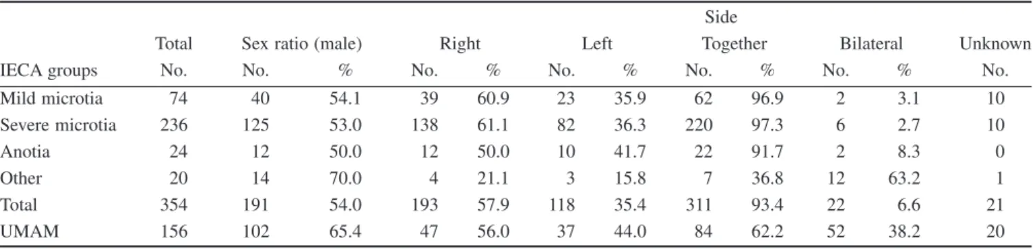
IECAs were diagnosed in liveborn infants. Of 354 cases with IECA, 74 and 236 had mild and severe microtia, respectively (Table 1).
Mild microtia did not associate with the atresia of the external auditory canal, but was associated with preauricular tag/pit/sinus in nine cases (12.2%). Severe microtia was associated with preauricu- lar tag/pit/sinus in 69 cases (29.2%), and 212 cases (89.8%) had also aural stenosis or atresia. Anotia occurred only 24 cases (6.8%), and all of these cases had aural stenosis or atresia, while eight (33.3) were affected with preauricular tag/pit/sinus as well. There is an obvious correlation between the severity of microtia/anotia and the frequency of above associated CAs. The fourth group included 20 cases with ‘other’ IECA, and of these 20 cases, 18 were personally examined. Five cases had middle ear CAs (atresia of auditory canal with fusion of ear ossicles in four cases, atresia of the auditory canal without membranous labyrinth in the middle ear and organ Corti in one case) and anotia/microtia; 12 cases were affected with aural atresia/stenosis and anotia in one side with microtia in the other side, and one case was affected with polyotia and microtia. Two cases had only medically recorded diagnosis of ear CAs without personal examination (inner ear CA with anotia in one case, and absence of Eustachian tube with microtia in another case). Thus, of 354 cases with IECA, each had microtia or anotia with or without other ear CAs.
Table 1 Distribution of cases with different groups/types of IECA and UMAM, in addition to their sex ratio and side distribution of ear CAs
IECA groups
Total Sex ratio (male)
Side
Right Left Together Bilateral Unknown
No. No. % No. % No. % No. % No. % No.
Mild microtia 74 40 54.1 39 60.9 23 35.9 62 96.9 2 3.1 10
Severe microtia 236 125 53.0 138 61.1 82 36.3 220 97.3 6 2.7 10
Anotia 24 12 50.0 12 50.0 10 41.7 22 91.7 2 8.3 0
Other 20 14 70.0 4 21.1 3 15.8 7 36.8 12 63.2 1
Total 354 191 54.0 193 57.9 118 35.4 311 93.4 22 6.6 21
UMAM 156 102 65.4 47 56.0 37 44.0 84 62.2 52 38.2 20
Of 156 cases with UMAM in the HCCSCA, four (2.6%) occurred in stillborn fetuses, and of 152 liveborn cases, 28 (18.4%) died during the infant period. Two, three, four to five and six to nine component CAs were recorded in 36 (23.1%), 41 (26.3%), 51 (32.7%) and 28 (32.7%) UMAM cases, respectively.
The sex ratio (i.e. the proportion of males) of IECA cases shows a slight male preponderance (Table 1), but the male excess was strong in the group of ‘other’ ear CAs. UMAM cases showed a more robust male predominance.
Laterality may also have some association with the origin of ear CAs. Microtia/anotia had an obvious predominance of unilateral manifestation, with some excess of the right side in cases with microtia (Table 1). However, this trend does not exist in the ‘other’
group, because nearly two-thirds of these cases had bilateral mani- festation of ear CAs. The less frequent occurrence of unilateral ear CA is characteristic for UMAM cases as well.
The maternal characteristics in the groups of cases with IECA and different controls are shown in Table 2. The number of controls was 38 151 and represented 1.8% of all Hungarian newborns. The mean maternal age in the case group of IECA did not differ signifi- cantly from control mothers. The mean birth order was higher in the mothers of IECA cases compared to the mothers of matched con- trols and all controls; however, there was no difference in the mean birth order of the mothers of IECA cases and malformed controls.
The rate of unmarried women was lower in the mothers of matched controls. The distribution of employment status was different between case mothers and matched or all control mothers, but not between cases and malformed control mothers. These differences reflect the lower proportion of the two groups of high socioeco- nomic status (i.e. professional and managerial employment in the mothers of IECA cases and malformed controls). The lowest pro- portion of professional women was found in the group of IECA cases. Of 354 case mothers, 190 (53.7%) used folic acid, while this rate was similar in matched (59.1%) and all (54.5%) control mothers but lower in the mothers of malformed controls (49.5%).
Of 354 mothers of IECA cases, 112 had personal information regarding smoking and drinking habits during pregnancy, 20 (17.8%) were smokers while two (1.8%) were regular drinkers (more than one drink per week). The proportion of smokers was 19.1% in the group of all controls and 21.2% in the mothers of malformed controls.
The maternal characteristics of 156 cases with UMAM were also evaluated (Table 3). There was no significant difference in the mean maternal age or birth order among the study groups. Marital status also did not show any significant difference. However, the distribution of employment status in the mothers of UMAM cases differed from control groups due to mainly the lowest proportion of managerial and skilled workers and the highest proportion of semiskilled workers and ‘other’ (mainly due to students). The use of folic acid was similar in the mothers of cases (53.8%), matched (53.8%) and all (54.5%) controls, but lower in the mothers of malformed controls (46.0%).
Table 4 summarizes the data of pregnancy complications in the mothers of IECA cases because some drug treatments might be connected with these pathological conditions. There was no asso- ciation of any pregnancy complication with higher risk of IECA.
Pregnancy complications in the mothers of UMAM cases are not shown here because the associated component CAs may have a stronger effect for these variables than ear CAs.
The number and incidence of acute maternal diseases were the following in the mothers of 354 cases with IECA: influenza/
common cold with secondary complications (75, 21.2%); and acute diseases of the respiratory system (43, 12.1%), digestive system
(2, 0.6%), urinary tract (32, 9.0%), genital organs (19, 5.4%) and others (14, 4.0%). Only high fever related to influenza/common cold showed association with higher risk of IECA (adjusted OR with 95% CI. 4.3, 1.9–7.4). The number and prevalence of chronic diseases, such as diabetes mellitus (3, 0.8%) and epilepsy (3, 0.8%) was low, essential hypertension (20, 5.6%), hemorrhoids (14, 4.0%), primary hypotension (13, 3.7%), varicose veins in the lower extremities (9, 2.5%) and migraine (6, 1.7%) occurred more fre- quently. However, there was no higher risk of IECA in cases born to mothers with these chronic diseases.
Two acute diseases occurred more frequently in the mothers of UMAM cases than in the mothers of controls. Six UMAM cases had severe skin scar and unilateral microtia born to mothers with vari- cella (chickenpox) during pregnancy. High fever related to influenza/
common cold with secondary complications and tonsillitis together occurred more frequently in the mothers of UMAM cases (16.7%) than in the mothers of matched controls (4.3%), all controls (6.0%) and malformed controls (11.1%). Maternal chronic diseases were also evaluated without any association with higher risk of UMAM.
The objective of the present study was the evaluation of different drugs in the origin of IECA or UMAM.
The frequency of drug treatments (used at least by five mothers of IECA cases) during the pregnancy in the group of cases and controls is shown in Table 5. There was no permanent higher risk of IECA in cases compared to all control groups; higher risk of some drugs was found only in separate comparisons. Allylestrenol was used more frequently by the mothers of IECA cases than by the mothers of matched controls. The occurrence of nitrofurantoin treatment was more common in the mothers of IECA cases than in mothers of all controls. However, two drugs, prednisolone or tribenoside were used more frequently by the mothers of IECA cases than by the mothers of all controls and malformed controls. These risk figures were 4.1 and 3.1 in prednisolone compared to all controls and malformed controls, while 2.6 in tribenoside compared both to all controls and malformed controls, thus we cannot neglect them.
In the next step the above drugs were evaluated therefore only in the second and/or third gestational month (i.e. in the critical period of IECA if at least three mothers of IECA cases who used them) (Table 5). There was no pregnant woman with prednisolone treat- ment in the second or third gestational month. The onset of pred- nisolone treatment was in the fifth, sixth, seventh and ninth months in two, two, one and one woman, respectively. Of seven pregnant women with tribenoside use, only one was treated with this drug in the critical period of IECA (i.e. from the first month to the ninth month), while the onset of tribenoside treatment was in the fourth, sixth and seventh month in one, three and two women, respectively.
Thus the causal association of these drug treatments with higher risk of IECA can be excluded. However, allylestrenol was used more frequently in the second and/or third gestational month by the mothers of IECA cases than by the mothers of matched controls.
In addition, the use of aminophenazone+carbromal (Demalgon, Chinoin, Budapest, Hungary) was also more frequent during the second and/or third gestational month in the mothers of IECA cases than in the mothers of all controls. However, these higher risks were not seen in the comparison of IECA cases and other controls; in addition, these significant associations disappeared if only medi- cally recorded allylestrenol or aminophenazone+carbromal treat- ments were evaluated.
The frequency of drug treatments (used by at least three case mothers) during pregnancy in women who had cases with UMAM is shown in Table 6. Aminophylline was used more frequently by the mothers of UMAM cases than by the mothers of matched controls. At the comparison of cases with UMAM and all controls,
Table2Age,birthorder,andsocioeconomicstatusofmothersinthestudygroups Demographicfactors CaseswithIECA (n=354) Matchedcontrols (n=511) Allcontrols (n=38151)
Malformedcontrols (n=21140) MaternalageNo.%No.%c2 2=P=No.%c2 2=P=No.%c2 2=P= 19orless3710.4509.832778.6231110.9 20–2925872.938675.50.80.662760272.32.50.291444568.33.90.14 30ormore5916.77514.7727219.1438420.7 t=P=t=P=t=P= Mean⫾SD25.2⫾5.224.9⫾4.60.90.3725.5⫾4.91.10.2525.5⫾5.31.10.29 Birthorder 115242.926652.1c2 1=7.01820947.7c2 1=3.2991046.9c2 1=2.2 2ormore20257.124547.9P=0.0081994252.3P=0.071123053.1P=0.14 t=P=t=P=t=P= Mean⫾SD1.9⫾1.01.7⫾0.93.10.0021.7⫾0.94.2<0.00011.9⫾1.10.01.00 SocioeconomicstatusORwith95%CIORwith95%CIORwith95%CI Unmarried205.6112.22.9,1.2–6.614713.91.3,0.8–2.111795.61.1,0.7–1.7 Employment Professional205.65611.0435311.417828.4 Managerial7621.513626.61013426.6459321.7 Skilledworker10028.214929.2c2 6=24.91169030.6c2 6=54.2588027.8c2 6=10.0 Semiskilledworker6518.48015.7P<0.0001578315.2P<0.00013.56416.9P<0.0001 Unskilledworker226.2214.118594.913946.6 Housewife4512.7285.520385.319449.2 Other267.3418.022946.019839.4
Table3MaternalcharacteristicsofcaseswithUMAMandoftheircontrols MaternalvariablesCaseswithUMAM(n=156)Matchedcontrols(n=277)Allcontrols(n=38151)Malformedcontrols(n=1193) QuantitativeNo.%No.%ComparisonNo.%ComparisonNo.%Comparison Maternalage(years) 19orless1710.9248.732778.614111.8 20–2910567.319570.4c2 2(2)=0.7 P=0.71 2760272.3c2 2=2.1 P=0.35
78565.8c2 2=0.2 P=0.92 30ormore3421.85820.9727219.126722.4 Mean⫾SD25.65.225.85.3T=0.4,P=0.7025.54.9t=0.3,P=0.8025.55.5T=0.2,P=0.83 Birthorderc2 1=0.1c2 1=0.1c2 1=0.1 17749.413247.7P=0.731820947.7P=0.6856947.7P=0.70 2ormore7950.614552.31994252.362452.3 Mean⫾SD1.81.11.81.1T=0.0,P=1.001.70.9t=1.4,P=0.171.91.2T=1.0,P=0.32 SocioeconomicstatusNo.%No.%OR,95%CINo.%OR,95%CINo.%OR,95%CI Unmarried127.7217.60.8,0.4–1.814713.91.8,0.9–3.2605.01.6,0.8–3.1 Employmentstatus Professional1710.93512.6435311.4826.9 Managerial3119.97326.41013426.626822.5 Skilledworker3723.78028.9c2 6=16.41169030.6c2 6=32.631226.2c2 6=15.2 Semiskilledworker3019.24014.4P=0.03578315.2P<0.000121017.6P=0.02 Unskilledworker85.1165.818594.9796.6 Housewife85.1155.420385.313111.0 Other2516.0186.522946.01119.3
only one medicinal product: combination of aminophenazone+ carbromal (Demalgon) was associated with a higher risk of UMAM. The use of paracetamol was more frequent in the mothers of four UMAM cases than in the mothers of all controls and mal- formed controls. The occurrence of penamecillin in the mothers of cases was associated with a higher risk of UMAM at the compari- son with matched and all controls. However, there was only one drug: hydroxyethylrutosidea, which had a significantly higher use by the mothers of UMAM cases than by the mothers of all the three groups of control mothers.
In the next step drugs were evaluated only in the second and/or third gestational month if at least four mothers of UMAM cases used (Table 6). Bromhexine and penamecillin were associated with a higher risk of UMAM at the comparison of case mothers and all control mothers. Dipyrone was associated with a higher risk of UMAM at the comparison of case mothers with matched control and all control mothers. Paracetamol was used only by three mothers of UMAM cases during the second and/or third gestational month, but its occurrence was higher in the mothers of UMAM cases than in the mothers of all control and malformed control mothers. However, when only medically recorded drug treatments were evaluated, these associations disappeared. However, two drugs, aminophenazone+carbromal in five mothers and hydroxy- ethylrutosidea in seven mothers of UMAM cases were used during the critical period of UMAM more frequently than by the mothers of all the three control groups. Of five case mothers with
aminophenazone+carbromal treatment, three were medically recorded in the prenatal maternity logbook, and if only medically recorded drug treatments were considered, the pervious association was diminished to the borderline level. However, of seven case mothers with hydroxyethylrutosidea treatment, six were medically recorded, thus the very high OR did not change. These cases are shown in Table 7 because the pattern of component CAs of these UMAM cases is characteristic: beyond anotia/microtia or complex ear CA, poly/syndactyly, genital organ CAs (hypospadias, pseudo- hermaphroditism, vaginal atresia, undescended testis) and anal atresia showed a higher observed number/occurrence than their expected number on the basis of the data set of the HCCSCA. Thus the combination of these component CAs is not random and it indicates a specific MCA-syndrome.
The well-known human teratogenic drugs were also evaluated independently on the number of mothers of UMAM cases. Of four multimalformed cases born to epileptic mothers, two had two com- ponent CAs (anotia+hypospadias after valproate monotherapy and severe microtia+nail hypoplasia after phenytoin mono- therapy), one case had five component CAs (severe microtia+ coloboma+patent ductus arteriosus+choanal atresia+syndactyly in feet: this case can be diagnosed as CHARGE-association and it was associated with the use of phenytoin, primidone, diazepam and mephenytoin) and one had six component CAs (severe microtia+stenosis of pulmonary artery+rectal stenosis+giant kidney+clubfoot+absence of vertebra after the maternal Table 4 Incidence of pregnancy complications in the mothers of IECA cases, matched controls, all controls and malformed controls
Pregnancy complications
Cases with IECA (n=354)
Matched controls (n=511)
All controls (n=38 151)
Malformed controls (n=21 140)
No. % No. % OR† 95%CI No. % OR† 95%CI No. % OR† 95%CI
Threatened abortion 51 14.4 94 18.4 0.8 0.6–1.2 6510 17.1 0.8 0.6–1.1 3193 15.1 1.0 0.7–1.3 Nausea/vomiting, severe 28 7.9 54 10.6 0.7 0.5–1.5 3856 10.1 0.8 0.7–1.0 1614 7.6 1.0 0.8–1.2
Preeclampsia 10 2.8 15 2.9 1.0 0.4–2.3 1156 3.0 1.0 0.5–1.8 621 2.9 1.0 0.5–1.8
Pregnancy related renal disease
6 1.7 11 2.2 0.8 0.3–2.1 492 1.3 1.3 0.6–3.0 312 1.5 1.1 0.5–2.5
Edema/excessive weight gain without
hypertension
2 0.6 14 2.7 0.2 0.0–1.0 912 2.4 0.2 0.1–0.9 405 1.9 0.3 0.1–1.1
Gestational diabetes 1 0.3 3 0.6 0.6 0.1–6.0 229 0.6 0.5 0.1–3.6 113 0.5 0.6 0.1–4.2
Placental disorders, including placenta previa, premature separation of placenta, antepartum hemorrhage
1 0.3 7 1.4 0.3 0.0–2.6 593 1.6 0.2 0.0–1.4 275 1.3 0.2 0.0–1.6
Polyhydramnios 1 0.3 2 0.4 0.7 0.1–8.1 191 0.5 0.6 0.1–4.0 155 0.7 0.4 0.1–2.7
Threatened preterm delivery, including cervical incompetence
43 12.1 75 14.7 0.9 0.6–1.3 5447 14.3 0.8 0.6–1.2 2417 11.4 1.1 0.8–1.5
Anemia 51 14.4 104 20.4 0.6 0.4–0.9 6358 16.7 0.8 0.6–1.1 2998 14.2 1.0 0.7–1.3
Other (e.g. trauma, poisoning, blood isoimmunization)
11 3.1 21 4.1 0.8 0.4–1.7 1770 4.6 0.7 0.4–1.2 824 3.9 0.8 0.4–1.5
†OR adjusted for maternal age and employment status, in addition to birth order and maternal diseases.
Table5Occurrenceoffrequentlyuseddrugsduringtheentirepregnancy(infiveormorecases)andinthesecondand/orthirdgestationalmonths(inthreeormorecases)inthe mothersofIECAcasesandofcontrols Drugs(oraltreatment) EntirepregnancyII-IIImonths (criticalperiod) CaseswithIECA (n=354) Matchedcontrols (n=511) Allcontrols (n=38151)
Malformedcontrols (n=21140) No.%No.%OR†95%CINo.%OR†95%CINo.%OR†95%CI Acetylsalicylicacid174.8173.31.40.7–2.715144.01.30.8–2.19924.71.00.6–1.6 II-IIImonths41.151.01.10.3–4.24351.11.00.4–2.83041.40.80.3–2.1 Allylestrenol5415.36111.91.61.0–2.4535714.01.10.8–1.5317915.01.00.8–1.3 II-IIImonths257.1224.32.31.2–4.422836.01.30.8–1.913686.51.10.7–1.7 Aminophenazone61.771.41.40.5–4.47301.90.90.4–2.14472.10.80.3–1.8 II-IIImonths30.930.61.90.3–9.02710.71.30.4–3.91830.91.00.3–3.0 Aminophenazone+carbromal‡61.720.43.40.7–17.33410.91.90.8–4.33481.61.00.4–2.2 II-IIImonths41.100.0––1160.33.71.4–10.11630.81.40.5–3.8 Aminophylline205.6397.60.80.4–1.422846.01.00.6–1.512836.10.90.6–1.4 Ampicillin236.5438.40.90.5–1.526246.91.00.6–1.515327.30.90.6–1.4 II-IIImonths61.7112.20.80.3–2.16371.71.10.5–2.44041.90.90.1–2.1 Clopamide92.5173.30.80.3–2.09592.51.00.5–2.04702.21.10.6–2.2 Clotrimazole308.5458.81.00.6–1.730778.11.10.8–1.615197.21.20.8–1.7 II-IIImonths30.961.20.80.2–3.77211.90.50.2–1.53771.80.50.2–1.5 Dexomethasone61.781.61.40.5–4.23310.92.00.9–4.51820.92.00.9–4.6 Diazepam5114.45611.01.40.9–2.2413010.81.41.0–1.9247611.71.30.9–1.7 II-IIImonths61.7122.40.80.3–2.27041.90.90.4–2.04782.30.70.3–1.7 Dimenhydrinate102.8244.70.60.3–1.417264.50.60.3–1.18474.00.70.4–1.3 II-IIImonths82.3183.50.80.3–1.813653.60.60.3–1.26613.10.70.3–1.4 Dipyrone226.2214.11.50.8–2.919115.01.30.8–2.012565.91.00.7–1.6 II-IIImonths82.361.21.80.6–5.97371.91.20.6–2.55402.60.90.4–1.8 Drotaverine287.9448.61.10.6–1.934929.20.80.6–1.218989.00.90.6–1.3 II-IIImonths92.571.42.40.9–6.810472.70.90.5–1.85732.70.90.5–1.8 Hydroxyethylrutosidea133.7122.31.80.8–4.311433.01.30.7–2.25162.41.50.9–2.7 Metronidazole102.8193.70.60.3–1.514163.70.80.4–1.48944.20.70.3–1.2 Nalidixicacid61.751.01.90.5–6.83771.01.70.8–3.92181.01.60.7–3.7 Nitrofurantoin185.1173.31.50.8–3.110792.81.81.1–2.86983.31.50.9–2.4 Paracetamol00.030.6–1730.5–1190.6– Penamecillin226.2356.81.00.6–1.822465.91.10.7–1.714486.90.90.6–1.4 II-IIImonths72.0102.00.90.3–2.46851.81.20.6–2.54822.30.90.4–1.8 Pholedrine185.1173.31.60.8–3.115094.01.30.8–2.07073.31.50.9–2.4 II-IIImonths41.151.01.10.3–4.55411.40.80.3–2.12541.20.90.3–2.4 Potassium+magnesium§102.8193.70.70.3–1.914053.70.80.4–1.57173.40.80.4–1.6 Prednisolone61.720.43.30.6–17.01720.54.11.8–9.31090.53.21.4–7.4 Promethazine5515.57614.91.10.7–1.7602515.81.00.7–1.3334915.81.00.7–1.3 II-IIImonths236.5234.51.70.9–3.218254.81.40.9–2.210745.11.30.9–2.0 Rutosidea51.471.41.20.4–4.25491.41.00.4–2.52121.01.50.6–3.6 Senna61.7142.70.70.2–2.09372.50.80.3–1.74662.20.80.4–1.8 II-IIImonths30.930.61.80.3–8.92690.71.40.4–4.21390.71.30.4–4.2 Terbutaline4412.45110.01.40.9–2.2399410.51.30.9–1.7212310.01.30.9–1.8 II-IIImonths51.420.43.50.6–19.32600.72.10.9–5.21600.81.90.8–4.7 Tribenoside72.081.61.60.5–5.62850.72.61.2–5.51540.72.61.2–5.7 Thiethylperazine72.0122.31.00.4–2.67462.01.00.5–2.23781.81.10.5–2.3 II-IIImonths51.491.80.90.3–3.05671.51.00.4–2.42721.31.10.4–2.6 Benzylpenicillin(parenteral)41.140.81.00.2–4.42050.52.20.8–5.91580.81.50.6–4.1 †Adjustedformaternalageandemploymentstatus,inadditiontobirthorderandmaternaldiseases;‡Demalgon;§Panagin. Boldnumbersshowsignificantassociations.
Table6Occurrenceoffrequentlyuseddrugsandknownhumanteratogenicdrugsduringtheentirepregnancy(infourormorecases)andinthesecondand/orthirdgestationalmonth (inthreeormorecases)inthemothersofUMAMcasesandofcontrols Drugs EntirepregnancyII-IIImonths CaseswithUMAM (n=156) Matchedcontrols (n=277) Allcontrols (n=38151)
Malformedcontrols (n=1193) FrequentlyuseddrugsNo.%No.%OR†95%CINo.%OR†95%CINo.%OR†95%CI Acetylsalicylicacid74.582.91.80.6–5.415144.01.30.6–2.7786.50.70.3–1.6 Allylestrenol2415.43613.01.00.6–1.9535714.01.20.8–1.922418.10.80.5–1.3 II-IIImonths117.1176.10.90.4–2.222836.01.30.7–2.3937.80.90.5–1.8 Aminophenazone53.251.81.80.5–6.67301.91.80.7–4.5352.91.20.5–3.1 Aminophenazone+carbromal‡53.241.42.80.6–12.63410.93.71.5–9.1242.01.70.6–4.4 II-IIImonths53.210.410.21.1–90.51160.311.04.4–27.3131.12.91.0–8.3 Aminophylline138.3114.02.41.0–5.722846.01.50.8–2.6584.91.80.9–3.4 Ampicillin95.8196.90.90.4–2.126246.90.80.4–1.6806.70.90.4–1.7 Bromhexine63.831.14.00.9–17.18002.11.80.8–4.2221.82.10.8–5.4 II-IIImonths31.910.45.00.5–52.51820.54.31.4–13.760.53.20.8–13.3 Clotrimazole117.1207.20.90.4–2.030778.10.90.5–1.6816.81.00.5–1.9 II-IIImonths42.641.41.60.4–6.97211.91.40.5–3.8242.01.20.4–3.6 Diazepam74.5238.30.50.2–1.2413010.80.40.2–0.921217.80.20.1–0.5 II-IIImonths42.620.73.10.5–18.67041.91.40.5–3.8383.20.80.3–2.4 Dipyrone138.3124.32.30.9–5.319115.01.70.9–3.0917.61.10.6–2.1 II-IIImonths85.141.44.41.3–15.57371.92.71.3–5.6373.11.70.8–3.7 Drotaverine149.0279.70.90.5–1.934929.21.00.6–1.712110.10.90.5–1.7 II-IIImonths85.193.31.50.5–4.110472.71.90.9–3.9363.01.80.8–4.0 Hydroxyethylrutosidea117.172.52.81.0–5.911433.02.61.4–4.8272.33.81.8–8.0 II-IIImonths74.500.0––1970.59.03.3–19.920.223.37.2–81.1 Metronidazole85.1155.40.70.3–1.714163.71.40.7–2.8524.41.20.5–2.5 Paracetamol42.610.4––1730.54.81.7–13.370.63.91.1–13.7 II-IIImonths31.900.0––510.110.83.3–35.930.36.31.2–32.3 Penamecillin159.693.33.51.4–8.622465.91.81.1–3.21119.31.10.6–1.9 II-IIImonths63.931.13.60.9–15.26851.82.41.0–5.4433.61.20.5–2.8 Pholedrine53.262.21.50.4–5.215094.00.90.4–2.1383.21.10.4–3.0 Promethazine2818.04114.81.20.6–2.1602515.81.20.8–1.821618.11.00.7–1.6 II-IIImonths106.4114.02.00.8–5.318254.81.40.7–2.7726.01.10.5–2.2 Senna74.5114.01.00.4–2.99372.52.00.9–4.3272.32.00.9–4.8 II-IIImonths31.941.41.00.2–4.82690.73.00.9–9.570.63.30.8–1.32 Terbutaline138.3217.61.30.6–2.8399410.50.80.5–1.417014.30.60.3–1.0 Humanteratogenicdrugs Antiepileptics4§2.610.46.51.2–28.5530.124.61.9–42.830.38.91.8–39.1 II-IIImonths4§2.610.46.51.2–28.5530.124.61.9–42.830.38.91.8–39.1 Oxytetracycline21.300.0––1900.52.80.7–11.6121.01.30.3–5.9 Cotrimoxazole¶42.620.73.20.6–18.04431.22.20.8–6.1252.11.30.4–3.7 VitaminA10.600.0–930.22.70.4–19.310.18.00.5–129.5 †Adjustedformaternalageandemploymentstatus,inadditionbirthorderandmaternaldiseases;‡Demalgon;§valproate,phenytoin,primidone+phenobarbital+phenacemide,phenytoin+primidone+diazepam +mephenytoin;¶sulfamethoxazole+trimethoprim. Boldnumbersshowsignificantassociations.
Table7DataofcaseswithUMAMborntomotherswithhydroxyethylrutosideatreatment No.
Timeof treatment (month)Indication
Otherdiseases (month)OthertreatmentsSexofcasesComponentCAsofUMAMEarCAOtherCAs 1I-III†VVLECommoncold(I)Promethazine(I-III)MAnotiaVentricularseptaldefect 2II-V†VVLEPsoriasis(I-IX) Vulvovaginitis(II) Bronchitis(VI) Clotrimazole(II) Metronidazole(II) Ampicillin(VI) Paracetamol(VI) MSeveremicrotiaHypospadias+Syndactyly 3II-V†VVLEHemorrhoid(I-IX) Commoncold(VI)
Reparon§ Sulphamethoxydiazine(VI) Dipyrone(VI)
MComplexPoly/syndactyly 4III-IXVVLE––FAnotiaPoly/syndactyly+Pseudohermaphroditism 5I-VI†VVLE Phlebitis(II-III)
Hemorrhoid(I-IX) Pharyngitis(I)
Oxytetracycline(II-III)MSeveremicrotiaMicrocephaly+Hypospadias+Anal atresia+Congenitaldislocationof hip+Syndactylyinfeet 6III-VI†VVLL Phlebitis(VI)
––MAnotiaCongenitalhydrocephalus, Coloboma+Dysmorphicface (unspecified)+Atrialseptaldefect,type II+Hypospadias+Analatresia 7III-VIII†VVLL––MMildmicrotiaMicrophthalmia-coloboma+Anal atresia+Undescendedtestis+Poly/ syndactyly 8VII-IXVVLL––MAnotiaBranchialcyst+Macrostomia+Vaginal atresia+Syndactyly 9IV-VI†‡PhlebitisUrinarytract infection(IV) Influenza(V) Migraine(VIII) Nitrofurantoin(IV) Penicillin(V) Kefalgin(VIII)¶
FAnotiaCleftpalate+Anal atresia+Clubfoot+Polydactyly 10IV-VII†VVLLVulvovaginitis(VII)Clotrimazole(VII)MMildmicrotiaPoly/syndactyly 11IV-IX†VVLL––FSeveremicrotiaCleftlipandcleftpalate+Ventricular septaldefect+Clubfoot †Medicallyrecordedinprenatalmaternitylogbook;‡Stillbornfetus;§Reparonsuppository:Bacteriumcoli+phenol;¶Kefalgin:ergotamine+aminophenazone+caffeine+extr. belladonnaesiccum. F,female,M,male;VVLL,varicoseveinsoflowerextremities.