Antibiotics 2021, 10, 1134. https://doi.org/10.3390/antibiotics10091134 www.mdpi.com/journal/antibiotics
Article
No Correlation between Biofilm Formation, Virulence Factors, and Antibiotic Resistance in Pseudomonas aeruginosa:
Results from a Laboratory-Based In Vitro Study
Márió Gajdács 1,2,*, Zoltán Baráth 3, Krisztina Kárpáti 4, Dóra Szabó 2, Donatella Usai 5, Stefania Zanetti 5 and Matthew Gavino Donadu 5,6
1 Department of Oral Biology and Experimental Dental Research, Faculty of Dentistry, University of Szeged, Tisza Lajos körút 63., 6720 Szeged, Hungary
2 Institute of Medical Microbiology, Faculty of Medicine, Semmelweis University, Nagyvárad tér 4., 1089 Budapest, Hungary; szabo.dora@med.semmelweis-univ.hu
3 Department of Prosthodontics, Faculty of Dentistry, University of Szeged, Tisza Lajos körút 62–64, 6720 Szeged, Hungary; barzol34@gmail.com
4 Department of Orthodontics and Pediatric Dentistry, Faculty of Dentistry, University of Szeged, Tisza Lajos körút 62–64, 6720 Szeged, Hungary; drkarpatik@gmail.com
5 Department of Biomedical Sciences, University of Sassari, 07100 Sassari, Italy;
dusai@uniss.it (D.U.); zanettis@uniss.it (S.Z.); mdonadu@uniss.it (M.G.D.)
6 Department of Chemistry and Pharmacy, University of Sassari, 07100 Sassari, Italy
* Correspondence: mariopharma92@gmail.com or gajdacs.mario@stoma.szote.u-szeged.hu;
Tel.: +36-62-342-532
Abstract: Pseudomonas aeruginosa (P. aeruginosa) possesses a plethora of virulence determinants, in- cluding the production of biofilm, pigments, exotoxins, proteases, flagella, and secretion systems.
The aim of our present study was to establish the relationship between biofilm-forming capacity, the expression of some important virulence factors, and the multidrug-resistant (MDR) phenotype in P. aeruginosa. A total of three hundred and two (n = 302) isolates were included in this study.
Antimicrobial susceptibility testing and phenotypic detection of resistance determinants were car- ried out; based on these results, isolates were grouped into distinct resistotypes and multiple anti- biotic resistance (MAR) indices were calculated. The capacity of isolates to produce biofilm was assessed using a crystal violet microtiter-plate based method. Motility (swimming, swarming, and twitching) and pigment-production (pyoverdine and pyocyanin) were also measured. Pearson correlation coefficients (r) were calculated to determine for antimicrobial resistance, biofilm- formation, and expression of other virulence factors. Resistance rates were the highest for ceftazidime (56.95%; n = 172), levofloxacin (54.97%; n = 166), and ciprofloxacin (54.64%; n = 159), while lowest for colistin (1.66%; n = 5); 44.04% (n = 133) of isolates were classified as MDR. 19.87%
(n = 60), 20.86% (n = 63) and 59.27% (n = 179) were classified as weak, moderate, and strong biofilm producers, respectively. With the exception of pyocyanin production (0.371 ± 0.193 vs. non-MDR:
0.319 ± 0.191; p = 0.018), MDR and non-MDR isolates did not show significant differences in expression of virulence factors. Additionally, no relevant correlations were seen between the rate of biofilm formation, pigment production, or motility. Data on interplay between the presence and mechanisms of drug resistance with those of biofilm formation and virulence is crucial to address chronic bacterial infections and to provide strategies for their management.
Keywords: Pseudomonas aeruginosa; antimicrobial resistance; biofilm; pigment; motility
Citation: Gajdács, M.; Baráth, Z.;
Kárpáti, K.; Szabó, D.; Usai, D.;
Zanetti, S.; Donadu, M.G. No Correlation between Biofilm-Formation, Virulence Factors and Antibiotic Resistance in Pseudomonas aeruginosa: Results from a Laboratory-Based In Vitro Study.
Antibiotics 2021, 10, 1134. https://
doi.org/10.3390/antibiotics10091134
Academic Editor: Elena De Vecchi
Received: 31 August 2021 Accepted: 19 September 2021 Published: 20 September 2021
Publisher’s Note: MDPI stays neu- tral with regard to jurisdictional claims in published maps and institu- tional affiliations.
Copyright: © 2021 by the authors. Li- censee MDPI, Basel, Switzerland.
This article is an open access article distributed under the terms and con- ditions of the Creative Commons At- tribution (CC BY) license (http://crea- tivecommons.org/licenses/by/4.0/).
1. Introduction
Pseudomonas aeruginosa (P. aeruginosa) is a motile, non-fermenting Gram-negative bacillus with non-fastidious growth requirements, which is ubiquitous in various aquatic environments, in addition to being commonly involved in healthcare-associated infections (HAIs) [1,2]. P. aeruginosa is considered as an opportunistic pathogen (8–20% of hospitalized individuals are colonized), as it is more commonly found in patients affected by invasive surgical interventions, immunosuppression (associated with malignancies and their treatment, HIV infection), or other underlying diseases (e.g., diabetes) [3–5]. This microorganism has been associated with a wide variety of hard-to-treat infections, such as ventilator-associated pneumonia (VAP), sepsis, skin and soft tissue infections (linked to burn injuries or pressure ulcers), bone and joint infections, otitis externa, and keratitis [6,7]; in addition, multisite infections are also fairly common. Pseudomonas spp. are also frequent colonizers of the airways in patients affected by cystic fibrosis, contributing to acute exacerbations and the progressive decrease in lung function [8].
Although P. aeruginosa (and non-fermenters in general) is considered a low-grade pathogen, it possesses a plethora of virulence determinants, including its production of pigments (pyoverdine, pyocyanine and pyomelanin), exotoxins (such as exotoxins A, S, T and U), proteases (leading to tissue destruction), siderophores, lectins, flagella, and various secretion systems (most notably the Type III “injectasome”) [9,10]. However, the production of biofilm is probably the most important factor in the survival of P. aeruginosa in harsh environmental conditions (such as in nosocomial settings, like intensive care units [ICUs] and surgical theaters), facilitating the establishment of chronic infections and persistence in vivo [11,12]. Biofilms are composed on mono- or multispecies aggregates of bacterial communities, various exopolysaccharides (EPS), environmental DNA, and other biomolecules (lipids, proteins, carbohydrates) [13]. Biofilms provide protection from sheer forces, drying and immune cells, while also leading to the adaptation of P. aeruginosa into metabolically-inactive persister cells [14]. In addition to persister (or small colony variant;
SCV) formation, the chemical composition of biofilms inhibits the diffusion of antimicrobials (acting as a pharmacokinetic barrier to these drugs) and disinfectants/biocides [15]. Consequently, the minimum inhibitory concentrations (MICs) of bacteria embedded in biofilm may be 101–104 times higher, compared to their planktonic counterparts [16].
The emergence of antimicrobial resistance (AMR) has become a critical issue for healthcare professionals worldwide, as their therapeutic armamentarium has become severely limited to address infections caused by multidrug-resistant (MDR) bacteria [17,18]. As a consequence, infections caused by MDR pathogens are associated with increased mortality rates and hospitalization costs, and decreased quality of life (QoL) in affected patients [19]. P. aeruginosa possesses intrinsic resistance to a wide range of antimicrobials, and due to its pronounced genomic plasticity and rich resistome, it has a particular propensity of acquire mechanisms of resistance (through horizontal gene transfer) to several, structurally-distinct antimicrobial drugs [9,20]. As a result, P.
aeruginosa isolates with high-level resistance to fluoroquinolones, aminoglycosides, and carbapenems are increasingly common worldwide [21]. The association between biofilm- forming capacity, virulence factor expression, and the MDR phenotype in pathogenic bacteria has been studied extensively [22,23]; however, the topic is still a contentious issue, as many authors—using various methodologies and involving different species of clinically-relevant bacteria—have come to markedly different conclusions. With this in mind, the aim of our present study was to establish the relationship between biofilm- forming capacity, the expression of some important virulence factors, and the MDR phenotype in P. aeruginosa.
2. Materials and Methods 2.1. Isolates
A total of three hundred and two (n = 302) P. aeruginosa isolates were included in this study, which were kindly provided from the strain collections of a Hungarian and Italian tertiary care hospital. The study uses a cross-sectional design, with microorganisms that were isolated between 1 Janurary 2019 and 1 Janurary 2020., originating from various types of clinical specimens, being randomly selected to be included in our experiments.
During the study, P. aeruginosa ATCC 27853 (MDR isolate, weak biofilm producer) and P.
aeruginosa PAE 170022 (susceptible isolate, strong biofilm producer) were used as control strains, obtained from the American Type Culture Collection (Manassas, VI, USA) [24].
Stock cultures were stored at −80 °C in a cryopreservation medium (700 µL trypticase soy broth + 300 µL 50% glycerol) until use.
2.2. Re-Identification of Isolates
All isolates included in our study were re-identified as P. aeruginosa before further experiments. Re-identification of isolates was carried out using matrix-assisted laser desorption/ionization time-of-flight mass spectrometry (MALDI-TOF MS; Bruker Daltonics, Bremen, Germany); to perform the MALDI-TOF assay, bacterial cells from fresh overnight cultures on agar plates were transferred to a stainless steel target with a sterile toothpick. An on-target extraction was performed by adding 1 µL of 70% formic acid prior to the matrix. After drying at room temperature, the cells were covered with 1 µL matrix (α-cyano-4-hydroxy cinnamic acid in 50% acetonitrile/2.5% trifluoro-acetic acid; Bruker Daltonics, Bremen, Germany). Mass spectrometry analyses were performed by a MicroFlex MALDI Biotyper (Bruker Daltonics, Bremen, Germany) in positive linear mode across the m/z range of 2 to 20 kDa; for each spectrum, 240 laser shots at 60 Hz in groups of 40 shots per sampling area were collected [22]. For analyses of spetra, the MALDI Biotyper RTC 3.1 software and the MALDI Biotyper Library 3.1 (Bruker Daltonics.
Bremen. Germany) were utilized. After analysis, a log(score) value was assigned to all isolates, indicating the reliability of MALDI-TOF MS identification. The log(score) values were evaluated as follows: a log(score) <1.69 showed unreliable identification, 1.70–1.99 corresponded to probable genus-level identification, 2.00–2.29 corresponded to reliable genus-level identification, while a score ≥2.30 corresponded to reliable species-level identification [25].
2.3. Antimicrobial Susceptibility Testing, Resistotyping
Antimicrobial susceptibility testing for respective isolates was carried out using the Kirby-Bauer disk diffusion method (Oxoid, Basingstoke, UK), and in subsequent experiments (when relevant) with E-tests (Liofilchem, Roseto degli Abruzzi, Italy) on Mueller-Hinton agar (MHA) (bioMérieux, Marcy-l’Étoile, France) plates in case of ceftazidime (CAZ; 10 µg), cefepime (FEP; 30 µg), imipenem (IMI; 10 µg), meropenem (MER; 10 µg), ciprofloxacin (CIP; 5 µg), levofloxacin (LEV; 5 µg), gentamicin (GEN; 10 µg), and amikacin (AMI; 30 µg), respectively. Colistin (COL) susceptibility was performed using the broth microdilution method in cation-adjusted Mueller–Hinton broth (MERLIN Diagnostika GmbH, Bremen, Germany). Intermediate results were grouped with and reported as resistant [26]. The isolates were grouped into distinct resistotypes based on the presence of phenotypic resistance to relevant antimicrobials [27]. Interpretation of the results were based on the recommendations of the European Committee on Antimicrobial Susceptibility Testing (EUCAST) relevant at the time of isolation [28]. Classification of the isolates as (MDR; resistance to at least one agent in ≥3 antibiotic groups) was based on Magiorakos et al. [29]; in subsequent analyses, isolates were divided as non-MDR and MDR. A multiple antibiotic resistance (MAR) index—ranging between 0 and 1—was calculated by dividing the total number of detected resistance to antimicrobials for each isolate by the total number of tested antimicrobials [27].
2.4. Phenotypic Detection of AmpC Overexpression and Carbapenemase Production
In CAZ-resistant isolates, the overexpression of AmpC β-lactamase enzymes was tested by an agar plate method, where the agar base was supplemented with cloxacillin (250 µg/mL); during this assay, the property of cloxacillin—capable of inhibiting the effects of AmpC β-lactamases—was used [26,30]. A two-fold difference in CAZ MICs (determined by E-tests; Liofilchem, Roseto degli Abruzzi, Italy) with or without the pres- ence of cloxacillin, was considered as positivity for AmpC overexpression [26,30]. Pheno- typic screening for carbapenemase-production—performed if MER disk diameters were either in the intermediate (23–18 mm) or resistant (<19 mm) categories [28]—was assessed by the modified Hodge (cloverleaf) test (MHT), optimized for P. aerugniosa, as previously described [31]. In the assay, MER disks (10 µg; Oxoid, Basingstoke, UK) were utilized and Escherichia coli ATCC 25922 was used as an indicator organism.
2.5. Phenotypic Detection of Bacterial Efflux Pumps Contributing to the Drug-Resistant Phenotype
The effect of phenylalanine-arginine β-naphthylamide (PAβN)—a compound with well-known efflux pump inhibitory activity—on the susceptibility of tested antimicrobials was detected using the agar dilution method described previously [32]. The experiments were performed in case of isolates resistant to MER and/or CIP, based on the disk diffu- sion-based susceptibility tests. During the experiments, the concentration of PAβN was 40 µg/mL in the agar base; a two-fold decrease in MER and/or CIP MICs (determined by E-tests; Liofilchem, Roseto degli Abruzzi, Italy) in the presence of PAβN, compared to the MIC values without the inhibitor, was considered as positivity for efflux pump overex- pression [32]. P. aeruginosa ATCC 27853 was used as a control strain.
2.6. Biofilm Production
The capacity of respective P. aeruginosa isolates to produce biofilm was assessed using a microtiter-plate based method previously described by Ramos-Vivas et al. [33]. In brief, overnight P. aeruginosa cultures (grown on Luria–Bertani [LB] agar) were inoculated into 5 mL of Luria-Bertani (LB) broth, and incubated overnight at 37 °C. The following day, 180 µL of LB broth and 20 µL of P. aeruginosa suspension (set at 106 CFU/mL) were measured onto 96-well flat-bottomed microtiter plates, to a final volume of 200 µL, and incubated for 24 h at 37 °C. After the incubation period, the supernatants were discarded and the wells were washed three times using 200 µL of phosphate buffered saline (PBS;
pH at 7.2), to remove planktonic cells that may interfere with the the interpretation of the results. After washing, the wells were fixed with 250 µL of methanol (Sigma-Aldrich, St.
Louis, MO, USA) for 10 min and stained with a 1.0% crystal violet (CV; Sigma-Aldrich, St.
Louis, MO, USA) solution for 15 min. Subsequently, the CV solution was discarded, and the wells were washed three times with purified water to remove excess stain. The con- tents of the wells were re-suspended in 250 µL of 33% V/V% glacial acetic acid (Sigma- Aldrich, St. Louis, MO, USA) and absorbance at 570 nm (OD570) was measured using a microtiter plate reader. All experiments were carried out in triplicate. The interpretation of the results was carried out based on the recommendations of Ansari et al., i.e., isolates were classified as weak/non-biofilm producers with OD570 values <0.12, moderate biofilm producers with OD570 =0.12–0.24, and strong biofilm producers with OD570 values >0.24, respectively [34].
2.7. Motility (Swimming, Swarming, and Twitching) Assays
For motility assays, bacterial cultures grown overnight were diluted to a density of 105 CFU/mL and transferred to Petri dishes containing Tryptic Soy Agar (TSA) medium, with different concentrations of agar (0.3%, 0.8% and 2% for swimming, swarming, and twitching motility, respectively). The bacterial suspension was transferred into the agar medium by puncture using a pipette tip (at 1/2 depth for swimming and swarming mo- tility and at full depth for twitching motility) [35,36]. The plates were then incubated at 37
°C for 24 h (swimming and swarming motility) and 48 h (twitching motility). After the incubation period, the diameter of the growth zones (mm) were measured; in case of swimming and swarming motility, the measurements were made directly, while in case of twitching motility, the agar layer was removed and the bottom of the plates were stained directly with 0.01% CV solution (Sigma-Aldrich, St. Louis, MO, USA) [35,36]. All experiments were carried out in triplicate.
2.8. Production of Pyoverdine and Pyocyanin Pigments
An overnight grown bacterial culture was diluted to 105 CFU/mL in TSB, transferred to 24-well culture plates (Sarstedt, Nümbrecht, Germany), and incubated at 37 °C for 48 h. Following incubation, each bacterial suspension was collected in an Eppendorf tube and centrifuged at 10,000 RPM. The supernatants were transferred to transparent 96-well plates for pyocyanin measurements, while they were transferred to black 96-well plates for pyoverdine measurements. The absorbance for pyocyanin (OD686) was measured at λ
= 686 nm, while the fluorescence of pyoverdine at λex/em = 395/460 nm (EM460 at EX395), using a microtiter plate reader [37,38]. All experiments were carried out in triplicate.
2.9. Statistical Analyses
Descriptive statistical analysis (including means with ranges and percentages to char- acterize data) was performed using Microsoft Excel 2013 (Redmond, WA, USA, Microsoft Corp.). Independent sample t-tests were performed to compare measurements of OD570
(for biofilm production), growth zones (for swimming, swarming and twitching motility), OD686 (for pyocyanin production) and EM460 (for pyoverdine production) between MDR and non-MDR P. aeruginosa isolates. A correlation matrix was calculated to determine the association between the measurements (i.e., numerical data OD570, growth zones in mm, OD686 and EM460, respectively) corresponding to the expression of virulence-determinants.
In addition, correlation between the presence of resistance against tested antibiotics and biofilm formation was also determined, where isolates received a score of 0/1 depending on the susceptibility/resistance to an antimicrobial, and 0/0.5/1, based on the classification of the biofilm-forming capacity (weak/moderate/strong) of the isolate, respectively. Based on the value of the Pearson-correlation coefficients (r), the relationship between the vari- ables was determined as follows: of |r| < 0.3 were denoted as weak correlation, 0.3 < |r|
< 0.5 as moderate correlation, and 0.5 < |r| < 0.85 as strong correlation [39]. Statistical analyses were performed with SPSS software version 22 (IBM Corp., Armonk, NY, USA).
3. Results
3.1. Antimicrobial Resistance of P. aeruginosa Isolates, Resistotyping
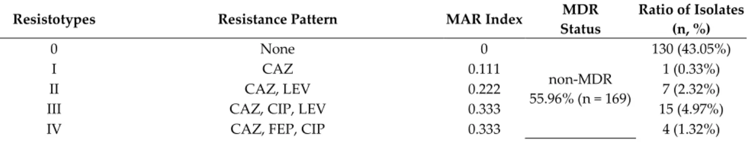
The antimicrobial resistance levels of the P. aeruginosa isolates included in the study were the following: CAZ 56.95% (n = 172), LEV 54.97% (n = 166), CIP 52.64% (n = 159), FEP 49.34% (n = 149), GEN 33.77% (n = 102), AMI 25.82% (n = 78), MER 23.17% (n = 70), IMI 21.19% (n = 64), and COL 1.66% (n = 5; MIC> 2 mg/L); overall, 44.04% (n = 133) of isolates were classified as MDR. The distribution of the various resistotypes detected among P.
aeruginosa isolates are presented in Table 1.
Table 1. Resistotype distribution and MAR indices of respective isolates.
Resistotypes Resistance Pattern MAR Index MDR
Status
Ratio of Isolates (n, %)
0 None 0
non-MDR 55.96% (n = 169)
130 (43.05%)
I CAZ 0.111 1 (0.33%)
II CAZ, LEV 0.222 7 (2.32%)
III CAZ, CIP, LEV 0.333 15 (4.97%)
IV CAZ, FEP, CIP 0.333 4 (1.32%)
V CAZ, FEP, LEV 0.333 4 (1.32%)
VI CAZ, FEP, GEN 0.333 5 (1.66%)
VII CAZ, FEP, CIP, LEV 0.444 3 (0.99%)
VIII CAZ, FEP, CIP, LEV, GEN 0.555
MDR 44.04% (n = 133)
5 (1.66%)
IX CAZ, FEP, CIP, LEV, IMI 0.555 10 (3.31%)
X CAZ, FEP, CIP, LEV, MER 0.555 10 (3.31%)
XI CAZ, FEP, CIP, LEV, GEN, IMI 0.666 7 (2.32%)
XII CAZ, FEP, CIP, LEV, GEN, MER 0.666 7 (2.32%)
XIII CAZ, FEP, CIP, LEV, IMI, MER 0.666 16 (5.29%)
XIV CAZ, FEP, CIP, LEV, GEN, AMI 0.666 23 (7.62%)
XV CAZ, FEP, CIP, LEV, GEN, AMI, IMI 0.777 16 (5.29%)
XVI CAZ, FEP, CIP, LEV, GEN, AMI, MER 0.777 22 (7.28%)
XVII CAZ, FEP, CIP, LEV, GEN, AMI, COL 0.777 2 (0.67%)
XVIII CAZ, FEP, CIP, LEV, GEN, AMI, IMI, MER 0.888 12 (3.97%)
XIX CAZ, FEP, CIP, LEV, GEN, AMI, IMI, MER, COL 1.000 3 (0.99%)
3.2. Detection of AmpC Overexpression, Carbapenemase Production, and Efflux Pump Overexpression Using Phenotypic Methods
To ascertain the contribution of various resistance mechanisms in the drug resistance of the relevant P. aeruginosa isolates, phenotypic tests were utilized. Using the cloxacillin plate-based assay, AmpC overexpression—corresponding to a two-fold decrease in the CAZ MICs—was noted in 48.26% (n = 83; 27.48% overall) of isolates; CAZ resistance was characteristic to all (83/83) of these isolates, while 61/83, 13/83, and 11/83 were resistant to FEP, MER, and IMI, respectively. The MHT test was used to detect for the production of carbapenemases: the test was positive in 25.71% (n = 18; 5.96% overall) of cases, when non- susceptibility to MER (or both) was seen. The effects of efflux pump overexpression (based on the PAβN screening agar, demonstrated by the two-fold decrease in MICs in the pres- ence of the inhibitor) were noted in 31.42% (n = 22; 7.28% overall) with regards to MER resistance and in 54.72% (n = 87; 28.8% overall) regarding CIP resistance, respectively. In the case of n = 3 isolates AmpC-hyperproduction and MHT-positivity, while in n = 6 iso- lates, efflux pump overexpression and MHT positivity were noted; in n = 3 isolates, efflux pump overexpression, AmpC hyperproduction, and MHT positivity were detected sim- ultaneously.
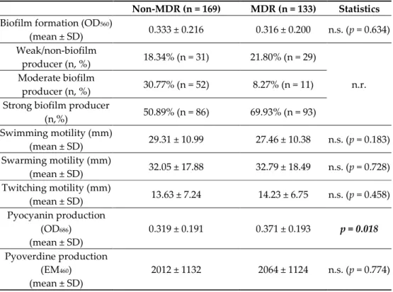
3.3. Biofilm Formation and Expression of Virulence Factors (Pigments, Motility) among Non- MDR and MDR P. aeruginosa
Biofilm formation assays were carried out in a microtiter plate-based platform, using CV staining. Based on the OD570 measurements, 19.87% (n = 60) of the isolates were weak/non-biofilm producers, 20.86% (n = 63) were moderate biofilm producers, while 59.27% (n = 179) were strong biofilm producers, overall. The distribution of isolates with different biofilm-forming capacities did not show pronounced differences among the MDR and non-MDR groups (Table 2). Additionally, while numerical differences may be seen, MDR and non-MDR isolates did not show significant differences in expression of virulence factors (measured via our in vitro experiments), with the exception of pyocyanin production (OD686), which was shown to be higher among MDR isolates (0.371 ± 0.193 vs.
non-MDR: 0.319 ± 0.191) (Table 2).
The correlation between the presence/absence (denoted as 0 and 1 in the analyses) of resistance to relevant antibiotics and biofilm-forming capacity (denoted as 0, 0.5 and 1 in the analyses for weak, moderate, and strong biofilm production, respectively) is presented in Table 3. Similarly to our previous findings (MDR/non-MDR), biofilm formation did not show significant correlation with the existence of resistance to any antibiotic tested, with
|r| values consistently < 0.3, with the exception of CIP (r = 0.309; p > 0.05) and LEV (r =
0.324; p > 0.05). The correlation matrix on the phenotypic expression of biofilm formation and other virulence factors is presented in Table 4; no relevant correlations (with |r| values consistently < 0.1) were seen between the rate of biofilm formation and pigment produc- tion or motility.
Table 2. Biofilm formation and expression of other virulence factors among non-MDR and MDR P.
aeruginosa.
Non-MDR (n = 169) MDR (n = 133) Statistics Biofilm formation (OD560)
(mean ± SD) 0.333 ± 0.216 0.316 ± 0.200 n.s. (p = 0.634) Weak/non-biofilm
producer (n, %) 18.34% (n = 31) 21.80% (n = 29)
n.r.
Moderate biofilm
producer (n, %) 30.77% (n = 52) 8.27% (n = 11) Strong biofilm producer
(n,%) 50.89% (n = 86) 69.93% (n = 93) Swimming motility (mm)
(mean ± SD) 29.31 ± 10.99 27.46 ± 10.38 n.s. (p = 0.183) Swarming motility (mm)
(mean ± SD) 32.05 ± 17.88 32.79 ± 18.49 n.s. (p = 0.728) Twitching motility (mm)
(mean ± SD) 13.63 ± 7.24 14.23 ± 6.75 n.s. (p = 0.458) Pyocyanin production
(OD686) (mean ± SD)
0.319 ± 0.191 0.371 ± 0.193 p = 0.018 Pyoverdine production
(EM460) (mean ± SD)
2012 ± 1132 2064 ± 1124 n.s. (p = 0.774) n.s.: not significant; n.r.: not relevant; values in boldface were p < 0.05
Table 3. Correlation matrix regarding the microbial categories (weak/moderate/strong) based on biofilm formation and resistance to antibiotics in P. aeruginosa isolates.
CAZ FEP IMI MER CIP LEV GEN AMI COL
Biofilm formation
0.123 0.134 0.101 0.124 0.309 0.324 0.139 0.113 −0.012
Pearson-corre- lation coeffi-
cient (r = ) 0.231 0.218 0.581 0.564 0.102 0.099 0.567 0.605 0.986 Statistics (p = ) Table 4. Correlation matrix regarding the phenotypic expression of biofilm formation and other virulence factors among tested P. aeruginosa isolates.
Biofilm Formation
(OD570)
Swimming Motility (mm)
Swarming Motility
(mm)
Twitching Motility
(mm)
Pyocyanin Production (OD686)
Pyoverdine Production
(EM460) Biofilm
formation (OD570)
X 0.981 0.580 0.518 0.581 0.373
Statistics (p = ) Swimming
motility (mm) −0.003 X 0.143 0.432 0.998 0.244
Swarming
motility (mm) 0.032 0.084 X 0.303 0.351 0.389
Twitching
motility (mm) −0.032 −0.045 −0.059 X 0.118 0.244
Pyocyanin production (OD686)
−0.0370 0.001 0.054 0.090 X 0.814
Pyoverdine production
(EM460)
0.051 −0.067 −0.050 −0.067 0.014 X
Pearson correlation coefficient (r = ) 4. Discussion, Review of the Literature
The increasing prevalence of MDR infections globally—especially in non-fermenting Gram-negative bacteria—may be considered as one of the hallmarks of the 21st century, especially when it comes to the procurement of safe and effective medical care [40,41]. The injudicious use of antibiotics and the lack of newly developed antibiotics are important hallmarks of this impending crisis [42,43]. Based on the projections of the “Burden of An- timicrobial Resistance Collaborative Group”, corresponding to the European Union (EU) and the European Economic Area (EEA), over 700,000 infections, >33,000 deaths, and
~900,000 disability-adjusted life years (DALY) were attributable to MDR bacteria, in the year 2015 alone [44]. Non-fermenting Gram-negative bacteria extensively contribute to the overall infectious disease burden—especially in immunocompromised individuals—
which is now showing an increasing trend of morbidity and mortality (HAIs are associ- ated with an overall mortality rate of 20–60%), due to the developments in resistance rates [45,46]. This has been confirmed by the World Health Organization’s (WHO) published report, in which carbapenem-resistant Acinetobacter baumannii complex (CR-AB), car- bapenem-resistant P. aeruginosa (CR-PA), and carbapenem-resistant and/or ESBL-produc- ing Enterobacterales were all identified as “critical priority” pathogens for the develop- ment of novel antibiotics and alternative anti-infective treatment strategies [47].
P. aeruginosa—especially under strong selection pressures in nosocomial environ- ments—has the capacity of becoming resistant to most relevant antimicrobials [9,48]. This includes intrinsic non-susceptibility to many orally-available agents (e.g., a chromoso- mally-encoded AmpC β-lactamase, which may be stably de-repressed), and the acquisi- tion of novel resistance genes through the means of horizontal gene transfer (i.e., in- tegrons, plasmids, or transposons), leading to resistance to the four major group of anti- pseudomonal agents, namely relevant cephalosporins (and cephalosporin/β-lactamase combinations), carbapenems, aminoglycosides, respiratory fluoroquinolones (ciprofloxa- cin, levofloxacin, and moxifloxacin), and colistin [49]. Based on the data from the ECDC Surveillance Atlas of Infectious Diseases for 2019, resistance rates in P. aeruginosa ranged between 3.5−52.2%, 4.5–52.2%, 0.3–48.9% and 0–55.4% for ceftazidime, fluoroquinolones, aminoglycosides, and carbapenems in EU countries, respectively [50]. In addition to these genetically-determined resistance mechanisms (leading to the expression of mutated tar- get proteins or inactivating enzymes, the relevance of so-called “adaptive” mechanisms also needs to be discussed; strong biofilm formation and metabolic downregulation to- wards the emergence of SCVs are difficult to study in vitro, but their relevance in in vivo infection dynamics must not be underestimated, as they lead to therapy-resistant, recalci- trant infections [51].
In clinical practice, MDR Pseudomonas infections are most often treated by car- bapenem antibiotics, as these drugs are often considered the last safe and effective alter- natives to treatment, especially in some patients (e.g., the elderly), where other drugs would be contraindicated due to their toxic adverse events or in cases (e.g., critically ill patients) where pathophysiological changes may significantly alter antibacterial pharma- cokinetics, making the dosing of other drugs difficult [52]. Thus, the growing rate of CR- PA in nosocomial infections (with rates ranging between 0–60%, showing pronounced
geographical differences) is a chilling concept; in a recently published meta-analysis, it was found that in addition to a history of carbapenem use, the prior use of piperacil- lin/tazobactam or vancomycin were all relevant risk factors for the acquisition of CR-PA [53,54]. Phenotypic carbapenem resistance is often due to a combination of more than one mechanism of resistance, which may be mediated via modifications in the transpeptidases (or penicillin-binding proteins; PPB2 and 3) [55], outer membrane impermeability (often due to OprD porin deficiency or loss) [56], overexpression of efflux pump systems [57], and the production of carbapenemases. Carbapenemases are β-lactamases with versatile hydrolytic capabilities; they can hydrolyze penicillins, cephalosporins, monobactams, and carbapenems [58]. Based on their biochemical characteristics and substrate profile, car- bapenemases are classified into serine (including Ambler Class A [KPC], C [AmpC] and D [OXA-48-like family]) and metallo-β-lactamases (MBLs; Ambler Class B) [59]. These en- zymes are often encoded by horizontally-transferable genes, which are often also associ- ated with resistance determinants to other classes of antimicrobial drugs [60]. Currently, the propagation of carbapenemase-producing organisms, especially in non-fermenting Gram-negative bacteria, is a public health issue these resistance-determinants may rapidly disseminate, leading to outbreaks of bacteria with extensive resistance [61]. While Class A and D carbapenemases have also been described, the metallo-β-lactamases blaVIM and blaIMP are the most prevalent in Pseudomonas spp. [62,63]. The treatment of CR-PA infec- tions heavily rely on last-resort agent that are either associated with more serious adverse events (colistin), or that are relatively new, with limited clinical experience in these infec- tions (ceftolozane/tazobactam, ceftazidime/avibactam, meropenem/vaborbactam, plazmomycin) [64]. In addition, the therapy of metallo-β-lactamases is especially difficult, as currently there are no β-lactamase-inhibitors authorized for clinical use that would be effective against these enzymes. For example, in a study by O’Neall et al., n = 250 car- bapenem non-susceptible P. aeruginosa isolates were tested for their susceptibility for ceftolozane/tazobactam and ceftazidime/avibactam, showing that these drugs presented with good in vitro efficacy against these isolates, but only as long as they did not produce carbapenemases; on the other hand, isolates carrying blaVIM or blaNDM were resistant to these new antibiotic combinations [65]. Thus, it is unsurprising that many clinical studies have highlighted that CR–PA infections are associated with worse clinical outcomes, com- pared to their carbapenem-susceptible counterparts [66].
In studies trying to ascertain the relationship between biofilm formation and drug resistance, phenotypic or a combination of phenotypic and molecular-based (PCR) meth- odologies are used, involving a wide variety of isolates from distinct clinical origins [67,68]. The majority of these studies have highlighted that around 75–99% of Pseudomonas spp. are biofilm producers, including 8–50% being characterized as strong biofilm pro- ducers [23]. As a part of our study, a large pool (n = 302) of P. aeruginosa isolates from diverse geographical and clinical origins were subjected to phenotypic tests to ascertain the possible correlation between biofilm-forming capacity, pigment production and mo- tility (chosen as representative expressions of virulence), and antimicrobial resistance in these isolates. In our pool of isolates, resistance rates were the highest for ceftazidime and the fluoroquinolones, while >44% was classified as MDR. Over 25% isolates were charac- terized by the expression of AmpC β-lactamase, ~6% was positive for carbapenemase- production, and in ~7%, carbapenem non-susceptibility was affected by efflux pump over- expression, respectively (based on the phenotypic tests applied). Majority of isolates (59.27% overall) were strong biofilm producers, both in the MDR and non-MDR groups.
Based on our results, no relevant correlation was seen between the resistance to individual antibiotics—or the presence of the MDR phenotype as a whole—and biofilm-forming ca- pacity. Similarly—with the exception of pyocyanin production—no association was noted for other virulence factors and drug resistance. Based on our experimental data, we also did not observe a notable relationship between biofilm formation and other virulence fac- tors. It is interesting to note that there was no association between the levels of different motilities expressed by Pseudomonas; this may be explained by the fact that—although all
of these motility strategies have important physiological roles—their differential expres- sion is needed to accommodate the adaptation of this pathogen to different niches [9,69].
Pyocyanin is a blue-green, lipid soluble pigment, which has redox-active properties, being a major electron acceptor for molecular O2; the synthesis of this pigment is mediated by PhzM (N-methyltransferase) and PhzS (flavin-dependent hydroxylase) [70]. This pig- ment has been described as an essential factor in the virulence of Pseudomonas, especially in skin and soft tissue infection and invasive infections of the lungs. Pyocyanin may often be detected in high concentrations in the airway tissues and sputum of CF patients, where it contributes to chronic lung infection and bronchiectasis by facilitating mucus overpro- duction, inhibition of ciliary activity and the α1-protease inhibitor (leading to excess tissue injury), and inhibition of IL-2 expression, leading to increased IL-8 release [71]. Pyocyanin may also have inhibitory properties on other bacterial species, aiding Pseudomonas in the competition in a given ecological niche; among others, this phenomenon was demon- strated in the decreased diversity of microbial communities in the sputa of patients, where pyocyanin concentrations were high [72,73]. Our experiments found that the production of pyocyanin was a distinctive hallmark between the MDR and non-MDR group, with isolates in the former group showing higher levels of pigment production. During the experiments of Fuse et al., reverse-phase high-performance liquid chromatography (RP- HPLC) was used for the quantitative extraction of pyocyanine in P. aeruginosa; their study concluded that MDR (including many MBL-producers) reduced pyocyanine expression, compared to the non-MDR group [74]. With similar aims to our study, Gholami et al. in- cluded 100 P. aeruginosa isolates from clinical and environmental origins; they found blaTEM and blaSHV positivity in 92%/16% and 20/6% of the isolates, respectively, with clinical isolates having higher proclivity to becoming MDR. 70% and 28% (with 98% and 70% of these isolates carrying the algD and algU genes) of these isolates were classified as strong biofilm producers; however, no association between the MDR phenotype and bio- film-formation was found [75]. Yamani et al. utilized phenotypic and genotypic methods to characterize 66 Pseudomonas isolates: in their study, 53.03%/24.24%/22.73% of isolates were classified as strong/moderate/weak biofilm producers, respectively. By measuring the expression levels of 10 virulence genes (exoT, exoS, exoU, phZ, las and pil genes), they found that—although all associated genes were present in all isolates—all biofilm and virulence-associated genes were significantly upregulated in non-MDR isolates, demon- strating an inverse correlation [76]. The experiments of Kamali et al. included 80 isolates, from which 50% were characterized as moderate or strong biofilm producers; they found that 87.5% possessed all three genes relevant for biofilm formation (algD, pslD, and pelF) and a positive correlation between biofilm formation and the presence of these genes, while no association was noted between biofilm formation and drug resistance [77].
Jabalameli et al. studied the carriage of Type III secretory toxin-encoding genes and their cytotoxic action on A549 human lung cancer cell lines in 96 clinical P. aeruginosa isolates, finding that the carriage of exoT, exoY, exoU, and exoS were 100%, 95%, 64.5%, and 29%, respectively. 47%/26% of the isolates were strong/moderate biofilm producers, respec- tively; no association was seen between resistance phenotype and biofilm formation, and interestingly, neither between the presence of toxin-encoding genes and the extent of toxic effects seen on the cell lines [78].
In contrast, the study of Abidi et al., which included 22 P. aeruginosa isolated from keratitis, concluded that strong biofilm producers were significantly more common among MDR isolates [79]. Choy et al. involved 77 Pseudomonas isolates originating from keratitis of contact lens and non-contact lens origin; while no correlation was seen be- tween drug resistance and biofilm formation, the presence of the exoU gene (mostly found in contact lens-related isolates) and strong biofilm formation showed strong positive cor- relations [80]. In a study involving 74 P. aeruginosa from the sputum of CF and non-CF patients, Perez et al. noted that no differences in biofilm formation were observed among isolates from different patient origins (although 85.7% of CF isolates presented as the mu- coid phenotypic variant), MBL positivity showed strong association with strong biofilm
production [81]. In a study involving n = 190 isolates, Eladawy et al. found no relevant associations between antimicrobial resistance, biofilm formation, and the presence of genes encoding for selected virulence factors (i.e., alignate, biofilm, exotoxin A, L-orni- thine monooxygenase, phospholipases T3SS, proteases, and pyocyanin) [82]. da Costa Lima et al. used molecular methods to assess the correlation between the presence of quorum sensing (QS) genes and biofilm formation in 40 P. aeruginosa isolates: while all isolates carried these genes, no association was found with the capacity to produce biofilm [83]. Subedi et al. included 22 isolates from eye infections and CF sputa for testing: the prevalence of exoU was 61.5%, and isolates carrying exoU showed higher rates of re- sistance and higher rates of mutations in the quinolone resistance determining region (QRDR) [84]. In a study involving 78 clinical and environmental isolates, Karami et al.
found MDR rates of 48.7% and a strong positive association was shown between biofilm- formation and the MDR status [85]. Bahador et al. noted a positive association between biofilm formation and the presence of the exoU and exoS genes (36.6% and 55.7%, respec- tively), but not with antimicrobial resistance (16% of isolates were extensively drug re- sistant [XDR]) in the 75 pseudomonads tested [86]. In a comprehensive laboratory study by Milojkovic et al. involving 94 isolates—similarly to our study—no correlation was noted between antibiotic resistance, genetic composition, pigment production, serotypes, and biofilm formation [87]. Cho et al. assessed biofilm formation and MBL carriage in carbapenem-resistant P. aeruginosa isolates; the overwhelming majority of isolates (>92%) were biofilm producers. Furthermore, the presence of the pslA gene and the MDR pheno- type all showed positive association with biofilm-forming capacity [88]. Eighty-six P. ae- ruginosa isolates (involving >31% MDR and >12% XDR), collected from various clinical materials, were included in the study of Zahedani et al., where strong correlations were found between the expression of efflux pumps and biofilm formation (22.47%/21.34%/11.23% were strong/moderate/weak biofilm producers); additionally, the presence of some efflux pumps (MexEF-OprN) reduced the virulence of these isolates, while others (MexAB-OprM, MexCD-OprJ) did not have the same effect [89]. Rodulfo et al. involved n = 176 strains (isolated between 2009 and 2016) in their experiments, showing high levels of drug resistance (38.1% MDR), and resistance rates showed strong associa- tion with the presence of exoU and hemolysin, while negative associations with twitching motility [90]. In isolates originating from endotracheal aspirates from ICU patients, Fricks- Lima et al. found strong and significant correlations between ceftazidime and imipenem- resistance and strong biofilm-forming capacity [91].
Biofilms are characterized by a protective form of growth (essential for survival) in a nosocomial environment, which may also maintain an inflammatory environment in vivo [91,92]. Nevertheless, it is obvious that the continuous, long-term expression of antibiotic resistance-determinants and virulence factors is detrimental for the microorganism’s abil- ity to adapt to environmental changes [93,94]. For example, in chronic infections, where P. aeruginosa establishes long-term persistence in biofilm, the expression of virulence fac- tors is downregulated, to accommodate for the lower metabolic activity in the exopoly- saccharide matrix [95]. QS is one of the principal mechanisms responsible for—among others—the regulation of virulence factor expression and biofilm formation. QS in Pseu- domonas consist of a three interconnected systems, namely the Las, Rhl, and Pqs systems;
these systems facilitate the changes needed for the pathogens’ survival, by the sensing of diffusible signal molecules found in their surroundings, which is a proxy for the popula- tion density in the niche [96–98]. However, many antibiotics also have the ability to affect these QS systems in Pseudomonas, either by directly acting on gene expression or by the degradation of these signal molecules [99]. The emergence of P. aeruginosa isolates with different biofilm-forming capacities may also inform the genetic heterogeneity within these species, which is an important factor for successful infection in humans [9]. Never- theless, differences in the phenotypes and susceptibility trends in these isolates may also stem from their different geographical origin and the sampling frame used [100]. The main limitation of our study was the reliance on the use of phenotypic methods only during the
assessment of the resistance, biofilm-forming, and virulence phenotype of the isolates;
therefore, we did not have data from molecular methods or on the clonal complexes (CCs) for the isolates implicated in the assays. On the other hand, our data are generated from a considerably large number of geographically-distinct isolates.
5. Conclusions
P. aeruginosa is an opportunistic pathogen, which is often implicated in infectious pa- thologies, leading to a considerable disease burden worldwide. Along with its intrinsic resistance to many antibiotics, Pseudomonas infections are further complicated by the in- creasing prevalence of carbepenem and colistin non-susceptibility in these strains. Pseu- domonas species possess many important virulence factors in their repertoire, in addition to their propensity to form biofilms (75–100% are biofilm producers). Data on interplay between the presence and mechanisms of drug resistance with those of biofilm formation and virulence is crucial to address chronic bacterial infections (leading to therapeutic fail- ure and decreased quality of life in the affected patients), and to provide strategies for their management. Our study has showed that isolates with the MDR and non-MDR phe- notype did not differ significantly in the context of the virulence determinants studied (biofilm, various motilities, pyoverdine), with the exception of pyocyanin production.
Similarly, no statistically-relevant interplay was observed between the individual viru- lence factors. The latter may either be explained by the adaptation of the microorganism to express their virulence factors only in situations where they are necessary for survival, or our in vitro methodologies utilized were not sophisticated enough to detect their asso- ciation. As presented in our paper, many authors have aimed to provide clarity on asso- ciation between biofilm-forming capacity, virulence factor-expression, and the MDR phe- notype in P. aeruginosa, but the presently-available results do not yet allow for a conclu- sion to be made. Additional experiments—preferably involving genomics and/or in vivo methods—are needed to provide further clarity and insights into this field.
Author Contributions: M.G., M.G.D., and D.U. conceived and designed the study. M.G.D., M.G., K.K., D.U., and S.Z. were involved in the collection of isolates and in performing the experiments.
M.G.D., Z.B., and S.Z. provided resources, D.S. and S.Z. supervised the project. M.G. and M.G.D.
wrote the initial draft of the manuscript. All authors have read and agreed to the published version of the manuscript.
Funding: M.G. was supported by the János Bolyai Research Scholarship (BO/00144/20/5) of the Hun- garian Academy of Sciences. The research was supported by the ÚNKP-21-5-540-SZTE New Na- tional Excellence Program of the Ministry for Innovation and Technology from the source of the National Research Development and Innovation Fund. M.G. would also like to acknowledge the support of ESCMID’s “30 under 30” Award.
Institutional Review Board Statement: The study was conducted in accordance with the Declara- tion of Helsinki and national and institutional ethical standards. Ethical approval for the study pro- tocol was obtained from the Human Institutional and Regional Biomedical Research Ethics Com- mittee, University of Szeged (registration number: 140/2021-SZTE (5019)).
Informed Consent Statement: Not applicable.
Data Availability Statement: All data generated during the study are presented in this paper.
Conflicts of Interest: The authors declare no conflict of interest, monetary or otherwise. The authors alone are responsible for the content and writing of this article.
References
1. Whistler, T.; Sangwichian, O.; Jorakate, P.; Sawatwong, P.; Surin, U.; Piralam, B.; Thamthitiwat, S.; Promkong, C.; Peruski, L.
Identification of Gram negative non-fermentative bacteria: How hard can it be? PLoS Neglect. Pathog. 2019, 13, e0007729.
2. Fiscarelli, E.; Rossitto, M.; Rosati, P.; Essa, N.; Crocetta, V.; Di Giulio, A.; Lupetti, V.; Di Bonaventura, G.; Pompilio, A. In vitro newly isolated environmental phage activity against biofilms preformed by Pseudomonas aeruginosa from patients with cystic fibrosis. Microorganisms 2021, 9, 478, https://doi.org/10.3390/microorganisms9030478.
3. Klockgether, J.; Tömmler, B. Recent advances in understanding Pseudomonas aeruginosa as a pathogen. F100Research 2017, 6, e1261.
4. Çiçek, A..; Ertürk, A.; Ejder, N.; Rakici, E.; Kostakoğlu, U.; Yıldız, I.E.; Özyurt, S.; Sönmez, E. Screening of antimicrobial re- sistance genes and epidemiological features in hospital and community-associated carbapenem-resistant pseudomonas aeru- ginosa infections. Infect. Drug Resist. 2021, 14, 1517–1526, https://doi.org/10.2147/idr.s299742.
5. Migiyama, Y.; Yanagihara, K.; Kaku, N.; Harada, Y.; Yamada, K.; Nagaoka, K.; Morinaga, Y.; Akamatsu, N.; Matsuda, J.; Izumi- kawa, K.; et al. Pseudomonas aeruginosa bacteremia among immunocompetent and immunocompromised patients: Relation to initial antibiotic therapy and survival. Jpn. J. Infect. Dis. 2016, 69, 91–96, https://doi.org/10.7883/yoken.jjid.2014.573.
6. Yang, K.; Xiao, T.; Shi, Q.; Zhu, Y.; Ye, J.; Zhou, Y.; Xiao, Y. Socioeconomic burden of bloodstream infections caused by car- bapenem-resistant and carbapenem-susceptible Pseudomonas aeruginosa in China. J. Glob. Antimicrob. Resist. 2021, 26, 101–107, https://doi.org/10.1016/j.jgar.2021.03.032.
7. Hilliam, Y.; Kaye, S.; Winstanley, C. Pseudomonas aeruginosa and microbial keratitis. J. Med. Microbiol. 2020, 69, 3–13, https://doi.org/10.1099/jmm.0.001110.
8. Vongthilath, R.; Thiriez, B.R.; Dehillotte, C.; Lemonnier, L.; Guillien, A.; Degano, B.; Dalphin, M.-L.; Dalphin, J.-C.; Plésiat, P.
Clinical and microbiological characteristics of cystic fibrosis adults never colonized by Pseudomonas aeruginosa: Analysis of the French CF registry. PLoS ONE 2019, 14, e0210201, https://doi.org/10.1371/journal.pone.0210201.
9. Behzadi, P.; Baráth, Z.; Gajdács, M. It’s not easy being green: A narrative review on the microbiology, virulence and therapeutic prospects of multidrug-resistant pseudomonas aeruginosa. Antibiotics 2021, 10, e42.
10. Veesenmeyer, J.L.; Hauser, A.R.; Lisboa, T.; Rello, J. Pseudomonas aeruginosa virulence and therapy: Evolving translational strat- egies. Crit. Care Med. 2009, 37, 1777–1786, https://doi.org/10.1097/ccm.0b013e31819ff137.
11. Ciofu, O.; Rojo-Molinero, E.; Macia, M.D.; Oliver, A. Antibiotic treatment of biofilm infections. APMIS 2017, 125, 304–319, https://doi.org/10.1111/apm.12673.
12. Azeredo, J.; Azevedo, N.F.; Briandet, R.; Cerca, N.; Coenye, T.; Costa, A.R.; Desvaux, M.; Di Bonaventura, G.; Hébraud, M.;
Jaglic, Z.; et al. Critical review on biofilm methods. Crit. Rev. Microbiol. 2017, 43, 313–351.
13. Jamal, M.; Ahmad, W.; Andleeb, S.; Jalil, F.; Imran, M.; Nawaz, M.A.; Hussain, T.; Ali, M.M.; Rafiq, M.; Kamil, M.A. Bacterial biofilm and associated infections. J. Chin. Med. Assoc. 2018, 81, 7–11, https://doi.org/10.1016/j.jcma.2017.07.012.
14. Lebeaux, D.; Ghigo, J.-M.; Beloin, C. Biofilm-related infections: Bridging the gap between clinical management and fundamental aspects of recalcitrance toward antibiotics. Microbiol. Mol. Biol. Rev. 2014, 78, 510–543, https://doi.org/10.1128/mmbr.00013-14.
15. Maurice, N.M.; Bedi, B.; Sadikot, R.T. Pseudomonas aeruginosa biofilms: Host response and clinical implications in lung infections.
Am. J. Respir. Cell Mol. Biol. 2018, 58, 428–439, https://doi.org/10.1165/rcmb.2017-0321tr.
16. Ciofu, O.; Tolker-Nielsen, T. Tolerance and resistance of Pseudomonas aeruginosa biofilms to antimicrobial agents—How P. aeru- ginosa can escape antibiotics. Front. Microbiol. 2019, 10, e913.
17. Zhen, X.; Lundborg, C.S.; Sun, X.; Gu, S.; Dong, H. Clinical and economic burden of carbapenem-resistant infection or coloni- zation caused by Klebsiella pneumoniae, Pseudomonas aeruginosa, Acinetobacter baumannii: A multicenter study in China. Antibiotics 2020, 9, e514.
18. Gajdács, M.; Albericio, F. Antibiotic resistance: From the bench to patients. Antibiotics 2019, 8, 129, https://doi.org/10.3390/anti- biotics8030129.
19. Amsalu, A.; Sapula, S.A.; Lopes, M.D.B.; Hart, B.J.; Nguyen, A.H.; Drigo, B.; Turnidge, J.; Leong, L.E.; Venter, H. Efflux pump- driven antibiotic and biocide cross-resistance in Pseudomonas aeruginosa isolated from different ecological niches: A case study in the development of multidrug resistance in environmental hotspots. Microorganisms 2020, 8, 1647, https://doi.org/10.3390/mi- croorganisms8111647.
20. Zhang, Y.; Li, Y.; Zeng, J.; Chang, Y.; Han, S.; Zhao, J.; Fan, Y.; Xiong, Z.; Zou, X.; Wang, C.; et al. Risk factors for mortality of inpatients with Pseudomonas aeruginosa bacteremia in China: Impact of resistance profile in the mortality. Infect. Drug Resist.
2020, 13, 4115–4123, https://doi.org/10.2147/idr.s268744.
21. Bonomo, R.A.; Szabo, D. Mechanisms of multidrug resistance in acinetobacter species and Pseudomonas aeruginosa. Clin. Infect.
Dis. 2006, 43, S49–S56, https://doi.org/10.1086/504477.
22. Tahaei, S.A.S.; Stájer, A.; Barrak, I.; Ostorházi, E.; Szabó, D.; Gajdács, M. Correlation between biofilm-formation and the antibi- otic resistant phenotype in Staphylococcus aureus isolates: A laboratory-based study in Hungary and a review of the literature.
Infect. Drug Resist. 2021, 14, 1155–1168, https://doi.org/10.2147/idr.s303992.
23. MirzaHosseini, H.K.; Hadadi-Fishani, M.; Morshedi, K.; Khaledi, A. Meta-analysis of biofilm formation, antibiotic resistance pattern, and biofilm-related genes in Pseudomonas aeruginosa isolated from clinical samples. Microb. Drug Resist. 2020, 26, 815–
824, https://doi.org/10.1089/mdr.2019.0274.
24. Saeki, E.K.; Yamada, A.Y.; de Araujo, L.A.; Anversa, L.; Garcia, D.D.O.; de Souza, R.L.B.; Martins, H.M.; Kobayashi, R.K.T.;
Nakazato, G. Subinhibitory concentrations of biogenic silver nanoparticles affect motility and biofilm formation in Pseudomonas aeruginosa. Front. Cell. Infect. Microbiol. 2021, 11, https://doi.org/10.3389/fcimb.2021.656984.
25. Schubert, S.; Kostrzewa, M. MALDI-TOF MS in the microbiology laboratory: Current trends. Curr. Issues Mol. Biol. 2017, 23, 17–
20, https://doi.org/10.21775/cimb.023.017.
26. Gajdács, M. Carbapenem-resistant but cephalosporin-susceptible Pseudomonas aeruginosa in urinary tract infections: Oppor- tunity for Colistin Sparing. Antibiotics 2020, 9, 153, https://doi.org/10.3390/antibiotics9040153.
27. Sadat, A.; El-Sherbiny, H.; Zakaria, A.; Ramadan, H.; Awad, A. Prevalence, antibiogram and virulence characterization of Vibrio isolates from fish and shellfish in Egypt: A possible zoonotic hazard to humans. J. Appl. Microbiol. 2020, 131, 485–498, https://doi.org/10.1111/jam.14929.
28. EUCAST Clinical Breakpoints and Dosing. Available online: https://www.eucast.org/clinical_breakpoints (accessed on 5 Au- gust 2021).
29. Magiorakos, A.P.; Srinivasan, A.; Carey, R.B.; Carmeli, Y.; Falagas, M.E.; Giske, C.G.; Paterson, D.L. Multidrug-resistant, exten- sively drug-resistant and pandrug-resistant bacteria: An international expert proposal for interim standard definitions for ac- quired resistance. Clin. Microbiol. Infect. 2012, 18, 268–281.
30. Khalili, Y.; Yekani, M.; Goli, H.R.; Memar, M.Y. Characterization of carbapenem-resistant but cephalosporin-susceptible Pseu- domonas aeruginosa. Acta Microbiol. Immunol. Hung. 2019, 66, 529–540, https://doi.org/10.1556/030.66.2019.036.
31. Chou, C.-H.; Lai, Y.-R.; Chi, C.-Y.; Ho, M.-W.; Chen, C.-L.; Liao, W.-C.; Ho, C.-M.; Chen, Y.-A.; Chen, C.-Y.; Lin, Y.-T.; et al.
Long-term surveillance of antibiotic prescriptions and the prevalence of antimicrobial resistance in non-fermenting gram-neg- ative bacilli. Microorganisms 2020, 8, 397, https://doi.org/10.3390/microorganisms8030397.
32. Akhi, M.T.; Khalili, Y.; Chotaslou, R.; Yousefi, S.; Kafil, H.S.; Naghili, B.; Sheikhalizadeh, V. Evaluation of carbapenem resistance mechanisms and its association with Pseudomonas aeruginosa infection in the northwest of Iran. Microb. Drug Res. 2018, 24, 126–
135.
33. Ramos-Vivas, J.; Chapartegui-González, I.; Fernández-Martı́nez, M.; González-Rico, C.; Fortún, J.; Escudero, R.; Marco, F.; Li- nares, L.; Montejo, M.; Aranzamendi, M.; et al. Biofilm formation by multidrug resistant Enterobacteriaceae strains isolated from solid organ transplant recipients. Sci. Rep. 2019, 9, e8928.
34. Ansari, M.A.; Khan, H.M.; Khan, A.A.; Cameotra, S.S.; Saquib, Q.; Musarrat, J. Gum Arabic capped-silver nanoparticles inhibit biofilm formation by multi-drug resistant strains of Pseudomonas aeruginosa. J. Basic Microbiol. 2014, 54, 688–699, https://doi.org/10.1002/jobm.201300748.
35. Ha, D.-G.; Kuchma, S.L.; O’Toole, G.A. Plate-based assay for swimming motility in Pseudomonas aeruginosa. In Pseudomonas Methods and Protocols; Filloux, A., Ramos, J.L., Eds.; Humana Press Inc.: New York, NY, USA, 2014.
36. Turnbull, L.; Whitchurch, C.B. Motility assay: Twitching motility. In Pseudomonas Methods and Protocols; Filloux, A., Ramos, J.- L., Eds.; Humana Press Inc.: New York, NY, USA, 2014; pp. 73–86.
37. Das, T.; Manefield, M. Pyocyanin promotes extracellular DNA release in Pseudomonas aeruginosa. PLoS ONE 2012, 7, e46718, https://doi.org/10.1371/journal.pone.0046718.
38. Markwitz, P.; Olszak, T.; Gula, G.; Kowalska, M.; Arabski, M.; Drulis-Kawa, Z. Emerging phage resistance in Pseudomonas aeru- ginosa PAO1 is accompanied by an enhanced heterogeneity and reduced virulence. Viruses 2021, 13, 1332, https://doi.org/10.3390/v13071332.
39. Mukaka, M.M. A guide to appropriate use of correlation coefficient in medical research. Malawi Med. J. 2012, 24, 69–71.
40. Donadu, M.; Usai, D.; Pinna, A.; Porcu, T.; Mazzarello, V.; Fiamma, M.; Marchetti, M.; Cannas, S.; Delogu, G.; Zanetti, S.; et al.
In vitro activity of hybrid lavender essential oils against multidrug resistant strains of Pseudomonas aeruginosa. J. Infect. Dev.
Ctries. 2018, 12, 009–014, https://doi.org/10.3855/jidc.9920.
41. Amorese, V.; Donadu, M.; Usai, D.; Sanna, A.; Milia, F.; Pisanu, F.; Molicotti, P.; Zanetti, S.; Doria, C. In vitro activity of essential oils against Pseudomonas aeruginosa isolated from infected hip implants. J. Infect. Dev. Ctries. 2018, 12, 996–1001, https://doi.org/10.3855/jidc.10988.
42. Aslam, A.; Gajdács, M.; Zin, C.S.; Ab Rahman, N.S.; Ahmed, S.I.; Zafar, M.Z.; Jamshed, S. Evidence of the practice of self-med- ication with antibiotics among the lay public in low- and middle-income countries: A scoping review. Antibiotics 2020, 9, 597, https://doi.org/10.3390/antibiotics9090597.
43. O’Neill, J. Antimicrobial Resistance: Tackling a Crisis for the Health and Wealth of Nations. Available online: https://wellcom- ecollection.org/works/rdpck35v/items (accessed on 5 August 2021).
44. Cassini, A.; Högberg, L.D.; Plachouras, D.; Quattrocchi, A.; Hoxha, A.; Simonsen, G.S.; Colomb-Cotinat, M.; Kretzschmar, M.;
Devleesschauwer, B.; Cecchini, M.; et al. Attributable deaths and disability-adjusted life-years caused by infections with antibi- otic-resistant bacteria in the EU and the European Economic Area in 2015: A population-level modelling analysis. Lancet Infect.
Dis. 2018, 19, 56–66, https://doi.org/10.1016/s1473-3099(18)30605-4.
45. Rojas, A.; Palacios-Baena, Z.; López-Cortés, L.; Rodríguez-Baño, J. Rates, predictors and mortality of community-onset blood- stream infections due to Pseudomonas aeruginosa: Systematic review and meta-analysis. Clin. Microbiol. Infect. 2019, 25, 964–970, https://doi.org/10.1016/j.cmi.2019.04.005.
46. Carmeli, Y.; Troillet, N.; Karchmer, A.W.; Samore, M.H. Health and economic outcomes of antibiotic resistance in Pseudomonas aeruginosa. Arch. Intern. Med. 1999, 159, 1127–1132, https://doi.org/10.1001/archinte.159.10.1127.
47. World Health Organisation. Global Priority List of Antibiotic-Resistant Bacteria to Guide Research, Discovery, and Development of New Antibiotics; World Health Organisation: Geneva, Switzerland, 2017; pp. 1–7.
48. Poole, K. Pseudomonas Aeruginosa: Resistance to the max. Front. Microbiol. 2011, 2, e65.
49. Nguyen, L.; Garcia, J.; Gruenberg, K.; MacDougall, C. Multidrug-resistant pseudomonas infections: Hard to treat, but hope on the horizon? Curr. Infect. Dis. Rep. 2018, 20, 23, https://doi.org/10.1007/s11908-018-0629-6.
50. ECDC Surveillance Atlas of Infectious Diseases. Available online: https://atlas.ecdc.europa.eu/public/index.aspx?Da- taset=27&HealthTopic=4 (accessed on 5 August 2021).
![maltophilia ;nevertheless,otherlessfrequentlyisolatedgenerashouldalsobetakeninto functions[ , ].NFGNBincludesomecommonlyisolatedgenera,suchas Pseudomonas , inanoxidativefashion)rods.Inaddition,theyareallsimilarinthattheyareunableto Burkholderiamallei , an](data:image/gif;base64,R0lGODlhAQABAIAAAP///wAAACH5BAEAAAAALAAAAAABAAEAAAICRAEAOw==)