R E S E A R C H A R T I C L E Open Access
Size-dependent activity of silver
nanoparticles on the morphological switch and biofilm formation of opportunistic
pathogenic yeasts
Bettina Szerencsés1†, Nóra Igaz2†, Ákos Tóbiás2, Zsombor Prucsi2, Andrea Rónavári3, Péter Bélteky3, Dániel Madarász3, Csaba Papp1, Ildikó Makra2, Csaba Vágvölgyi1, Zoltán Kónya3,4, Ilona Pfeiffer1†and Mónika Kiricsi2*†
Abstract
Background:Dimorphism and biofilm formation are important virulence factors of some opportunistic human pathogenic yeasts. Such species commensally colonize skin or mucosal surfaces generally in yeast form, but under particular circumstances, convert into virulent hyphae and disseminate internal organs or cause mucocutaneous infections. The yeast-to-hypha shape-conversion promotes the development of a biofilm, a thick extracellular matrix with sessile cells within. The biofilm is capable to prevent the penetration of antifungal drugs, rendering the surviving biofilm-resident cells intrinsic sources of recurrent infections. The aim of this study was to evaluate the ability of silver nanoparticles (AgNPs) to attenuate the morphological switch and biofilm formation of several opportunistic pathogenic yeasts and to determine whether this feature depends on the nanoparticle size.
Results:AgNPs in three different sizes were prepared by chemical reduction approach and characterized by transmission electron microscopy, ultraviolet–visible spectroscopy and dynamic light scattering. The antifungal activity was evaluated by the microdilution method, the inhibitory capacity on biofilm formation and the biofilm degradation ability of differently sized AgNPs was assessed by viability assay. The morphological state of
opportunistic pathogenic yeast cells in monoculture and in co-culture with human keratinocytes in the presence of AgNPs was examined by flow cytometry and scanning electron microscopy. All the three AgNPs inhibited the growth of the examined opportunistic pathogenic yeasts, nevertheless, AgNPs with the smallest diameter exhibited the most prominent toxic activities. AgNPs attenuated the biofilm formation in a nanoparticle size-dependent manner; however, their biofilm destruction capacity was negligible. AgNPs with the smallest size exerted the most significant effect on suppressing the morphological change of pathogens in monoculture as well as in a co-culture with keratinocytes.
(Continued on next page)
© The Author(s). 2020Open AccessThis article is licensed under a Creative Commons Attribution 4.0 International License, which permits use, sharing, adaptation, distribution and reproduction in any medium or format, as long as you give appropriate credit to the original author(s) and the source, provide a link to the Creative Commons licence, and indicate if changes were made. The images or other third party material in this article are included in the article's Creative Commons licence, unless indicated otherwise in a credit line to the material. If material is not included in the article's Creative Commons licence and your intended use is not permitted by statutory regulation or exceeds the permitted use, you will need to obtain permission directly from the copyright holder. To view a copy of this licence, visithttp://creativecommons.org/licenses/by/4.0/.
The Creative Commons Public Domain Dedication waiver (http://creativecommons.org/publicdomain/zero/1.0/) applies to the data made available in this article, unless otherwise stated in a credit line to the data.
* Correspondence:kiricsim@gmail.com;kiricsim@bio.u-szeged.hu
†Bettina Szerencsés, Nóra Igaz, Ilona Pfeiffer and Mónika Kiricsi contributed equally to this work.
2Department of Biochemistry and Molecular Biology, Faculty of Science and Informatics, University of Szeged, Közép fasor 52, Szeged H-6726, Hungary Full list of author information is available at the end of the article
(Continued from previous page)
Conclusions:Our results confirm that AgNPs are capable to hinder yeast-to-hypha morphological conversion and biofilm formation of opportunistic pathogens and this biological effect of AgNPs is size-dependent.
Keywords:Pathogenic yeasts, Silver nanoparticles, Morphological switch, Biofilm formation, Human keratinocytes, Toxicity
Background
The incidence of human infections caused by yeasts or other fungi increased worldwide during the last decades, generating significant health problems [1]. Although the symptoms of fungal diseases are often mild and superficial (e.g. irritation, itching and swelling), on occasion, life- threatening invasive infections develop. The clinical mani- festations of such infections are strongly related to host im- munity and to the particular physiological condition of the patient [2, 3]. Despite antifungal therapy, invasive fungal diseases are associated with high mortality rate causing the death of about one and half million people in a year, mainly due to complications attributed to the infection [4]. Espe- cially immune-compromised and severely ill patients are at high risk and exhibit increased susceptibility to systemic fungal infections [5]. The most common pathogens gener- ating invasive infections belong toCandida, Cryptococcus, AspergillusandPneumocystisgenera; however, several spe- cies from Lodderomyces, Pichia, Rhodotorulaand Trichos- poron genera have recently been implicated in the development of systemic infections [6–12].
Numerous factors influence the pathogenic capacity of the above mentioned species, such as secretion of spe- cific enzymes, thermotolerance, toxin production, as well as morphologic transformation [2]. Indeed, several op- portunistic pathogenic yeasts present dimorphism.
Under certain conditions, these yeast cells experience a special morphological alteration and turn into elongated cells called hyphae. It has been shown that such a con- version is a fundamental feature of the pathophysiology and the virulence of these strains [13,14]. As a matter of fact, opportunistic pathogens commensally colonize the skin or mucosal surfaces of the gastrointestinal and uro- genital tracts mostly in yeast form, then yeasts switch to virulent hyphae which can disseminate internal organs or cause mucocutaneous infections [15].
Phenotypic switching requires a massive shift in the gene expression pattern to support the extensive remod- eling of the cell wall and of the cytoskeleton, and to pro- duce specific proteins to promote host adhesion and invasion. For example, hyphae are capable of releasing hydrolytic enzymes to help them penetrate into the host tissue more easily, but these filamentous cells can also assist other, non-dimorphic species upon host invasion [16]. Therefore, such a morphological switch represents an important virulence factor of the pathogens.
The yeast-to-hypha shape-conversion also promotes the development of a biofilm either on inert (medical de- vices, such as catheters, shunts and stents) or on bio- logical surfaces, like skin or mucosa [17]. Biofilms are well-structured communities wherein cells are sur- rounded by a thick extracellular matrix [18]. Since this matrix prevents the penetration of drugs, sessile cells within the biofilms are usually protected from antifungal agents, thus the surviving biofilm-resident cells are in- trinsic sources of recurrent infections [19].
The limited number and the moderate or low efficacy of antifungal drugs, the severe adverse effects related to their administration and the emergence of resistant strains are still main challenges in the treatment of fun- gal diseases, and stressed the need to develop new, highly effective drugs to combat pathogenic species. Sil- ver nanoparticles (AgNPs) fulfill such requirements, since they exhibit outstanding biological activities, such as anti-cancer, antibacterial, antiviral and antifungal ef- fects [20–25]. Exploiting these advantageous properties, the applications of AgNPs are widespread: as disinfecting materials, in cosmetic industry, wastewater treatment but also for a broad range of biomedical applications [20, 26]. As a consequence, there is a huge global de- mand to increase the production of nanosized silver, using conventional chemical methods or alternative, en- vironmentally safe and sustainable, green synthetic ap- proaches [27–31].
It was demonstrated on different human cell lines and even on plant and bacterial cells, that the biological ef- fect of silver nanoparticles depends on the particle size [32–36]. Although the antifungal activity of AgNPs was proven on numerous species, the impact of nanoparticle size on the morphological switch of dimorphic yeasts has not been addressed yet. Since yeast-to-hypha shift is a significant virulence factor and is directly related to biofilm formation, it is fundamental to understand how the size of AgNPs could affect this feature. Therefore, in this present study we carried out a complex screening on selected dimorphic yeasts to determine the size- dependent effects of AgNPs on morphological change of these species. For this purpose, we synthesized AgNPs in three different sizes and treated dimorphic yeasts with the obtained nanoparticles. The morphology of the cells, the degree of biofilm development and the viability of the cells within the biofilm were analyzed after silver
nanoparticle applications. To evaluate whether the effects of differently sized AgNPs on yeast-to-hypha switch mani- fest on biotic surface colonizing species as well, a co- culture system consisting of human keratinocyte cells and dimorphic yeasts was established and treated with AgNPs.
Results
Characterization of nanoparticles
Silver nanoparticles in three different sizes were pre- pared by chemical reduction in a size-controlled, seed- mediated growth approach and were subsequently char- acterized by transmission electron microscopy (TEM), ultraviolet–visible (UV-Vis) spectroscopy and dynamic light scattering (DLS).
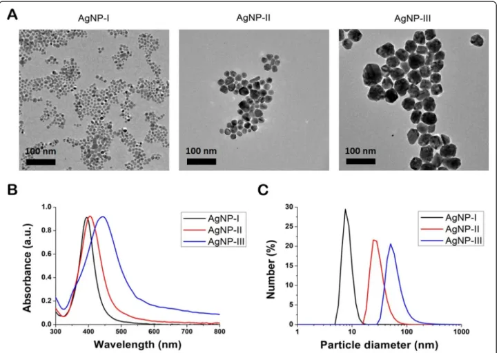
TEM images indicated that all three nanoparticle sam- ples contained quasi-spherical AgNPs (Fig.1a). Based on image analysis only minor polydispersity was observed and the average size of the AgNPs proved to be 7.0 ± 1.6 nm for AgNP-I, 21.4 ± 4.6 nm for AgNP-II and 50.3 ± 7.9 nm for AgNP-III, respectively. UV-Vis spectra of AgNPs showed absorption peak maxima at 395 nm in
case of AgNP-I, at 404 nm for AgNP-II and at 442 nm for AgNP-III, characteristic to surface plasmon reson- ance, which further supported the formation of metallic silver nanoparticles and confirmed the difference in average nanoparticle diameters between the three sam- ples (Fig.1b). According to DLS measurements (Fig.1c), the average hydrodynamic particle size of AgNP-I was between 5 and 12 nm diameter, that of AgNP-II was be- tween 15 and 50 nm, whereas the size of AgNP-III nano- particles was around 35–90 nm, providing further evidence of the nanoparticle size difference in the ob- tained three silver colloid solutions.
Antifungal activity of the differently sized AgNPs
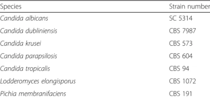
The potential toxic effect of AgNPs with different sizes was tested againstCandida,LodderomycesandPichiaspe- cies (Table1). We found that all three AgNP specimens inhibited the growth of the examined strains but in a dif- ferent extent. AgNP-I proved to be the most effective one, while AgNP-III exerted the least toxic activity. Pichia membranifaciens CBS 191 was the most susceptible for
Fig. 1Physicochemical properties of citrate-stabilized silver nanoparticles of three different sizes. Transmission electron microscopic images (a) and UV-Vis spectra (b) of the synthetized, differently sized AgNPs. Size distribution of the nanoparticles determined by dynamic light scattering measurements (c)
the AgNPs as the lowest minimal inhibitory concentra- tions (MIC) were detected for this strain (Table2). Unfor- tunately, in many cases the minimal inhibitory concentrations could not be established (Table3) because of the low concentrations of the AgNP stock solutions.
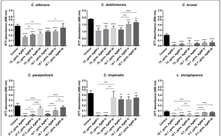
The effect of AgNPs on biofilm formation and destruction In order to examine whether AgNPs are capable of modu- lating the propensity of dimorphic yeasts to build biofilms, secondly, to test whether the degree of such a modulatory action would depend on the nanoparticle diameter, we treated Candida and Lodderomyces cells with differently sized AgNPs in two concentrations and detected the via- bility of fungal cells within the biofilms by 2,3-bis-(2- methoxy-4-nitro-5-sulfophenyl)-2H-tetrazolium-5-car- boxanilide (XTT) assay. The higher dose corresponded to 75μg/mL in case ofC. albicans, C. dubliniensis, C. krusei, C. tropicalis, 37.5μg/mL forC. parapsilosisand 18.75μg/
mL for Lodderomyces sp., respectively. The lower dose was generally set as 18.75μg/mL, except forLodderomyces elongisporus,where it was set to 9.3μg/mL.
AgNPs exerted inhibitory effect on the biofilm forma- tion of every examined Candida and Lodderomyces strain. In most cases the observed repression on biofilm development was dose- and size-dependent, and gener- ally, nanoparticles with the smallest average diameter (AgNP-I) hampered the biofilm formation more effi- ciently than AgNP-II or AgNP-III solutions (Fig. 2).
Moreover, treatments of the biofilms with AgNPs in
higher concentrations could enhance their inhibitory ef- fect on biofilm development (Fig.2).Pichia membranifa- ciens CBS 191 cells did not adhere to the surface of the microplates, and did not develop a biofilm suitable for examination.
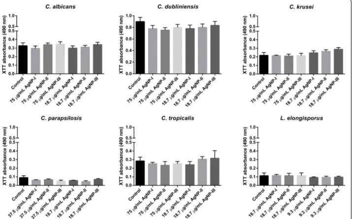
As we found that AgNPs inhibited biofilm formation quite efficiently, next we examined whether AgNPs were capable to destroy already existing fungal biofilms as well. The biofilm degradation ability of differently sized AgNPs was screened using matured biofilms ofCandida and Lodderomyces species. Silver nanoparticles were ap- plied on biofilms in two concentrations (the same high and low doses were used as upon biofilm formation ex- periments). Our results indicated that significant biofilm degradation could not be achieved using any of these citrate-stabilized AgNPs. Smaller nanoparticles applied in high concentration (AgNP-I, 75μg/mL) were just as non-effective on the stable fungal biofilm as large AgNPs (Fig.3).
Morphology of AgNP-treated strains
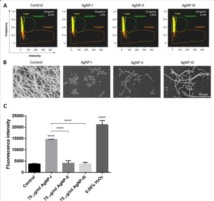
Since the generation of elongated cells by yeast-to-hypha conversion is a prerequisite of biofilm formation by most dimorphic yeasts, the shape of C. albicans (Fig.4) cells treated with differently sized AgNPs were analyzed first by flow cytometry, then with scanning electron micros- copy (SEM). Using flow cytometry, the almost spherical yeasts can be discriminated from the elongated hypha in the supernatant of the sample, thus the differing mor- phological states of potentially pathogenic species can be quantified in suspension. By SEM on the other hand, the morphology of surface attached individual cells or cells within the biofilm can be visualized. The percentage of elongated C. albicans hyphae in the suspension of the untreated control was 10.2%, whereas in the supernatant of AgNP-I-treated samples only 2.4% of the cells were in hypha form. We quantified 5.05% elongated cells in the AgNP-II-, and 11.3% in the AgNP-III-treated sample su- pernatants by flow cytometry (Fig.4a). These results in- dicate that small sized AgNPs (AgNP-I) were highly effective in suppressing the morphological conversion of dimorphic yeasts. On the other hand, the largest AgNPs Table 1List of the tested strains
Species Strain number
Candida albicans SC 5314
Candida dubliniensis CBS 7987
Candida krusei CBS 573
Candida parapsilosis CBS 604
Candida tropicalis CBS 94
Lodderomyces elongisporus CBS 1072
Pichia membranifaciens CBS 191
CBSCentraalbureau voor Schimmelcultures,SCSquibb Institute for Medical Research, New Brunswick, New Jersey, USA
Table 2Minimal inhibitory concentrations of the synthesized AgNPs against yeasts with pathogenic potential
Species AgNP-I (μg/mL) AgNP-II (μg/mL) AgNP-III (μg/mL)
Candida albicansSC 5314 > 75 > 75 > 75
Candida kruseiCBS 573 > 75 > 75 > 75
Candida parapsilosisCBS 604 37.5 > 75 > 75
Candida tropicalisCBS 94 > 75 > 75 > 75
Candida dubliniensisCBS 7987 > 75 > 75 > 75
Lodderomyces elongisporusCBS 1072 37.5 18.75 37.5
Pichia membranifaciensCBS 191 4.6875 18.75 37.5
(of approx. 50 nm diameter) had no effect on yeast-to- hypha transformation, asC. albicanscultures exposed to AgNP-III contained comparable amounts of elongated hypha forms in the supernatant as the untreated control.
SEM images taken of AgNP-treated and untreated C.
albicanscells (Fig.4b) confirmed the morphological dif- ferences determined by flow cytometry. Mostly yeast forms were found in the AgNP-I-exposed samples, how- ever, shorter or longer hyphae appeared in samples treated
with AgNP-II and AgNP-III (representative images are shown in Fig. 4b). SEM images revealed massive biofilm formation in untreated C. albicanssamples, where almost exclusively elongated hyphae were present. These results suggest that small-sized silver nanoparticles (AgNP-I) ex- hibit the highest potential to suppress yeast-to-hypha mor- phological switch of the opportunistic pathogenic yeast strains. Moreover, our data indicate that with increasing nanoparticle diameters the capacity of AgNPs to inhibit the morphological switch of dimorphic fungi decreases substantially.
Since the inhibition of morphological switch can be the consequence of oxidative stress induced by AgNP treatments, we investigated the reactive oxygen species (ROS) generating potential of differently sized AgNPs in C. albicans cultures. Cells were treated with AgNP-I, AgNP-II and AgNP-III, respectively, and were stained with 2′,7′–dichlorofluorescein diacetate (DCFDA). 2′, 7′–dichlorofluorescein (DCF) fluorescence intensity values show that small-sized AgNPs (AgNP-I) induce significant ROS production compared to untreated con- trol samples, however, medium-sized AgNPs are less
Fig. 2Inhibitory effect of differently sized AgNPs on biofilm formation of opportunistic pathogenic yeasts. Inhibitory effect of AgNPs on the formation of biofilms was evaluated by determining the viability of the variousCandidaandLodderomycescells in the biofilm using XTT assay after 72-h treatments with AgNP-I, AgNP-II or AgNP-III in the indicated concentrations. Suspensions without AgNPs were used as growth control.
The values represent the mean ± standard deviation calculated from three independent experiments (*,p≤0.05, **,p≤0.01, ***,p≤0.001, ****, p≤0.0001, unpairedttest)
Table 3The inhibition rate (%) in the presence of differently sized AgNPs applied in 75μg/mL concentration
Species AgNP-I AgNP-II AgNP-III
Candida albicansSC 5314 75 50 25
Candida kruseiCBS 573 90 90 10
Candida parapsilosisCBS 604 100 90 65
Candida tropicalisCBS 94 90 40 40
Candida dubliniensisCBS 7987 60 20 0
Lodderomyces elongisporusCBS 1072 100 100 100 Pichia membranifaciensCBS 191 100 100 100
potent and AgNP-III is a very weak inducer of ROS gen- eration (Fig.4c). Hence, the different degree of oxidative stress triggered by differently sized AgNPs can form the molecular basis for the morphology switch inhibiting po- tential of AgNPs inC. albicanscells.
Efficiency of AgNPs to modulate the morphological switch of opportunistic pathogenic yeasts on biotic surfaces The effects of differently sized AgNPs on yeast-to-hypha switch of opportunistic pathogenic yeasts were then deter- mined in a co-culture system, consisting of human keratino- cyte cells and dimorphic yeasts, generated with the aim to model fungal infection on biotic surfaces, like human skin.
First, the toxicity of the synthesized silver colloids was assessed on human HaCaT keratinocytes by 3-(4,5-di- methylthiazol-2-yl)-2,5 diphenyl tetrazolium bromide (MTT) viability assay. As expected, AgNPs decreased cell viability in size-, and concentration-dependent man- ner. The calculated half maximal inhibitory concentra- tion (IC50) values indicated that the strongest toxicity on human keratinocytes was achieved upon small nanopar- ticle (AgNP-I) exposures (IC50= 8.02 ± 1.7μg/mL), while lower extent of cell death was induced by medium-sized AgNPs (AgNP-II, IC50= 42.6 ± 18.3μg/mL). The smallest
decrease in viability was detected following AgNP-III nanoparticle treatments (IC50= 91.3 ± 6.1μg/mL).
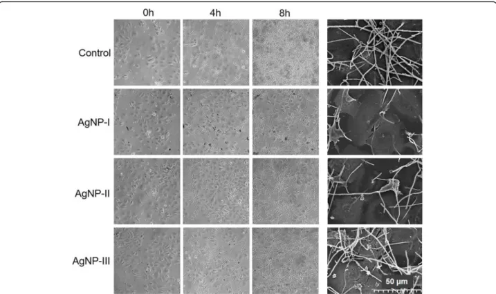
Then, co-cultures of HaCaT human keratinocytes and C. albicans cells (in 1:5 human/yeast cell ratio) were established, and exposed to differently sized AgNPs in 75μg/mL concentration for 8 h. During treatment hypha formation of Candida cells was followed microscopic- ally. C. albicans cells were present in filamentous form in the untreated co-cultures, and after 8-h incubation a thick biofilm could be observed over and around kerati- nocytes (Fig. 5). AgNP treatments clearly reduced hypha formation of C. albicans, since less elongated cells were found in AgNP-exposed cultures after 4 and 8 h incuba- tion, compared to the corresponding untreated control.
Importantly, inhibition of the yeast-to-hypha switch in the co-culture proved to be nanoparticle size-dependent.
We noticed that AgNP-I administration attenuated hypha formation as well as the biofilm generation of C.
albicans with the highest degree on the surface of the keratinocyte layer. This inhibitory effect was smaller when AgNP-II was applied, and resulted to be the least pronounced upon AgNP-III administration (Fig.5). SEM images taken from AgNP-treatedC. albicansand HaCaT keratinocyte cells in the co-culture supported the above
Fig. 3Effect of differently sized AgNPs on biofilm degradation. To determine the ability of AgNPs to destroy existing biofilms, three-day old matured biofilms ofCandidaandLodderomycesstrains were treated for 72 h with differently sized AgNPs in the indicated concentrations and the viability of the cells was assessed by XTT assay. The values represent the mean ± standard deviation calculated from three
independent experiments
described morphological differences (Fig.5). It is import- ant to note that throughout the co-culture experiments, also upon AgNP treatments (8-h incubation of the co- cultures with AgNPs was monitored microscopically) keratinocyte morphology, the integrity of HaCaT cell layer as well as cell-cell contacts were not compromised.
All these results indicate that the morphological switch of dimorphic, potentially pathogenic fungal cells can be
attenuated by AgNPs not only on abiotic, but also on bi- otic surfaces such as skin, and smaller particle diameters are more advantageous to suppress the formation of virulent hyphae.
Discussion
Morphological plasticity, such as the capability to undergo a yeast-to-hypha transformation, contributes
Fig. 4Morphology of control and AgNP-treatedC. albicanscells. Cell morphology of untreated and AgNP-exposedC. albicanswas detected by flow cytometry after 72-h incubation in the presence of differently sized AgNPs in 75μg/mL concentration. The percentage of aggregated, yeast- shaped and elongated cells were quantified by IDEAS 6.2 software (Amnis-EMD Millipore, Burlington, MA, USA) and the percentage of elongated cells is indicated on the figures (a). Morphology of the surface-anchored cells after 72-h incubation in the presence of AgNPs was analyzed using scanning electron microscopic images (b). In order to assess the degree of reactive oxygen species formation, mean DCF fluorescence intensity was determined on DCFDA-stained, AgNP-treatedC. albicanscells. Values represent the mean ± standard deviation calculated from three independent experiments (****,P< 0.0001, unpairedttest) (c)
significantly to the virulence of many opportunistic hu- man pathogenic yeasts. In response to the changing en- vironmental conditions within the host, planktonic yeast cells convert to filamentous hyphae, and the emerging elongated cells facilitate the pathogen penetration and potentiate host invasion [37]. Filament formation is also an indispensable factor for the generation of structured microbial communities called biofilm. Hyphae provide the structural integrity and the multilayered architecture for mature biofilms [38]. Biofilm-located cells express decreased sensitivity to antifungal compounds compli- cating the treatment of fungal diseases. Therefore, the inhibition of the morphological switch and thereby bio- film development could decrease the virulence of these pathogens and promotes the eradication of the infection.
The antiseptic application of silver goes back to an- cient times, however, the emergence of various antibi- otics confined its usage substantially [39]. In the last two decades, silver has been re-introduced to broad range medical applications in the nanoparticle formulation.
AgNPs perform well against numerous Gram-positive and Gram-negative bacteria, several viruses and patho- genic fungi as well. The mechanism of action behind the anti-pathogenic activity of AgNPs has not been eluci- dated in every detail. Nevertheless, it is generally ac- cepted that AgNPs exert their antimicrobial effects
through microbial membrane damage, caused by the at- tachment of AgNPs on the cell surface, which leads to structural as well as functional alterations in the mem- brane (membrane destabilization, formation of pores and cytoplasm leakage). This action is complemented by the release of Ag+ ions from the nanoparticle surface, which provokes the generation of reactive oxygen spe- cies [39]. The latter will ultimately induce sub-cellular structure damage by inactivation and denaturation of es- sential microbial macromolecules (proteins, enzymes, and nucleotides) and leads to apoptotic cell death [40].
Although innovative AgNP synthesis methods emerge daily, and the as-prepared nanomaterials are now rou- tinely tested for cytotoxic, antibacterial or antifungal ac- tivities [41–43], the scientific literature is scarce in reports describing the effects of AgNPs on yeast cell ultrastructure and biofilm development. In fact, only a few authors examined the AgNP-induced morphological changes of Candida albicans. They demonstrated that silver nanoparticles hamper C. albicans biofilm forma- tion in vitro via inhibition of filament formation and by destruction and of the cell wall integrity. Lara and co- workers applied extremely small, in average 1 nm sized AgNPs on C. albicans cells [38], whereas Muthamil et al. used relatively big (40–55 nm) AgNPs [44], and fi- nally, Jalal et al. utilized polydispersed biosynthesized
Fig. 5Size-dependent modulation of the morphological switch of opportunistic pathogenic yeasts co-cultured with keratinocytes by AgNPs.
Morphology ofC. albicanscells in co-culture with keratinocytes was monitored by light microscopy after 4 and 8 h, as well as by scanning electron microscopy after 8 h of AgNP-I, AgNP-II and AgNP-III treatments
AgNPs [45]. However, none of these studies investigated the size-dependent effect of the AgNPs on this species, nor tested the actions of AgNPs on dimorphic yeasts of other genus.
Therefore, in the present work we studied the size- dependent action of silver nanoparticles on the morpho- logical transition and subsequent biofilm formation of dif- ferent dimorphic yeast species. To achieve this aim citrate- coated AgNPs in three different sizes (AgNP-I: ~ 7.0 nm;
AgNP-II: ~ 21 nm; AgNP-III: ~ 50 nm) were successfully prepared by one of the most widely utilized chemical re- duction approaches [46]. The sizes of the different AgNP samples were verified by TEM and DLS measurements.
UV-Vis spectra of the prepared colloids also supported the synthesis of AgNPs based on previously reported spectral characteristics of particles with similar morphologies [47].
The antifungal activity test revealed that silver nanoparticles with the smallest size (AgNP-I) were the most effective against the examined opportunistic pathogenic species. C.
parapsilosisand rare pathogens such asL. elongisporusand P. membranifaciens proved to be the most sensitive to AgNP exposures [48].
Importantly, biofilm development of theCandidaspe- cies andL. elongisporuswas substantially inhibited in the presence of the nanoparticles, and in this respect again AgNP-I proved to be the most potent, as treatments with small nanoparticles led to the most significant de- cline in the viability of biofilm-located cells. The strong capacity of small AgNPs to inhibit biofilm formation correlated well with the attenuated hypha formation of AgNP-I-exposed dimorphic yeasts, demonstrated by FACS and SEM data. Most probably the formation of re- active radicals and the consequent oxidative stress in- duced by small AgNPs lay behind this outstanding potential of small-sized nanoparticles to hamper mor- phological switch in dimorphic yeasts.
The observed inhibitory feature of AgNPs on yeast-to- hypha morphological conversion holds great potential in nanomedicine, if this ability of AgNPs manifests on bio- logical surfaces. Therefore, we tested the capability of AgNPs to suppress the morphological transformation of C. albicansin a co-culture system with human keratino- cytes. As before, considerably decreased hypha formation was detected in the presence of AgNP-I compared to untreated control cultures. These results suggest that the capability of AgNPs, especially those with small di- ameters, to modulate the morphological transformation and thereby the virulence of dimorphic pathogenic yeasts is maintained on biotic surfaces, like skin and mu- cosa, which feature can be exploited further in biomed- ical applications. On the other hand, it is important to note, that in contrast to the findings by Lara et al. [38], in our study AgNPs could not disintegrate already devel- oped, matured biofilms, since we found that the biofilm
destruction ability of the differently sized, citrate-coated AgNPs was negligible. Our findings imply the prophylac- tic application of citrate-coated AgNPs in order to take advantage of their antifungal capacities against di- morphic yeasts.
Normally, a sequence of tightly regulated events leads to successful biofilm generation. Yeast cells colonize and ad- here to soft or hard surfaces mediated by specific adhesion factors. In case ofC. albicans, key regulators, like Bcr1, as well as its downstream targets such as cell wall proteins Als1, Als3 have been implicated as required elements for the early steps of the process [49]. Cell-cell communica- tion, especially quorum sensing, is also crucial at this stage to prevent overpopulation and competition for nutrients, and for the induction of hypha-formation-associated genes. After initial adherence and focal attachment of in- dividual cells, a basal layer is formed, which is then sub- jected to cell proliferation, early-stage filamentation, followed by the production of exopolymeric substance by sessile cell communities in the multilayered network regu- lated by factors like Rlm1 and Zap1 in C. albicans [49].
Exposure of dimorphic fungi to AgNPs and the produced reactive radicals might affect most of these events, never- theless, the precise mechanisms at the molecular level be- hind the inhibitory action of AgNPs on the morphological switch and biofilm formation of these opportunistic path- ogens is yet to be demonstrated. In fact, we have already started to establish a set of mutant Candida strains for testing which molecular event related to morphological conversion is perturbed by AgNP exposures or by the trig- gered oxidative stress.
Our results indicate that virulence factors of opportunistic pathogens, like morphological switch and biofilm formation can be efficiently modulated by the utilization of AgNPs.
However, it has to be stressed that in order to achieve the desired effects, nanomaterials have to be designed properly.
We found that size indeed matters when AgNPs are intended to act on dimorphic pathogenic yeasts. The per- formance of larger nanoparticles (AgNP-III) in impairing yeast-to-hypha conversion and biofilm generation was con- siderably worse than medium (AgNP-II) and small-sized (AgNP-I) counterparts, which phenomenon might be ex- plained by their different abilities in ROS production. Since AgNP size seems to have a defining role in determining and fine-tuning the biological activity of nanoparticles, hence, more careful considerations are required upon nanomaterial design and production and thorough screening regimens of the obtained nanomaterials are recommended to estimate their efficiencies.
Conclusions
Our data confirm that citrate-coated AgNPs are able to inhibit the morphological switch and biofilm formation of dimorphic yeasts, however, they are not effective
against fully developed biofilms. AgNPs with the smal- lest size performed especially well in all experiments fur- ther supporting the fact that particle size has significant impact on the biological activity of these nanomaterials.
Our results might influence potential strategies and op- portunities for the clinical translation of AgNP applica- tion in the field of medical mycology.
Methods
Synthesis of silver nanoparticles
All chemicals for the nanoparticle synthesis were ob- tained from Merck (Darmstadt, Germany).
Citrate-stabilized silver nanoparticles in three different sizes were prepared by chemical reduction using sodium borohydride, according to Wan et al. with slight adjust- ments [46]. Twenty mL of 1% w/v citrate solution and 75 mL of water were added in a beaker and the mixture was heated to 70 °C. Then, 2 mL of 1% w/v AgNO3solution were introduced to the mixture, followed by drop-wise addition of 2 mL of 0.1% w/v freshly prepared sodium bo- rohydride solution. The reaction solution was kept at 70 °C under vigorous stirring for 1 h and was then cooled to room temperature. The resulting AgNPs, denoted as AgNP-I, were used as starter seeds for AgNP-II.
To obtain larger AgNPs in a size-controlled manner, a seed-mediated growth approach was employed. For the synthesis of AgNP-II with an average diameter of 20 nm, 2 mL of 10% w/v citrate solution was mixed with 75 mL of water and brought to 80 °C. Next, 10 mL of starter seed solution (AgNP-I) were added, followed by the sup- plementation of 2 mL of 1% w/v AgNO3 solution while vigorous mechanical stirring for 2 h, then the resulting suspension was cooled to room temperature.
For the production of AgNP-III with an average size of 50 nm, the 20 nm AgNPs (AgNP-II), obtained in the pre- vious synthesis procedure, were used as seeds, and the above described growth procedure steps were repeated.
The final colloid samples were stored at 4 °C in all cases.
After synthesis, the nominal concentrations of the three AgNP samples were about 128μg/mL, 157μg/mL and 160μg/mL, respectively. For an easier handling throughout the experiments, we decided to set the con- centration of all three samples to 150μg/mL. Concen- trating AgNP-I was achieved by placing the sample in a drying oven on 40 °C overnight, then measuring its vol- ume and adding distilled water if necessary, to get a final volume of about 85 mL. The latter two samples after similar procedures were diluted to 93 and 95 mL final volume respectively, thus the standard 150μg/mL con- centration for all three samples was achieved. The calcu- lations (see supplementary material) show how the nominal concentrations could be assessed for each sam- ple and the V150values highlight the final volume of the
standardized AgNP samples that were used for the sub- sequent in vitro experiments.
Characterization of nanoparticles
The morphological characteristics of AgNPs were exam- ined by transmission electron microscopy applying a FEI Tecnai G2 20× microscope (FEI Corporate Headquar- ters, Hillsboro, OR, USA) at an acceleration voltage of 200 kV. The optical properties of nanoparticles were assessed by spectral analysis, where the absorbance spec- tra of nanoparticles were recorded using an Ocean Op- tics 355 DH-2000-BAL UV–Vis spectrophotometer (Halma PRC, Largo, FL, USA) within the 300–800 nm range. The size distribution of the nanoparticle samples was examined by DLS analysis using a Zetasizer Nano ZS Instrument (Malvern, Worcestershire, UK).
Antifungal activity
The antifungal susceptibility test was carried out using the three differently sized silver nanoparticles (AgNP-I, AgNP-II and AgNP-III) on each potentially pathogenic yeast species listed in Table 1. The minimal inhibitory concentration (MIC) was determined with the microdi- lution method in 96-well microplates. To 50μL of stan- dardized cell suspension (5 × 10⁴ cell/mL in Dulbecco’s Modified Eagle Medium (DMEM) supplemented with 1% fetal bovine serum (FBS) 50μL silver nanoparticle solution was added in serially two-fold-diluted concen- trations (in the range of 75 and 4.69μg/mL in the above described medium) and incubated for 48 h at 30 °C (20 °C in the case of Pichia membranifaciens). AllCan- didaandLodderomyces species grow well at 30 °C, how- ever, the growth of C. parapsilosis and Lodderomyces elongisporus is somewhat hampered at 37 °C. Thus, for this experiment 30 °C incubation temperature was ap- plied to establish the MIC values in vigorously growing cultures. The control samples contained 50μL cell sus- pension and 50μL medium without nanoparticles. The optical density of the cultures was detected at 620 nm in SPECTROstar Nano plate reader (BMG LabTech, Offen- burg, Germany). The experiments were carried out at least three times always in triplicates.
Effect of AgNPs on biofilm formation
The biofilm formation and the viability of the cells in the biofilm were tested after AgNP treatments. Briefly, suspensions from each strain in 5 × 10⁴ cell/mL density were prepared in DMEM medium supplemented with 1% FBS. Then, 50μL of the cell suspension were trans- ferred into wells of a microtiter plate and 50μl of either AgNP-I, AgNP-II or AgNP-III were added in 75 (high dose) or in 18.7μg/mL (lower dose) concentrations, re- spectively. In some cases where preliminary experimen- tal results indicated high susceptibility, the AgNP
concentrations corresponding to high dose and/or low dose were set differently. Therefore, 37.5 and 18.7μg/
mL were applied forC. parapsilosisand 18.7 and 9.3μg/
mL concentrations forL. elongisporus. Suspensions with- out AgNPs were used as growth control, while the medium without cells and without AgNPs was used as negative control. Plates were incubated for 72 h at 37 °C in 5% CO2 level, then the biofilms were washed twice with phosphate buffered saline (PBS) to remove slightly attached cells and 2,3-bis-(2-methoxy-4-nitro-5-sulfo- phenyl)-2H-tetrazolium-5-carboxanilide (XTT) reduc- tion assay was used to detect the viability of the biofilm- associated cells. XTT was prepared at 0.5 mg/mL con- centration in PBS supplemented with 1μM menadion.
After adding 100μL XTT solution to each well, the plates were incubated for 2 h at 37 °C in dark. There- after, 80μL of each supernatant were transferred to new 96-well plates and the absorbance of each solution was measured at 490 nm using SPECTROstar Nano plate reader (BMG LabTech, Offenburg, Germany). The ex- periments were carried out three times always in 8 bio- logical replicates.
Determination of biofilm degradation ability
The biofilm formation was initiated by inoculating 100–
100μL cell suspensions (prepared in DMEM medium supplemented with 1% FBS in 5 × 10⁴ cell/mL density) from each strain into the wells of flat-bottomed 96-well microtiter plates. Plates were incubated for 72 h at 37 °C in an atmosphere containing 5% CO2. After biofilm for- mation, the medium was removed and silver nanoparti- cle solutions (diluted in DMEM medium) were added to the biofilms in 75 or 18.7μg/mL final concentrations.
For C. parapsilosis, high dose corresponded to 37.5μg/
mL while low dose was 18.7μg/mL concentration and in case of L. elongisporus 18.7 and 9.3μg/mL concentra- tions were set as high and low dose. Then plates were further incubated for 72 h at 37 °C. A series of only DMEM-treated biofilms and biofilm-free wells contain- ing DMEM medium served as positive and negative con- trols. Viability of the cells was detected by XTT reduction assay, as described above. All the assays were carried out three times in 8 replicates.
Morphological characterization of fungal cells
C. albicans was selected for morphological analysis of cells treated with differently sized AgNPs. C. albicans cells were inoculated into DMEM medium supple- mented with 1% FBS in 5 × 10⁴ cell/mL concentration and were cultured for 24 h at 37 °C in the absence, as well as in the presence of either AgNP-I, AgNP-II or AgNP-III particles in 75 and 18.75μg/mL final concen- trations. The proportion of yeast cells and hyphae was determined from the sample supernatant by flow
cytometry (FlowSight®, Amnis-EMD Millipore, Burling- ton, MA, USA). Cells cultured without AgNPs were used as control.
Detection of reactive oxygen species
ROS production upon AgNP treatments was detected by DCFDA staining. C. albicans cells were inoculated into DMEM medium supplemented with 1% FBS in 5 × 10⁴ cell/mL concentration and were cultured for 24 h at 37 °C in the absence, as well as in the presence of either AgNP- I, AgNP-II or AgNP-III particles of 75μg/mL final con- centrations. Cells cultured with H2O2in 0.08% concentra- tion were used as positive control. After the treatments all cultures were incubated with DMEM containing 10μM DCFDA (Sigma-Aldrich) in the dark for 1 h. The total fluorescence intensity of the samples was measured by flow cytometry (FlowSight®, Amnis-EMD Millipore, Burlington, MA, USA). Measurements were repeated three times using 3 independent biological replicates.
Keratinocyte cell culture
HaCaT immortalized human keratinocyte cells were purchased from ATCC (Manassas, VA, USA) and main- tained in DMEM medium containing 4.5 g/L glucose (Sigma-Aldrich, Saint Louis, MO, USA), supplemented with 10% FBS, 2 mM L-glutamine, 0.01% streptomycin and 0.005% penicillin (Sigma-Aldrich, Saint Louis, MO, USA). Cells were cultured in a 37 °C incubator at 5%
CO2in 95% humidity.
Measurement of keratinocyte viability
HaCaT cell viability was measured after AgNP-I, AgNP- II and AgNP-III treatments. For this HaCaT cells were seeded into 96-well plates in 10,000 cells/well density, and treated on the following day with silver nanoparti- cles of three different sizes in increasing concentrations.
After 24-h treatments, HaCaT cells were washed with PBS and incubated for an hour at 37 °C with 0.5 mg/mL MTT reagent (SERVA, Heidelberg, Germany) diluted in culture medium. Formazan crystals were solubilized in DMSO (Sigma-Aldrich, Saint Louis, MO, USA) and ab- sorption was measured at 570 nm using a Synergy HTX plate reader (BioTek-Hungary, Budapest, Hungary). Ex- periments were performed at least three times using four independent biological replicates. IC50 values were ob- tained based on the results of MTT assays.
Co-culture system
HaCaT immortalized human keratinocyte cells were used in a co-culture system together with C. albicans cells in order to model fungal infection of human skin.
To investigate the hyphae formation of yeast cells within co-cultures upon AgNP treatments, HaCaT cells were seeded into 6-well plates and left to grow until they
reached confluence to provide a continuous, consistent biosurface. Then, when this biofilm layer was completely established, 5 × 10⁵ C. albicans cells were added to the wells, and the co-cultures were exposed to 75μg/mL AgNPs. The cultures were continuously monitored under the microscope, throughout the investigation period (maximum 8 h). Photographs were taken after 0;
4; and 8-h treatments using Nikon Eclipse TS100 inverted light microscope (Nikon, Minato, Japan).
Scanning electron microscopy
A.: The ultrastructure of the AgNP-treated and non- treatedC. albicansbiofilms formed on sterile glass coverslips (Sigma-Aldrich, Saint Louis, MO, USA) was examined by scanning electron microscopy.
Biofilm formation was carried out as described above in the relatedMethodssection. Cell suspen- sions were treated with either AgNP-I, AgNP-II or AgNP-III in 75μg/mL concentration for 72 h at 37 °C, then cells were washed with PBS and fixed in 2.5% glutaraldehyde at 4 °C overnight.
B.: SEM images were taken of untreated as well as of AgNP-treated HaCaT -C. albicansco-cultures. For this, HaCaT cells were seeded onto plastic coverslips (Sar- stedt, Nümbrecht, Germany) placed in 6-well plates.
On the following day 5 × 10⁵C. albicansyeast cells were added to each well and the co-cultures were treated with differently sized AgNPs in 75μg/mL concentration.
After 8 h of treatment, cells were washed with PBS and fixed in 2.5% glutaraldehyde at 4 °C overnight.
All samples (prepared as described in part A or B) were dehydrated by increasing percentage of ethanol di- luted in water (50, 70, 80, 90, 95, 98, 100%, for 15 min each), followed by a series of tert-butanol:ethanol mix- ture (1:2, 1:1, 2:1 volume ratio) at room temperature.
Then the cells were incubated with absolute tert-butanol overnight at 4 °C and were finally lyophilized. The cover- slips were mounted on specimen stubs with electrically conductive adhesive tape and the samples were covered with a thin metal palladium-gold layer. SEM imaging was performed by a Hitachi S4700 electron microscope (AuroScience, Budapest, Hungary) using 10 kV acceler- ating voltage and 10μA emission current.
Supplementary information
Supplementary informationaccompanies this paper athttps://doi.org/10.
1186/s12866-020-01858-9.
Additional file 1.
Abbreviations
AgNPs:Silver nanoparticles; CBS: Centraalbureau voor Schimmelcultures;
DCF: 2′,7′–dichlorofluorescein; DCFDA: 2′,7′–dichlorofluorescein diacetate;
DLS: Dynamic light scattering; DMEM: Dulbecco’s Modified Eagle Medium;
FBS: Fetal bovine serum; IC50: Half maximal inhibitory concentration;
MIC: Minimal inhibitory concentration; MTT: 3-(4,5-dimethylthiazol-2-yl)-2,5 diphenyl tetrazolium bromide; PBS: Phosphate buffered saline; ROS: Reactive oxygen species; SC: Squibb Institute for Medical Research; SEM: Scanning electron microscopy; TEM: Transmission electron microscopy; UV- Vis: Ultraviolet–visible; XTT: 2,3-bis-(2-methoxy-4-nitro-5-sulfophenyl)-2H- tetrazolium-5-carboxanilide
Acknowledgments Not applicable.
Authors’contributions
BS was responsible for microbial strains and performed the basic antimicrobial screens, NI maintained and treated human keratinocytes and carried out the cytotoxicity assays, performed co-culture studies; ÁT and ZP performed biofilm inhibition and destruction experiments, AR and PB pre- pared and characterized the nanoparticles. DM made scanning electron microscopic images, CP performed flow cytometry experiments. IM prepared samples for scanning electron microscopy, executed the statistical analyses, prepared the figures. CV made substantial contribution to the interpretation of the data and was revising the manuscript; ZK characterized the nanoparti- cles, coordinated the related research activities, drafted the manuscript. IP and MK designed the project, coordinated the studies, contributed substan- tially to the interpretation of the data and drafted the manuscript. All authors read and approved the final manuscript.
Funding
This research was supported by the Hungarian Government and the European Union within the frames of the Széchenyi 2020 Programme through grants GINOP-2.3.2-15-2016-00035 and GINOP-2.3.2-15-2016-00038 and UNKP-18-3-I-SZTE-42 (N.I.). The funding bodies provided financial support to purchase the chemicals required for the project, however, had no influ- ence on the study design, on the collection, analysis and interpretation of the data nor had a role in writing the manuscript.
Availability of data and materials
All original data of this manuscript are available from the corresponding author on reasonable request.
Ethics approval and consent to participate Not applicable.
Consent for publication Not applicable.
Competing interests
The authors report no conflicts of interest in this work.
Author details
1Department of Microbiology, University of Szeged, Szeged, Hungary.
2Department of Biochemistry and Molecular Biology, Faculty of Science and Informatics, University of Szeged, Közép fasor 52, Szeged H-6726, Hungary.
3Department of Applied and Environmental Chemistry, University of Szeged, Szeged, Hungary.4HAS-USZ Reaction Kinetics and Surface Chemistry Research Group, Szeged, Hungary.
Received: 25 June 2019 Accepted: 16 June 2020
References
1. Brown GD, Denning DW, Levitz SM. Tackling human fungal infections.
Science. 2012;336(6082):647.
2. Badiee P, Hashemizadeh Z. Opportunistic invasive fungal infections:
diagnosis & clinical management. Indian J Med Res. 2014;139(2):195–204.
3. Morrell M, Fraser VJ, Kollef MH. Delaying the empiric treatment of Candida bloodstream infection until positive blood culture results are obtained: a potential risk factor for hospital mortality. Antimicrob Agents Chemother.
2005;49(9):3640–5.
4. Bongomin F, Gago S, Oladele R, Denning D. Global and multi-national prevalence of fungal diseases—estimate precision. J Fungi. 2017;3(4):57.
5. Vallabhaneni S, Mody RK, Walker T, Chiller T. The global burden of fungal diseases. Infect Dis Clin North Am. 2016;30(1):1–11.
6. Hazen KC. New and emerging yeast pathogens. Clin Microbiol Rev. 1995;
8(4):462–78.
7. Bhally HS, Jain S, Shields C, Halsey N, Cristofalo E, Merz WG. Infection in a neonate caused byPichia fabianii: importance of molecular identification.
Med Mycol. 2006;44(2):185–7.
8. Paula C, Krebs V, Auler M, Ruiz L, Matsumoto F, Silva E, et al. Nosocomial infection in newborns byPichia anomalain a Brazilian intensive care unit.
Med Mycol. 2006;44(5):479–84.
9. Adler A, Hidalgo-Grass C, Boekhout T, Theelen B, Sionov E, Polacheck I.
Pichia farinosabloodstream infection in a lymphoma patient. J Clin Microbiol. 2007;45(10):3456–8.
10. de Almeida JN, Hennequin C. Invasive trichosporon infection: a systematic review on a re-emerging fungal pathogen. Front Microbiol. 2016;7:1629.
11. Al-Obaid K, Ahmad S, Joseph L, Khan Z.Lodderomyces elongisporus: a bloodstream pathogen of greater clinical significance. New Microbes New Infect. 2018;26:20–4.
12. Wirth F, Goldani LZ. Epidemiology of Rhodotorula: an emerging pathogen.
Interdiscip Perspect Infect Dis. 2012;2012:465717.
13. Brand A. Hyphal growth in human fungal pathogens and its role in virulence. Int J Microbiol. 2012:517529.
14. Jiang C, Li Z, Zhang L, Tian Y, Dong D, Peng Y. Significance of hyphae formation in virulence ofCandida tropicalisand transcriptomic analysis of hyphal cells. Microbiol Res. 2016;192:65–72.
15. Goyal S, Castrillón-Betancur JC, Klaile E, Slevogt H. The interaction of human pathogenic fungi with C-type lectin receptors. Front Immunol. 2018;9:1261.
16. Dalle F, Wächtler B, L’Ollivier C, Holland G, Bannert N, Wilson D, et al.
Cellular interactions ofCandida albicanswith human oral epithelial cells and enterocytes. Cell Microbiol. 2010;12(2):248–71.
17. Kojic EM, Darouiche RO. Candida infections of medical devices. Clin Microbiol Rev. 2004;17(2):255–67.
18. Silva S, Negri M, Henriques M, Oliveira R, Williams DW, Azeredo J.
Adherence and biofilm formation of non-Candida albicansCandida species.
Trends Microbiol. 2011;19(5):241–7.
19. Silva S, Rodrigues C, Araújo D, Rodrigues M, Henriques M. Candida species biofilms’antifungal resistance. J Fungi. 2017;3(1):8.
20. Tran QH, Nguyen VQ, Le AT. Silver nanoparticles: synthesis, properties, toxicology, applications and perspectives. Adv Nat Sci Nanosci Nanotechnol.
2013;4:033001.
21. Mohanpuria P, Rana NK, Yadav SK. Biosynthesis of nanoparticles:
technological concepts and future applications. J Nanopart Res. 2008;10(3):
507–17.
22. Ivask A, Elbadawy A, Kaweeteerawat C, Boren D, Fischer H, Ji Z, et al.
Toxicity mechanisms inEscherichia colivary for silver nanoparticles and differ from ionic silver. ACS Nano. 2014;8(1):374–86.
23. Rai M, Yadav A, Gade A. Silver nanoparticles as a new generation of antimicrobials. Biotechnol Adv. 2009;27(1):76–83.
24. Seil JT, Webster TJ. Antimicrobial applications of nanotechnology: methods and literature. Int J Nanomedicine. 2012;7:2767–81.
25. Flores-López LZ, Espinoza-Gómez H, Somanathan R. Silver nanoparticles:
electron transfer, reactive oxygen species, oxidative stress, beneficial and toxicological effects. Mini review. J Appl Toxicol. 2019;39(1):16–26.
26. Kovács D, Igaz N, Keskeny C, Bélteky P, Tóth T, Gáspár R, et al. Silver nanoparticles defeat p53-positive and p53-negative osteosarcoma cells by triggering mitochondrial stress and apoptosis. Sci Rep. 2016;6:27902.
27. Sharma VK, Yngard RA, Lin Y. Silver nanoparticles: green synthesis and their antimicrobial activities. Adv Colloid Interf Sci. 2009;145(1-2):83–96.
28. Narayanan KB, Sakthivel N. Green synthesis of biogenic metal nanoparticles by terrestrial and aquatic phototrophic and heterotrophic eukaryotes and biocompatible agents. Adv Colloid Interf Sci. 2011;169(2):59–79.
29. Boroumand Moghaddam A, Azizi S, Namvar F, Moniri M, Mohamad R, Md.
Tahir P. Nanoparticles biosynthesized by fungi and yeast: a review of their preparation, properties, and medical applications. Molecules. 2015;20(9):
16540–65.
30. Rónavári A, Kovács D, Igaz N, Vágvölgyi C, Boros IM, Kónya Z, et al.
Biological activity of green-synthesized silver nanoparticles depends on the applied natural extracts: a comprehensive study. Int J Nanomedicine. 2017;
12:871–883.
31. Rónavári A, Igaz N, Gopisetty MK, Szerencsés B, Kovács D, Papp C, et al.
Biosynthesized silver and gold nanoparticles are potent antimycotics against
opportunistic pathogenic yeasts and dermatophytes. Int J Nanomedicine.
2018;13:695–703.
32. Liu W, Wu Y, Wang C, Li HC, Wang T, Liao CY, et al. Impact of silver nanoparticles on human cells: effect of particle size. Nanotoxicology. 2010;
4(3):319–30.
33. Scherer MD, Sposito JCV, Falco WF, Grisolia AB, Andrade LHC, Lima SM, et al. Cytotoxic and genotoxic effects of silver nanoparticles on meristematic cells ofAllium ceparoots: A close analysis of particle size dependence. Sci Total Environ. 2019;660:459–67.
34. Wu M, Guo H, Liu L, Liu Y, Xie L. Size-dependent cellular uptake and localization profiles of silver nanoparticles. Int J Nanomedicine. 2019;14:
4247–59.
35. Choi O, Hu Z. Size dependent and reactive oxygen species related nanosilver toxicity to nitrifying bacteria. Environ Sci Technol. 2008;42(12):
4583–8.
36. Gliga AR, Skoglund S, Odnevall Wallinder I, Fadeel B, Karlsson HL. Size- dependent cytotoxicity of silver nanoparticles in human lung cells: the role of cellular uptake, agglomeration and Ag release. Part Fibre Toxicol. 2014;11:
11.
37. da Silva Dantas A, Lee KK, Raziunaite I, Schaefer K, Wagener J, Yadav B, et al.
Cell biology ofCandida albicans–host interactions. Curr Opin Microbiol.
2016;34:111–8.
38. Lara HH, Romero-Urbina DG, Pierce C, Lopez-Ribot JL, Arellano-Jiménez MJ, Jose-Yacaman M. Effect of silver nanoparticles onCandida albicansbiofilms:
an ultrastructural study. J Nanobiotechnology. 2015;13:91.
39. Maillard JY, Hartemann P. Silver as an antimicrobial: facts and gaps in knowledge. Crit Rev Microbiol. 2013;39(4):373–83.
40. Burdușel A-C, Gherasim O, Grumezescu AM, MogoantăL, Ficai A, Andronescu E. Biomedical applications of silver nanoparticles: an up-to-date overview. Nanomaterials. 2018;8(9):681.
41. Gurunathan S. Rapid biological synthesis of silver nanoparticles and their enhanced antibacterial effects againstEscherichia fergusoniiand Streptococcus mutans. Arab J Chem. 2019;12(2):168–80.
42. Hamouda RA, Hussein MH, Abo-elmagd RA, Bawazir SS. Synthesis and biological characterization of silver nanoparticles derived from the cyanobacteriumOscillatoria limnetica. Sci Rep. 2019;9(1):13071.
43. Korshed P, Li L, Liu Z, Mironov A, Wang T. Size-dependent antibacterial activity for laser-generated silver nanoparticles. J Interdiscipl Nanomed.
2019;4(1):24–33.
44. Muthamil S, Devi VA, Balasubramaniam B, Balamurugan K, Pandian SK.
Green synthesized silver nanoparticles demonstrating enhanced in vitro and in vivo antibiofilm activity againstCandida spp. J Basic Microbiol. 2018;58(4):
343–57.
45. Jalal M, Ansari MA, Alzohairy MA, Ali SG, Khan HM, Almatroudi A, et al.
Anticandidal activity of biosynthesized silver nanoparticles: effect on growth, cell morphology, and key virulence attributes of Candida species.
Int J Nanomedicine. 2019;14:4667–79.
46. Wan Y, Guo Z, Jiang X, Fang K, Lu X, Zhang Y, et al. Quasi-spherical silver nanoparticles: aqueous synthesis and size control by the seed-mediated Lee-Meisel method. J Colloid Interface Sci. 2013;394:263–8.
47. Huang T, Xu XHN. Synthesis and characterization of tunable rainbow colored colloidal silver nanoparticles using single-nanoparticle plasmonic microscopy and spectroscopy. J Mater Chem. 2010;20(44):9867–76.
48. Cendejas-Bueno E, Gomez-Lopez A, Mellado E, Rodriguez-Tudela JL, Cuenca-Estrella M. Identification of pathogenic rare yeast species in clinical samples: comparison between phenotypical and molecular methods. J Clin Microbiol. 2010;48(5):1895–9.
49. Gulati M, Nobile CJ.Candida albicansbiofilms: development, regulation, and molecular mechanisms. Microbes Infect. 2016;18(5):310–21.
Publisher’s Note
Springer Nature remains neutral with regard to jurisdictional claims in published maps and institutional affiliations.