III./8.3.: Diagnosis of spinal disorders
The spinal cord is located in the vertebral canal – in a bony structure, which makes the diagnosis of spinal disorders difficult.
Key words: X-ray, spinal CT, myelo-CT, spinal MRI
Among the traditional imaging methods, X-ray was the only diagnostic help for about a 100 years. The bony parts of the vertebral column are well seen by X-ray, but the spinal cord and nerve roots are not
visualized.
Degenerative diseases of the vertebral column, such as scoliosis, kyphosis, spondylosis, osteoporosis, spondylolysthesis, or fractures and dislocations due to trauma can be diagnosed by X-ray. Some vertebral tumors may also be seen.
Note: X-ray gives indirect information about the nervous system
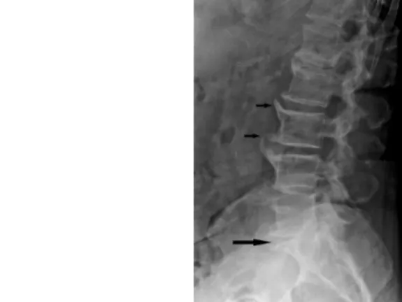
However, X-ray gives only indirect information about the other structures of the nervous system. For example, an intervertebral discal herniation, a common disorder, is suggested only by the decrease of the gap between the two adjoining vertebrae, the herniation itself is not visible.
Fig. 1: Discal herniation suggested only by the decrease of the gap between the two adjoining vertebrae on X-ray image
Myelography – a special X-ray method
When the herniation is small, but its location is critical (e.g.
intraforaminal), the gap remains normal and X-ray appears to be negative, despite marked symptoms.
A special X-ray technique, myelography was later developed, which is able the visualize the intraspinal space. A contrast material is injected via a lumbar puncture into the cerebrospinal fluid, which outlines the vertebral canal and the nerve roots. Malformations, such as tumors or discal herniation are seen as areas outlined by the contrast material on myelography. The method can be combined with classic tomography.
Fig. 2: Discal herniation is seen as a negative sign on myelography
Angiography is a rarely used method in the diagnosis of spinal disorders. It is mainly used in the diagnosis of spinal arterio-venous malformations, or to detect the source of spinal bleeding.
Advantages and disadvantages of spinal CT
The ability of CT to differentiate between tissues is better than that of X-ray; it is useful to study the bones and intervertebral discs. CT is able to make images only in the axial plain; image reconstruction is possible in other planes, however with decreased quality. Furthermore, only a few spinal segments can be examined in one session, thus the clinician must precisely indicate the region of interest for a CT examination.
It has limited value in the diagnosis of spinal cord lesions. The sensitivity of CT may be enhanced by using a contrast material (myelo-CT).
CT is widely used in emergency medicine (traumatology) and after vertebral colon surgeries for follow-up.
MRI: the ideal choice The main advantage of MRI is that it does not have any harmful effect on living organisms. Long segments of the spine, or even the whole spinal column can be examined by MRI, and in different plains with the same quality. MRI provides high-resolution images of soft tissues, the spinal cord and intramedullary disorders, and the nerve roots.
Skeletal structures are not a source of disturbing artifacts on MRI. One disadvantage of MRI is that compact bones are not depicted well due to their low signal intensity. Nonetheless, MRI is the imaging
technique of first choice in spinal disorders.
Fig. 3: Cauda compression due to a ruptured herniated disc on MRI
Postoperative MRI
Even modern, MRI-compatible metal implants can cause disturbing artifacts on MRI, therefore CT scans are recommended for
postoperative follow-up.
Contraindications of MRI
CT+MRI If MRI is contraindicated (pacemaker, non-compatible metal implant, severe obesity, claustrophobia), CT or myelography are alternative methods.
In special cases, both CT and MRI are required for the final diagnosis.
Evaluation of spinal function
Evoked potentials are methods that provide information about the function of the spinal cord and peripheral nerves. They have an important role in the process of establishing the indication of a neurosurgical intervention in clinically dubious cases. Somatosensory evoked potential (SEP) examines the sensory, while motor evoked potential (MEP) by transcranial magnetic stimulation examines the motor system.