III./8.4.1.: Developmental malformations of the spine and the spinal cord
This chapter summarizes the malformations of the spine and the spinal cord.
Key words: Chiari malformation, neural tube, syrinx Chiari (Arnold-Chiari) malformation
space-occupying defect in the foramen magnum with syrinx in the cervical spine
Arnold-Chiari malformation is a developmental malformation of the craniospinal region. The cerebellar tonsils alone (Chiari I) or in combination with the medulla oblongata (Chiari II) herniate into the foramen magnum, which has a space-occupying effect. Arnold-Chiari malformation is often associated with hydrocephalus (enlargement of the ventricles), syringomyelia (a cavity formed within the spinal cord), and meningocele (a neural tube defect) due to the disturbance of circulation of the cerebrospinal fluid.
The aim of surgical intervention is to eliminate the space-occupying effect and to restore normal circulation of the cerebrospinal fluid by the removal of the cerebellar tonsils or of the bone at the base of the skull (replaced by a dural graft). If syringomyelia developed, removal of the tonsils or surgical decompression at the base of the skull is usually sufficient to halt progression. If it is insufficient, a shunt may be placed connecting the cavity of the syrinx and the subarachnoid space (syringosubarachnoid shunt).
Fig. 4: Arnold-Chiari malformation with syrinx formation on MRI
Meningocele, meningomyelocele
Neural tube closing defect
Developmental malformations of the spinal cord, the meninges or the bony spine are the consequence of neural tube closure defects. The most common form is spina bifida (open spine due to a vertebral defect). It is called meningocele if the meninges herniate through the vertebral defect, or meningomyelocele if the sac-like bulge contains neural tissues as well. Spina bifida in itself does not require surgical intervention, but surgery is often necessary after birth if meningocele or meningomyelocele is present. The skin is usually thin above the lesion, which leads to ulceration and infection. These malformations are often associated with hydrocephalus, which requires the placement of a shunt.
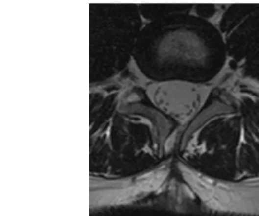
Fig. 5: Spina bifida and meningocele at the level of L4 vertebra on MRI
Syringomyelia, syringobulbia
Cavity within the spinal cord
Syringomyelia and syringobulbia refer to the formation of an
abnormal cavity within the spinal cord or the medulla oblongata. The cavity usually communicates with the central canal, however, it has no inner ependymal layer. Caused by the pressure of the cavity, the main symptom of syringomyelia is dissociated sensory disturbance at the lower cervical region: loss of pain, temperature and touch sensation on the hands, with preserved proprioceptive sensation. Atrophy and weakness of small hand muscles eventually follow.
Basilar impression
Basilar impression is the invagination of the upper cervical spine into the posterior fossa. The axis of the dens is located higher than normal, resulting in brainstem compression. Progressive symptoms may require surgical intervention (resection of the apex of the dens via a transoral exploration, or bone removal and decompression at the base of the skull).
Klippel-Feil syndrome
Klippel-Feil syndrome is characterized by the congenital fusion of cervical vertebrae due to an inappropriate segmentation of the cervical spine. It may be combined with other spinal developmental
malformations, such as basilar impression or atlanto-occipital fusion.
Narrow spinal canal syndrome
The congenital narrowness of the spinal canal occurs mainly at the lumbar and the cervical part of the vertebral column. In patients with congenital narrow spinal canal, space-occupying lesions cause spinal cord or nerve root compression earlier than normally.
Tethered cord syndrome
Tethered cord syndrome refers to the condition where the spinal cord is pulled down by the filum terminale below the level of L2 vertebra.
It is often associated with intradural lipoma or meningomyelocele. Its first symptoms are typically pain, weakness of both legs, gait disorder,
The spinal cord is tethered to the spinal canal.
and autonomic dysfunction. It is treated by the surgery of the filum terminale.
Fig. 6: Sacral fistula and tethered cord on MRI
Split cord malformation
The split cord malformation has several subtypes. In type I
(diastematomyelia), the spinal cord is split in the midline and the two halves are found in separate dural sacs. At the level of the
hemisection, vertebral malformations (absence of cartilage,
hyperostosis) may be seen. In type II (diplomyelia), the hemicords are separated by a fibrous median septum within a single dural sac.