III./8.4.7 Spinal tumors
Introduction
Twenty percent of all deaths are caused by tumors; 2% of tumor related deaths are due to primary tumors of the central nervous system.
Eighty-five percent of CNS tumors are intracranial and 15% are spinal.
The classification of CNS tumors is based on the WHO histological criteria. In addition to morphological and immuno-histochemical
examinations, molecular biological examinations (e.g. FISH; fluorescent in situ hybridization) play an increasing role in tumor analysis. Similarly, new therapeutic methods that exert their effects through specific molecular targets are being developed. The prognosis of a tumor depends on its biological behavior and location. The aim of treatment is to maintain quality of life, not only to prolong the survival.
Keywords: spinal tumor, intradural, extradural, intramedullary
classification
Based on location, spinal tumors are divided into three categories.
Extradural tumors (55%)
The most common extradural tumors are metastases
Extradural tumors are primarily vertebral metastases, leading to vertebral compression and paravertebral – epidural spread, which causes spinal cord or nerve root compression. Metastases may be solitary or multiple. Eighty percent of spinal metastases are due to primary cancers in the lung, breast, GI tract, and the prostate. Furthermore, malignant melanoma, lymphomas and multiple myeloma are also common sources of vertebral metastases.
Pain is the first symptom in 85% of the cases, which is not alleviated by rest. Radicular pain is the first to appear, followed by signs of spinal cord compression and slowly developing transverse lesion. Motor symptoms are usually more pronounced than sensory symptoms, due to the sensitivity of corticospinal tract. At the time of diagnosis, paraparesis is seen in 76% of the cases.
Neurological signs occur in 20-40% of spinal metastases. The chance of a successful surgical intervention decreases with the deterioration of neurological symptoms. No benefit is expected from surgery if more than 48 hours have elapsed after the development of a transverse lesion. The vertebral abnormality can be detected by X-ray examination in 67-85% of cases.
Importance of MRI in diagnosis
MRI examination has the main role in diagnosis, but CT and bone scan may provide additional information. The aim of surgical intervention is to resolve the instability of the spine and to achieve decompression, with the aim of improving quality of life. However, survival is not increased by surgery. In addition to surgery, radiotherapy is also an option for spinal metastases. Radiotherapy is the treatment of first choice in metastases of small cell lung cancer, if pathological vertebral fracture and spinal cord compression are not present. Chemotherapy depends on the type of the primary tumor. In inoperable multiple vertebral metastases, vertebroplasty may be performed to alleviate local pain. Vertebroplasty is a procedure where bone cement is injected into the fractured vertebra.
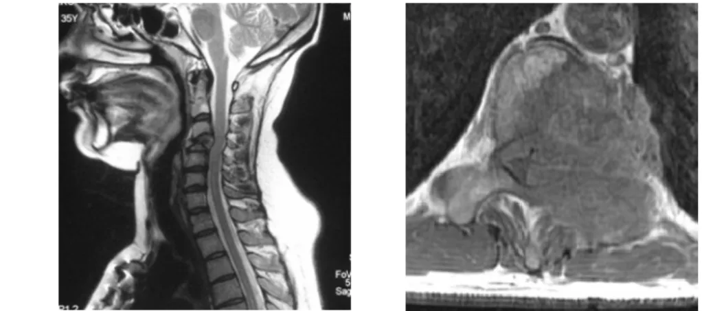
Fig. 25: Pathological fracture of C4 vertebra due to breast cancer metastasis on sagittal MRI
image
Fig. 26: Lung cancer metastasis in the Th11 vertebra with spinal cord compression
Rare extradural tumors:
Osteoid osteomas, osteoblastomas, chondromas, neurofibromas and hemangiomas are less common extradural tumors. Neurofibromas have a shape of a sandglass, growing out from the spinal canal to the thorax or the abdomen through an enlarged intervertebral foramen.
Fig. 27: Sandglass tumor on axial MRI image
Intradural – extramedullary tumors (40%)
Intradural – extramedullary tumors are most often neurinomas or
meningiomas
Intradural – extramedullary tumors are located on the lateral or posterior surface of the spinal cord. They usually cause chronic radicular pain first, lasting for months. Later on signs of nerve root or spinal cord dysfunction also appear.
These tumors histologically are neurinomas or meningiomas. Multiple neurinomas indicate neurofibromatosis (Recklinghausen’s disease).
Ependymomas, originating from the filum terminale, are also
extramedullary tumors. Rarely metastases or lipomas may occur in the extramedullary space. Extramedullary lesions are characterized by radicular or local pain, which precedes the development of neurological signs. Treatment is usually surgical. Radiotherapy is recommended in malignant tumors and in some cases of CSF metastases (e.g.
medulloblastoma, ependymoma). Intrathecal chemotherapy may be applied in disseminated leptomeningeal lymphomas.
Fig. 28: Calcified thoracic meningioma on 3D reconstructed CT scan
Fig. 29: Upper thoracic neurinoma on coronal MRI image
Intramedullary tumors (5%)
Intramedullary tumors are characterized by signs of slowly progressing transverse lesion, reduced pain and temperature sensation below the level of the lesion, and the lack of radicular pain. A tumor in the conus cause
“saddle” type hypesthesia and autonomic disturbances. Thirty-seven percent of conus tumors are ependymoma, 24% astrocytomas, 11%
hemangioblastomas, 5% lipomas, 4% metastases, and 1.5% dermoid or epidermoid teratomas and lymphomas. The treatment of intramedullary tumors is usually surgical. Radiotherapy may be effective in malignant lesions (e.g. malignant glioma).
Rare tumors
Two-thirds of
intramedullary tumors are ependymomas or astrocytomas
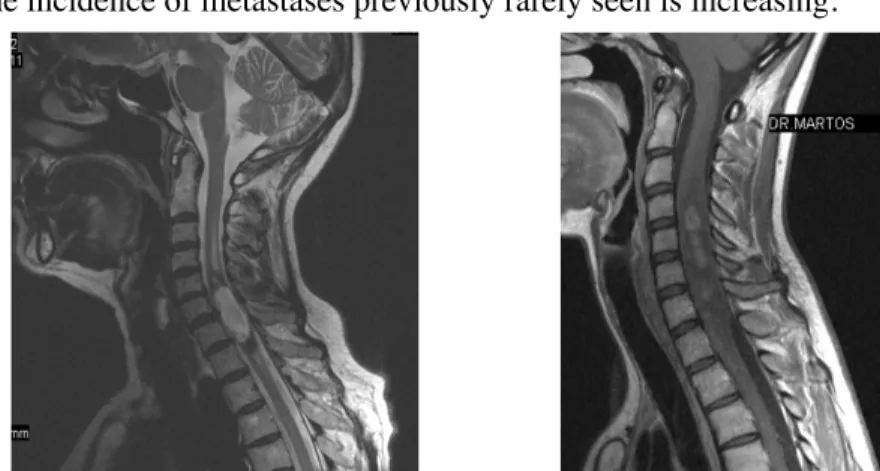
The incidence of metastases previously rarely seen is increasing.
Fig. 30: Intramedullary ependymoma at C5-6 level on sagittal MRI image
Fig. 31: Diffuse malignant astrocytoma in the cervical spinal cord on contrast enhanced
sagittal MRI images
The treatment and follow up of the patients with central nervous system tumors involve the interdisciplinary efforts of neurologists, oncologists and neurosurgeons.