PREDICTORS OF MORTALITY AND LOCAL RECURRENCE IN THE SURGICAL
MANAGEMENT OF PRIMARY TUMORS OF THE SPINE
PhD thesis
Zsolt Szövérfi MD
Semmelweis University
Doctoral School of Clinical Medicine
Supervisor: Dr. Áron Lazáry, Ph.D.
Official reviewers:
Dr. Imre Antal, Ph.D.
Dr. Attila Schwarcz, Ph.D.
Head of the Final Examination Committee:
Prof. Dr. Zoltán Sápi, Ph.D.
Members of the Final Examination Committee:
Dr. Zsolt Vendégh, Ph.D.
Dr. Gergely Pánics, Ph.D.
Budapest, 2016
1
Table of Contents
1. The list of Abbreviations ... 3
2. Introduction (with the background of the technical literature) ... 6
2.1. Clinical context ... 6
2.2. Epidemiology ... 8
2.3. Clinical manifestation and diagnosis ... 13
2.4. Staging and principals of spinal surgical oncology ... 16
2.5. Surgical therapy ... 22
2.6. Neo-adjuvant and adjuvant treatment possibilities ... 24
2.7. Outcome ... 27
2.8. Prognostic factors in primary spine tumor surgery ... 28
3. Objectives ... 31
4. Methods ... 32
4.1. Study design ... 32
4.2. Data collection ... 35
4.2.1. Preoperative data ... 35
4.2.2. Intraoperative data ... 35
4.2.3. Postoperative data ... 36
4.3. Data analysis and statistics... 37
4.3.1. Primary Spinal Tumor Mortality Score: development of a prognostic scoring system for survival at PST patients. ... 37
4.3.2. Prognostic variables for local recurrence and overall survival at surgically treated sacral chordoma patients ... 39
5. Results ... 40
5.1. Primary Spinal Tumor Mortality Score: development of a prognostic scoring system for survival at PST patients. ... 40
5.1.1. Demographic data and clinical characteristics ... 40
5.1.2. Study design and variable selection ... 44
5.1.3. Survival analysis ... 45
5.1.4. Prognostic score development ... 47
5.1.5. Internal validation of the PSTMS... 50
2
5.2. Prognostic variables for local recurrence and overall survival at surgically treated
sacral chordoma patients ... 52
5.2.1. Demographics... 52
5.2.2. Variable selection ... 55
5.2.3 Local recurrence analysis ... 55
5.2.4 Survival analysis ... 57
6. Discussion ... 59
6.1. Primary Spinal Tumor Mortality Score: development of a prognostic scoring system for survival at PST patients. ... 59
6.2. Prognostic variables for local recurrence and overall survival at surgically treated sacral chordoma patients ... 62
7. Conclusions ... 66
7.1. Principal results ... 66
7.1.1. Primary Spinal Tumor Mortality Score: development of a prognostic scoring system for survival at PST patients. ... 67
7.1.2. Prognostic variables for local recurrence and overall survival at surgically treated sacral chordoma patients ... 68
7.2. Future directions ... 69
8. Summary ... 70
9. Összefoglaló ... 71
10. Bibliography ... 72
11. Bibliography of the candidate’s publications ... 87
11.1. Publications related to the PhD thesis ... 87
11.2. Publications not related to the thesis ... 88
12. Acknowledgments ... 89
3
1. The list of Abbreviations
ABC - aneurismal bone cyst AUC - area under curve B - parameter estimate
bFGF - basic fibroblast growth factor Ch - chordoma
ChS - chondrosarcoma CI - confidence interval CT - computer tomography D - death
df - degrees of freedom EA - Enneking appropriate EI - Enneking inappropriate ES - Ewing's sarcoma GCT - giant cell tumor Gd. - gadolinium HR - hazard ratio
IMRT - intensity-modulated radiation therapy
K-M - Kaplan Maier LR - local recurrence
LRFS - local recurrence free survival MFH - malignant fibrous histiocytoma MPNST - malignant peripheral nerve sheathe tumor
MRI - magnetic resonance imaging NCSD - National Center for Spinal Disorders
OS - overall survival OST - osteosarcoma
PCNA - proliferating cell nuclear antigen expression
PEEK - polyether ether ketone PET - positron emission tomography PNET - primitive neuroectodermal tumor
PRBC - packed red blood cells PST - primary spinal tumor
PSTMS - primary spinal tumor mortality score
QOL - quality of life SE - standard error
SEER - Surveillance, Epidemiology, and End Results
SINS - Spinal Neoplastic Instability Score
SOSG - Spine Oncology Study Group SOSGOQ - Spine Oncology Study Group Outcomes Questionnaire SPECT - single photon emission computed tomography
SS - synovial sarcoma
SSCCC - Symptomatic spinal cord or cauda equina compression
WBB - Weinstein-Boriani-Biagini
6
2. Introduction (with the background of the technical literature)
2.1. Clinical context
Management of primary spinal tumors (PST) is a challenging issue of spine care [1]. The clinical behavior of these lesions depends mainly on the biological nature of the tumor [2]. However, clinical experience shows that the localization, the local dimensions of the neoplasm, and its relationship with the surrounding nerve structures and organs are also important factors influencing the PST associated morbidity and mortality [3-5]. In spite of the multidisciplinary cooperation and the acceptance of different diagnostic and treatment protocols the management of the PSTs remains controversial [1, 6, 7]. As the effectiveness of the chemo- or radiotherapy is still limited in the majority of the tumor types, surgical intervention still has the highest role in the treatment of PSTs [1, 8]. In the past decades, the surgical treatment of PST has undergone a substantial paradigm shift from palliative procedures to total en bloc removal of the tumor, despite the fact that extended surgeries can result in increased perioperative morbidity [9, 10]. Scientific data suggest that surgical resection is effective in the improvement of short term local control, but the long term effects are less favorable, and it has not been proved yet whether the surgical resection is associated with improved overall survival [6, 11, 12]. However, for certain tumor types, the positive effect of surgical intervention on survival was previously reported, and the possible impact of other, pre- and postoperative factors has been also investigated [6, 13]. In different medical fields, various prognostic scoring systems have been developed to risk stratify patients, and subsequently guide therapy [14]. In spine tumor surgery, the development of similar scoring systems had been limited mainly to metastatic lesions of the spine. For instance, the Tomita, the Tokuhashi scores, the SINS score and the more recently published Oswestry Risk Index are frequently used in the management of spinal metastatic lesions [15-19]. In comparison, the literature is scarce about the predictive factors which influence the survival of the PST patients. Generally, the published studies draw conclusions from underpowered analyses, presenting small case series of PSTs [4, 20]. The exceptions are the publications from the SEER database
7
which are based on large retrospective datasets [6, 13, 21-23]. However, they have also some limitations like the heterogeneity of the data, the inconsistent or not reported treatment methods and the lack of a rigorous follow up. Relying on this database McGirt et al. developed the only scoring system so far that aims to predict the prognosis in patients undergoing surgical resection for malignant primary osseous spinal neoplasms [13]. Their study determined the effect of five variables on survival for three tumor types (chordoma, chondrosarcoma and osteosarcoma).
Chordoma is a particular chapter of spine oncology. It is a unique malignant tumor, arising from notochordal remnants, thus it is located almost exclusively in the axial skeleton [24]. It has an overall incidence of 0.08 per 100,000 individuals and accounts for 40% of all primary sacral tumors [25]. Sacral chordoma is a typically slow growing and locally aggressive tumor, with a reduced ability to metastasize [26]. The diagnosis is often delayed because of the long standing, nonspecific initial symptoms, allowing the tumor to reach large sizes [27]. Because chordomas have shown to have a poor sensitivity towards radiotherapy and chemotherapy, they are mainly treated by surgical resection, in spite of the complex, resource intensive, and impairment inducing nature of the procedures [28]. Enneking oncologic management principles would recommend wide surgical en bloc resection of chordomas; however, this is difficult, even in the hands of the most experienced spine oncology surgeons [29]. Wide resection is not uniformly achieved in 35-75% of cases, primarily due to the relatively inaccessible anatomical location, preference for neurological preservation and large size at the time of diagnosis [11, 30-35]. The fact that chordomas grow in a lobulated fashion and have distant microscopic tumor outgrowths also makes wide surgical resection difficult [36]. Based on low quality evidence insufficient tumor resection is probably the main cause of local recurrence and subsequently death [11, 32, 35]. Other factors that possibly influence survival and local recurrence have been previously reported and include increased age, high sacral localization, lack of radiotherapy, prior resections, higher tumor grade, and increasing extent of tumor invasion [3, 4, 11, 13, 29, 32, 37-40]. Based on the dire consequences of sacral chordomas management (high mortality and morbidity) higher levels of evidence are needed to improve decision making and consequently patient outcome.
8
2.2. Epidemiology
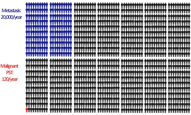
Primary tumors of the spine are rare [41]. They account for less than 5% of all osseous neoplasms and less than 0.2% of all cancers [37]. The incidence of the disease is 0.08-8 new cases per 100 000 individuals [1]. In the United States of America approximately 120 new cases are diagnosed every year (Figure 1). The incidence of benign spinal tumors is higher, but not as high as the incidence of metastatic spinal disease (20 000 new cases/year).
Figure 1 The comparison of the incidence of metastatic spinal tumors with the incidence of primary spinal tumors in the USA
The most frequent primary benign spinal tumors are schwannoma, hemangioma, osteoblastoma, osteoid osteoma, giant cell tumor and aneurismal bone cyst of the spine [42, 43].
Schwannomas are tumors arising from the nerve sheath cells. They grow slowly, but malign transformation can occur [44]. Majority of them are intradural tumors causing only nerve compression and damage, but can have extradural origin. In extremely rare cases can even have osteal origin [45]. Extradural spinal schwannomas present as dumbbell shaped in 10-15% of the cases [46].
9
The histological appearance of osteoblastoma and osteoid osteoma is similar.
They can be differentiated by their size, a lesion with a nidus >2 cm is classified as osteoblastoma [47]. The incidence of osteoblastoma is around 1% of overall incidence of bone tumors, and only 30-40% of them have spinal localization. In contrast osteoid osteoma’s incidence is higher, is around 5% of all bone tumors. The spinal occurrence of osteoid osteoma is 7-10 %. Both lesions are more frequent in men (2-3:1) [48].
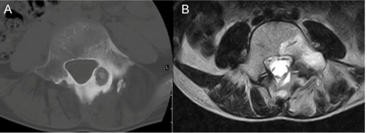
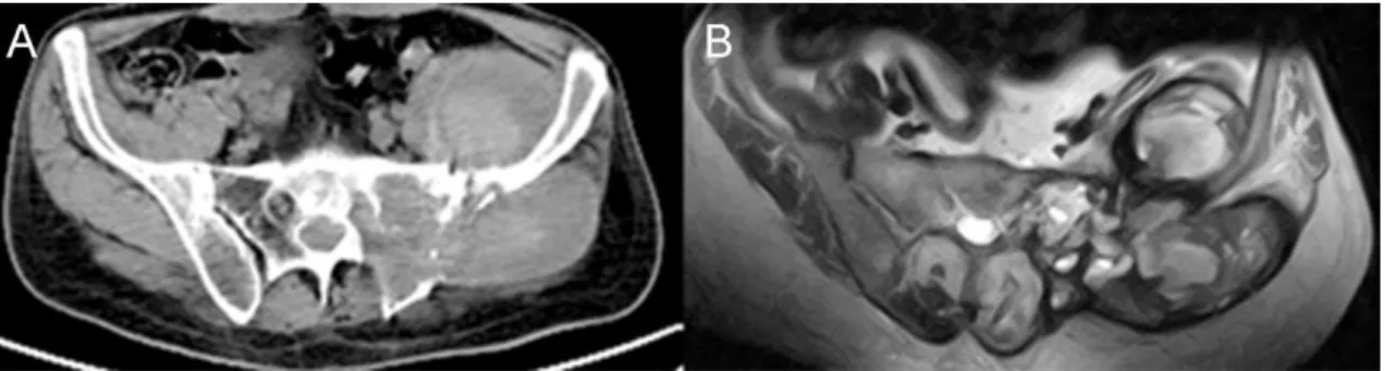
Osteoid osteomas occur predominantly in the young. They can appear on any spinal level, but frequently involve the posterior elements of the lumbar spine (Figure 2).
Figure 2 Osteoid osteoma of the posterior elements of the spine, A. axial CT image, B.
axial MRI image (T2 sequence)
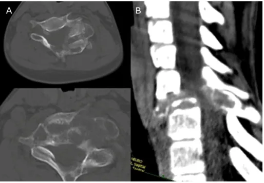
Figure 3 GCT of the sacrum, A. axial MRI image (T1 sequence), B. sagittal CT image, C. coronal CT image, D. axial CT image
Giant cell tumor (GCT) rarely involves the spine, it usually occurs in the metaphysis of the long bones. The spinal involvement can be between 7 to 10% [47] of all cases (Figure 3). GCT is the second most common primary bone tumor of the sacrum behind chordoma [43]. Usually is diagnosed at adults after the skeletal
10
maturation, it has a slight female predominance. In rare cases malignant transformation can occur.
The spinal manifestation of hemangioma is high. According to autopsy reports the incidence of spinal hemangiomas can be between 10 to 27 percent [49]. The majority of these tumors are asymptomatic and they are diagnosed incidentally. There is however a small subset of hemangiomas which can cause symptoms due to excessive growth or due to pathological fractures.
The incidence of aneurismal bone cyst (ABC) is 0.14/100 000/year. Usually they occur as a primary lesion, but can appear secondary to hemangiomas or osteoblastomas (Figure 4). ABC is a disease of the young, majority of the cases present before the age of 30. It has a slight female predominance. Most of the cases affect the lumbar spine, and the sacral localization is rare.
Figure 4 Radiological appearance of an ABC secondary to cervical osteoblastoma (CVII) A. axial CT images, B. sagittal CT reconstruction
Spinal malignant primary bone tumors are rare, they are accounting for less than 5% of all osseous neoplasms, and less than 0.2% of all cancers. According to the results of large scale population registries, the incidence of primary spinal tumors varies between 32% and 71% of all primary spinal tumors [23, 51]. The most common
11
primary malignant tumors of the spine are chordoma and sacral sarcomas like chondrosarcoma, osteosarcoma and Ewing sarcoma [22].
Chordoma is the most common primary spinal tumor with an overall incidence of 0.08 per 100 000 individuals accounting for 40% of all primary sacral tumors [52].
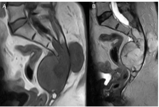
The male:female prevalence ratio is 2:1 with an increasing incidence after the fourth decade [25]. These lesions arise from notochordal remnants within the vertebral bodies and sacrum and are considered slow growing, locally aggressive lesions. The most common localization of chordoma is the skull base (clivus) and sacrum (Figure 5).
Median overall survival is estimated to 7.7 years in the Surveillance, Epidemiology, and End Results (SEER) Program of the National Cancer Institute (1973-2009) [52].
Figure 5 Sacral chordoma, sagittal MRI images A. T1, B. T2 sequence
Chondrosarcoma has an overall incidence of 0.5 per 100 000 per year [53]. It is more common in males aged between 30-70 years, with a peek in the fifth decade.
Chondrosarcoma may arise as primary tumor or as secondary transformation of an osteochondroma or enchondroma [54].
12
Ewing's sarcoma and the PNET-group are the second most frequent primary malignant bone cancers in children and adolescents with an overall incidence is less than 0.2 per 100 000 per year [55]. They involve the spine primarily in 3 to 10% of cases [56], sacrum being the most involved spinal level [57]. The male female ratio for Ewing’s sarcoma is 3:1, it affects young people between 5 to 30 years. Seventy five percent of this tumor occurs in the first two decade [56].
Osteosarcoma is the most common primary malignant bone tumor but rarely affects the spine [58]. Many of the osteosarcomas that occur in the sacrum (Figure 6) are secondary to degeneration of Paget disease [56].
Figure 6 Sacral osteosarcoma: A. axial CT image, B. axial MRI image
13
2.3. Clinical manifestation and diagnosis
The clinical presentation of a spinal tumor depends mainly on the anatomical location of the lesion [59]. Majority of the patients initially report back pain but a painless visible mass can also be the first sign of the disease. Persistent, non-mechanical back pain must be distinguished from common back pain [60]. Night pain and thoracic spine pain are both important symptoms because they suggest a neoplastic origin of the pain.
The pain is secondary due to the mass effect of the tumor, the erosion and impingement of the surrounding structures or the pathological fracture of the affected vertebrae [61].
Pain may be present with or without neurologic symptoms. Numbness, loss of sensation, decreased reflexes, sphincter dysfunction or motor deficit can be also the first clinical signs of a PST. At patients with cervical and thoracic spinal tumors physical examination, can reveal severe neurologic disturbances, signs of spinal cord compression (positive Hoffman or Babinski sign, spastic weakness as well as hyperreflexia in the extremities, and gait instability) [62, 63]. A specific concordance of sensory, motor, and vegetative symptoms may suggest the development of another severe neurologic entity the cauda equina syndrome, requiring urgent surgical intervention [64]. The patient may present with weight loss, general weakness and other general neoplastic signs, but these are rather the characteristic of metastatic lesions [65].
Each patient suspected of having a spinal tumor should undergo a thorough local and systemic work-up. Imaging studies give information about the extension of the tumor, but the most important element of the staging procedure is the biopsy [29]. Plain radiography is often the first imaging modality performed but it has limited sensitivity [56]. Visualization of an infiltrated body on plain radiography requires at last a 50%
destruction of the vertebral body [66]. Thus a pathological fracture can be easily identified. More accurate visualization of the spinal malformation can be obtained by using computerized tomography (CT) or magnetic resonance imaging (MRI). In most cases, both should be performed, because of the different characteristics of the two methods (Figure 2) [61]. CT provides superior information on cortical bone and tumor calcification, while MRI is excellent at delineating soft tissue, neural involvement, bone marrow infiltration, and epidural extension [67]. Additionally, the possibility of three- dimensional reconstruction is a great advantage of the CT scan. Although, some spinal
14
tumors have specific CT or MRI signs [25, 54, 56, 68], it is only sufficient to provide a presumptive diagnosis (Table 1).
Table 1 Diagnostic characteristics of Malignant Primary Sacral Tumors [29], *Gd:
Gadolinium
Tumor CT MR
Chordoma
Expansive
Lytic
Sclerotic
Intratumoral calcifications
T1 hypointense
T2 hyperintense
Gd* enhancement
Chondrosarcoma
Expansive
Lytic
Bone destruction
Soft tissue expansion
T1 hypointense to isointense
T2 hyperintense
Gd ‘‘rings and arcs’’ pattern Ewing sarcoma Lytic
Sclerotic
T1 isointense
T2 isointense to hyperintense
Gd enhancement Osteosarcoma
Lytic
Destructive
Matrix mineralization
T1 hypointense
T2 hyperintense
Bone scintigraphy is useful to determine whether the spinal lesion is localized or it is multiple, and to search for the primary tumor or metastases [69]. Although most spinal tumors have an increased uptake on bone scan, it lacks specificity to identify the nature of an abnormality. A more advance form of scintigraphy is the SPECT (Single Photon Emission Computed Tomography), which has higher specificity and sensitivity.
It even can detect lesions otherwise missed on CT or MRI examinations [70]. Until recently, PET has rarely been used to assess spinal tumors, but having even higher specificity and sensitivity can be helpful in detecting micrometastases, or the exact extent of paravertebral, epidural tumor growth [71].
The final diagnosis of primary spinal tumor can be made after a biopsy and an accurate histological examination. There are four main biopsy techniques: fine needle aspirate biopsy (FNAB), core needle biopsy, incisional biopsy, and excisional biopsy [67]. Incisional “open” biopsy was considered to be the gold standard for the diagnosis of bone lesions, with 98% accuracy [72], but several studies demonstrated that it significantly increases the risk of recurrence [73, 74]. Recently, percutaneous CT guided core needle biopsy has gained popularity, showing a good accuracy with a less invasive
15
procedure. Furthermore, Saad et al. reported the superiority of the FNAB, the procedure having a low complication rate and a lower likelihood of an extralesional spread of tumor cells [75]. A meta-analysis of spinal percutaneous biopsies estimated its accuracy to 92% [76]. Although the risk of tumor cell contamination is lessened by the core biopsy and FNAB approaches, resection of the biopsy tract is still mandatory [73, 77].
For tumors limited to the posterior elements, an excisional biopsy can be both diagnostic and therapeutic [1].
16
2.4. Staging and principals of spinal surgical oncology
Before any therapeutic intervention an oncological staging of the patient is critical. A bone scan is an important tool in establishing the solitary nature of the lesion.
Additionally, conventional radiological staging before surgery generally includes a CT scan of the head, chest, abdomen, and pelvis. Osteoporosis is a global condition that may affect the surgeon’s reconstructive options after the tumor resection. When osteodensitometry reveals a T-score of less than -2.0, reconstructive possibilities may become limited [78]. Another preoperative factor which has to be investigated is the general health condition of the patient. As several studies have shown that comorbidities can increase the risk of perioperative complications, they must be accurately identified and minimized by multidisciplinary consultation [79-81].
According to the “International Union Against Cancer”, the objectives of cancer staging are aiding the planning course of treatment, providing insight into the prognosis, assisting in the evaluation of the treatment results, facilitating the interinstitutional communication, and contributing to continuing cancer research [82, 83]. Based on these principles Dr. William Enneking introduced a surgical staging system for the management of appendicular musculoskeletal tumors in 1980 [84]. As, it was originally developed for extremities the adoption of this classification in the management of primary spine tumors is difficult (the epidural compartment, the sacrifice of the neural elements, and the restoration of spinal stability are not considered) [85]. To overcome this paucity Boriani et al. proposed a modification of the original Enneking staging system applicable for spinal tumors [86, 87]. They introduced the following concepts to uniformise the terminology: intralesional resection (piecemeal debulking or curettage), marginal resection (lesion shelled out leaving pseudocapsule or reactive zone), wide resection (intracompartmental en bloc resection), and radical resection (extracompartmental excision).
According to the Enneking classification benign tumors are divided into three categories (Table 2); S1 (latent or inactive stage), S2 (active stage), S3 (aggressive stage) [86]. In the S1 stage the tumor is not growing, or is growing very slowly, has well defined margins or capsule, and causes few or no symptoms [88]. Thus no
17
treatment is required unless palliative surgery for decompression or stabilization.
Tumors in the S2 stage are characterized by slow growth and mild clinical symptoms. In this stage bone scans are usually positive. Intralesional resection is the treatment of choice in this stage. Although the recurrence rate is low it can be further decreased by local adjuvant treatment (cryotherapy, embolization, radiotherapy) [89]. Tumors in the S3 stage are rapidly growing benign tumors. Their capsule is thin, discontinuous or absent, and is usually surrounded by wide reactive hypervascualrized tissue [67]. Thus they are frequently not confined to the vertebra, invading the epidural or paravertebral space. They should be treated by marginal or wide resections.
Table 2 The Enneking Surgical Staging of benign spinal tumors
Staging Description Treatment Example
S1 Latent
Well-defined margins or capsule No or very slow growth
Nonoperative, unless decompression or stabilization is needed
Schwannoma Hemangioma Osteochondroma
S2 Active
Thin capsule
Reactive pseudocapsule Slow growth
Intralesional curettage Osteoid osteoma Osteoblastoma
S3
Aggressive
Very thin or incomplete capsule Wide reactive pseudocapsule Rapid growth
Marginal or wide resection
ABC GCT
In the case of malignant tumors three stages are used (Table 3). Stage I for low grade tumors, stage II for high grade tumors. Each stage is further divided into two subcategories based on the local extent of tumor (A: confined to the vertebral body, B:
the tumor involves the paravertebral, epidural compartments). Stage III represents any tumor with distant metastasis [86].
A stage I tumor does not have a true capsule, but it is surrounded with a thick pseudocapsule. The pseudocapsule can contain small microscopic tumor islands. In the case of stage II tumors the tumor growth is so rapid that there is no time for a pseudocapsule formation. These tumors can produce skip metastases [89]. Stage I, II tumors should be treated by wide en bloc resection. Based on the individual tumor characteristics adjuvant therapy may be beneficial to decrease the local recurrence.
Patients with stage III tumors are candidates only for palliative surgery and subsequent adjuvant therapy [67].
18
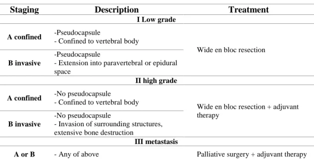
Table 3 The Enneking Surgical Staging of malignant primary spinal tumors
Staging Description Treatment
I Low grade A confined -Pseudocapsule
- Confined to vertebral body
Wide en bloc resection B invasive
-Pseudocapsule
- Extension into paravertebral or epidural space
II high grade A confined -No pseudocapsule
- Confined to vertebral body
Wide en bloc resection + adjuvant therapy
B invasive
-No pseudocapsule
- Invasion of surrounding structures, extensive bone destruction
III metastasis
A or B - Any of above Palliative surgery + adjuvant therapy
As the use of the Enneking Classification in the management of primary bone tumors of the appendicular skeleton has resulted in a significant improvement in survival, many oncology spine experts started to adopt Enneking principles in their everyday practice. Fisher et al. even introduced the terminology of “Enneking appropriate” (EA, surgical margin as recommended by the Enneking Classification) and
“Enneking inappropriate” (EI, surgical margin not recommended by Enneking Classification), to assess the successfulness of the surgery [12]. According to this the surgery is performed based on the Enneking recommendations, and the resulting surgical margin is categorized by the pathologist as intralesional, marginal or wide. If this corresponds with the Enneking recommendation, then the surgery is considered EA, if not than EI.
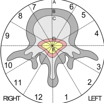
As the Enneking staging system was developed primary for the appendicular skeleton its main shortcoming is that it does not addresses the spinal canal. To overcome this Weinstein in collaboration with the Rizzoli Institute created the Weinstein-Boriani-Biagini staging system (Figure 7) [86, 90]. The fundamental concept of this system is to ensure the sparing of spinal cord without compromising the surgical tumor margins [85]. The staging system records the tumor propagation on an axial view of an MRI and CT exam. In the axial plane the vertebra is divided into 12 radiating zones (numbered 1 to 12 in a counter-clockwise order) and into five layers (A to E,
19
from the paravertebral region to the dural involvement). The longitudinal extent of the tumor is recorded by listing the caudal and proximal involved vertebral levels [86].
Figure 7 The Weinstein-Boriani-Biagini staging system
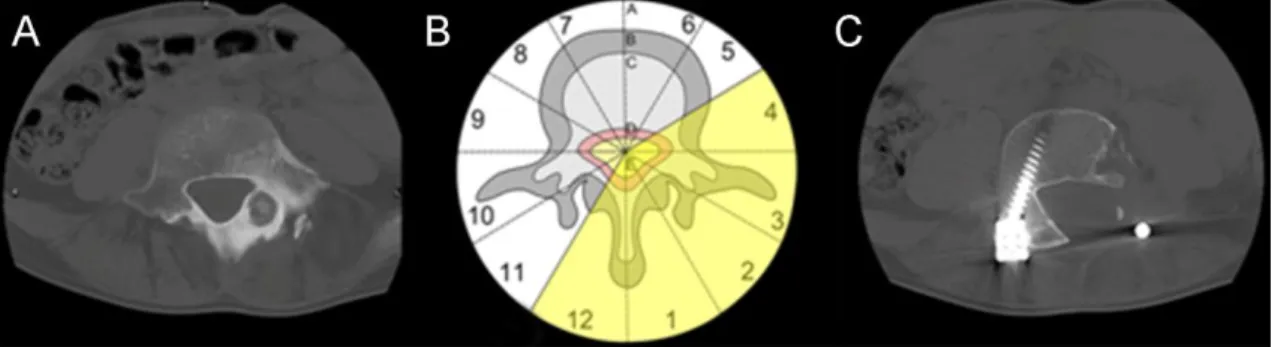
Boriani et al. proposed three resection types based on the tumor localization [86]. If the tumor is confined to the zones 4-8 or 5-9 then an en bloc vertebrectomy should be performed on one or two stages. If the tumor is localized in the zones 3-5 or 8-10 then a wide or marginal “sagittal resection” should be attempted. This should be performed from a combined anterior and posterior approach. If the tumor is localized in the zones 10-3, then marginal or wide en bloc resection can be performed by a posterior approach (Figure 8).
Unfortunately, the WBB classification was developed to be used on the mobile spine, thus it cannot be applied for sacral tumors. The sacral region is anatomically very complex, the surgeon needs to take in consideration other critical structures (including the rectum, cauda equina and iliac vessels) and the preservation or reconstruction of the lumbo-pelvic junctions stability [91]. Currently, there are no validated and widely used surgical staging systems which take in account all these issues. Recently Zhang et al.
based on own clinical experience proposed a novel classification system for sacral tumors [91]. The classification system is a combination of the WBB and Enneking tumor staging methods, and contains 16 possible categories. Sacral tumors are divided
20
into 2 major types (above or below S2) and then 4 further subtypes (based on the extension of the tumor in the pelvic cavity: < 5 cm or ≥ 5 cm). A further subdivision (similar to the WBB system) is then added according to the axial plane anatomy (3 zones: anterior sacrum, posterior sacrum, and lateral sacrum).
Figure 8 En bloc resection by posterior approach: A. axial CT image of an LV osteoid osteoma, B. the planning phase of the surgery according to the Weinstein-Boriani- Biagini staging system, C. postoperative axial CT image of the LV vertebrae.
In the planning process of the surgical treatment, the classification described by Fourney et al. (Figure 9) (based on the level of nerve root sacrifice) could be useful in the everyday clinical experience [27].
They categorized sacral resections into two groups, midline tumors and eccentric lesions. The midline group included low, middle, and high sacral amputations, total sacrectomy, and hemicorporectomy. In the case of low sacral amputation, the resection was performed at the level of the S4 nerve roots, in the case of midsacral amputation the resection was at the S3 nerve roots, and in the case of high sacral amputation at the level of the S2 nerve roots. If the tumor reached the S1 nerve roots, then total sacrectomy was the treatment of choice. Hemicorporectomy (translumbar amputation) was indicated for localized, aggressive tumors that had spread beyond the sacrum to the lumbar spine. If the tumor was located in unilateral position and the planned resection does not exceed the midline, they introduced the term “eccentric resection” including tumors overgrowing the sacroiliac joint and penetrating to the pelvic bones or to the extraosseal compartments.
21
Figure 9 Categorization of sacral resections after Fourney et al. [27]; A. Low sacral amputation - the sacrifice of S4 nerve roots B. Midsacral amputation - the sacrifice of the S3 nerve roots C. High sacral amputation - the sacrifice of the S2 nerve roots D.
Total sacrectomy - the sacrifice of the S1 nerve roots E. Hemicorporectomy (translumbar amputation) - for aggressive tumors that had spread beyond the sacrum to the lumbar spine F. Eccentric resection - for tumors that does not exceed the midline
22
2.5. Surgical therapy
The importance of a multidisciplinary management in PST patients cannot be overestimated. Surgeons of multiple specialties (spine, musculoskeletal, vascular, gastrointestinal, plastic surgery, and urology specialists) must be involved into the planning of the surgery [78].
The primary goal of the surgical therapy is oncologic control. However, with the exception of the benign tumors this can be achieved only by en bloc resection [73]. The procedure of en bloc tumor resection (Figure 10) is complex and can have a significant morbidity and mortality. Restoration of neurological function, pain control, deformity correction and stabilization are only secondary goals of surgery [62]. One of the most important issues of surgical planning is informing the patient and his family about the tradeoffs of en bloc resection (increased survival vs. high planed morbidity) [67].
The surgical treatment of PSTs is a complex procedure and demands expert surgical skills. The preferred surgical approach has to be decided on an individual basis because of a high variability of tumor morphology, location and pathology [92-94]. It should be kept in mind that the biopsy tract should always be included in the resection.
Therefore, the surgeon should be involved into the planning of the biopsy, assuring that the biopsy tract will be excised en bloc with the tumor specimen [61].
Depending on tumor morphology surgical approaches include posterior decompression, posterior decompression with stabilization and fusion, posterior en bloc or intralesional resection (± stabilization and fusion), posterior en bloc or intralesional corpectomy (± stabilization and fusion), corpectomy from thoracotomy or retroperitoneal approach with or without posterior stabilization and fusion [62].
Recently all these approaches were attempted from minimal invasive approaches with varying success [95]. A detailed description of the surgical techniques is far beyond the scope of this chapter.
In cases where excessive bone resection must be performed biomechanical reconstruction of the spinal column is mandatory. This can be achieved by posterior pedicle screw and rod stabilization, with or without anterior column reconstruction, with
23
or without prefabricated or custom made implants [7]. Due to large bone defects, cytotoxic adjuvant therapy and radiotherapy achieving bony fusion may be challenging [67]. As the patient may permanently rely on implanted instrumentation to maintain stability, the fusion rate can be facilitated by the implantation of tricortical iliac crest strut graft, allograft or vascularized fibula graft.
Figure 10 En bloc resection of a sacral tumor: A. the resected specimen, B. closed-loop reconstruction of the spino-pelvic junction after tumor resection
After the en bloc resection of a large tumor, one of the greatest difficulties is the closure of the surgical site. Several reconstructive techniques are used for soft tissue reconstruction to prevent wound healing complications. Paraspinous muscle, trapezius muscle, and latissimus dorsi muscle flaps can be used on the thoracic and lumbar spine [96], vertical rectus abdominis myocutaneous flap and gluteus maximus adipomuscular flaps can be applied after sacrectomies [97-100].
24
2.6. Neo-adjuvant and adjuvant treatment possibilities
The primary goal of the therapeutic process is curative. In the majority of the cases this can be reached only by complete surgical excision of the tumor. In addition, majority of primary spinal tumors are chemo- and radiotherapy resistant. In this setting the role of adjuvant treatment is still unclear and varies by pathology [62].
In the case of benign tumors, the treatment protocols are well defined. Majority of them can be treated with good efficiency by surgical intervention (marginal en bloc resection or intralesional curettage) [67]. Usually after surgery they do not require adjuvant therapies. There are however some exceptions, like the denosumab treatment of GCT and the serial embolization of ABC. Although GCT is a benign tumor it may became locally aggressive, and even can give distant metastases in small number of cases [50]. The treatment of choice of these lesions is en bloc surgical resection, to avoid local recurrence [101]. Denosumab is a newly developed monoclonal antibody which has already been demonstrated to induce clinical and radiographic tumor remission [102]. Although the effectiveness of denosumab was demonstrated in several clinical studies, the role in the treatment algorithm of GCTs of the spine has not yet been defined [102, 103]. ABCs are benign but locally aggressive tumors containing thin walled, blood-filled cystic cavities [101]. Traditionally, ABC was treated by simple curettage or complete excision. Recurrence rates after curettage were reported as much as 50% [104]. With en bloc resection recurrence rate can be minimized, but this treatment possibly exposes the patient to high surgical morbidity [105]. As ABC is heavily vascularized, embolization of the tumor before surgery is common. In the Rizzoli Orthopedic Institute, a group of surgeons started to perform serial embolization of the tumor without surgery [106]. Boriani et al. reported, that serial embolization can be as effective as surgery, while being less invasive [107].
Treatment options of malignant PSTs should be discussed by a multidisciplinary team (oncologists, radiologists, radiotherapists and surgeons). They should decide on the optimal treatment strategy, including chemo-, radiotherapy and the surgical intervention. The decision depends on the location, extent and biological aggressiveness of the lesion and it is influenced by the general condition of the patient [29].
25
Majority of primary spinal tumors, including chordoma and chondrosarcoma, are relatively resistant to the conventional radio- or chemotherapy, although radiotherapy can be used as an adjunctive treatment in case of intralesional surgical resection [61]. In the case series of York et al. adjuvant radiotherapy tripled the disease-free survival time in chordomas [32]. Biologically higher radiation doses can be achieved with charged particle beam radiation therapies (i.e., protons, helium, neon, and carbon ions). Due to increased effective doses and the lower incidence of side effects, carbon-ion radiotherapy [108, 109], and proton/photon therapy [110], were reported to have better results compared with conventional radiotherapy. In contrast to conventional radiotherapy, where the full dose is delivered to the spine, cauda equina and the surrounding soft tissues, intensity-modulated radiation therapy (IMRT), stereotactic radiosurgery and the CyberKnife can deliver a high-dose single fraction to the target tissue sparing most of the adjacent neural or visceral elements [111-113]. The effect of the different radiation therapies can be further enhanced by the utilization of radiosensitizing agents like razoxane [114, 115]. Chemotherapy has never played a significant role in the treatment of low-grade spinal malignancies. Reports of tumor responses to regimens, including anthracyclines, cisplatin and alkylating agents, are only anecdotal [116]. Recently, medical oncologists have pointed out the apparent sensitivity of chordoma to new molecular-targeted agents like imatinib, cetuximab and gefitinib [117]. Unfortunately, these novel drugs are only accessible in clinical studies, and only for patients with unresecteble or metastatic tumors [118]. Chemotherapy is not effective in chondrosarcoma, however new chemotherapeutic agents like pemetrexed or sumantinib are currently evaluated [119, 120].
A decade ago the treatment of choice in high-grade primary malignant sacral tumors, like Ewing sarcoma and osteosarcoma was surgical intervention [121]. Today, due to the development of novel chemotherapeutic agents the surgical intervention has become the last step. In a systematic review, Sciubba et al. concluded that in the case of spinal Ewing sarcoma and osteosarcoma neoadjuvant chemotherapy and multimodality management offers a significant improvement in local control and long-term survival [121]. Surgery plus modern multidrug chemotherapy has dramatically increased the 5- year disease-free survival rate of osteosarcoma patients to 60-70%, and in the case of Ewing sarcoma patients to 80% [122, 123]. Although the treatment of choice of Ewing
26
sarcoma and osteosarcoma is chemotherapy, even with effective chemotherapy, these tumors are rarely cured without surgical resection [124, 125].
27
2.7. Outcome
Clinical outcome of primary spinal tumor surgeries needs to be evaluated in three dimensions: surgical outcome (complications), oncological outcome (survival and local recurrence) and functional outcome (disability, pain, etc). Surgical outcome can be evaluated in reflection of the intraoperative and postoperative complications which occur with high incidence in these extended surgeries.
As primary spinal tumor surgery is characterized by complex surgical techniques, prolonged operating time and severe bleeding, the likelihood of perioperative complications is high. During the surgery, unplanned nerve root resections, visceral and vascular perforations may occur and intraoperative death is also a possible severe complication. In the early postoperative period the development of different wound or surgical site infections may require additional surgical interventions.
In primary spinal neoplasms, oncological outcome was reported to be associated with the tumorous involvement of the resection margins several times [40]. En bloc resection of the tumor with wide margins results in the lowest risk for local recurrence and systemic spread of the disease, but to achieve it can be very challenging even impossible in certain cases. In general, functional outcome is the most important for the patient. Development of any neurologcial deficit (motor-, sensor- and vegetative disturbance) is strongly determined by the level of the nerve root sacrifice; however, ambulation ability and local pain is also associated with the stability of the spine as well as the success of the soft tissue reconstruction. For the evaluation of the neurological outcome, the modified Biagini scale can be used [27, 126] however the overall functional outcome is a more complex dimension. So far, no validated measurement tool for the evaluation of the functional outcome has been published. On the other hand, the cross-culturally adapted versions of the SOSGOQ (Spine Oncology Study Group Outcomes Questionnaire) which was originally developed for metastatic spinal lesions [127], seem to be an optimal tool for the follow-up of the functional outcome after primary spinal tumor resections too.
28
2.8. Prognostic factors in primary spine tumor surgery
Survival analysis is generally defined as a set of statistical methods to analyze the time (the outcome variable) to the occurrence of an event of interest (such as death or recurrence of a tumor etc.) [128]. For example, if the event of interest is the local recurrence, then the “survival time” is time (in months, years) from the start of the observation (surgery) until the appearance of the local recurrence [129]. In this case the studied time period is named local recurrence free survival (LRFS). If the event of interest is death, than the survival period is called overall survival (OS). Survival analysis requires special techniques because the event of interest does not necessarily occur for all patients before the end of the study (e.g. some patients are still alive or tumor free at the end of the study) . This is called censoring; meaning that the observation period ended without observing the event of interest or the patient is lost to follow-up. Unlike ordinary regression models, survival methods correctly incorporate information from both censored and uncensored observations in estimating important model parameters. The simplest form of survival analysis the Kaplan Meier method is widely used to estimate and graph survival probabilities as a function of time. It can be used to obtain univariate descriptive statistics for survival data, including the median survival time, and compare the survival for two or more groups of subjects. For more detailed analysis the Cox proportional hazards regression model can be used [131].
This method it allows testing for differences in survival times of two or more groups of interest, while allowing adjusting for covariates of interest. The Cox regression model provides useful in interpreting information regarding the relationship of the outcome variable and different predictors.
The literature about the predictive factors which influence the survival and local recurrence of the PST patients is scarce. Majority of the published studies (Table 4) draw conclusions from small retrospective PST case series which result in underpowered analyses [4, 20]. Furthermore, these studies use only the Kaplan Maier test to identify the prognostic factors for OS or LRFS.
29
Table 4 Literature review on prognostic factors for OS and LRFS of primary spinal tumors; R: local recurrence, D: death, LRFS: local recurrence free survival, OS:
overall survival, Ch: chordoma, ChS: chondrosarcoma, OST: osteosarcoma, ES : Ewing sarcoma, SS : synovial sarcoma, MPNST: malignant peripheral nerve sheathe tumor, MFH : malignant fibrous histiocytoma, GCT: giant cell tumor, PCNA : proliferating cell nuclear antigen expression, bFGF: basic fibroblast growth factor, KM: Kaplan Maier analysis, COX: Proportional hazards model
Author Type N# R D Stat. Prognostic factors
1993
Samson et al.
Ch 21 13 11 KM LRFS: age (marginally significant)
1999
Cheng et al.
Ch 23 13 11 KM LRFS: High sacral localization, age OS: High sacral localization
1999
York et al.
Ch 27 18 15 KM LRFS: surgical margins, lack of radiotherapy
2000
Bergh et al.
Ch 39 17 16 COX LRFS: Invasive diagnostic procedure outside tumor center, surgical margins and tumor necrosis
OS: Larger tumor size and surgical margins
2001
Bergh et al.
ChS 69 17 26 COX LRFS: surgical margins, primary treatment outside tumor center
OS: Tumor grade
2005
Fuchs et al.
Ch 52 23 19 KM LRFS: surgical margins
OS: age, marginal or intralesional excision
2009
Yang et al.
Ch 22 8 - KM LRFS: surgical margins
OS: higher tumor location and higher expressions of PCNA and bFGF
2010
Stacchiotti et al.
Ch 138 69 82 COX LRFS: surgical margins OS: larger tumor size
2010
Ruggieri et al.
Ch 56 24 19 KM LRFS: surgical margins, previous intralesional surgery
2010
Cheng et al.
Ch 36 16 6 COX LRFS: muscle invasion, surgical margins
2010
Zhou et al.
Ch 37 25 12 COX LRFS: surgical margins, multiple vertebral levels OS: upper cervical spine, multiple vertebral levels
2013
Cho et al.
Ch, ChS OST, ES SS, MFH MPNST
29 23 16 KM OS: distant metastasis
2013
Xu et al.
GCT 102 38 7 COX LRFS: age >40 year, subtotal resection, lack of bisphosphonate treatment
2014
Yin et al.
ChS 98 42 32 COX LRFS: surgical margins
OS: tumor grade, surgical margins
2015
Wang et al.
MPNST 43 22 22 COX LRFS: osteolytic destruction, tumor grade, S100, SMA, CD57 biomarkers
OS: osteolytic destruction, tumor grade, S100, Ki67 biomarkers
2015
Meng et al.
Ch 153 51 42 COX LRFS: dediferentaited chordoma, level, surgical margin, Frankel scores A-C
OS: surgical margins, Karnofsky score <80
30
The exceptions are the publications from the SEER database (Table 5) which are based on large retrospective datasets [6, 13, 21-23]. However, they have also some limitations like the heterogeneity of the data, the inconsistent or not reported treatment methods and the lack of a rigorous follow up. Several publications tried to identify prognostic factors, but the majority of these studies are statistically underpowered.
Table 5 Population based studies from the SEER registry; LR: local recurrence, D:
death, LRFS: local recurrence free survival, OS: overall survival, Ch: chordoma, ChS:
chondrosarcoma, OS: osteosarcoma, ES: Ewing sarcoma, KM: Kaplan Maier analysis, COX: Proportional hazards model
Author Type N# LR D Stat. Prognostic factors
2009 Jawad et al.
Ch 962 - 577 (10y) COX OS: lack of surgery, age >59 year, tumor size > 8cm
2011
McGirt et al.
Ch ChS OS
114 - - COX OS: sacral localization, more recent year of diagnosis, age and increasing extent of tumor invasion
2011
Mukherjee et al.
Ch ChS OS ES
1892 1116 KM OS: tumor invasion beyond
periosteum
2012
Mukherjee et al.
Ch ChS OS ES
827 401 KM OS: non-surgical therapy
2012
Lee et al.
Ch 409 199 COX OS: non-Hispanic race, low socio- economic status, large tumor, non- surgical treatment
31
3. Objectives
As seen in the previous chapter surgical therapy of PSTs is the only curative treatment option. However, en bloc surgical resection has a high morbidity and mortality rate. Thus appropriate patient selection is essential, only those patients should undergo extensive surgeries who clearly would benefit from it. This setting is complicated by the rarity and heterogeneity of PSTs, thus studying them is difficult.
The purpose of the present thesis is to investigate the possible effects of several clinical parameters on survival and local recurrence in a large institutional cohort of PST patients, and subsequently in a multicenter cohort of surgically treated sacral chordoma patients.
Our objectives were:
1. To investigate the demographics of a large single institutional cohort of surgically treated primary spinal tumor patients.
2. To investigate the effect on postoperative survival of several preoperative clinical parameters in a large single institutional cohort of surgically treated primary spinal tumor patients
3. To create a prognostic scoring system which can predict the postoperative survival based on preoperative parameters at primary spinal tumor patients.
4. To investigate the demographics of a large multicenter cohort of surgically treated sacral chordoma patients.
5. To investigate the effect of several clinical parameters on the postoperative survival of sacral chordoma patients.
6. To investigate the effect of several clinical parameters on local recurrence of sacral chordoma patients.
32
4. Methods
4.1. Study design
National Center for Spinal Disorders (NCSD), a tertiary care spine referral center in Hungary for a population of 10 million, is the main oncologic spine surgery center in Central Europe. In 2007 based on the Spine Oncology Study Group’s (SOSG, an international panel of spine oncology experts) guidelines an institutional database was built (containing clinical and outcome data about surgically treated primary spinal tumor and tumor-like lesion cases). Patient data between 1995 and 2007 was collected in a retrospective fashion, but from 2007 a prospective data collection of clinical data was started (Figure 11). The database is regularly updated even today.
Figure 11 Ambispective data collection with cross sectional follow up on vital status.
From 2010 the members of the SOSG continued their work under the umbrella of AOSpine International Knowledge Forum Tumor. In 2011 they started one of the first multicenter studies on primary spinal tumors [132]. An ambispective cohort study was performed by thirteen leading spine oncology referral centers (Figure 12).
Seven centers were from North America (Johns Hopkins University School of Medicine, Baltimore, USA; University of British Columbia, Vancouver, Canada; MD Anderson Cancer Center, Houston, USA; University of Toronto, Toronto, Canada;
33
Memorial Sloan-Kettering Center, New York, USA; Mayo Clinic, Rochester, USA;
University of California San Francisco, San Francisco, USA), five from Europe (National Center for Spinal Disorders, Budapest, Hungary; Rizzoli Institute, Bologna, Italy; Queens Medical Centre, Nottingham, UK; Instituto Ortopedico Galeazzi, Milan, Italy; Oxford University Hospital NHS Trust, Oxford, UK), and one from Australia (Princess Alexandra Hospital, Brisbane, Australia).
Figure 12 Thirteen leading spine oncology referral centers: 1 University of British Columbia 2 University of Toronto; 3 University of California San Francisco; 4 Mayo Clinic; 5 MD Anderson Cancer Center; 6 Johns Hopkins University; 7 Memorial Sloan- Kettering Center; 8 Queens Medical Centre; 9 Oxford University Hospital NHS Trust;
10 Instituto Ortopedico Galeazzi; 11 Rizzoli Institute; 12 National Center for Spinal Disorders; 13 Princess Alexandra Hospital
The majority of the data was collected retrospectively, but smaller part was collected prospectively (ambispective design). To prevent loss to follow-up bias, a cross-sectional follow-up of the vital status was performed at the end of the study period (December 2012). Patients met the inclusion criteria if they were diagnosed with a primary spinal tumor, received a surgical resection, and participated in at least one clinical follow-up. Patients with a secondary spinal tumor, spinal cord tumor, spinal lymphoma, or myeloma were not included in the study. Subjects who had only biopsy or had insufficient clinical data were also excluded. The NCSD contributed with 300
34
PST cases to the AOSpine’s Retrospective database (Figure 13). As the two databases are similar the data transfer had been easily performed.
1182 spinal tumor patients with
surgical intervention or biopsy in the NCSD (1995-2012)
392 primary spinal tumor (PST) patients
323 PST until 2012 366 PST patients with
good data quality
637 metastasis &
153 myeloma/
lymphoma
26 patients with insufficient
dataset
43 PST patients, with
only biopsy 300 patients
from the NCSD
323 final PST cohort 1195 patients
from the other centers
1495 PST patients
344 Chordoma
167 Final cohort
of sacral chordoma 173 Sacral chordoma 1151
Other tumor
171 Mobile spine
chordoma
6 patients with
Enneking grade III
tumors
A.
B.
Figure 13 Flow-chart for patient selection A. NCSD primary spinal tumor cohort, B.
AOSpine sacral chordoma cohort.
The present thesis is about two analyses, one from the NCSD Primary Spinal Tumor Database (all cases), and one from the AOSpine Knowledge Forum Tumor Primary Spinal Tumor Retrospective database (sacral chordoma cases).
35
4.2. Data collection
Study data were collected and managed using a secure, web-based application, the REDCap electronic data capture system [133]. The databases were hosted at the National Center for Spinal Disorders (the institutional database only) and the AOSpine International. Data about demographics, baseline patient and tumor characteristics, surgical treatment, local disease recurrence, morbidity, and cross-sectional survival were gathered and entered into a database. The studies were approved by the Ethics Committee of the Hungarian Ministry of Health (49777/2012/EKU; 751/PI/12).
4.2.1. Preoperative data
Preoperative inpatient and outpatient clinical records were used to identify demographic and clinical data including age, gender, detailed medical history, preoperative symptoms, presence of pathologic vertebral fractures, and different neurological signs. Previous tumor surgery was defined as a surgical intervention beyond biopsy before the surgical resection. Neoadjuvant treatment methods were also recorded. Motor deficit was assessed according to the Frankel scale. Signs of spinal cord compression and/or vegetative dysfunction due to cauda equina compression were also recorded. Results from imaging (CT, MRI, X-Ray, bone scan, PET-CT) and histological diagnosis were used to determine the localization, the local extension, and the oncologic stage of the tumor. The staging was performed according to the main categories of the Enneking surgical staging system if it was applicable [84].
4.2.2. Intraoperative data
Intraoperative surgical data including surgical approach, nerve root and cauda equina sacrifice, type of resection, type of reconstruction, and the amount of blood loss were recorded. The parameters for type of resection (wide, marginal, intralesional or palliative) were determined by the surgeon. The surgeon’s impression about the surgical margins was validated by the pathologist during the histological analysis. The resections were also categorized according to the Enneking principles [12]. Tumor volume was measured on the histopathologic specimens. The height, width, and depth of the tumor
36
were recorded, and the volume was calculated using the formula of an ellipsoid mass (volume=π/6×height×width×depth) [134]. Tumor volume was transformed into a categorical variable where tumors were grouped as <100 cm3 and ≥100 cm3.
4.2.3. Postoperative data
Follow-up data were obtained by direct examination of the patient and by performing the required imaging modalities. Follow-up data included any early and late postoperative complications, adjuvant radiation and chemotherapy, local recurrence, any further surgeries for complications or recurrence, and current vital status.
Postoperative complications were considered “early” if they occurred within six weeks after surgery and “late” if they occurred more than six weeks postoperative.
At the end of the study period, a cross-sectional follow-up of the vital status was performed in the form of an outpatient visit, telephone interview or accessing governmental vital statistic databases, if necessary.
37
4.3. Data analysis and statistics
Statistical analyses were performed using SPSS 20.0, Statsoft Statistica 10 and STATA 12.0 software. Demographic data was analyzed by descriptive and none parametric statistics. Survival analysis (Kaplan-Meier method, Mantel-Cox log-rank test, univariate and multivariate Cox proportional hazards regression) was used to identify the prognostic factors for OS and LRFS. In the regression analyses, significant prognostic variables were identified when p ≤ 0.05.
4.3.1. Primary Spinal Tumor Mortality Score: development of a prognostic scoring system for survival at PST patients.
Patients were divided into a training cohort (n = 273) and a validation cohort (n = 50) using a randomization procedure. Factors prognostic for poor survival were identified in the training cohort and combined into a scoring system, which was validated in the validation cohort (Figure 14/green). The Kaplan-Meier method (K-M) was used to estimate the primary outcome of interest, the overall survival. Survival was defined as the length of time from the spine tumor surgery to death [130]. Observations were censored when the patient was alive at the time of last clinical follow-up (Figure 14/blue).
Based on relevant literature (as described in section 1.8), thirteen pre-operative variables were identified from the REDCap database (age, gender, previous tumor surgery, pain, pathologic fracture, motor deficit, sings of spinal cord and cauda equina compression, time elapsed from first symptoms to the surgery, spinal level, tumor grade, tumor invasion, tumor volume). First we assessed the predictive proprieties of each variable with standard Kaplan-Maier method (K-M). Univariate association of each pre-operative variable with overall survival was determined using Cox proportional hazards regression [128]. All variables with at least a marginally significant effect on survival (p<0.1) were selected for the multivariate proportional hazards regression modeling. Variables were entered into the model in a backward stepwise fashion where the significance of the individual variables, and the model were determined by likelihood Chi2 statistics. The significant predictors (p<0.05) in the
![Table 1 Diagnostic characteristics of Malignant Primary Sacral Tumors [29], *Gd:](https://thumb-eu.123doks.com/thumbv2/9dokorg/1377665.113354/13.892.120.760.287.581/table-diagnostic-characteristics-malignant-primary-sacral-tumors-gd.webp)
![Figure 9 Categorization of sacral resections after Fourney et al. [27]; A. Low sacral amputation - the sacrifice of S4 nerve roots B](https://thumb-eu.123doks.com/thumbv2/9dokorg/1377665.113354/20.892.128.725.123.522/figure-categorization-sacral-resections-fourney-sacral-amputation-sacrifice.webp)
