Journal Pre-proofs
Research report
Increased level of NEAT1 long non-coding RNA is detectable in peripheral blood cells of patients with Parkinson’s disease
Fanni Annamária Boros, Rita Török, László Vécsei, Péter Klivényi
PII: S0006-8993(20)30028-7
DOI: https://doi.org/10.1016/j.brainres.2020.146672
Reference: BRES 146672
To appear in: Brain Research Received Date: 23 October 2019 Revised Date: 21 December 2019 Accepted Date: 12 January 2020
Please cite this article as: F. Annamária Boros, R. Török, L. Vécsei, P. Klivényi, Increased level of NEAT1 long non-coding RNA is detectable in peripheral blood cells of patients with Parkinson’s disease, Brain Research (2020), doi: https://doi.org/10.1016/j.brainres.2020.146672
This is a PDF file of an article that has undergone enhancements after acceptance, such as the addition of a cover page and metadata, and formatting for readability, but it is not yet the definitive version of record. This version will undergo additional copyediting, typesetting and review before it is published in its final form, but we are providing this version to give early visibility of the article. Please note that, during the production process, errors may be discovered which could affect the content, and all legal disclaimers that apply to the journal pertain.
© 2020 Published by Elsevier B.V.
Increased level of NEAT1 long non-coding RNA is detectable in peripheral blood cells of patients with Parkinson’s disease
Fanni Annamária Borosa, Rita Törökab, László Vécseiab, Péter Klivényia*
aDepartment of Neurology, Albert Szent-Györgyi Clinical Center, Faculty of Medicine, University of Szeged, Semmelweis u. 6. H-6725, Szeged, Hungary;
bMTA-SZTE Neuroscience Research Group, Semmelweis u. 6. H-6725, Szeged, Hungary
*Corresponding author:
Péter Klivényi, MD, PhD, DSc
Department of Neurology, Albert Szent-Györgyi Medical Center, Faculty of Medicine, University of Szeged
P.O. Box: 427, H-670l, Szeged, Hungary Tel/Fax: +36 62545-351; +36 62545-597 E-mail: klivenyi.peter@med.u-szeged.hu
The current work was supported by Hungarian Brain Research Program [Grant number 2017- 1.2.1-NKP-2017-00002 NAP VI/4] and by Economic Development and Innovation Operational Programme [Grant number GINOP-2.3.2-15-2016-00034].
The authors declare no conflict of interest regarding the publication of this article.
Abstract
Parkinson’s disease (PD) is the second most prevalent neurodegenerative disorder that poses serious burden to individuals and society as well. Although a number of PD associated genetic factors have been identified, the molecular mechanism of the disease so far has not been completely elucidated. Involvement of long non-coding RNAs (lncRNAs) in the pathology of neurodegenerative disorders is attracting increased interest because of the diverse mechanisms lncRNAs affect gene expression and cellular homeostasis at different levels. We aimed to test the feasibility of detecting alterations in lncRNA levels in easily accessible samples of PD patients by routine laboratory technique. By narrowing the number of selected lncRNAs implicated in neurodegeneration and increasing the number of PD samples included, we found one out of 41 lncRNAs readily detectable in increased level in peripheral blood of PD patients.
We detected NEAT1 to be significantly up-regulated in PD patients in multiple comparisons.
NEAT1 is the core element of nuclear paraspeckles and it plays role in regulation of
transcription, mRNA and miRNA levels, mitochondrial and cellular homeostasis. Our finding is in accord with recent data demonstrating changes in the level of NEAT1 in neurons of PD patients and in several models of the disease. However, to our knowledge this is the first study to report NEAT1 up-regulation in blood of PD patients. Identification of altered expression of this lncRNA in the periphery might help to a better understanding of the mechanisms
underlying PD, and can contribute to the identification of new therapeutic targets and disease markers.
Keywords: Long non-coding RNA, Parkinson’s disease, NEAT1, neurodegeneration
Highlights:
NEAT1 lncRNA regulates cellular and mitochondrial homeostasis.
Changes in NEAT1 level were reported in PD brain and in models of the disease.
We detected up-regulated NEAT1 level in leukocytes of PD patients.
NEAT1 up-regulation was most prominent among patients with long disease duration.
Abbreviations
PD: Parkinson’s disease
PARK: Parkinson’s disease associated genes lncRNA: long non-coding RNA
AD: Alzheimer’s disease HD: Huntington’s disease
ALS: amyotrophic lateral sclerosis DBS: deep brain stimulation
Uchl1 AS: carboxy-terminal hydrolase L1 antisense transcript
MALAT1: Metastasis Associated Lung Adenocarcinoma Transcript 1 SNGH1: Small Nucleolar RNA Host Gene 1
NEAT1: Nuclear Paraspeckle Assembly Transcript 1 MS: multiple sclerosis
PCR: polymerase chain reaction EOPD: early onset Parkinson’s disease LOPD: late onset Parkinson’s disease DD: disease duration
SDD: short disease duration LDD: long disease duration
TUG1: Taurine Up-Regulated Gene 1
PTENP1-AS: Phosphatase and Tensin Homolog Pseudogene 1 Antisense RNA SN: substantia nigra
NONO: non-POU domain containing octamer binding SFPQ: splicing factor proline glutamine rich
PINK1: PTEN-induced kinase 1
PTEN: Phosphatase and tensin homolog deleted on chromosome 10
1. Introduction
Parkinson’s disease (PD) is the second most common neurodegenerative disease affecting approximately 1-2% of the population over the age of 65 (Goedert, 2001). In PD leading motor symptoms, such as bradykinesia, rigidity, and tremor are often associated with non-motor symptoms, such as sleep- and mood disorders, depression and dementia. The progression of PD is a great burden for the patients, for their families and society as well. Although only a
relatively small fraction of PD cases is familiar, a number of genetic factors are believed to play direct or indirect role in PD etiology. Among the Parkinson’s disease associated genes (PARK) identified so far several are involved in mitochondrial energy conversion, oxidative stress response and apoptosis (reviewed in (Benson and Huntley, 2019)).
Long non-coding RNAs (lncRNAs) are frequently, although not obligatorily polyadenylated RNA polymerase II transcripts, which are over 200 nucleotides in length by definition with no identifiably open reading frame(s). In recent years lncRNAs attracted particular attention since they seem to play roles in regulating gene expression and cellular homeostasis at several levels and by diverse mechanisms. LncRNAs can modulate gene expression at transcriptional level by acting themselves as repressors or activators, by modulating regulatory factor availability, by serving as transcriptional co-factors, or by modulating RNA polymerase II activity. They can also act as post-transcriptional modulators by regulating mRNA availability, editing and
degradation, or by modulating miRNA mediated functions ((Feng et al., 2006; Martianov et al., 2007; Wang et al., 2008) for a review, see (Li et al., 2019)). Additionally, lncRNAs can exert regulation through epigenetic mechanisms (Brockdorff et al., 1992; Brown et al., 1992; Butler et al., 2019). Alterations in lncRNA levels have been shown in several neurological diseases.
An increasing body of evidence is accumulating the involvement of lncRNAs in Alzheimer’s disease (AD), Huntington’s disease (HD) and amyotrophic lateral sclerosis (ALS) (D. Q. Wang et al., 2018) Furthermore, possible connections between specific non-coding transcripts and PD
have also been suggested: Soreq et al. investigated the lncRNA profile of PD patients’
leukocytes by whole transcriptome sequencing (Soreq et al., 2014). They found that over 6000 detected leukocyte lncRNAs, 13 had altered expression in PD patients as compared to healthy controls. The majority of these lncRNAs - 8 out of 13 - showed increased expression. In the cases of four lncRNAs, deep brain stimulation (DBS) treatment resulted in a decrease in expression. According to this study RP11-462G22.1 (lnc-FRG1-3), an anti-sense transcript of the FRG1 gene, is significantly upregulated in PD. As the FRG1 gene is associated with facioscapulohumeral muscular dystrophy 1, the FRG1 lncRNA might contribute to the muscle rigidity seen in PD patients. Expression of the spliceosome component U1 was also
significantly upregulated in PD samples supporting the notion that the modulation of splicing might be involved in the course of the disease (Soreq et al., 2014). Strong down-regulation of the Ubiquitin carboxy-terminal hydrolase L1 antisense transcript (Uchl1 AS) in in vitro and in vivo PD models was reported in 2015 by Carrieri and colleagues (Carrieri et al., 2015). Uchl1 AS is transcribed on the same region but in opposite direction as the protein-coding UCHL1, alias PARK5 gene is, which has been shown to carry mutations in rare cases of early-onset familial PD. Moreover, the expression of Uchl1 AS is under the control of the transcription factor NURR1 that is involved in the maintenance and differentiation of dopaminergic cells and for which mutations have also been associated with PD (Grimes et al., 2006; Xu et al., 2002).
Normally Uchl1 AS is located in the nucleus however, under certain stress conditions it is transported to the cytoplasm, where it facilitates UCHL1 mRNA expression, leading to a rise in the level of UCHL1 protein. The overexpression of the UCHL1 protein has a neuroprotective effect, thus the increased expression of Uchl1 AS can be part of a cell-salvage mechanism (Carrieri et al., 2015).
Recently, Kraus and colleagues compared lncRNA expression levels in brain tissue of PD patients and healthy control individuals (Kraus et al., 2017). They found that out of 90 non-
coding transcripts investigated in their study only 5 had significantly altered levels in patients compared to healthy controls. The lncRNA H19 upstream conserved regions 1 and 2 were significantly downregulated, while lincRNA-p21, MALAT1 (Metastasis Associated Lung Adenocarcinoma Transcript 1), SNHG1 (Small Nucleolar RNA Host Gene 1), and NEAT1 (alias TncRNA) were found to have increased expression. In line with these findings, more recently Simchovitz et al. reported a significant elevation in the expression level of NEAT1 in the substantia nigra of PD patients (Simchovitz et al., 2019).
With the aim to test the feasibility of detecting alterations in lncRNA using easily accessible samples by routine laboratory technique we compared leukocyte-derived lncRNA levels in PD patients. Data obtained by this approach might provide details on the pathological mechanism underlying this neurodegenerative disease and indicate the possibility of lncRNA detection as diagnostic marker.
Blood samples are accessible by minor invasive procedures and offer the possibility of a cheap, feasible and quick way of identifying disease-related biomarkers. Nucleated leukocytes are informative both in respect to genetic and gene expression analysis and have the advantage that they reflect the status and changes occurring throughout the body due to their interactions with most of the tissues and organs (Soreq et al., 2014). Experimental data show that impacts
affecting the central nervous system, such as for example DBS, can cause changes in leukocyte gene expression (Soreq et al., 2012). Moreover, existing correlations demonstrated between peripheral blood-based and brain derived biomarkers in neuropsychiatric disorders (Harris et al., 2012), as well as identified blood-based biomarkers in AD (Fehlbaum-beurdeley et al., 2012), multiple sclerosis (MS) (Nickles et al., 2013) and schizophrenia (Harris et al., 2012) provide grounds for attempting such investigations in the case of PD as well. In this respect it is important to note that while the prominent motor symptoms of PD are primarily due to the
neuronal loss in the central nervous system, more and more observations suggest that it has more systemic effects, influencing functions of the peripheral nervous system as well (Berstad and Berstad, 2017; Dzamko et al., 2015; Hawkes et al., 2007; Liu et al., 2003; Svensson et al., 2015; Weller et al., 2005; Westfall et al., 2017).
The data we report here indicate a significant increase in NEAT1 lncRNA level in peripheral blood cells of PD patients. This observation correlates well both with recently published data on the up-regulation of this lncRNA in PD models and tissue samples and also with the suggested role of NEAT1 lncRNA in cellular functions affected in PD such as mitochodrial homeostasis, oxidative stress response, apoptosis. (Simchovitz et al., 2019; Y. Wang et al., 2018). Despite several recent reports on possible functions of NEAT1, whether it contributes primarily to neurodegeneration or neuroprotection is still unclear. It is well documented, that NEAT1 increases the stability of PINK1 protein, and by this, it facilitates mitophagy. As this might affect damaged and healthy mitochondria as well, this effect is considered more to be neurodegenerative (Oe et al., 2019)(Yan et al., 2018). On the other hand, in cellular and animal models, NEAT1 upregulation provides protection against oxidative stress of mitochondria by a LRRK2 mediated pathway, and based on this, its neuroprotective role is suggested (Simchovitz et al., 2019).
2. Results
2.1. LncRNAs detectable in peripheral blood samples of PD patients
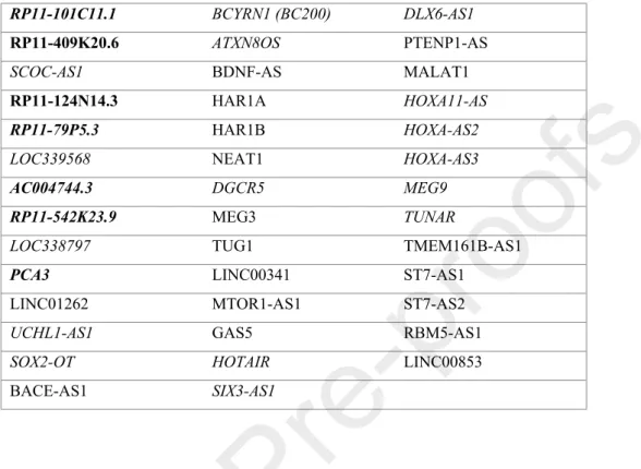
By reviewing literature data we selected 41 lncRNAs which have been implicated in
neurodegenerative malignancies (Table 1.). Nine of these were directly linked to PD (RP11- 101C11.1, RP11-409K20.6, RP11-124N14.3, RP11-79P5.3, AC004744.3, RP11-542K23.9, PCA3 (Soreq et al., 2014), NEAT1 (Kraus et al., 2017; Simchovitz et al., 2019) and MALAT1
(Liu et al., 2017)), while others were associated with AD (BC200, BACE1-AS (Feng et al., 2018; Lukiw et al., 1992)), HD (MEG3, TUG1 (Taurine Up-Regulated Gene 1), LINC00341, HAR-1A (Chanda et al., 2018; Johnson, 2012; Johnson et al., 2010; Wang et al., 2014)), and/or were found to be involved in mechanisms that are likely related to neurodegeneration. We attempted to detect these lncRNAs in peripheral blood cells of controls and PD patients (n=3 in each group) by qRT-PCR. Those lncRNAs, which had a Ct larger than 35 we excluded from the further analysis since the low expression level makes their detection by this technique unreliable. The levels of lncRNAs deemed to be detectable in our first approach were compared within larger groups of healthy and PD samples (n=15 and 18, respectively) (validation study I.). In this comparison we found the level of lncRNAs BC200, PTENP1-AS (Phosphatase and Tensin Homolog Pseudogene 1 Antisense RNA) and MEG3 to be below reliable detection level (Ct over 35). These RNAs therefore were omitted from the further analysis. The expression level of GAS5 showed minimal variation in both PD and control samples, therefore we selected GAS5 as an internal control for reference. Stable expression of GAS5 lncRNA has also been noted by both Santoro et al. and Kraus et al. (Kraus et al., 2017; Santoro et al., 2016). Applying GAS5 normalization, we found the expression of NEAT1 significantly up-regulated among PD patients (fold increase=1.93; p=0.035) compared to the control group (Table 2.). Similarly, we detected a significant up-regulation of the lncRNA TUG1 by comparing PD patients to the control cohort (fold increase=1.71; p=0.036) (Table 2.). Besides NEAT1 and TUG1, no other lncRNA was found to exhibit a significant difference in expression in comparison between this PD group and control cohort.
2.2. Comparison of NEAT1 and TUG1 lncRNA levels between study groups including larger number PD patients and controls
Based on the findings of validation study I., we investigated the expression of NEAT1 and TUG1 lncRNAs in study groups including larger number PD patients (n=43) and controls (n=36) (validation study II.). Applying GAS5 normalization, we found significant up-
regulation of NEAT1 expression among PD patients compared to controls (fold increase= 1.62;
p=0.0019) (Suppl. Table 1., Figure 1.) As DBS treatment was reported to influence lncRNA expression in leukocytes (Soreq et al., 2012), we compared our samples in respect of this treatment. The difference in NEAT1 expression between healthy control and PD patients was significant both in comparisons including and not including DBS patients (fold increase= 1.61 and 1.62; p=0.0021 and 0.0071, respectively) (Suppl. Table 1., Figure 1B and C). Between patients with- and without DBS we did not find significant difference in NEAT1 level (Suppl.
Table 1., Figure 1.). NEAT1 expression was significantly up-regulated as compared to the control group in both EOPD and LOPD group (fold change= 1.5 and 1.82; p=0.0181 and 0.0073, respectively). Between the EOPD and LOPD group however, there was no significant difference in NEAT1 level (Suppl. Table 1., Figure 1E-G,). Comparison of female PD patients to female control individuals revealed significantly up-regulated NEAT1 level among PD patients (fold increase= 1.72; p= 0.0073). Though the difference was not significant, up- regulation could also be observed among male PD patients compared to control male individuals (Suppl. Table 1., Figure 2H and I).
Comparison between subgroups SDD and LDD indicated slightly up-regulated NEAT1 level among patients of the LDD group, however, the difference was not significant. Comparisons of subgroups of SDD and LDD patients to the control group revealed significant up-regulated NEAT1 RNA levels in both cases (fold change= 1.57 and 1.74; p= 0.028 and 0.0008,
respectively) with more prominent NEAT1 up-regulation among LDD patients (Suppl. Table 1., Figure 2J-L.).
After performing Bonferroni correction the difference remained significant in pair wise
comparisons between the control group and PD group-,or patients with DBS or the LDD group.
In the expression of TUG1 we detected no significant difference in either of the comparisons described above (Suppl. Table 2.).
3. Discussion
With this study, we aimed to identify alterations in lncRNA expression in PD that could help to understand the underlying disease mechanisms, may help to find potential therapeutic targets, and can be potential biomarker for diagnosis. Our aim was also to use easily accessible sample to this analysis and reliable experimental approach that is readily available in a clinical
laboratory. We selected for a first comparison 41 lncRNAs which have been reported to be related to neurodegenerative diseases. Among these 7 lncRNAs (indicated in bold in Table 1.) had already been reported by Soreq et al. to have altered expression in PD (Soreq et al., 2014).
From these 7 lncRNAs, we detected only 2 (RP11-409K20.6 and RP11-124N14.3) by our assay, the other 5 had a Ct >40 (indicated in italics in Table 1.). The difference between our and the published data could be explained by the different methods used for expression monitoring:
while Soreq and colleagues carried out whole-transcriptome RNA-Seq analysis, we used real- time PCR. Another explanation could be in a high inter-individual expression variability of lncRNAs (Kornienko et al., 2016), and also the small number of analysed samples. Based on the data from this preliminary study we selected 12 lncRNAs for comparison involving a larger sample number (validation study I.). In this analysis we detected low expression levels for BC200, PTENP1-AS, and MEG3, therefore these transcripts had to be considered undetectable.
Our failure to detect these lncRNAs is unlikely because of technical reason. BC200 lncRNA is expressed predominantly in the brain, specifically in the hypothalamus, but shows low, or no
expression in other tissues (Castle et al., 2010; Tiedge et al., 1993). MEG3 is a candidate tumour suppressor. According to lncRNA database (http://www.lncrnadb.org) MEG3 is not expressed in human white blood cells. PTENP1-AS also acts as a tumour suppressor. Poliseno and colleagues, described relatively low levels of PTENP1-AS under physiological conditions in various human tissues, including peripheral blood leukocytes (Poliseno et al., 2010). In our assays the average Ct of the PTENP1-AS transcript was only slightly above the threshold (37.2
± 1.2 in the control group, and 37.9 ± 2 among PD patients).
Out of the well detectable 9 neurodegeneration-related lncRNAs, we found NEAT1 and TUG1 to be significantly up-regulated in PD patients compared to control group. We tested the expression of these two lncRNAs in a third set of samples (validation study II.). Results on this larger number of PD and control groups however revealed no significant difference in the expression of TUG1 between PD patients and non-PD controls. TUG1 lncRNA acts as transcriptional repressor, that upon DNA damage suppresses the progression of the cell-cycle (Khalil et al., 2009). It achieves this by forming PcG bodies (Polycomb group protein
containing nuclear repressive foci) on the promoters of cell cycle genes in interaction with a component of the Polycomb Repressive complex 1 (PRC1) (Yang et al., 2011). With respect to neurodegenerative diseases, TUG1 was found to be up-regulated in HD (Johnson, 2012), but no other data have been reported regarding TUG1 expression in neurodegenerative diseases, including PD.
We found NEAT1 to be significantly up-regulated in PD patients in multiple comparisons. The most prominent differences were observable by comparing all PD patients to all control
individuals (fold change= 1.62; p= 0.0019), PD patients with DBS to the control group (fold change= 1.61; p= 0.0021), and LDD patients’ group to control group (fold change= 1.74; p=
0.0008). In each of these comparisons the difference remains significant after Bonferroni
correction. In the case of comparison of PD patients with DBS to controls however, the small sample size of PD patients should be taken into account. The finding that the expression of NEAT1 was significantly up-regulated among LDD patients compared to control individuals suggests that the amount of the lncRNA increases with disease progression. This novel notion could be interesting in relation to PD pathology, however, whether the change in NEAT1 level is coincidental or in causative relation with disease progression remains to be explored.
In either way, NEAT1 elevated expression in PD patients deserves attention because this lncRNA has been shown to modulate cellular functions by several mechanisms and it might affect disease pathology via a number of these.
NEAT1 lncRNA is expressed in two forms: a 3756 nucleotide long NEAT1_1, also known as MEN ε, and as a 27 kb long NEAT1_2, alias MEN β. Although the smaller form corresponds to the 5’ end of the longer and both NEAT1 isoforms are involved in paraspeckle formation, the two differ in functions (Sunwoo et al., 2009). In this study we used primers which target the 5’region of NEAT1, consequently permit the detection of both NEAT1 isoforms.
NEAT1 lncRNA was first identified as a virus inducible gene product that was up-regulated in mouse central nervous system after virus infection (Saha et al., 2006). More recent data
demonstrated that HIV-1 (Zhang et al., 2013), influenza virus and herpes simplex virus infection also enhanced NEAT1 expression, and NEAT1 promoted the expression of antiviral genes, such as Interleukin-8 (IL-8) (Imamura et al., 2014). The involvement of NEAT1 in inflammatory processes raises the possibility that this lncRNA plays role in the reported increase of inflammatory markers in the peripheral blood of PD patients (King and Thomas, 2017). Recently various mechanisms have been described by which NEAT1 regulates cellular functions. Most of the effects mediated by NEAT1 are due its involvement in paraspeckle formation. Paraspeckles are approximately 0.5 µm size membraneless subnuclear particles
consisting of a large number of proteins and NEAT1 RNAs as a scaffold. Paraspeckles are responsible for the retention of the A-to-I hyperedited mRNAs in the nucleus (Mao et al., 2011). Through this they play a pivotal role in cellular response to stress. Under cellular stress conditions, such as transcription inhibition or exposure to the combination of
lipopolysaccharide and interferon-γ, specific A-to-I hyperedited RNA types can be rapidly transported into the cytoplasm where they are used for protein synthesis (Prasanth et al., 2005).
By this mechanisms the cell produces a rapid stress response since it can save time by not synthesizing de novo mRNAs, but using already synthesized transcripts for protein synthesis in order to respond to stress stimuli (Nakagawa and Hirose, 2012). By retaining hyperedited mRNAs in the nucleus paraspeckles play role in cell differentiation as well (Bond and Fox, 2009). Recently NEAT1 involvement in regulating mitochondrium homeostasis through the nuclear retention of mRNAs encoding proteins with mitochondrial function (mito-mRNA) has been shown. Paraspeckle-enriched mito-RNAs exhibited enhanced nucleo-cytoplasmic export in NEAT1 knockout cell suggesting a quality control mechanism that prohibit unnecessary translation of mito-mRNAs following stress (Y. Wang et al., 2018). Paraspeckles also regulate gene expression by retaining regulatory proteins. Sequestration and/or liberation of
transcription factors to and from paraspeckles offer rapid ways for modulating gene expression.
The possible roles of NEAT1 in the human central nervous system attracted attention in the last decade. Investigation of human nucleus accumbens samples of heroin abusers showed a
significant up-regulation of the gene as compared to drug-free controls (Michelhaugh et al., 2011). The association of NEAT1 lncRNA expression with neurodegenerative diseases was also examined in ALS (Nishimoto et al., 2013), HD (Johnson, 2012) and PD (Kraus et al., 2017). In 2013 Nishimoto and colleagues reported that NEAT1_2 transcript was up-regulated in ALS patients compared to the control group (Nishimoto et al., 2013). Similarly, NEAT1 up- regulation was found in HD patients (Johnson, 2012). According to a recent study by Kraus et
al., the lncRNA NEAT1 was significantly up-regulated in the anterior cingulate gyrus of PD patients as well as compared to healthy controls (Kraus et al., 2017). More recently Simchovitz et al. reported significant up-regulation of NEAT1 in the substantia nigra (SN) of PD patients compared to non-PD controls. In the SN, dopaminergic neurons were identified as the main NEAT1 expressers and paraspeckle forming cells. Cell culture and murine model studies showed that under oxidative stress conditions the expression of the NEAT1 transcript was enhanced. Genetic ablation of NEAT1 led to a reduction in the number of paraspeckle forming cells and also in the number of paraspeckles in the nucleus, together with a diminishment in the survival of cells pre-treated with oxidative stress agents (Simchovitz et al., 2019).
Interconnection between several PD associated genes and NEAT1 lncRNA has been
demonstrated in a number of PD models. Mutations of the gene encoding LRRK2 protein is among the most frequently identified genetic alterations in both familial and sporadic PD (Kumari and Tan, 2009). LRRK2 is involved in mitophagy and LRRK2 mutation delays the arrest of dysfunctional mitochondria (see (Grünewald et al., 2019)). Simchovitz and colleagues showed that NEAT1 could serve as a natural inhibitor of LRRK2 by retaining it in paraspeckles through its interaction with non-POU domain containing octamer binding (NONO) and splicing factor proline glutamine rich (SFPQ) proteins (Simchovitz et al., 2019).
Studies of MPTP/MPP+ induced mouse and cell culture models of PD yielded seemingly contrasting results regarding the role of NEAT1 in the course of PD. Recent studies involving murine and cell models reported up-regulation of NEAT1 after treatment with MPTP or MPP+, accompanied by decreased cell viability. On the other hand, genetic inhibition of NEAT1 promoted cell survival (Geng et al., 2019; Liu and Lu, 2018; Yan et al., 2018). Various mechanisms have been described by which NEAT1 regulates apoptosis, oxidative stress and neuroinflammation (Figure 2.). Yan et al. showed that in PD mice both NEAT1 and a PD
susceptibility gene, PTEN-induced kinase 1 (PINK1) expression was up-regulated. In vitro findings revealed that whereas genetic ablation of NEAT1 significantly decreased the otherwise enhanced apoptosis rate following MPP+ treatment and down regulated PINK1 expression, overexpression of the lncRNA inhibited the cycloheximide induced degradation of PINK1. The beneficial effects of NEAT1 knockdown were abolished via overexpressing PINK1. Based on these findings the authors proposed that by direct binding to PINK1, NEAT1 stabilizes the protein, thus mediates autophagy and neuronal injury (Yan et al., 2018). NEAT1 down- regulation was also found to decrease α-synuclein expression, and ablation of the lncRNA in SH-SY5Y cells increased cell viability. However, up-regulation of α-synuclein reversed the beneficial changes in apoptosis rate upon genetic inhibition of NEAT1 (Liu and Lu, 2018).
NEAT1 was also proposed to take part in the course of PD via a miR-221 related mechanism.
Recent findings revealed miR-221 down-regulation in serum samples of PD patients and proposed its potential role as a biomarker for the disease (Ding et al., 2016; Ma et al., 2016).
The expression of the micro RNA was also found to be reduced in cellular models of PD and overexpression of miR-221 promoted cell viability. A direct target of miR-221 is PTEN (Phosphatase and tensin homolog deleted on chromosome 10) which in addition to its tumorsuppressor function also has been linked to the pathogenesis of neurodegenerative disorders such as AD, ALS and PD (Ismail et al., 2012). Recently a study of SH-SY5Y cells revealed the down-regulation of miR-221 in parallel with NEAT1 up-regulation following MPP+ treatment (Geng et al., 2019). On the opposite, NEAT1 knockdown caused increased expression of the micro RNA. Overexpressing miR-221 prior to MPP+ treatment led to a decrease in ROS generation, LDH release and down-regulation of IL-1β, IL-6 and TNFα.
NEAT1 was identified as a molecular sponge for miR-221 that observation led to the
conclusion that MPP+ induced neuronal damage alleviation by NEAT1 ablation was partly due to the decreased sponging of miR-221 by NEAT1 (Geng et al., 2019).
All combined NEAT1 seems to be in a central position to regulate several of those cellular functions which have been shown to be altered in PD. To determine, which of the effects exerted by NEAT1 through modulating mitochondrial homeostasis, apoptosis, stress response, mRNA and miRNA availability are specific to unique cells in the central nervous system or mediate functions of different cell types, among them white blood cells, requires further
studies. Nonetheless, the change of NEAT1 level in peripheral blood cells in PD patients can be well accommodated with the described roles of the lncRNA and the effects it shows in PD models. However, whether the changes of NEAT1 levels that we observed in blood cells of PD patients are consequential or in causative relation with the disease needs further elucidation.
Answer to this question should be sought with keeping in mind the recent shift in the paradigm of PD aetiology that instead of viewing the disease specific to the central nervous system views it as a systemic ailment.
4. Conclusions
In easily accessible peripheral blood cells of PD patients we detected an increased NEAT1 lncRNA level. In light of the diverse mechanisms by which NEAT1 affects cellular functions, the increased level of the lncRNA can be causally or coincidently linked to PD. Change in NEAT1 lncRNA expression in brain tissue of PD patients has been reported recently (Kraus et al., 2017; Simchovitz et al., 2019). Our finding that the change in NEAT1 level can be detected in blood samples might open possibilities to find signs of developing PD by investigating samples more easily accessible than the nervous tissue. By uncovering the cause and effects of the change of NEAT1 level in PD progression could lead to a better understanding of the underlying mechanisms of the disease and to the identification of new potential therapeutic targets to interfere with this devastating neurodegeneration.
5. Experimental procedure 5.1. Study design
For a preliminary study we selected 41 lncRNAs, each of which has been implicated in
neurodegenerative disease (Table 1.) and examined their levels in blood cells of a small number of PD patient versus control samples (n=3 and 3) with real-time polymerase chain reaction (RT-PCR). Based on this preliminary result we narrowed the set of neurodegeneration-related lncRNAs to 12 (validation study I., Table 2.) and analysed the expression levels of these lncRNAs in a second, larger group of PD patients and non-PD controls (n=18 and 15). Based on the results of these assays 2 lncRNAs out of the 12 were selected for further study and the level of these was investigated a third larger group of patients and controls consisting 43 and 36 individuals, respectively (validation study II.).
5.2. Participiants
Both PD patients and non-PD volunteers were Hungarians of Caucasian origin. The diagnosis of PD was set up based on medical history and physical examination carried out by movement disorder specialists. Individuals with known other malignancies or with cancerous disease in their history were excluded. Validation study group I. composed of 9 women and 9 men, the mean age of the cohort was 60.3±5.7 years. The average age at disease onset was 52.5±5.6 years. Validation study group II consisted of 43 PD patients. The mean age of this group of patients was 63.3±11.4 years, the male/female ratio: 24/19, the average age at disease onset was 54.8±12.6 years. Out of the 43 participants, 6 reported first symptoms to appear before the age of 40 years (mean age at disease onset 35±7.8 years). One of these patients reported the possibility of positive family history regarding PD: the main symptom of this participant was left side dominant tremor, and tremor was reported to be present in family members both of the
maternal and paternal side, however, to our knowledge no definite diagnosis of PD was set up for the relatives. Genetic screening of this patient (pathogenic LRRK2, parkin and DJ-1 mutations) yielded negative results.
For data analysis patients were further divided into two cohorts: the early onset PD group (EOPD) consisted those, who had a disease onset before or at the age of 60 years (n=27; age:
57.6±9.8 years), while the late onset PD group (LOPD) consisted those with disease onset after 60 years (n=16; age: 73±5.9 years). The average age at the onset of the disease in EOPD and LOPD groups was 47.5±10.2 and 66.5±4.0 years, respectively. Based on disease duration (DD) patients were also separated into two subgroups. Out of the 42 patients, 27 belonged to the
‘short DD’ (SDD) subgroup with DD <10 years (age: 62.9±11.9 years, DD: 4.9±2.8 years), while the ‘long DD’ - DD≥10 years - subgroup (LDD) consisted of 15 individuals (age:
63.7±10.9 years, DD: 14.6±5.0 years). One patient had to be excluded from this analysis as no information was available upon DD. In validation study I. there were no patients who had DBS treatment. Among PD patients of validation study II., 8 out of 43 participants had gone under DBS surgery (PD DBS n=8, age: 64.3±7.1 years; PD no DBS n=35, age: 63.1±12.2 years).
Out of the 43 PD patients 26 individuals went under genetic testing regarding the monogenic forms of the disease ((LRRK2; parkin and DJ-1), of which all tests yielded negative results.
The majority of patients - 30 out of 43 - presented tremor. Modified Hoehn and Yahr scale varied between 1 and 5, mean 1.9±0.8. UPDRS motor score of the patients at the time of sample collection ranged from 2 to 48 points, the mean score was 12±8.9 points. 36 out of 43 PD patients received levodopa as treatment, and the mean time of treatment duration at sample collection was 6.4 ±4.1 years. Further details of clinical data are presented in Supplementary Table 4.
In validation study I. control group, the male/female ratio was 6 to 9, and the mean age was 61.3±9.9 years. The validation study II. control group consisted of 36 individuals (male/female:
16/20), the mean age was 57.6±18.0 years (for detailed demographic data see Suppl. Table 3.).
Before participating, informed consent was obtained from each participant included in the study. The study protocol was approved by the Medical Research Council Scientific and Research Ethics Committee and was in full accordance with the Declaration of Helsinki ethical principles for medical research involving human subjects.
5.3. Methods
Blood samples were collected from members of patient and control groups in EDTA containing blood tubes. White blood cells of 5 ml blood samples were separated by centrifugation and lysed in 1 ml of TRI Reagent (Sigma) by repetitive pipetting within 2 hours of collection.
Samples were then stored at -80oC. Total RNA isolation was carried out following the Trizol RNA purification protocol according to the manufacturer’s instructions (Sigma). RNA concentration was determined with a MaestroNano micro-volume spectrophotometer. In the preliminary and first validation study complementary DNA (cDNA) was synthesized from 500 ng of extracted RNA with the use of RT2 First Strand Kit (Qiagen) following the
manufacturer’s instructions. Real-timePCR was performed with the use of RT2 SYBR Green Mastermix (Qiagen). Equal volumes of cDNA samples were aliquoted into the wells of a specifically designed Custom RT2 PCR Array (Qiagen) containing lncRNA specific primer pairs (25 ul final volume). Two arrays were designed and used for the groups of 41 and 12 lncRNAs (Table 1 and 2, respectively).
For validation study II commercially available NEAT1 and TUG1 gene-specific primers were used (Qiagen). cDNA was synthesized from 1000 ng of RNA with the use of Revert Aid First Strand cDNA Synthesis Kit (Thermo Scientific). Real-time PCR was performed with the use of
RT2 SYBR Green Mastermix (qPCRBIO) in 25 µl final volume. PCR reactions were carried out in a CFX96 thermocycler (Bio-Rad). Cycling conditions are available upon request.
5.4. Data analysis and statistics
Statistical analysis of PCR results was performed using RT2 PCR analysis web portal (http://pcrdataanalysis.sabiosciences.com/pcr/arrayanalysis.php) and GraphPad Prism 6.01 statistics software using the ΔΔCt method. In short, ΔCt was calculated as the difference between a gene of interest and the average of reference gene, ΔΔCt was calculated as ΔCt (patient) – average ΔCt (control) and fold change was determined as 2-ΔΔCt value (Livak and Schmittgen, 2001). For the identification of the outliers among 2-ΔΔCt replicates the ROUT method was used. D’Agostino and Pearson omnibus normality test was used for the analysis of data distribution. If the data showed normal distribution, we implemented unpaired t-test, while in the case of non-normal distribution Mann-Whitney U test was performed. P value under 0.05 was considered significant. Due to the multiple comparisons, Bonferroni correction was
implemented. Following this, p value under 0.004 was considered significant.
Data Availability
The data that support the findings of this study are available from the corresponding author.
Acknowledgements
The current work was supported by Hungarian Brain Research Program [Grant number KTIA_13_NAP-A-II/17] and by Economic Development and Innovation Operational Programme [Grant number GINOP-2.3.2-15-2016-00034].
Conflict of interest
The authors declare no conflict of interest regarding the publication of this article.
Compliance with Ethical Standards
Informed consent was obtained from all individual participants included in the study. All procedures performed in studies involving human participants were in accordance with the ethical standards of the institutional and national research committee and with the 1964 Helsinki declaration and its later amendments or comparable ethical standards.
References
Benson, D.L., Huntley, G.W., 2019. Are we listening to everything the PARK genes are telling us? J. Comp. Neurol. 527, 1527–1540. https://doi.org/10.1002/cne.24642
Berstad, K., Berstad, J.E.R., 2017. Parkinson’s disease; the hibernating spore hypothesis. Med.
Hypotheses 104, 48–53. https://doi.org/10.1016/j.mehy.2017.05.022
Bond, C.S., Fox, A.H., 2009. Paraspeckles : nuclear bodies built on long noncoding RNA. J.
Cell Biol. 186, 637–644. https://doi.org/10.1083/jcb.200906113
Brockdorff, N., Ashworth, A., Kay, G.F., Mccabe, V.M., Norris, D.P., Cooper, P.J., Swift, S., 1992. The product of the mouse Xist gene is a 15 kb inactive X-specific transcript containing no consewed ORF and located in the nucleus. Cell 71, 515–526.
https://doi.org/10.1016/0092-8674(92)90519-I
Brown, C.J., Hendrich, B.D., Rupert, J.L., Xing, Y., Lawrence, J., Willard, F., 1992. The
Human XIST Gene: Analysis of a 17 kb Inactive X-Specific RNA That Contains Conserved Repeats and Is Highly Localized within the Nucleus. Cell 71, 527–542.
https://doi.org/10.1016/0092-8674(92)90520-M
Butler, A.A., Johnston, D.R., Kaur, S., Lubin, F.D., 2019. Long noncoding RNA NEAT1 mediates neuronal histone methylation and age-related memory impairment. Sci. Signal.
12, eaaw9277. https://doi.org/10.1126/scisignal.aaw9277
Carrieri, C., Forrest, A.R.R., Santoro, C., Persichetti, F., Carninci, P., Zucchelli, S., Gustincich, S., 2015. Expression analysis of the long non-coding RNA antisense to Uchl1 ( AS Uchl1 ) during dopaminergic cells ’ differentiation in vitro and in neurochemical models of Parkinson ’ s disease 9, 1–11. https://doi.org/10.3389/fncel.2015.00114
Castle, J.C., Armour, C.D., Lower, M., Haynor, D., Biery, M., Bouzek, H., Chen, R., Jackson, S., Johnson, J.M., Rohl, C.A., Raymond, Christopher, K., 2010. Digital Genome-Wide ncRNA Expression, Including SnoRNAs, across 11 Human Tissues Using PolyA-Neutral Amplification. PLoS One 5, 1–9. https://doi.org/10.1371/journal.pone.0011779
Chanda, K., Das, S., Chakraborty, J., Bucha, S., Maitra, A., Chatterjee, R., Mukhopadhyay, D., Bhattacharyya, N.P., 2018. Altered levels of long ncRNAs Meg3 and Neat1 in cell and animal models of Huntington’s disease. RNA Biol. 15, 1348–1363.
https://doi.org/10.1080/15476286.2018.1534524
Ding, H., Huang, Z., Chen, M., Wang, C., Chen, X., Chen, J., Zhang, J., 2016. Identification of a panel of five serum miRNAs as a biomarker for Parkinson’s disease. Park. Relat. Disord.
22, 68–73. https://doi.org/10.1016/j.parkreldis.2015.11.014
Dzamko, N., Geczy, C.L., Halliday, G.M., 2015. Inflammation is genetically implicated in Parkinson’s disease. Neuroscience 302, 89–102.
https://doi.org/10.1016/j.neuroscience.2014.10.028
Fehlbaum-beurdeley, P., Sol, O., Laurent, D., Lemari, J.C., Zhou, W., Hampel, H., Einstein, R., 2012. Validation of AclarusDx TM , a Blood-Based Transcriptomic Signature for the Diagnosis of Alzheimer’s Disease. J. Alzheimer’s Dis. 32, 169–181.
https://doi.org/10.3233/JAD-2012-120637
Feng, J., Bi, C., Clark, B.S., Mady, R., Shah, P., Kohtz, J.D., 2006. The Evf-2 noncoding RNA is transcribed from the Dlx-5 / 6 ultraconserved region and functions as a Dlx-2
transcriptional coactivator. Genes Dev. 20, 1470–1484.
https://doi.org/10.1101/gad.1416106.
Feng, L., Liao, Y.T., He, J.C., Xie, C.L., Chen, S.Y., Fan, H.H., Su, Z.P., Wang, Z., 2018.
Plasma long non-coding RNA BACE1 as a novel biomarker for diagnosis of Alzheimer disease. BMC Neurol. 18, 1–8. https://doi.org/10.1186/s12883-017-1008-x
Geng, L., Zhao, J., Liu, W., Chen, Y., 2019. Knockdown of NEAT1 ameliorated MPP+ - induced neuronal damage by sponging miR-221 in SH-SY5Y cells. RSC Adv. 9, 25257–
25265. https://doi.org/10.1039/c9ra05039f
Goedert, M., 2001. Alpha-synuclein and neurodegenerative diseases. Neurosci. 2, 492–501.
https://doi.org/10.1038/35081564
Grimes, D.A., Han, F., Panisset, M., Racacho, L., Xiao, F., Zou, R., Westaff, K., Bulman, D.E., 2006. Translated Mutation in the Nurr1 Gene as a Cause for Parkinson’s Disease. Mov.
Disord. 21, 906–909. https://doi.org/10.1002/mds.20820
Grünewald, A., Kumar, K.R., Sue, C.M., 2019. New insights into the complex role of mitochondria in Parkinson’s disease. Prog. Neurobiol. 177, 73–93.
https://doi.org/10.1016/j.pneurobio.2018.09.003
Harris, L.W., Pietsch, S., Cheng, T.M.K., Schwarz, E., Guest, P.C., Bahn, S., 2012.
Comparison of Peripheral and Central Schizophrenia Biomarker Profiles. PloS one 7, 1–
9. https://doi.org/10.1371/journal.pone.0046368
Hawkes, C.H., Del Tredici, K., Braak, H., 2007. Parkinson’s disease: a dual-hit hypothesis.
Neuropathol. Appl. Neurobiol. 33, 599–614. https://doi.org/10.1111/j.1365- 2990.2007.00874.x
Imamura, K., Imamachi, N., Akizuki, G., Kumakura, M., Kawaguchi, A., Nagata, K., Kato, A., Kawaguchi, Y., Sato, H., Yoneda, M., Kai, C., Yada, T., Suzuki, Y., Yamada, T., Ozawa, T., Kaneki, K., Inoue, T., Kobayashi, M., Kodama, T., Wada, Y., Sekimizu, K., Akimitsu, N., 2014. Long Noncoding RNA NEAT1-Dependent SFPQ Relocation from Promoter Region to Paraspeckle Mediates IL8 Expression upon Immune Stimuli. Mol. Cell 53, 393–
406. https://doi.org/10.1016/j.molcel.2014.01.009
Ismail, A., Ning, K., Al-Hayani, A., Sharrack, B., Azzouz, M., 2012. PTEN: a molecular target for neurodegenerative disorders. Transl. Neurosci. 3, 132–142.
https://doi.org/10.2478/s13380-012-0018-9
Johnson, R., 2012. Neurobiology of Disease Long non-coding RNAs in Huntington’s disease neurodegeneration. Neurobiol. Dis. 46, 245–254.
https://doi.org/10.1016/j.nbd.2011.12.006
Johnson, R., Richter, N., Jauch, R., Gaughwin, P.M., Zuccato, C., Cattaneo, E., Stanton, L.W., 2010. Human accelerated region 1 noncoding RNA is repressed by REST in Huntington’s disease. Physiol. Genomics 41, 269–274.
https://doi.org/10.1152/physiolgenomics.00019.2010.
Khalil, A.M., Guttman, M., Huarte, M., Garber, M., Raj, A., Rivea, D., Thomas, K., Presser,
A., Bernstein, B.E., Oudenaarden, A. Van, Regev, A., Lander, E.S., Rinn, J.L., 2009.
Many human large intergenic noncoding RNAs associate with chromatin-modifying complexes and affect gene expression. PNAS 106, 11667–11672.
https://doi.org/10.1073/pnas.0904715106
King, E., Thomas, A., 2017. Systemic Inflammation in Lewy Body Diseases. Alzheimer Dis.
Assoc. Disord. 31, 346–356. https://doi.org/10.1097/WAD.0000000000000211 Kornienko, A.E., Dotter, C.P., Guenzl, P.M., Gisslinger, H., Gisslinger, B., Cleary, C.,
Kralovics, R., Pauler, F.M., Barlow, D.P., 2016. Long non-coding RNAs display higher natural expression variation than protein-coding genes in healthy humans. Genome Biol.
17, 1–23. https://doi.org/10.1186/s13059-016-0873-8
Kraus, T.F.J., Haider, M., Spanner, J., Steinmaurer, M., Dietinger, V., Kretzschmar, H.A., 2017. Altered Long Noncoding RNA Expression Precedes the Course of Parkinson’s Disease-a Preliminary Report. Mol. Neurobiol. 54, 2869–2877.
https://doi.org/10.1007/s12035-016-9854-x
Kumari, U., Tan, E.K., 2009. LRRK2 in Parkinson’s disease: genetic and clinical studies from patients. FEBS 276, 6455–6463. https://doi.org/10.1111/j.1742-4658.2009.07344.x Li, Z., Zhao, W., Wang, M., Zhou, X., 2019. The role of long noncoding RNAs in gene
expression regulation. Gene Expr. Profiling Cancer 1–17.
https://doi.org/10.5772/intechopen.81773
Liu, B., Gao, H., Hong, J., 2003. Parkinson’s Disease and Exposure to Infectious Agents and Pesticides and the Occurrence of Brain Injuries: Role of Neuroinflammation. Environ.
Health Perspect. 111, 1065–1073. https://doi.org/10.1289/ehp.6361
Liu, W., Zhang, Q., Zhang, J., Pan, W., Zhao, J., Xu, Y., 2017. Long non-coding RNA
MALAT1 contributes to cell apoptosis by sponging miR-124 in Parkinson disease. Cell Biosci. 7, 1–9. https://doi.org/10.1186/s13578-017-0147-5
Liu, Y., Lu, Z., 2018. Long non-coding RNA NEAT1 mediates the toxic of Parkinson’s disease induced by MPTP/MPP+ via regulation of gene expression. Clin. Exp. Pharmacol.
Physiol. 45, 841–848. https://doi.org/10.1111/1440-1681.12932
Livak, K.J., Schmittgen, T.D., 2001. Analysis of relative gene expression data using real-time quantitative PCR and the 2-ΔΔCT method. Methods 25, 402–408.
https://doi.org/10.1006/meth.2001.1262
Lukiw, W.J., Handley, P., Wong, L., Mclachlan, C.D.R., 1992. BC200 RNA in normal human neocortex, non-Alzheimer dementia (NAD), and senile dementia of the Alzheimer type (AD). Neurochem. Res. 17, 591–597. https://doi.org/10.1007/BF00968788
Ma, W., Li, Y., Wang, C., Xu, F., Wang, M., Liu, Y., 2016. Serum miR-221 serves as a biomarker for Parkinson’s disease. Cell Biochem. Funct. 34, 511–515.
https://doi.org/10.1002/cbf.3224
Mao, Y.S., Zhang, B., Spector, D.L., 2011. Biogenesis and function of nuclear bodies. Trends Genet. 27, 295–306. https://doi.org/10.1016/j.tig.2011.05.006
Martianov, I., Ramadass, A., Barros, A.S., Chow, N., Akoulitchev, A., 2007. Repression of the human dihydrofolate reductase gene by a non-coding interfering transcript. Nature 445, 666–670. https://doi.org/10.1038/nature05519
Michelhaugh, S.K., Lipovich, L., Blythe, J., Jia, H., Kapatos, G., Bannon, M.J., 2011. Mining Affymetrix microarray data for long non-coding RNAs: altered expression in the nucleus accumbens of heroin users. J. Neurochem. 116, 459–466. https://doi.org/10.1111/j.1471- 4159.2010.07126.x
Nakagawa, S., Hirose, T., 2012. Paraspeckle nuclear bodies-useful uselessness? Cell. Mol. Life Sci. 69, 3027–3036. https://doi.org/10.1007/s00018-012-0973-x
Nickles, D., Chen, H.P., Li, M.M., Khankhanian, P., Madireddy, L., Caillier, S.J., Santaniello, A., Cree, B.A.C., Pelletier, D., Hauser, S.L., Oksenberg, J.R., Baranzini, S.E., 2013. Blood RNA profiling in a large cohort of multiple sclerosis patients and healthy controls. Hum.
Mol. Genet. 22, 4194–4205. https://doi.org/10.1093/hmg/ddt267
Nishimoto, Y., Nakagawa, S., Hirose, T., Okano, H.J., Takao, M., Shibata, S., Suyama, S., Kuwako, K., Imai, T., Murayama, S., Suzuki, N., Okano, H., 2013. The long non-coding RNA nuclear-enriched abundant transcript 1 _ 2 induces paraspeckle formation in the motor neuron during the early phase of amyotrophic lateral sclerosis. Mol. Brain 6, 1–18.
https://doi.org/10.1186/1756-6606-6-31
Oe, S., Kimura, T., Yamada, H., 2019. Regulatory non-coding RNAs in nervous system development and disease. Front. Biosci. 24, 1203–1240. https://doi.org/10.2741/4776 Poliseno, L., Salmena, L., Zhang, J., Carver, B., Haveman, W.J., Pandolfi, P.P., 2010. A
coding-independent function of gene and pseudogene mRNAs regulates tumour biology.
Nature 465, 1033–1038. https://doi.org/10.1038/nature09144
Prasanth, K. V, Prasanth, S.G., Xuan, Z., Hearn, S., Freier, S.M., Bennett, C.F., Zhang, M.Q., Spector, D.L., 2005. Regulating Gene Expression through RNA Nuclear Retention. Cell 123, 249–263. https://doi.org/10.1016/j.cell.2005.08.033
Saha, S., Murthy, S., Rangarajan, P.N., 2006. Identification and characterization of a virus- inducible non-coding RNA in mouse brain. J. Gen. Virol. 87, 1991–1995.
https://doi.org/10.1099/vir.0.81768-0
Santoro, M., Nociti, V., Lucchini, M., Fino, C. De, Losavio, F.A., Mirabella, M., 2016.
Expression Profile of Long Non-Coding RNAs in Serum of Patients with Multiple Sclerosis. J. Mol. Neurosci. 59, 18–23. https://doi.org/10.1007/s12031-016-0741-8
Simchovitz, A., Hanan, M., Niederhoffer, N., Madrer, N., Yayon, N., Bennett, E.R., Greenberg, D.S., Kadener, S., Soreq, H., 2019. NEAT1 is overexpressed in Parkinson’s disease
substantia nigra and confers drug-inducible neuroprotection from oxidative stress. FASEB J. 33, 11223–11234. https://doi.org/10.1096/fj.201900830r
Soreq, L., Bergman, H., Goll, Y., Greenberg, D.S., Israel, Z., Soreq, H., 2012. Deep brain stimulation induces rapidly reversible transcript changes in Parkinson’s leucocytes. J. Cell.
Mol. Med. 16, 1496–1507. https://doi.org/10.1111/j.1582-4934.2011.01444.x
Soreq, L., Guffanti, A., Salomonis, N., Simchovitz, A., Israel, Z., Bergman, H., Soreq, H., 2014. Long Non-Coding RNA and Alternative Splicing Modulations in Parkinson’s Leukocytes Identified by RNA Sequencing. PLoS Comput. Biol. 10, 1–22.
https://doi.org/10.1371/journal.pcbi.1003517
Sunwoo, H., Dinger, M.E., Wilusz, J.E., Amaral, P.P., Mattick, J.S., Spector, D.L., 2009. MEN e/b nuclear-retained non-coding RNAs are up-regulated upon muscle differentiation and are essential components of paraspeckles. Genome Res. 3, 347–359.
https://doi.org/10.1101/gr.087775.108.
Svensson, E., Horvath-Puho, E., Thomsen, R.W., Djurhuus, J.C., Pedersen, L., Borghammer, P., Sørensen, H.T., 2015. Vagotomy and Subsequent Risk of Parkinson’s Disease. Ann.
Neurol. 78, 522–529. https://doi.org/10.1002/ana.24448
Tiedge, H., Chen, W., Brosius, J., 1993. Primary structure, neural-specific expression, and dendritic location of human BC200 RNA. J. Neurosci. 13, 2382–2390.
Wang, D.Q., Fu, P., Yao, C., Zhu, L.S., Hou, T.Y., Chen, J.G., Lu, Y., Liu, D., Zhu, L.Q.,
2018. Long non-coding RNAs, novel culprits, or bodyguards in neurodegenerative diseases. Mol. Ther. - Nucleic Acids 10, 269–276.
https://doi.org/10.1016/j.omtn.2017.12.011
Wang, F., Fischhaber, P.L., Guo, C., Tang, T.S., 2014. Epigenetic modifications as novel therapeutic targets for Huntington’s disease. Epigenomics 6, 287–297.
https://doi.org/10.2217/epi.14.19
Wang, X., Arai, S., Song, X., Reichart, D., Du, K., Pascual, G., Tempst, P., Rosenfeld, M.G., Glass, C.K., Kurokawa, R., 2008. Induced ncRNAs allosterically modify RNA binding proteins in cis to inhibit transcription. Nature 454, 126–130.
https://doi.org/10.1038/nature06992.Induced
Wang, Y., Hu, S.-B., Wang, M.-R., Yao, R.-W., Wu, D., Yang, L., Chen, L.-L., 2018. Genome- wide screening of NEAT1 regulators reveals cross-regulation between paraspeckles and mitochondria. Nat. Cell Biol. 20, 1145–1158. https://doi.org/10.1038/s41556-018-0204-2 Weller, C., Oxlade, N., Dobbs, S.M., Dobbs, R.J., Charlett, A., Bjarnason, I.T., 2005. Role of
inflammation in gastrointestinal tract in aetiology and pathogenesis of idiopathic parkinsonism. FEMS Immunol. Med. Microbiol. 44, 129–135.
https://doi.org/10.1016/j.femsim.2005.01.011
Westfall, S., Lomis, N., Kahouli, I., Dia, S.Y., 2017. Microbiome, probiotics and neurodegenerative diseases: deciphering the gut brain axis. Cell. Mol. Life Sci.
https://doi.org/10.1007/s00018-017-2550-9
Xu, P., Liang, R., Jankovic, J., Hunter, C., Zeng, Y.X., Ashizawa T, Lai, D., Le. W D, 2002.
Association of homozygous 7048G7049 variant in the intron six of Nurr1 gene with Parkinson’s disease. Neurology 58, 881–884. https://doi.org/10.1212/WNL.58.6.881
Yan, W., Chen, Z.Y., Chen, J.Q., Chen, H.M., 2018. LncRNA NEAT1 promotes autophagy in MPTP-induced Parkinson’s disease through stabilizing PINK1 protein. Biochem. Biophys.
Res. Commun. 496, 1019–1024. https://doi.org/10.1016/j.bbrc.2017.12.149
Yang, L., Lin, C., Liu, W., Zhang, J., Ohgi, K.A., Grinstein, J.D., Dorrestein, P.C., 2011. Gene Relocation between Nuclear Structures Mediates Gene Activation Programs. Cell 147, 773–788. https://doi.org/10.1016/j.cell.2011.08.054
Zhang, Q., Chen, C.-Y., Yedavali, V.S.R.K., Jeang, K.-T., 2013. NEAT1 Long Noncoding RNA and Paraspeckle Bodies Modulate HIV-1 Posttranscriptional Expression. MBio 4, 1–
9. https://doi.org/10.1128/mBio.00596-12.
Figure 1. Comparisons of NEAT1 lncRNA level between controls (n=36) and PD patients (n=43) and their subgroups. Fold regulation are shown with standard deviation. Significant up-regulation of NEAT1 expression was detected in comparisons between PD and control group (A), PD patients with or without DBS and control group (B and C). Comparison between patients with and without DBS does not shown significant difference in NEAT1 level (D). NEAT1 was found to be significantly upregulated in EOPD and LOPD groups as compared to control group (E and F), while no significant difference was detected between EOPD and LOPD patients (G). NEAT1 was found to be up-regulated both in female PD to female control and male PD to male control comparisons (H and I), however the difference was not significant in the latter. Neither was significant difference
detectable in NEAT1 level between SDD and LDD patient groups (J). NEAT1 was found significantly up-regulated in comparisons between both SDD and LDD groups and controls (K and L). The difference of NEAT1 expression between PD group vs. control group, patients with DBS vs. control group and LDD vs. control group remained significant after Bonferroni correction.
Abbreviations: PD: Parkinson’s disease; Ctrl: control; DBS: deep brain stimulation; EOPD:
early onset Parkinson’s disease; LOPD: late onset Parkinson’s disease; SDD: short disease duration; LDD: long disease duration; ns.: non-significant; *: p<0,05; **: p<0,01; ***:
p<0,001; #: p value significant after Bonferroni correction.
Figure 2. Mechanisms by which NEAT1 might effect cell viability and PD.
NEAT1 lncRNA, a major constituent of paraspeckles, plays divers regulatory roles by modulating the availability of mRNAs, miRNAs and transcription factors. By the nuclear retention of mito-mRNAs - mRNAs encoding proteins with mitochondrial function – NEAT1
directly affects mitochondrium homeostasis. (Y. Wang et al., 2018).
In the pathogenesis of PD (boxed) NEAT1 was suggested to participate by regulating autophagy, neuroinflammation and neuronal cell injury via stabilizing PINK1 (Yan et al., 2018), influencing SNCA expression (Liu and Lu, 2018) and sponging miR-221 (Geng et al., 2019). NEAT1 was also proposed to be a bona fide LRRK2 inhibitor acting via its nuclear retention (Simchovitz et al., 2019).
Abbreviations: NEAT1: Nuclear paraspeckle assembly transcript 1; PINK1: PTEN-induced kinase 1; SNCA: α-synuclein; LRRK2: Leucine-rich repeat kinase 2; mito-mRNAs: messenger RNAs encoding proteins with mitochondrial function.
Figure 1. Comparisons of NEAT1 lncRNA level between controls (n=36) and PD patients (n=43) and their subgroups.
Fold up/down regulation
PD total
(n=43) Ctrl total (n=36) 0
1 2 3 4 5
#
**
A
Fold up/down regulation
PD D BS
(n=8) Ctrl total
(n=36) 0
1 2 3 4 5
#
**
B
Fold up/down regulation
PD no DBS (n=3
5) Ctrl tot
al
(n=36) 0
1 2 3 4
5 **
C
Fold up/down regulation
PD D BS (n=8)
PD no DBS (n=3
5) 0
1 2 3 4 5
D ns.
Fold up/down regulation
EOPD(n=2 7)
Ctrl total (n=36) 0
1 2 3 4
5 *
E
Fold up/down regulation
LOPD (n=1
6)
Ctrl total (n=36) 0
1 2 3 4
5 *
F
Fold up/down regulation
EOP D
(n=27) LOPD
(n=16) 0
1 2 3 4
5 ns.
G
Fold up/down regulation
PD fema le
(n=19)
Ctrl fema le
(n=20) 0
1 2 3 4 5
H **
Fold up/down regulation
PD male
(n=24) Ctrl ma
le
(n=16) 0
1 2 3 4
5 ns.
I
Fold up/down regulation
SDD
(n=27) LDD
(n=15) 0
1 2 3 4 5
J ns.
Fold up/down regulation
SDD (n=27)
Ctrl total (n=36) 0
1 2 3 4
5 *
K
Fold up/down regulation
LDD (n=1
5)
Ctrl tot al (n=36) 0
1 2 3 4 5
#
L ***
Figure 2. Mechanisms by which NEAT1 might effect cell viability and PD.
Sponging of miR-221 PINK1 stabilization
NEAT1
Modulation of the availability of mito- mRNAs, mRNAs, miRNAs and
transcriptional regulators
LRRK2 inhibition (?) Modulation of gene expression and
mitochondrial homeostasis
NEAT1
SNCA
Increase of αsynuclein expression PINK1
NEAT1
NEAT1 miR-221
NEAT1 LRRK2
Modulation of cell viability
Paraspeckle formation
Table 1. Neurodegeneration implicated lncRNAs included in the perliminary study (control n=3, PD n=3).
Bold: lncRNAs reported to have altered expression in PD (Soreq et al., 2014). Italics: lncRNAS detected in low level (Ct>40), bold: lncRNAs, which were reported to have altered expression in PD by Soreq and collegues (Soreq et al., 2014).
RP11-101C11.1 BCYRN1 (BC200) DLX6-AS1
RP11-409K20.6 ATXN8OS PTENP1-AS
SCOC-AS1 BDNF-AS MALAT1
RP11-124N14.3 HAR1A HOXA11-AS
RP11-79P5.3 HAR1B HOXA-AS2
LOC339568 NEAT1 HOXA-AS3
AC004744.3 DGCR5 MEG9
RP11-542K23.9 MEG3 TUNAR
LOC338797 TUG1 TMEM161B-AS1
PCA3 LINC00341 ST7-AS1
LINC01262 MTOR1-AS1 ST7-AS2
UCHL1-AS1 GAS5 RBM5-AS1
SOX2-OT HOTAIR LINC00853
BACE-AS1 SIX3-AS1
Table 2. LncRNAs included in validation study I. (control n=15, PD n=18) and their expression
changes.
LncRNAs detected in Ct >35 were excluded from further analysis.
Abbreviations: PD: Parkinson’s disease; Ctrl: control.
Average Ct
Gene Symbol PD Ctrl
Fold change (PD/ Ctrl)
P value
RP11-409K20.6 34.21 34.63 1.53 0.88
GAS5 27.73 27.53 n.a. n.a.
RP11-124N14.3 34.36 34.73 1.48 0.95
LINC00341 33.77 34.31 1.66 0.55
PINK1-AS 34.23 34.93 1.86 0.79
NEAT1 26.70 27.46 1.93 0.035*
MALAT1 31.95 32.36 1.52 0.07
MTOR-AS1 34.96 34.97 1.15 0.57
TUG1 30.35 30.93 1.71 0.037*
BC200 >35 >35 n.a. n.a.
PTENP1-AS >35 >35 n.a. n.a.
MEG3 >35 >35 n.a. n.a.