Association of Skin Examination Behaviors and Thinner Nodular vs Superficial Spreading Melanoma at Diagnosis
Clio Dessinioti, MD; Alan C. Geller, RN, MPH; Aravella Stergiopoulou, MSc; Susan M. Swetter, MD;
Eszter Baltas, MD; Jonathan E. Mayer, MD, MPH; Timothy M. Johnson, MD; John Talaganis, MD;
Myrto Trakatelli, MD; Dimitrios Tsoutsos, MD; Gerasimos Tsourouflis, MD; Alexander J. Stratigos, MD
IMPORTANCEEarly melanoma detection strategies include skin self-examination (SSE), physician skin examination (PSE), and promotion of patient knowledge about skin cancer.
OBJECTIVETo investigate the association of SSE, PSE, and patient attitudes with the detection of thinner superficial spreading melanoma (SSM) and nodular melanoma (NM), the latter of which tends to elude early detection.
DESIGN, SETTING, AND PARTICIPANTS This cross-sectional, questionnaire-based, multicenter study identified patients with newly diagnosed cutaneous melanoma at 4 referral hospital centers in the United States, Greece, and Hungary. Among 920 patients with a primary invasive melanoma, 685 patients with SSM or NM subtype were included.
INTERVENTIONSA standardized questionnaire was used to record sociodemographic information, SSE and PSE practices, and patient perceptions in the year prior to diagnosis.
MAIN OUTCOMES AND MEASURESData were analyzed according to histologic thickness, with a 2-mm cutoff for thinner SSM and NM.
RESULTSOf 685 participants (mean [SD] age, 55.6 [15.1] years; 318 [46%] female), thinner melanoma was detected in 437 of 538 SSM (81%) and in 40 of 147 NM (27%). Patients who routinely performed SSE were more likely to be diagnosed with thinner SSM (odds ratio [OR], 2.61; 95% CI, 1.14-5.40) but not thinner NM (OR, 2.39; 95% CI, 0.84-6.80). Self-detected clinical warning signs (eg, elevation and onset of pain) were markers of thicker SSM and NM.
Whole-body PSE was associated with a 2-fold increase in detection of thinner SSM (OR, 2.25;
95% CI, 1.16-4.35) and thinner NM (OR, 2.67; 95% CI, 1.05-6.82). Patient attitudes and perceptions focusing on increased interest in skin cancer were associated with the detection of thinner NM.
CONCLUSIONS AND RELEVANCEOur findings underscore the importance of complementary practices by patients and physicians for the early detection of melanoma, including regular whole-body PSE, SSE, and increased patient awareness.
JAMA Dermatol. 2018;154(5):544-553. doi:10.1001/jamadermatol.2018.0288 Published online April 18, 2018.
Author Affiliations:Author affiliations are listed at the end of this article.
Corresponding Author:Alexander J.
Stratigos, MD, First Department of Dermatology–Venereology, Andreas Sygros Hospital, University of Athens, 5 I. Dragoumi St, 16121 Athens, Greece (alstrat@hol.gr).
JAMA Dermatology | Original Investigation
I
n patients with cutaneous melanoma, tumor thickness is the strongest independent predictor of survival. Twenty- year survival approaches 96% in patients with thin mela- noma (Breslow thickness ≤1 mm), while thicker melanomas are associated with higher mortality risk.1,2In 6 populations of European heritage, predictive models suggest continuous increases in incidence of cutaneous melanoma through 2031, which highlights the need for melanoma control strategies.3Widespread early detection efforts have contributed to the rapidly rising incidence of thin melanoma in Australia,4the United States,5and Europe.6,7Factors associated with the de- tection of thin melanoma (≤1 mm) in a multicenter observa- tional US study showed that patients who underwent a full- body physician skin examination (PSE) in the year before diagnosis were twice as likely to have thin melanoma.8In a sub- sequent study of Greek patients, thin melanoma was associ- ated with female sex, married status, and performing careful skin self-examination (SSE).9In all of these studies, however, there are limited data correlating these diagnostic and behav- ioral factors with different histologic subtypes of cutaneous melanoma, including the most common, superficial spread- ing melanoma (SSM), and the most commonly fatal, nodular melanoma (NM).
In a population-based, prospective melanoma registry study among 26 736 patients with thin melanoma (<0.75 mm), NM subtype was among the factors associated with increased risk of death.2,10Because NM accounts for 40% to 50% of mela- nomas with Breslow thickness greater than 2 mm11,12and is of- ten described as a rapidly growing tumor,13there may be a nar- rower window for detection of NM in its thinner phases. In general, NM exhibits distinct characteristics from SSM: it oc- curs more frequently in older men,11,14has higher growth ki- netics and mitotic rate, and presents with clinical character- istics that tend to elude early detection (eg, amelanosis, symmetry, and border regularity).11-13,15,16
Few studies have as- sessed factors associated with the detection of thinner NM (≤2 mm).12,14,17,18
The aim of this multicenter study was to investigate skin examination and behavioral patterns in patients with thinner vs thicker melanoma and examine how they may differ be- tween NM and SSM subtypes.
Methods
Participation Centers and Patients
Pooled data were collected from 3 studies that used the same protocol among 4 dermatology-based melanoma referral centers at Stanford University and the University of Michi- gan in the United States,8at the University of Athens and collaborating centers in Greece,9and at the University of Szeged in Hungary from January 2015 to December 2015.
Institutional review board and ethics approval and informed patient consent were obtained at all sites. The participants were consecutive, newly diagnosed, predominantly Cauca- sian patients 18 years or older with primary invasive mela-
noma. As the study aimed to explore differences in tumor thickness at diagnosis between NM and SSM, only patients with these histopathological subtypes were included.
Patients with melanoma in situ, multiple primary melano- mas, or noncutaneous melanoma were excluded.
Patient Interview and Data Collection
All questions concerned the year before diagnosis. The same structured questionnaire was used, based on the study by Swetter et al,8after translation into Greek and Hungarian. A sample of randomly selected questions were translated back to English to validate the accuracy of the translation. The der- matologist or an appropriately trained physician, nurse, or re- search assistant administered the questionnaires. Investi- gated variables included demographic information (age, sex, education, and marital status); phenotypic characteristics (skin color and skin reaction to first sun exposure during summer);
and melanoma history (previous melanoma and family his- tory of melanoma in a first-degree relative). Questionnaire items included attitudes and perceptions reflecting mela- noma awareness, SSE and PSE practices, and mode of mela- noma discovery.
We categorized SSE in 3 ways as previously described in the study by Pollitt et al19: (1) routine examination of any of 13 specific body areas, (2) frequency of mole examination, and (3) use of a picture aid illustrating a melanoma tumor. The first measure asked patients to identify which of 13 areas of their skin they routinely examined. This measure was also dichoto- mized by whether patients routinely examined their skin on some and/or all areas or no areas. The second measure as- sessed the frequency with which patients carefully exam- ined their moles, categorized as every 1 to 2 months, every 6 months, every year, and never. The third item assessed whether patients ever used a picture of melanoma to help them look at their skin.
Patients were asked about self-detected clinical changes in the lesion that turned out to be melanoma (color, border, thickness/elevation, pain, itching, bleeding, different than it used to be), whether they could easily see the lesion, and whether they noticed a change in any of their moles.
Physician skin examination in the year before diagnosis was assessed, as previously described in the study by Swetter et al,8by asking patients whether they had a usual
Key Points
QuestionWhat examination practices and patient attitudes are associated with the detection of thinner nodular melanoma (NM) and superficial spreading melanoma (SSM)?
FindingsIn this cross-sectional pooled analysis of 685 patients, whole-body physician skin examination was associated with thinner NM and SSM, while skin self-examination was associated with thinner SSM only. Increased skin cancer awareness was associated with thinner NM.
MeaningBecause NM is typically detected at greater than 2 mm thickness, understanding these factors for earlier detection may improve survival.
place to go when sick or in need of health advice, whether they had a physician for routine care, whether a physician examined their skin for cancer during any visits, why the physician examined their skin for cancer, and whether the physician examined the patient’s whole skin or just a par- ticular lesion.
In addition, 1 composite variable assessed successful SSE as self-detection of thinner melanoma in patients who regu- larly performed SSE. A second variable assessed successful PSE as the detection of thinner melanoma by physicians in pa- tients who received a PSE.
Clinical examination of patients was conducted by a der- matologist who provided the count of total nevi and clini- cally atypical (dysplastic) nevi. Anatomic location and histo- pathological characteristics of melanoma were classified according to the 2009 American Joint Committee on Cancer melanoma staging and classification guidelines.1Accepted cri- teria for histopathologic classification of SSM vs NM subtype were used.20
For thinner NM, a cutoff of 2 mm or less was used be- cause only 4 NM in the entire data set were diagnosed with a thickness of 1 mm or less, which precluded any reliable analy- sis of the examined factors. For thinner SSM, the primary out- come of Breslow thickness of less than or equal to 2 mm was used to define thin melanoma to be consistent with the defi- nition of thinner NM. Thinner SSM were further investigated in a secondary analysis with the use of a cutoff of less than or equal to 1 mm, while maintaining the cutoff of 2 mm or less for thinner NM.
Statistical Analysis
Descriptive statistics for the characteristics of patients were calculated. Continuous data are presented as mean (SD) for normally distributed variables and were compared using the studentttest. Categorical data are presented as numbers and frequencies.
Association between the 2 melanoma subtypes and each variable was investigated by exploratory analysis with a χ2 test or a Fisher exact test, as appropriate, and with univari- ate logistic regression analysis. To investigate the association of thinner melanoma with every variable, multiple logistic regression analysis was carried out with different models for the outcomes of NM or SSM melanoma subtypes, including statistically significant variables from the univariate analy- sis. Multivariate analysis for SSM was adjusted for age, sex, and education, and multivariate analysis for NM included patient age and sex, as no factors were statistically signifi- cant in the univariate analysis for thinner NM. Adjustment for country (US, Greece, or Hungary) showed similar results for NM and SSM, so this variable was not included in the final parsimonious model (data not shown).
As the detection of thinner melanoma was the outcome of this study, the odds ratios are reported as the odds of thin- ner melanoma compared with those of thicker melanoma. All Pvalues were 2-sided, and the significance level wasP< .05.
Analyses were carried out using STATA statistical software, version 13 (StataCorp).
Results
Patient and Melanoma Characteristics
Overall, there were 920 patients with a cutaneous melanoma diagnosis. Of these, 235 patients were excluded (207 with mela- noma of other histological types, and 28 for missing or “don’t know” answers in variables of interest). Exclusion rates per country were as follows: United States, 30%; Greece, 18%; and Hungary, 18%. Included patients per country were as follows:
395 from the United States, 165 from Greece, and 125 from Hun- gary. In the total of 685 included patients with SSM and NM, 437 of 538 (81.0%) had thinner SSM (≤2 mm) and 40 of 147 (27.2%) had thinner NM (≤2 mm) (Figure).
The mean (SD) age of participants was 55.6 (15.1) years, and 318 of 685 (46%) were female. Sociodemographic variables and nevus count by melanoma thickness are presented inTable 1.
In comparison with patients with SSM, patients with NM were older (mean age, 58.79 years vs 54.71 years;P= .004) and more likely to be male (62% vs 51%;P= .02). For thinner vs thick NM, there were no significant associations by age, sex, marital sta- tus, education, or location of melanoma. Phenotypic factors such as skin color, number of nevi, and number of atypical nevi were not associated with melanoma thickness.
Clinical and Behavioral Traits Associated With the Detection of Thinner NM
Routine SSE of some (≥1) or all body parts was not associated with thinner NM (odds ratio [OR], 2.39; 95% CI, 0.84-6.80).
There were no self-detected clinical changes of the lesion that Figure. Flowchart of Patients With Melanoma Included in the Study
920Assessed for eligibility 566From United States 202From Greece 152From Hungary
538Diagnosed with SSM 317From United States 122From Greece
99From Hungary
147Diagnosed with NM 78From United States 43From Greece 26From Hungary 685Included
235Excluded
207Did not meet inclusion criteria 67Lentigo maligna melanoma 32Acral lentiginous melanoma 26Desmoplastic melanoma 82Other or unknown
histologic subtype Excluded for other reasons 28
Of 920 patients assessed for eligibility, 685 were included in the study.
Participants were consecutive, newly diagnosed, predominantly Caucasian patients 18 years or older with primary invasive melanoma. As the study aimed to explore differences in tumor thickness at diagnosis between nodular melanoma (NM) and superficial spreading melanoma (SSM), only patients with these histopathological subtypes were included. Of 685 patients, 147 were diagnosed with NM, and 538 were diagnosed with SSM.
were associated with the detection of thinner NM, except for patients who reported noticing a change in any of their moles (OR, 2.62; 95% CI, 1.21-5.67) (Table 2).
In the multivariate analysis, receiving a PSE was associ- ated with the detection of thinner NM (OR, 2.21; 95% CI, 1.04- 4.69), especially when the physician conducted a whole- body skin examination (OR, 2.67; 95% CI, 1.05-6.82) rather than examining a particular lesion. For patients with NM, having been told by their doctor that they were at risk for skin cancer was associated with thinner NM detection (OR, 5.32; 95% CI, 2.26-12.53). When PSE was part of the doctor’s routine physi- cal examination, it was not associated with the detection of thinner NM (OR, 2.26; 95% CI, 0.81-6.30); it only reached sig- nificance when PSE was prompted by increased patient or phy- sician concern or awareness (Table 3).
Thinner NM detection was significantly associated with pa- tients taking an interest in reading about skin cancer detec- tion (OR, 4.20; 95% CI, 1.62-10.87), thinking it was important
to look at skin for signs of melanoma (OR, 5.52; 95% CI, 2.09- 14.50), and believing it was important to have a health care pro- fessional examine the skin for signs of melanoma (OR, 4.10;
95% CI, 1.65-10.16) (Table 4).
Patients who were comfortable having a family member look at their moles and were comfortable undressing for a skin examination by a health care professional had an estimated 6-fold to 7-fold increased probability of being diagnosed with thinner NM compared with thick NM. Patients who never thought of themselves at risk for melanoma were at higher risk for thicker NM (Table 4).
Clinical and Behavioral Traits Associated With the Detection of Thinner SSM
Routine SSE of some (≥1) or all body parts was significantly as- sociated with thinner SSM (OR, 2.61; 95% CI, 1.14-5.40). Thin- ner SSM was associated with the regular SSE of body areas that were easy to self-examine (face, front of legs, chest, stomach, Table 1. Basic Characteristics by Melanoma Thickness in Patients With NM or SSM
Characteristic
Patients With NM (n = 147) Patients With SSM (n = 538) Tumor
≤2 mm
Tumor
>2 mm PValue
Tumor
≤2 mm
Tumor
>2 mm PValue
Patients, No. (%) 40 (27) 107 (73) 437 (81) 101 (19)
Age, mean (SD), y 58 (15) 59 (16) .72 53 (14) 61 (16) <.001a
Sex, No. (%) .50 .25
Male 23 (25) 68 (75) 219 (79) 57 (21)
Female 17 (30) 39 (70) 218 (83) 44 (17)
Marital status, No. (%)b .09 .62
Married 21 (22) 74 (78) 316 (82) 70 (18)
Widowed/single never-married/divorced or separated
18 (35) 33 (65) 116 (80) 29 (20)
Education, No. (%)c .45 <.001a
Associate’s degree/graduated from college/postgraduate
23 (30) 53 (70) 277 (87) 42 (13)
Secondary/high school 17 (25) 52 (75) 156 (73) 58 (27)
Skin color, No. (%) .30 .82
Very fair/fair 36 (29) 89 (71) 376 (81) 86 (19)
Dark/olive/very dark 4 (18) 18 (82) 61 (80) 15 (20)
No. of nevi, No. (%) .22 .49
0-20 22 (23) 75 (77) 270 (81) 64 (19)
20-50 12 (38) 20 (63) 100 (79) 26 (21)
>50 6 (33) 12 (67) 67 (86) 11 (14)
No. of atypical nevi, No. (%) .70 .94
0 19 (24) 61 (76) 226 (82) 49 (18)
1-5 8 (32) 17 (68) 97 (84) 19 (16)
>6 5 (28) 13 (72) 66 (83) 14 (17)
Histopathologic ulceration, No. (%)d
.002a,e <.001a,e
No 25 (39) 39 (61) 382 (88) 51 (12)
Yes 12 (16) 65 (84) 53 (53) 47 (47)
Unknown 2 (50) 2 (50) 1 (33) 2 (67)
Location of melanoma, No. (%)f .91 .05
Head/neck 8 (29) 20 (71) 39 (75) 13 (25)
Trunk 14 (24) 44 (76) 199 (84) 37 (16)
Upper extremity 6 (30) 14 (70) 81 (86) 13 (14)
Lower extremity 12 (30) 28 (70) 110 (81) 36 (25)
Abbreviations: NM, nodular melanoma; SSM, superficial spreading melanoma.
aStatistically significant.
bEight missing values.
cSeven missing values.
dFour missing values.
eFisher exact test.
fEleven missing values.
Table 2. Multivariate Logistic Regression Analysis of Patient Skin Self-Examination Practices Associated With Melanoma Thickness in Patients With NM or SSM
Variable
Patients With NM (n = 147) Patients With SSM (n = 538) Patients,
No.
Tumor ≤2 mm,
OR (95% CI)a PValue
Patients, No.
Tumor ≤2 mm,
OR (95% CI)b PValue Who first noticed the lesion that
turned out to be melanoma?c
Patient 92 1.19 (0.47-3.02) .71 265 0.66 (0.39-1.13) .13
Medical provider 20 1.77 (0.52-6.01) .36 111 2.48 (1.14-5.40) .02d
Spouse/family/friend/other 35 1 [Reference] 157 1 [Reference]
Routine self-examinatione Examination of some/all skin
107 2.39 (0.84-6.80) .10 400 2.61 (1.60-4.25) <.001d
No examination 34 1 [Reference] 125 1 [Reference]
Body areas routinely examined Areas that are easy to self-examinef
Yes 106 2.42 (0.85-6.90) .10 391 2.59 (1.58-4.23) <.001d
No 34 1 [Reference] 125 1 [Reference]
Areas that are difficult to self-examineg
Yes 79 2.61 (0.88-7.72) .08 326 2.82 (1.69-4.71) <.001d
No 34 1 [Reference] 125 1 [Reference]
Areas that are very difficult to self-examineh
Yes 32 1.59 (0.44-5.73) .48 157 3.28 (1.73-6.21) <.001d
No 34 1 [Reference] 125 1 [Reference]
If patient noticed changes that turned out to be melanoma, what were those changes?
Change in border
Yes 26 0.28 (0.08-1.01) .05 98 1.85 (0.92-3.70) .08
No 119 1 [Reference] 402 1 [Reference]
Different than it used to be
Yes 65 0.78 (0.37-1.66) .52 186 0.77 (0.48-1.25) .30
No 80 1 [Reference] 314 1 [Reference]
Change in thickness/elevation
Yes 83 0.45 (0.21-0.96) .04d 148 0.20 (0.12-0.34) <.001d
No 62 1 [Reference] 352 1 [Reference]
Onset of pain
Yes 22 0.21 (0.05-0.96) .04d 35 0.28 (0.13-0.61) .001d
No 123 1 [Reference] 465 1 [Reference]
Onset of itching
Yes 43 0.56 (0.23-1.40) .22 103 0.50 (0.27-0.82) .008d
No 102 1 [Reference] 397 1 [Reference]
Onset of bleeding
Yes 43 0.42 (0.17-1.06) .07 51 0.19 (0.10-0.36) <.001d
No 102 1 [Reference] 449 1 [Reference]
Not applicable or never noticed
Yes 12 0.53 (0.11-2.52) .42 80 3.51 (1.45-8.46) .005d
No 199 1 [Reference] 422 1 [Reference]
Lesion colori
Pigmented 97 1.16 (0.48-2.85) .74 44 1.53 (0.73-3.23) .26
Don’t know 13 0.23 (0.03-2.12) .20 42 4.36 (1.23-15.45) .02d
Pink/skin colored 33 1 [Reference] 436 1 [Reference]
(continued)
and front of arms) (OR, 2.59; 95% CI, 1.58-4.23), difficult to self- examine (neck, upper shoulders, upper back, lower back, back of legs and arms) (OR, 2.82; 95% CI, 1.69-4.71), or very diffi- cult to self-examine (scalp and bottom of feet) (OR, 3.28; 95%
CI, 1.73-6.21). Self-detected clinical warning signs (eg, eleva- tion, onset of pain, itching, and bleeding) were markers of thick SSM (Table 2).
Receiving a whole-body PSE was associated with thinner SSM (OR, 2.25; 95% CI, 1.16-4.35) (Table 3). There were no sig- nificant associations of patient attitudes and perceptions about melanoma with thinner SSM (Table 4).
A secondary analysis using a cutoff of 1 mm or less for the definition of thinner SSM showed similar statistically signifi- cant results (data not shown).
Mode of Thinner Melanoma Detection According to SSE and PSE Practices
The majority of all patients with NM and SSM (53%) first no- ticed their melanoma compared with those whose tumors were detected by spouses, partners, family, friends, and others (28%) or a physician (19%). When examining tumor thickness by the person who first noticed the melanoma, patient detection of melanoma was not associated with thinner tumors. How- ever, detection of melanoma by a medical provider was asso- ciated with thinner SSM (OR, 2.48; 95% CI, 1.14-5.40) but not with thinner NM (OR, 1.77; 95% CI, 0.52-6.01) (Table 2).
Skin self-examination is successful when it leads to de- tection of early melanoma. Among the 107 patients with NM who performed SSE, 67 (63%) self-detected their melanoma;
however, only 22 (33%) of the patients who self-detected their melanoma were able to self-detect an NM less than or equal
to 2 mm in thickness (P= .60). Among the 89 patients who self- detected NM (3 patients had missing data), only 24 (27%) self- detected thinner NM. Among the 24 patients with thinner NM, most performed SSE (n = 22; 92%) compared with those who did not perform SSE (n = 2; 8%) (P= .03). This implies that self- detection of thinner NM was achieved primarily through regular SSE, even though the overall rates of successful SSE for NM were low.
Discussion
For skin cancer screening, PSE and SSE practices are comple- mentary approaches that may reduce melanoma-associated morbidity and mortality.7,21,22Our study investigated PSE, SSE, and patient attitudes related to the detection of thinner mela- noma and explored differences in patients with SSM vs NM.
Our pooled analysis of data from expert centers in 3 different countries focused for the first time on NM, the most com- monly fatal melanoma subtype. For our primary study out- come, a cutoff of less than or equal to a Breslow thickness of 2 mm was selected for thinner NM, as there were only 4 NM measuring 1 mm or less, precluding any meaningful analysis in this thickness group. This fact highlights the challenge of detecting thin NM, due to their small size, morphology that often does not follow the ABCD (asymmetry, border irregular- ity, color variation, and diameter >6 mm) criteria, and the higher growth rate and tumor kinetics compared with SSM.12,13,15Recent survival data published in the new Ameri- can Joint Committee on Cancer classification demonstrate simi- lar 5-year survival rates for T1 and T2 melanomas, ie, 5-year Table 2. Multivariate Logistic Regression Analysis of Patient Skin Self-Examination Practices
Associated With Melanoma Thickness in Patients With NM or SSM (continued)
Variable
Patients With NM (n = 147) Patients With SSM (n = 538) Patients,
No.
Tumor ≤2 mm,
OR (95% CI)a PValue
Patients, No.
Tumor ≤2 mm,
OR (95% CI)b PValue Could patient easily see
the lesion?j
Yes 108 2.32 (0.86-6.21) .10 325 0.66 (0.40-1.08) .10
No/don’t know 37 1 [Reference] 197 1 [Reference]
How often patient carefully examined their molesi
Every 1-2 mo/every 6 mo 40 1.36 (0.60-3.08) .46 153 1.49 (0.84-2.62) .17
Every year 16 0.61 (0.16-2.38) .48 81 0.82 (0.43-1.55) .54
Never 88 1 [Reference] 295 1 [Reference]
Had patient ever noticed a change in any moles?k
Yes 72 2.62 (1.21-5.67) .02d 295 0.79 (0.50-1.26) .33
No 74 1 [Reference] 237 1 [Reference]
When patient first became concerned about the lesionk
Only at time of diagnosis 37 2.07 (0.52-8.31) .30 180 1.07 (0.45-2.52) .89
<4 mo before diagnosis 62 0.77 (0.16-3.71) .74 191 0.47 (0.19-1.15) .10 4-12 mo before diagnosis 34 1.30 (0.28-5.97) .73 109 2.00 (0.81-4.91) .13
1+ years before diagnosis 14 1 [Reference] 51 1 [Reference]
Ever used a melanoma picturei
Yes 23 2.48 (0.98-6.30) .06 112 1.62 (0.83-3.15) .16
No 121 1 [Reference] 417 1 [Reference]
Abbreviations: NM, nodular melanoma; OR, odds ratio;
SSM, superficial spreading melanoma.
aAdjusted for age (continuous), sex.
bAdjusted for age (continuous), sex, education.
cTwelve missing values.
dStatistically significant.
eNineteen missing values.
fIncluded face, front of legs, chest, stomach, and front of arms.
gIncluded neck, upper shoulders, upper back, lower back, back of legs, and back of arms.
hIncluded scalp and bottom of feet.
iSeven missing values.
jEighteen missing values.
kTwenty missing values.
survival of 99% for T1a melanoma, 99% for T1b, 96% for T2a, and 93% for T2b.23These findings support our analysis using the 2-mm cutoff for the study of thinner NM and SSM.
Most melanomas were first noticed by the patient in our study, as previously reported24-27; however, self-detection did not result in significantly thinner NM or SSM, at least in part because of the fact that most patients who self-detected mela- noma did not perform regular SSE.
Performing regular SSE was associated with thinner SSM but not thinner NM. Skin self-examination has been associ- ated with the detection of thinner melanoma,9,19,24,25,27
and with reduced melanoma incidence in a population-based case-control study28in the United States. Although SSE did not result in significant rates of thinner NM detection in our study, those few patients who reported self-detection of thinner NM achieved this through SSE. Performance of SSE Table 3. Multivariate Logistic Regression Analysis of Health Care Behaviors Associated With Melanoma Thickness in Patients With NM or SSM
Variable
Patients With NM (n = 147) Patients With SSM (n = 538) Patients,
No.
Tumor ≤2 mm,
OR (95% CI)a PValue
Patients, No.
Tumor ≤2 mm,
OR (95% CI)b PValue
Had a usual place to go when sick/needed health advicec
No usual place to go for care 116 1 [Reference] 65 1 [Reference]
Doctor’s office/clinic/health center/urgent care center/emergency department/other
30 1.03 (0.41-2.57) .95 464 2.20 (1.15-4.19) .02d
Had a physician for routine caree
No 54 1 [Reference] 392 1 [Reference]
Yes 93 1.34 (0.61-2.93) .47 137 1.71 (1.01-2.89) .04d
Visits to physician in the year before patient was diagnosed with melanomac
0 22 1 [Reference] 63 1 [Reference]
1 38 1.42 (0.38-5.32) .60 136 0.88 (0.40-1.95) .76
2-3 44 2.70 (0.77-9.45) .12 174 1.35 (0.61-3.01) .46
>3 42 1.63 (0.44-5.95) .46 156 1.51 (0.67-3.41) .33
Patient received a physician skin examination for cancerf
No/don’t know 83 1 [Reference] 266 1 [Reference]
Yes 59 2.21 (1.04-4.69) .04d 252 1.53 (0.96-2.46) .08
If patient had a physician examine skin for cancer, what was the reason for examination?f
Part of physician’s routine physical examination
No 122 1 [Reference] 419 1 [Reference]
Yes 19 2.26 (0.81-6.30) .12 98 1.18 (0.65-2.15) .58
Physician told patient he or she should be screened for skin cancer
No 132 1 [Reference] 480 1 [Reference]
Yes 9 6.27 (1.46-26.88) .01d 37 1.42 (0.52-3.85) .49
Patient was concerned about skin cancer
No 130 1 [Reference] 471 1 [Reference]
Yes 11 3.86 (1.05-14.23) .04d 46 0.91 (0.40-2.08) .82
Patient’s spouse, partner, or other person thought patient should be screened
No 133 1 [Reference] 485 1 [Reference]
Yes 8 5.08 (1.13-22.73) .03d 32 0.89 (0.34-2.31) .81
Type of skin examination performed by physiciang
None 86 1 [Reference] 269 1 [Reference]
Whole skin 26 2.67 (1.05-6.82) .04d 125 2.25 (1.16-4.35) .02d
Particular lesion 28 1.73 (0.67-4.49) .26 112 1.17 (0.66-2.10) .59
Don’t know 2 4.23 (0.25-72.67) .32 13 0.46 (0.13-1.54) .21
Did physician tell patient he or she was at risk for skin cancer?
No 117 1 [Reference] 402 1 [Reference]
Yes 30 5.32 (2.26-12.53) <.001d 127 1.98 (1.06-3.71) .03d
Abbreviations: NM, nodular melanoma; OR, odds ratio; SSM, superficial spreading melanoma.
aAdjusted for age (continuous), sex.
bAdjusted for age (continuous), sex, education.
cTwelve missing values.
dStatistically significant.
eNineteen missing values.
fSeven missing values.
gEighteen missing values.
does not ensure that patients will be able to self-detect thin- ner NM, as opposed to thicker NM that may exhibit more obvious detectable changes, such as bleeding, ulceration, and elevation.29,30In this study, no self-recognized clinical changes of the lesion were associated with the detection of thinner NM, with the exception of patients who noticed a change in any of their moles, affirming the value of educat- ing patients and providers regarding the outlier phenom- enon and the need to seek prompt medical attention for a changing lesion. Both the “ugly duckling” rule and the addi- tion ofE(forevolving) have provided important clinical warning signs to improve the recognition of NM.31-35Our findings support the importance of educating individuals on SSE practices, including thoroughness and frequency,19,36 and highlight the need for complementary practices such as PSE for the detection of thinner NM.
Physician detection of melanoma is associated with detection of thinner melanoma.24,26Whole-body PSE in the year before melanoma diagnosis was associated with a 2.5-
fold increased probability of thin melanoma detection in US patients.8A population-based, case-control study in Austra- lia reported that whole-body PSE was associated with a 38%
higher probability of being diagnosed with a thin melanoma (≤0.75 mm).37A risk-stratified approach to skin cancer screening with whole-body examination by a dermatologist, supported by total-body photography and sequential digital dermoscopy imaging, was effective for the early detection of melanoma in a prospective 5-year study.38,39Notably, in our study, whole-body PSE was associated with more than a 2-fold increased likelihood of detection of thinner tumors for both SSM and NM, while the examination of only a par- ticular lesion was not. Physician examination may have an indirect effect on thinner melanoma detection by increasing overall patient awareness rather than focusing on a specific suspicious lesion. Training of physicians focusing on whole- body PSE for skin cancer, possibly with the assistance of der- moscopy, smart phone applications, or even artificial intelli- gence, may enhance thinner NM detection.40
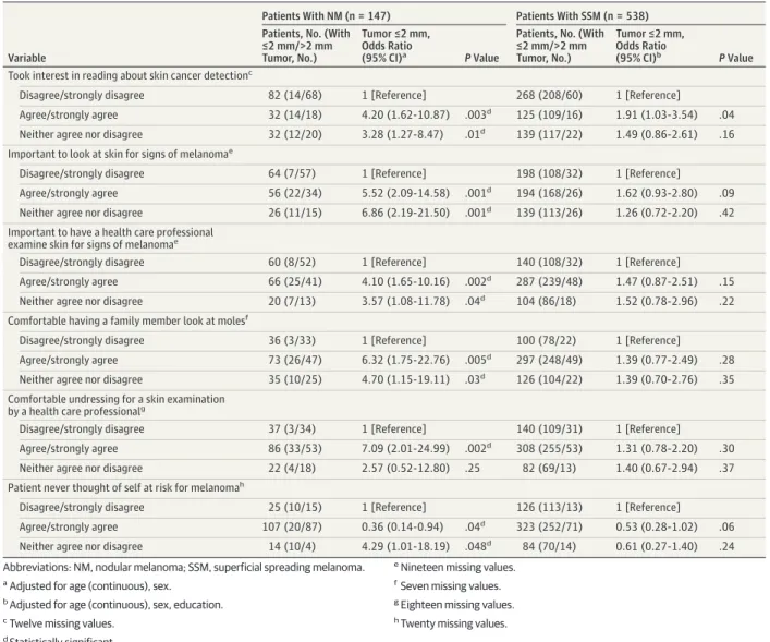
Table 4. Multivariate Logistic Regression Analysis of Attitudes About Melanoma Associated With Tumor Thickness in Patients With NM or SSM Subtypes
Variable
Patients With NM (n = 147) Patients With SSM (n = 538) Patients, No. (With
≤2 mm/>2 mm Tumor, No.)
Tumor ≤2 mm, Odds Ratio
(95% CI)a PValue
Patients, No. (With
≤2 mm/>2 mm Tumor, No.)
Tumor ≤2 mm, Odds Ratio
(95% CI)b PValue Took interest in reading about skin cancer detectionc
Disagree/strongly disagree 82 (14/68) 1 [Reference] 268 (208/60) 1 [Reference]
Agree/strongly agree 32 (14/18) 4.20 (1.62-10.87) .003d 125 (109/16) 1.91 (1.03-3.54) .04
Neither agree nor disagree 32 (12/20) 3.28 (1.27-8.47) .01d 139 (117/22) 1.49 (0.86-2.61) .16 Important to look at skin for signs of melanomae
Disagree/strongly disagree 64 (7/57) 1 [Reference] 198 (108/32) 1 [Reference]
Agree/strongly agree 56 (22/34) 5.52 (2.09-14.58) .001d 194 (168/26) 1.62 (0.93-2.80) .09
Neither agree nor disagree 26 (11/15) 6.86 (2.19-21.50) .001d 139 (113/26) 1.26 (0.72-2.20) .42 Important to have a health care professional
examine skin for signs of melanomae
Disagree/strongly disagree 60 (8/52) 1 [Reference] 140 (108/32) 1 [Reference]
Agree/strongly agree 66 (25/41) 4.10 (1.65-10.16) .002d 287 (239/48) 1.47 (0.87-2.51) .15
Neither agree nor disagree 20 (7/13) 3.57 (1.08-11.78) .04d 104 (86/18) 1.52 (0.78-2.96) .22 Comfortable having a family member look at molesf
Disagree/strongly disagree 36 (3/33) 1 [Reference] 100 (78/22) 1 [Reference]
Agree/strongly agree 73 (26/47) 6.32 (1.75-22.76) .005d 297 (248/49) 1.39 (0.77-2.49) .28
Neither agree nor disagree 35 (10/25) 4.70 (1.15-19.11) .03d 126 (104/22) 1.39 (0.70-2.76) .35 Comfortable undressing for a skin examination
by a health care professionalg
Disagree/strongly disagree 37 (3/34) 1 [Reference] 140 (109/31) 1 [Reference]
Agree/strongly agree 86 (33/53) 7.09 (2.01-24.99) .002d 308 (255/53) 1.31 (0.78-2.20) .30
Neither agree nor disagree 22 (4/18) 2.57 (0.52-12.80) .25 82 (69/13) 1.40 (0.67-2.94) .37
Patient never thought of self at risk for melanomah
Disagree/strongly disagree 25 (10/15) 1 [Reference] 126 (113/13) 1 [Reference]
Agree/strongly agree 107 (20/87) 0.36 (0.14-0.94) .04d 323 (252/71) 0.53 (0.28-1.02) .06
Neither agree nor disagree 14 (10/4) 4.29 (1.01-18.19) .048d 84 (70/14) 0.61 (0.27-1.40) .24 Abbreviations: NM, nodular melanoma; SSM, superficial spreading melanoma.
aAdjusted for age (continuous), sex.
bAdjusted for age (continuous), sex, education.
cTwelve missing values.
dStatistically significant.
eNineteen missing values.
fSeven missing values.
gEighteen missing values.
hTwenty missing values.
Behavioral and attitudinal predispositions and intentions to perform a practice (such as receipt of screening) may be strong indicators of one’s actual practice. In our study, patients diag- nosed with thinner NM were more likely than patients with thick NM to read about skin cancer, express the importance of look- ing at the skin for signs of melanoma, and have a health care pro- fessional examine the skin, suggesting that awareness can be a behavioral driver for earlier discovery of NM.
Limitations
Study limitations include possible reporting and recall bias of the frequency or completeness of SSE and PSE practices in the year prior to diagnosis. The statistically significant differ- ences in awareness in patients with thinner NM had very wide confidence intervals because few patients had thinner NM across categories, and should therefore be interpreted with cau- tion. Also, a relatively small number of thinner NM were in- cluded; other studies of NM have also been hampered by small
sample sizes, emphasizing the need for expanded investiga- tion of larger cohorts with more thin and thick NM.
Conclusions
Our pooled analysis shows that receipt of a whole-body PSE in the year before diagnosis was associated with diagnosis of thinner NM, while recognition of clinical changes in the le- sion was not. Routine SSE was associated with the detection of thinner SSM but not thinner NM, although significantly more patients who performed SSE succeeded in self-detection of thinner NM than those who did not perform SSE. These find- ings underscore the challenges of early NM detection and high- light the importance of complementary practices that in- clude regular whole-body PSE and increased patient and family awareness and education about SSE and PSE practices to pro- mote earlier detection of NM and SSM.
ARTICLE INFORMATION
Accepted for Publication:February 3, 2018.
Published Online:April 18, 2018.
doi:10.1001/jamadermatol.2018.0288 Author Affiliations:First Department of Dermatology–Venereology, National and Kapodistrian University of Athens School of Medicine, Andreas Sygros Hospital, Athens, Greece (Dessinioti, Stergiopoulou, Talaganis, Stratigos);
Department of Social and Behavioral Sciences, Harvard T. H. Chan School of Public Health, Boston, Massachusetts (Geller); Department of
Dermatology, Pigmented Lesion and Melanoma Program, Stanford University Medical Center, Palo Alto, California (Swetter); Veterans Affairs Palo Alto Health Care System, Palo Alto, California (Swetter);
Department of Dermatology and Allergology, University of Szeged, Szeged, Hungary (Baltas);
Department of Dermatology, University of Colorado School of Medicine, Aurora (Mayer); Department of Dermatology, University of Michigan, Ann Arbor (Johnson); Second Department of Dermatology, Aristotle University Medical School, Papageorgiou General Hospital, Thessaloniki, Greece (Trakatelli);
Department of Plastic Surgery, General Hospital of Athens Georgios Gennimatas, Athens, Greece (Tsoutsos); Department of Surgery, Laiko Hospital, Athens, Greece (Tsourouflis).
Author Contributions:Drs Stratigos and Dessinioti had full access to all of the data in the study and take responsibility for the integrity of the data and the accuracy of the data analysis.
Study concept and design:Dessinioti, Geller, Swetter, Baltas, Johnson, Talaganis, Tsoutsos, Stratigos.
Acquisition, analysis, or interpretation of data:
Dessinioti, Stergiopoulou, Swetter, Baltas, Mayer, Talaganis, Trakatelli, Tsourouflis, Stratigos.
Drafting of the manuscript:Dessinioti, Talaganis, Tsoutsos, Stratigos.
Critical revision of the manuscript for important intellectual content:Dessinioti, Geller, Stergiopoulou, Swetter, Baltas, Mayer, Johnson, Talaganis, Trakatelli, Tsourouflis, Stratigos.
Statistical analysis:Dessinioti, Stergiopoulou, Mayer, Talaganis.
Obtained funding:Talaganis.
Administrative, technical, or material support:
Geller, Baltas, Talaganis, Trakatelli, Tsourouflis.
Study supervision:Johnson, Talaganis, Tsourouflis, Stratigos.
Conflict of Interest Disclosures:Dr Stratigos reports no conflicts relevant to this article. Outside the scope of this research, Dr Stratigos reports serving on advisory boards for Regeneron and Novartis; receiving honoraria from LeoPharma, Novartis, and Merck Sharp & Dohme; and receiving research support from Roche, Genesis Pharma, Janssen Cilag, and Abbvie. No other disclosures are reported.
Funding/Support:Dr Dessinioti received support for this research from the State Scholarships Program (IKY), the European Union (European Social Fund), and the Greek National Strategic Reference Framework. Dr Baltas received the János Bolyai research scholarship of the Hungarian Academy of Sciences.
Role of the Funder/Sponsor:The funders/
sponsors had no role in the design and conduct of the study; collection, management, analysis, and interpretation of the data; preparation, review, or approval of the manuscript; and decision to submit the manuscript for publication.
Additional Contributions:We are indebted to the patients who participated at this study for their consideration and interest.
REFERENCES
1. Balch CM, Gershenwald JE, Soong SJ, et al. Final version of 2009 AJCC melanoma staging and classification.J Clin Oncol. 2009;27(36):6199-6206.
2. Green AC, Baade P, Coory M, Aitken JF, Smithers M. Population-based 20-year survival among people diagnosed with thin melanomas in Queensland, Australia.J Clin Oncol. 2012;30(13):
1462-1467.
3. Whiteman DC, Green AC, Olsen CM. The growing burden of invasive melanoma: projections of incidence rates and numbers of new cases in six susceptible populations through 2031.J Invest Dermatol. 2016;136(6):1161-1171.
4. Baade P, Meng X, Youlden D, Aitken J, Youl P.
Time trends and latitudinal differences in
melanoma thickness distribution in Australia, 1990-2006.Int J Cancer. 2012;130(1):170-178.
5. Geller AC, Clapp RW, Sober AJ, et al. Melanoma epidemic: an analysis of six decades of data from the Connecticut Tumor Registry.J Clin Oncol. 2013;
31(33):4172-4178.
6. Minini R, Rohrmann S, Braun R, Korol D, Dehler S. Incidence trends and clinical-pathological characteristics of invasive cutaneous melanoma from 1980 to 2010 in the Canton of Zurich, Switzerland.Melanoma Res. 2017;27(2):145-151.
7. Brunssen A, Waldmann A, Eisemann N, Katalinic A. Impact of skin cancer screening and secondary prevention campaigns on skin cancer incidence and mortality: a systematic review.J Am Acad Dermatol.
2017;76(1):129-139.e10.
8. Swetter SM, Pollitt RA, Johnson TM, Brooks DR, Geller AC. Behavioral determinants of successful early melanoma detection: role of self and physician skin examination.Cancer. 2012;118(15):
3725-3734.
9. Talaganis JA, Biello K, Plaka M, et al.
Demographic, behavioural and physician-related determinants of early melanoma detection in a low-incidence population.Br J Dermatol. 2014;171 (4):832-838.
10. Balch CM, Balch GC, Sharma RR. Identifying early melanomas at higher risk for metastases.J Clin Oncol. 2012;30(13):1406-1407.
11. Chamberlain AJ, Fritschi L, Giles GG, Dowling JP, Kelly JW. Nodular type and older age as the most significant associations of thick melanoma in Victoria, Australia.Arch Dermatol. 2002;138(5):
609-614.
12. Kalkhoran S, Milne O, Zalaudek I, et al.
Historical, clinical, and dermoscopic characteristics of thin nodular melanoma.Arch Dermatol. 2010;146 (3):311-318.
13. Liu W, Dowling JP, Murray WK, et al. Rate of growth in melanomas: characteristics and associations of rapidly growing melanomas.Arch Dermatol. 2006;142(12):1551-1558.
14. Geller AC, Elwood M, Swetter SM, et al. Factors related to the presentation of thin and thick nodular
melanoma from a population-based cancer registry in Queensland Australia.Cancer. 2009;115(6):1318- 1327.
15. Chamberlain AJ, Fritschi L, Kelly JW. Nodular melanoma: patients’ perceptions of presenting features and implications for earlier detection.J Am Acad Dermatol. 2003;48(5):694-701.
16. Greenwald HS, Friedman EB, Osman I.
Superficial spreading and nodular melanoma are distinct biological entities: a challenge to the linear progression model.Melanoma Res. 2012;22(1):1-8.
17. Demierre MF, Chung C, Miller DR, Geller AC.
Early detection of thick melanomas in the United States: beware of the nodular subtype.Arch Dermatol. 2005;141(6):745-750.
18. Moreau JF, Weinstock MA, Geller AC, Winger DG, Ferris LK. Individual and ecological factors associated with early detection of nodular melanoma in the United States.Melanoma Res.
2014;24(2):165-171.
19. Pollitt RA, Geller AC, Brooks DR, Johnson TM, Park ER, Swetter SM. Efficacy of skin
self-examination practices for early melanoma detection.Cancer Epidemiol Biomarkers Prev.
2009;18(11):3018-3023.
20. Clark WH Jr, From L, Bernardino EA, Mihm MC.
The histogenesis and biologic behavior of primary human malignant melanomas of the skin.Cancer Res.
1969;29(3):705-727.
21. Whiteman DC, Baade PD, Olsen CM. More people die from thin melanomas (ⱕ1 mm) than from thick melanomas (>4 mm) in Queensland, Australia.J Invest Dermatol. 2015;135(4):1190-1193.
22. Curiel-Lewandrowski C, Kim CC, Swetter SM, et al; Melanoma Prevention Working
Group—Pigmented Skin Lesion Sub-Committee.
Survival is not the only valuable end point in melanoma screening.J Invest Dermatol. 2012;132 (5):1332-1337.
23. Gershenwald JE, Scolyer RA, Hess KR, et al;
American Joint Committee on Cancer Melanoma Expert Panel and the International Melanoma Database and Discovery Platform. Melanoma staging: evidence-based changes in the American Joint Committee on Cancer eighth edition cancer staging manual.CA Cancer J Clin. 2017;67(6):472-492.
24. Carli P, De Giorgi V, Palli D, et al; Italian Multidisciplinary Group on Melanoma.
Dermatologist detection and skin self-examination are associated with thinner melanomas: results from a survey of the Italian Multidisciplinary Group on Melanoma.Arch Dermatol. 2003;139(5):607-612.
25. Brady MS, Oliveria SA, Christos PJ, et al.
Patterns of detection in patients with cutaneous melanoma.Cancer. 2000;89(2):342-347.
26. Kantor J, Kantor DE. Routine dermatologist-performed full-body skin examination and early melanoma detection.Arch Dermatol. 2009;145(8):873-876.
27. Epstein DS, Lange JR, Gruber SB, Mofid M, Koch SE. Is physician detection associated with thinner melanomas?JAMA. 1999;281(7):640-643.
28. Berwick M, Begg CB, Fine JA, Roush GC, Barnhill RL. Screening for cutaneous melanoma by skin self-examination.J Natl Cancer Inst. 1996;88 (1):17-23.
29. De Giorgi V, Grazzini M, Rossari S, et al. Is skin self-examination for cutaneous melanoma detection still adequate? A retrospective study.
Dermatology. 2012;225(1):31-36.
30. Richard MA, Grob JJ, Avril MF, et al. Melanoma and tumor thickness: challenges of early diagnosis.
Arch Dermatol. 1999;135(3):269-274.
31. Friedman RJ, Rigel DS, Kopf AW. Early detection of malignant melanoma: the role of physician examination and self-examination of the skin.CA Cancer J Clin. 1985;35(3):130-151.
32. Rigel DS, Russak J, Friedman R. The evolution of melanoma diagnosis: 25 years beyond the ABCDs.CA Cancer J Clin. 2010;60(5):301-316.
33. Goldsmith SM, Cognetta AB Jr. Time to move forward after the report of the AAD Task Force for the ABCDEs of Melanoma.J Am Acad Dermatol.
2015;73(4):e149-e150.
34. Abbasi NR, Shaw HM, Rigel DS, et al. Early diagnosis of cutaneous melanoma: revisiting the ABCD criteria.JAMA. 2004;292(22):2771-2776.
35. Rigel DS, Friedman RJ, Kopf AW, Polsky D.
ABCDE—an evolving concept in the early detection of melanoma.Arch Dermatol. 2005;141(8):1032-1034.
36. Coups EJ, Manne SL, Stapleton JL, Tatum KL, Goydos JS. Skin self-examination behaviors among individuals diagnosed with melanoma.Melanoma Res. 2016;26(1):71-76.
37. Aitken JF, Elwood M, Baade PD, Youl P, English D. Clinical whole-body skin examination reduces the incidence of thick melanomas.Int J Cancer.
2010;126(2):450-458.
38. Sondak VK, Glass LF, Geller AC. Risk-stratified screening for detection of melanoma.JAMA. 2015;
313(6):616-617.
39. Moloney FJ, Guitera P, Coates E, et al.
Detection of primary melanoma in individuals at extreme high risk: a prospective 5-year follow-up study.JAMA Dermatol. 2014;150(8):819-827.
40. Esteva A, Kuprel B, Novoa RA, et al.
Dermatologist-level classification of skin cancer with deep neural networks.Nature. 2017;542 (7639):115-118.