Environmental and genetic factors in the pathogenesis of melanoma and
melanoma associated other primary malignancies
Ph.D. Thesis
Zsófia Borbála Hatvani M.D.
Semmelweis University
Doctoral School of Clinical Medicine
Supervisor: Sarolta Kárpáti M.D., D.Sc.
Official reviewers: Judit Oláh M.D., Ph.D.
Tibor Krenács M.D., Ph.D.
Head of the comprehensive exam committee:
Ilona Kovalszky M.D., D.Sc.
Members of the comprehensive exam committee:
Bálint Nagy Ph.D.
Zsuzsanna Szalai Med.habil.
Budapest, 2014
2
TABLE OF CONTENTS
ABBREVIATIONS ... 4
I. INTRODUCTION ... 7
I.1.HISTORICAL/CULTURAL ASPECTS OF PIGMENTED LESIONS ... 7
I.2.MALIGNANT MELANOMA ... 10
I.3.NON-GENETIC FACTORS IN THE ETIOLOGY OF MM ... 11
I.3.1. Environmental predisposing factors ... 12
I.3.2. Immunosuppression and MM development... 15
I.4.GENETICS OF MM ... 16
I.4.1. Germline alterations in MM genesis ... 17
I.4.2. Somatic mutations and polymorphisms in MM ... 24
I.5.MM-ASSOCIATED OTHER PRIMARY MALIGNANCIES ... 30
I.5.1. Multiple primary melanomas (MPM) ... 30
I.5.2. MM and co-aggregation of other cancers ... 34
I.5.3. Hereditary cancer predisposing syndromes and MM ... 37
I.6.CLASSIC GENODERMATOSES AND MM... 42
I.6.1. Epidermolysis bullosa (EB) ... 42
I.6.2. Ichthyoses ... 43
I.6.3. Dowling-Degos disease (DDD) ... 43
I.6.4. Hailey-Hailey disease (HHD) ... 43
II. AIMS ... 44
III. MATERIALS AND METHODS ... 45
III.1.CLINICAL DATA AND PATIENT CHARACTERISTICS OF MPM STUDY ... 45
III.2.CLINICAL DATA OF UNIQUE MM-ASSOCIATED CASES ... 46
III.2.1. Two cancer prone families (Figure 12) ... 46
III.2.2. Six primary MMs ... 48
III.2.3. MM and phenotype suggesting PHTS/CS ... 49
III.3.MUTATION ANALYSIS ... 53
III.4.STATISTICAL ANALYSIS ... 59
IV. RESULTS ... 60
3
IV.1.MPM STUDY ... 60
IV.1.1. Clinicopathological attributes ... 60
IV.1.2. Genetic results ... 63
IV.1.3. Analysis of MC1R variant status ... 64
IV.2.GENETIC RESULTS OF THE UNIQUE MM-ASSOCIATED CASES ... 66
IV.2.1. Two cancer prone families ... 66
IV.2.2. Six primary MMs ... 69
IV.2.3. MM and phenotype suggesting PHTS/CS ... 70
V. DISCUSSIONS ... 71
V.1.MPM STUDY ... 71
V.1.1. Clinicopathological attributes... 71
V.1.2. Genetic results and their clinicopathological relevance in MPM patients ... 72
V.1.3. Analysis of MC1R variants status ... 81
V.2.UNIQUE MM-ASSOCIATED CASES ... 82
V.2.1. Two cancer prone families ... 82
V.2.2. Six primary MMs ... 89
V.2.3. MM and phenotype suggesting PHTS/CS ... 92
VI. CONCLUSIONS ... 95
VI.1.MPM STUDY ... 95
VI.2.UNIQUE MM-ASSOCIATED CASES ... 96
VI.2.1. Two cancer prone families ... 96
VI.2.2. Six primary MMs ... 96
VI.2.3. MM and phenotype suggesting PHTS/CS ... 97
VI.2.4. Conclusions of Unique MM-associated cases: ... 97
VII. SUMMARY ... 99
VIII. ÖSSZEFOGLALÁS ... 100
IX. REFERENCES ... 101
X. OWN PUBLICATIONS ... 145
XI. ACKNOWLEDGEMENTS ... 147
4
ABBREVIATIONS
6,4 PP: ………..…….………6-, 4 pyrimidin-pyrimidon AC: ……….………….….……….Adenylate cyclase ACTH: ……….……….……….……….….Adrenocorticotropic hormone AD: ……….…….………….……….Autosomal dominant ALM: ……….…….……..….………Acral-lentiginous melanoma α-MSH: ……….…….……….……...Alfa-melanocyte stimulating hormone BCC: ……….….………….……….Basal cell cancer Bp:………..….……….………Base pair BRRS: …………..……….………..…….. Bannayan-Riley-Ruvalcaba syndrome CCUO: ………...…...Cancer of unknown origin CDK4: ……….….………Cyclin-dependent kinase 4 CDKN2A: …………..………..……….Cyclin-dependent kinase 2A CLL: ………...……..………….…Chronic lymphocytic leukemia CPD: ………...…..……..……….Cyclobutane pyrimidin dimer CREB: ………...….cAMP-response element binding protein CS: ………..………..………Cowden syndrome CLS: ………..………Cowden-like syndrome CSD: ………..……….………...……Chronic sun damage DDD: ………...……….Dowling Degos Disease DS: ………..….………...……Dermoscopy DSB: ………...………Double strand break EB:.………..………..………..Epidermolysis bullosa HDM2: ………..………..Human homolog of murine Mdm2 HGVS: ………..………….……Human genome variation society HHD: ………..………Hailey-Hailey Disease HR: ………..…...……….Homolog recombination IVS: ………..………... Intervening sequence LC: ………..………Lung cancer LFS: ………..……….Li-Fraumeni syndrome LI: ………...……….Lamellar ichthyosis LMM: ………...……….……..Lentigo maligna melanoma
5
MC1R: ………..………..……….Melanocortin 1 receptor MEN-1: …………...……….……….Multiple endocrine neoplasia-1 miRNA: ………..……….….microRNA Mis: ………...Melanoma in situ MITF: ……….………Microphtalmia associated transcription factor MM: ………...………...………Malignant melanoma MMF: ………...……….Mycophenolate mofetil MP: ………..………...Methylprednisolone MPM: ……….………Multiple primary melanoma mTOR: ………...………..mammalian target of rapamycin mTORi: ………...………….…….mammalian target of rapamycin inhibitor NER: ………..………..Nucleotide excision repair NHL: ………..……….….………..Non-Hodgkin lymphoma NLS: ………..………..Nuclear localization signal NM: ………..……….……….……….Nodular melanoma NMSC: ………..……….………Non-melanoma skin cancer NRHC: ………..………Non-red hair color OMIM: ………..…………...………On-line Mendelian Inheritance in Man OR: ………..………..Odds ratio OTR: ………..……….….………….Organ transplant recipient PaC: ………..………..Pancreatic cancer PHTS: ………...……….….PTEN hamartoma tumor syndrome PKA: ………..………cAMP dependent protein kinase A PLS: ………..……….………….Proteus-like syndrome PrC: ………..………..Prostate cancer PS: ………..………..….………Proteus syndrome PTEN: ………..………Phosphatase and ptensin-homolog Rb: ………..………..…Retinoblastoma protein RCC: ………..……….Renal cell cancer RHC: ………..……….Red hair color ROS: ………..………...………..Reactive oxygen species RR: ………..………..…………Relative risk
6
SIR: ………..………Standardized incidence ratio SPM: ………..………..Single primary melanoma SRL: ………..…..………Sirolimus SSM: ………..……….Superficial spreading melanoma SU: ………..……….Semmelweis University TAC: ………..………..………Tacrolimus TILs: ………..………..…Tumor infiltrating lymphocytes UTR: ………..……….Untranslated region UVA: ..………..………Ultraviolet-A UVB: ………..………..……….Ultraviolet-B UVR: ………..………..……….………Ultraviolet radiation WHO: ………..………..……….World Health Organization
7
I. INTRODUCTION
I.1. Historical/cultural aspects of pigmented lesions
Before the 18th century, in most cultures immaculate skin with the aesthetics of smooth, white complexions of its ladies represented high value, health, beauty, innocence and perfection. Also in the art, skin marks (moles, birthmarks) or any disfiguring skin changes hadn’t been portrayed on paintings (Figure 1).
There had been many beliefs concerning moles and birthmarks. Also in the first English book published on skin diseases (Turner 1714), an individual chapter analyses the
Figure 1. Representations of the skin on paintings between the 16th-18th century
8
concepts of birthmarks. That time, fetal skin development was considered to be directly associated with the mother’s mental condition, and birthmarks as a consequence of maternal harmful impressions (desire, fright, accidents). Already from the sixteenth century localization of moles served as a basis of divination. As a few example, a mole on the belly denoted whoredome, luxury and gluttony; while a mole on the throat threatened the person with diseases such as asphyxia, or violent death. Top-to-bottom, left-right orientation and gender differences were also considered in mole reading. For instance a man with a mole on the upper lip was believed to have good fortune, while a woman to be debauched (summarized from Connor 2004).
Interestingly, on the basis of the favorable meanings of mole readings, in the eighteenth century, application of beauty spots from black silk to the face became a trend of fashion (Figure 2).
Figure 2. Artificial moles - application of beauty spots in the 18th century
9
Later, from the late nineteenth century, as knowledge about dermatological conditions especially about moles and melanoma got augmented and accessible (first description of melanoma is from 1812 by Rene Laennac), visualization of pigmented lesions in art works became also more common. Unfortunately a parallel unfavorable trend of a new body culture evolved with a higher ultraviolet radiation (UVR) exposure of the skin. In the last decades, there is a significant awareness of pigmented lesions in the general public, which trend is also reflected in art works with a great importance regarding public health (Figure 3) (Sources of paintings are detailed in the references).
Figure 3. Moles as commonly visualized skin marks after the 19th century
10
I.2. Malignant melanoma
Malignant melanoma (MM) is a tumor originating from melanocytes, the pigment cells with major function of synthesizing melanin pigment that determinates skin, eye and hair pigmentation. During embryogenesis melanocytes are originated from neural crest cells then they migrate to their targeted ectodermal locations. There are still debated results whether MM cells originate from the differentiated melanocytes, or from undifferentiated melanocyte-precursor stem cells. In any ways, sequential genetic events, inherited and acquired, are required for MM development (reviewed in Meyle and Guldberg 2009). MM occurs not only in the skin, but in mucosal membranes and in the uvea of the eye; however they are not elemental topics of this work.
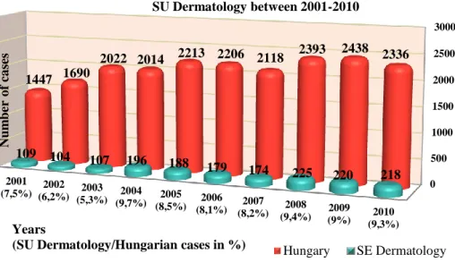
MM incidence has dramatically risen in the last decades, affecting even younger generations (Purdue et al. 2008). There are strong efforts to lower this incidence rates due early recognition and patient education, as therapy of advanced stage MM is still not that promising. Over the last 10 years in Hungary (National Cancer Registry), and also at Semmelweis University (SU) Department of Dermatology, Venereology and Dermatooncology (own data), the number of identified new cases of MM and melanoma in situ (Mis) cases clearly demonstrates a rising trend (Figure 4).
Figure 4. MM and Mis new cases in Hungary and at SU Dermatology between 2001 and 2010.
0 500 1000 1500 2000 2500 3000
2001 (7,5%) 2002
(6,2%) 2003 (5,3%) 2004
(9,7%) 2005 (8,5%) 2006
(8,1%) 2007 (8,2%) 2008
(9,4%) 2009 (9%) 2010
(9,3%) 1447 1690
2022 2014 2213 2206 2118 2393 2438 2336
109 104 107 196 188 179 174 225 220 218
Number of cases
Years
(SU Dermatology/Hungarian cases in %)
New cases of MM+Mis per years in Hungary and at SU Dermatology between 2001-2010
Hungary SE Dermatology
11
Using demographic data of Hungarian Central Statistical Office (KSH 2012), MM+Mis incidence rates within this time interval ranged between 12-20/100.000 with a rising trend, similarly to other Hungarian reports (Balatoni et al. 2011).
Traditional MM classification is based on tumor clinocopathology and includes major subtypes such as superficial spreading melanoma (SSM), nodular melanoma (NM), acrolentiginous melanoma (ALM) and lentigo maligna melanoma (LMM) (Clark 1967), supplemented with some less common subtypes (desmoplastic-, naevoid-, mucosal MM, MM arising from a blue naevus-, or from a congenital naevus, MM in childhood and persistent MM) (World Health Organization-WHO Classification 2006). However these subtypes own neither prognostic nor therapeutic importance. Recent molecular findings opened the opportunity of personalized targeted therapies with promising efficacy that points out the urgent need of a new up-to-date classification system (Scolyer et al. 2011). MM subgroups are also classified by body site and sun exposure (MM arising in chronically sun exposed, intermittently sun-exposed, or sun protected areas), moreover certain tumor locations show specific molecular characteristics regarding BRAF, RAS, c-KIT mutation status (see later in Table 3 on page 26).
I.3. Non-genetic factors in the etiology of MM
MM is a multifactorial cancer with identified environmental and hereditary predisposing factors. Prevalence rates and gene-environment interactions vary along geographical locations upon latitude. While the highest prevalence is observed in Australia and New-Zeeland, followed by the United States and some European populations (Little and Eide 2012), mutation frequencies of predisposing genes are detected inversely with the incidence rates, suggesting that in regions with the higher incidences, sporadic cases make up the majority.
12 I.3.1. Environmental predisposing factors
I.3.1.1. Ultraviolet light
Fundamental role of UVR in MM genesis has long been hypothesized (summary in Hocker and Tsao 2007). Besides the original concept, that history of sunburns are the predominant UV related risk factors (Gilchrest et al. 1999, Noonan et al. 2001), recently more lines of evidence suggest that all types (intermittent,
chronic, sunburns) of sun exposures together with the age at exposition may play role in MM genesis, especially those suffered during the most vulnerable early childhood.
Ninety-five percent of UVR reaching the ground is UVA (315-400nm) that is far less energetic than UVB (280-315nm). Previously, solar UVR induced DNA mutation formation and skin carcinogenesis was exclusively linked to UVB effects, however recently UVA has also been classified as class I carcinogen by the WHO (El Ghissassi et al. 2009).
I.3.1.1.1. UVB
Shortwave UV light generates DNA photoproducts by direct DNA absorption. These photoproducts are mainly cis-syn cyclobutane pyrimidine dimers (CPDs), pyrimidine 6- 4 pyrimidone photoproducts (6-4 PPs) and Dewar valence isomers (DewPPs). CPDs, mostly formed as C to T transitions (also known as UV signature mutations), are the most common UVB lesions. If DNA damage response mechanisms are sufficient, these premutagenic lesions are repaired, and don’t cause mutations and subsequent carcinogenesis (reviewed in Rünger and Kappes 2008). Upon different UVB exposures, activation of MAPK pathway, elevated tyrosinase expression in melanocytes, higher MM cell motility through interleukin-8 activation, and a number of clinical and experimental observations on animal models confirmed the pathogenic effects of UVB in MM genesis (reviewed by von Thaler et al. 2009).
Figure 5. Ferenc Gál: Beach (1925)
13 I.3.1.1.2. UVA
UVA was originally hypothesized to create different DNA lesions than UVB due to reactive oxygen species (ROS) induction. Recent findings suggest that the majority of UVA mutations particularly of UVA2 (315-340nm) induce also C to T transitions and subsequent CPDs. UVA also generates oxidative DNA damage through singlet oxygen or other ROS, predominantly on guanine. The most mutagenic product is 7,8-dihydro- 8-oxyguanin (8-oxoG). The carcinogenic effect of UVA is now mainly linked to CPDs and to the finding that UVA-generated CPDs are more mutagenic than UVB-induced ones (summarized in Rünger and Kappes 2008). This is also supported by our results, showing that antimutagenic cellular responses are much weaker upon UVA than UVB induction (Rünger et al. 2012), however on fibroblasts. When skin is exposed to the sun, the amount of UVB is sufficient to induce damage response mechanisms and also to minimize the effects of UVA induced damages. Pure UVA exposure (sunbed use, sun exposure through window glass, UVA phototherapy, use of non-broad spectrum sunscreens) is getting more frequent in the modern civilization, when the UVA induced DNA damage responses are usually not sufficient to prevent the mutation formation (reviewed in Rünger and Kappes 2008).
In primary MMs, besides oxidative DNA damages, predominantly UV signature mutations (C to T) are detected on tumor suppressor genes (p53, p16/INK4a, PTEN) at various percentages (52-68%). Whether these mutations are induced by UVA or UVB is unclear (reviewed in Rünger and Kappes 2008), however the role of UVA in MM genesis was repeatedly confirmed by clinico-epidemiological studies on sunscreen-, and sunbed use (Héry et al. 2010, Autier et al. 2011, Boniol et al. 2012).
I.3.1.2. Obesity
Obesity has been proven as a risk factor for several cancer types, although in terms of MM, debated results are available. A recent meta-analysis has proved the association with increased MM risk but only among men (Sergentanis et al. 2013).
I.3.1.3. Socioeconomic status, occupation
MM incidence is higher among people with larger income. The most likely explanation is the higher amount of recreational sun exposures during sunny holidays throughout a year (Kirkpatrick and White 1990, MacKie and Hole 1996). Moreover socioeconomic
14
status has been shown to have impact on MM survival as well due to better access to health services especially in countries where huge gap exist between the different socioeconomic layers (Quintella Mendes and Koifman 2013). As another factor, MM diagnosis in patients with lower socioeconomic level is more likely to be delayed with advanced Breslow thickness and disease stage (Pollitt et al. 2012).
Association between occupation and MM risk has been widely examined; early studies proved that indoor workers are at higher risk (Cooke et al. 1984), however if stratified those occupations by education level and training requirements, again higher socioeconomic status appeared to explain the differences. Indoor workers are more prone to get sunburn as they spend much less time in the sun than outdoor workers do.
A number of studies (Gundestrup and Storm 1999, Pukkala et al. 2012) reported that airline crew had a higher-than-expected rate of MM, and explained it by more recreational sun exposures between flights, but also by the regular exposure to cosmic radiation and magnetic field. Other studies did not confirm any difference between aircrew and random Icelandic population (Rafnsson et al. 2003).
Firefighters are reported to have an excess risk of certain cancers including MM. Mostly they are not related to carcinogenic inhalations (Ma et al. 1998, Milham 2009), but to exposures of electromagnetic field and radio-frequency radiation during their work, (Milham 2009).
Role of ionization radiation in skin cancer development is supported by the observed elevated incidence rates among radiologists (Wang et al. 1990). Studies on MM incidence among nuclear industry workers showed inconsistent results (Cardis et al.
2007, Gun et al. 2008). Our data from the Hungarian nuclear power plant did not support the occupational hazard to MM development (Tóth V. et al. 2013).
I.3.1.4. Pharmacological agents
I.3.1.4.1. Vitamin D
Serum 25-hydroxyvitamin D3 level and its role in cancer development and outcome is thoroughly investigated. Sun exposure is necessary to vitamin D synthesis, but also acts as a major risk factor for MM. Some studies indicate that increased level of vitamin D is associated with excess risk of MM (Afzal et al. 2013), while others fail to prove it (van der Pols et al. 2013).
15
I.3.1.4.2. Statins, non-steroidal anti-inflammatory agents, others Statins show no association between drug use and MM development (Jagtap et al.
2012).
Long term use of non-steroidal anti-inflammatory drugs (NSAIDs) decrease the MM risk (Curiel-Lewandrowsky et al. 2011a).
Oral contraceptives and hormone replacement therapy have no impact on MM risk. As phototoxic and photoallergic drugs intensify the UVR effect on the skin their contribution to MM development has been suspected; even short term use of quinolone and propionic acid derivative NSAIDs may increase the risk of MM (Siiskonen et al.
2013).
I.3.1.5. Pesticide exposure
More lines of evidence suggest that pesticide exposure may be additional risk factor for MM development (Leslie et al. 2010, reviewed by Weichenthal et al. 2012).
I.3.2. Immunosuppression and MM development
Organ transplant recipients (OTR) undergoing combined, long-term immunosuppressive therapy are prone to develop cancers, especially non-melanoma skin cancers (NMSC) (Otley and Pittelkov 2000). Although it is clearly documented that squamous cell carcinoma (SCC) (65-fold) (Jensen et al. 1999) and basal cell cancer (BCC) (10-fold) (Hartevelt et al. 1990) incidences are highly elevated among these patients, data on MM incidence rate is contradictory. Based on large studies, as high as 8-times elevated risk for MM development among OTRs have been observed (Le Mire et al. 2006), while in other studies the risk was not elevated at all (Lindelöf et al. 2000).
In general a three-, to five-fold elevated MM risk is concluded (Jensen et al. 1999, Hollenbeak et al 2005, Moloney et al. 2006, Zwald et al. 2010), and men are more frequently affected than women, reflecting the statistical fact, that most renal transplant recipients are men (Le Mire et al. 2006, Laing et al. 2006a). Reduction or cessation of immunosuppressive therapy results in a relative good outcome even in metastatic disease (Le Mire et al. 2006, Laing et al. 2006b). According to a Hungarian database, 3141 OTRs have been reported between 1973 and 2009, from who 7 developed MM during immunosuppressive therapy (Somlai et al. 2009).
16
The eruptive nevi phenomenon in immunosuppressed patients (López et al. 2010, de Boer and Kuyvenhoven 2011) further supports a significant association between melanocyte proliferation and immunosuppression. Chronic lymphocytic leukemia (CLL) or HIV positive patients are also more prone to MM development.
The suspected mechanisms behind the elevated cancer risks among OTRs are 1) a weaker immune surveillance against tumors and oncogenic viral agents, 2) direct oncogenic effects of certain immunosuppressive drugs, and 3) in rare cases transmission of primary MMs via transplanted organs.
I.4. Genetics of MM
The role of inherited and acquired genetic events in cancer development is well studied.
Cancer arises from clones of cells due to germline and/or somatically acquired mutations, and mature cancer clone development is further influenced by environmental factors and genomic sequence variants (reviewed in Stratton 2011).
Germline mutations are already present in the fertilized egg, and therefore in all somatic cells. These mutations are able to influence cancer susceptibility in a number of ways, like cancer clone development, mutation rate in somatic cells, and carcinogen metabolism.
Germline genetic variations include single nucleotide polymorphisms (SNPs), small microdeletions and/or insertions (indels), polymorphisms, and mutations according to their frequency and strength with disease penetrance. Certain regions in the genome may have copy-number polymorphisms that result in chromosomes with more than one or no copies of a certain gene. Epigenetic changes (DNA methylation, histone modification) and regulatory effects of miRNAs play also role in cancer development.
Both oncogenes and tumor suppressor genes might be targeted by genetic events.
Oncogenes are regulators of cell division; an activating mutation in only one allele is sufficient for initiating oncogenesis by bypassing the regulatory mechanisms and overactivation (dominant effect). In contrast, tumor suppressor genes inhibit cellular proliferation and initiate apoptosis if necessary. In general, loss-off function mutation only in one allele is insufficient to alter these functions, acting in a recessive manner, therefore mutations in both copies required to the oncogenic effect. More than 80% of the currently known cancer genes are dominantly acting. The proteins encoded by these
17
genes serve a wide variety of key functions in cell cycle regulation, apoptosis, chromatin modification and remodeling epigenetic processes (reviewed in Stratton 2011).
Before the era of next generation sequencing, the main purpose of cancer genetics studies was to identify rare, high penetrance gene mutations. With the new technical approaches, a huge number of high frequency, low penetrance, simultaneously inherited gene variants (not even mutations) are getting identified, and new model of cancer inheritance is emerging (‘common disease common variant’) (reviewed in Pfeifer and Hainaut 2011).
I.4.1. Germline alterations in MM genesis
I.4.1.1. High penetrance genes
A proportion of MM patients belong to MM prone families or have MPM; who are more prone to harbor rare, high-risk, high penetrance MM predisposing alleles. To date, two such genes are identified in MM genesis, cyclin-dependent kinase 2A (CDKN2A), and cyclin-dependent kinase 4 (CDK4).
I.4.1.1.1. CDKN2A gene
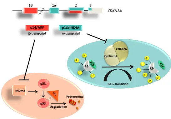
This locus (MIM#600160) is located on 9p21 and encodes two proteins through alternate reading frames, namely p16/INK4a and p14/ARF. The two tumor suppressor proteins act on different pathways, but both regulate cell cycle. As a result of two different promoters, p16 is formed from exon 1α exon 2 and 3, while p14/ARF is composed of a different exon 1 (1β) located 13 kb upstream of exon 1α and a shared exon 2 with an alternative reading frame.
The protein p16/INK4A consists of 156 amino acids and exhibits a structure with four ankyrin repeat motifs. As functionality, it inhibits the CDK4/6 mediated phosphorylation of retinoblastoma protein (Rb), thus resulting in a dephosphorylated, active Rb, that binds to E2F repressing its transcriptional function and arresting G1 checkpoint in cell cycle. When CDKN2A is mutated, phosphorylation of Rb is not inhibited, resulting in phosphorylated-inactive Rb state that cannot bind to E2F, so E2F is able to induce G1/S phase activating gene transcriptions and cells are undergoing uncontrolled cell divisions.
18
In contrast, p14/ARF is regulating p53 mediated apoptosis pathway through binding to and inhibiting the human homolog of murine Mdm2 (HDM2) that is involved in ubiquitin mediated degradation of proteins. Mutation in p14/ARF activates HDM2 function, so marking of proteins that undergo ubiquitination and subsequent degradation in proteasome becomes uncontrolled, resulting in p53 degradation and subsequent loss of p53 mediated apoptosis (summarized in Kim and Sharpless 2006)(Figure 6).
Figure 6. Structure and function of CDKN2A gene. Two distinct tumor suppressor proteins are encoded in the CDKN2A gene. P14/ARF (β-transcript-red) inhibits MDM2 mediated p53 ubiquitination, while p16/INK4A (α-transcript-blue) inhibits CDK4/6- mediated phosphorylation of Rb protein.
CDKN2A gene mutations may affect only p16/INK4A, only p14/ARF protein sequence or both; however those uniquely targeting p14/ARF (exon 1β) are relatively rare (Binni et al. 2010).
CDKN2A locus is frequently altered in many cancer types at somatic level, but the germline mutations are mostly associated with a phenotype of familiar MM or MPM and with an elevated risk of pancreatic cancer (PaC) (Lynch et al. 2002, 2008).
The mutation prevalence is about 0.2% among sporadic MM cases (Aitken et al. 1999);
while approximately 10-40% of MM prone families are carriers, with geographical
19
differences (Berwick et al. 2006, Goldstein et al. 2006, Goldstein et al. 2007). Areas with the highest MM incidence (Australia) own the lowest mutation rates with high penetrance. Within Europe, in populations with low MM incidences (South-Europe, Mediterranean countries), CDKN2A mutations are far more frequent (Goldstein et al.
2007). Patients carrying CDKN2A mutation have a younger age at diagnosis of first MM (Goldstein et al. 2007, Pedace et al 2011), are more prone to develop subsequent MMs (Pedace et al. 2011) and are more likely to have history of MM within their family (Pedace et al. 2011, Maubec et al. 2012).
I.4.1.1.2. CDK4 gene
CDK4 (MIM#123829) gene is located on 12q14 and contains 8 exons (Figure 7).
Figure 7. Structure of CDK4 gene. Red color represents transcribed exons.
The encoded oncogene is a downstream target of p16/INK4, therefore changes in structure and function may induce similar phenotypes to those caused by CDKN2A mutations.
Identified mutations occur solely in exon 2 at codon 24 (Zuo et al. 1996, Soufir et al.
1998), resulting in a majority of cases in R24C (Zuo et al. 1996) amino acid change, however R24H has been also detected in some families (Soufir et al. 1998, Molven et al. 2005). To date only a small number (<15) of MM families have been identified with CDK4 mutations (Zuo et al. 1996, Soufir et al. 1998, Helsing et al. 2008, Pjanova et al.
2007, Puntervoll et al. 2013). Phenotypes of these families do not differ from those of CDKN2A carriers’ (propensity to MPM, younger age of onset, increased occurrence of atypical nevi) (Puntervoll et al. 2013).
20 I.4.1.2. Intermediate penetrance genes
I.4.1.2.1. Melanocortin 1 receptor (MC1R) gene
MC1R (MIM#155555) gene is located on 16q24.3 and encodes for melanocortin 1 receptor, a 317 amino-acid G protein-coupled receptor with seven transmembrane domain that is expressed predominantly in epidermal and uveal melanocytes with a major role of regulating hair-, skin and eye pigmentation.
Expression is also detectable in pituitary cells, leukocytes, mast cells and pro-monocytes. The receptor is a member of the melanocortin receptor family that contains five differentially expressed G-protein-coupled receptors (MC1R- MC5R). Under physiological conditions MC1R is activated by binding of its ligands alfa- melanocytes stimulating hormone (α-MSH) or adrenocorticotropic hormone (ACTH). The subsequent cAMP production via adenylate
cyclase (AC) activates cAMP dependent protein kinase A (PKA) and a number of downstream targets like cAMP response element binding protein (CREB), microphtalmia-associated transcription factor (MITF). These events lead to proliferation and to a switch from red/yellow pheomelanin to the brown/black eumelanin production.
Eumelanin has a major role to protect DNA from UVR as a shield, while skin pigmentation is subsequently augmented (Figure 8). In contrast pheomelanin has a weak shield effect to protect DNA of melanocytes against UVR, and has shown to amplify UVA-induced ROS production (Rouzaud et al. 2005, Hill HZ and Hill GJ 2000).
The MC1R is a highly polymorphic gene (Gerstenblith et al. 2007). The non- synonymous variants have different effect on the receptor function. While gain-of- function mutations are not reported in human to date (reviewed in Rees 2004), loss-of-
Figure 8. MC1R receptor function; See details in text.
Abbreviations: Gs:G-proteins, AC: adenylate cyclase,
PKA: cAMP dependent protein kinase A.
21
function alleles can either result in impaired ability to bind α-MSH (R163Q) or to impaired activation of the G protein-coupled pathway of cAMP dependent kinases (V60L, R142H, R151C, R160W) (Ringholm et al. 2004). In some variants both distresses are detectable (D84E, V92M, D294H) (Ringholm et al. 2004). As carrying more variants is also common in homozygous or even compound heterozygous fashion, effect of the different allele combinations may result in a wide range of functional failures and in an elevated pheomelanin/eumelanin ratio in melanocytes.
The constitutive pheomelanin production by the genetically and subsequently functionally impaired receptor in certain MC1R variant carriers may manifests in red hair color (RHC) phenotype with fair skin, freckles and poor tanning ability (Valverde et al. 1995, Smith et al. 1998). These variants are referred as ‘R’ variants. Recent findings suggest that complete loss-of-function effects are rare even among these ‘R’
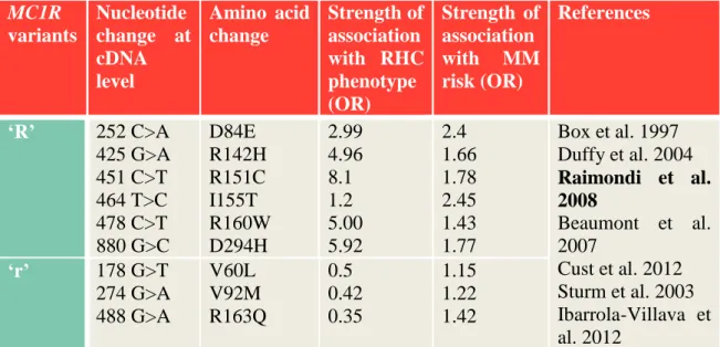
allele carriers (Newton et al. 2005). Variants with less destructive effects on receptor function and phenotype called non-RHC (NRHC) alleles, and labeled as ‘r’ alleles. The most frequent MC1R variants and their effect on RHC phenotype and MM risk are summarized in Table 1.
Table 1. The most frequent MC1R variants and their individual associations (Odds ratios: OR) to RHC phenotype and MM risk.
MC1R variants
Nucleotide change at cDNA level
Amino acid change
Strength of association with RHC phenotype (OR)
Strength of association with MM risk (OR)
References
‘R’ 252 C>A 425 G>A 451 C>T 464 T>C 478 C>T 880 G>C
D84E R142H R151C I155T R160W D294H
2.99 4.96 8.1 1.2 5.00 5.92
2.4 1.66 1.78 2.45 1.43 1.77
Box et al. 1997 Duffy et al. 2004 Raimondi et al.
2008
Beaumont et al.
2007
Cust et al. 2012 Sturm et al. 2003 Ibarrola-Villava et al. 2012
‘r’ 178 G>T 274 G>A 488 G>A
V60L V92M R163Q
0.5 0.42 0.35
1.15 1.22 1.42 Abbreviations: OR: odds ratio
22
It has long been suspected that MM risk among MC1R variant carriers isn’t only via an UV dependent way, as MMs often develop on sun protected body sites, moreover UV signature mutations are uncommon driver mutations (Curtin et al. 2005). The altered pheomelanin synthesis (pheomelanin or its any intermediate-, or by-product) might result in a perturbed intrinsic susceptibility of certain carcinogenesis (Mitra et al. 2012).
A number of meta-analyses specified the role of MC1R variants in MM development.
Certain ‘r’ variants carry also increased MM risk independently of the phenotype effects (Raimondi et al. 2008, Fargnoli et al. 2010, Kanetsky et al. 2010, Williams et al. 2011).
The number of ‘R’ or ‘r’ variant alleles may also enhance MM risk (Goldstein et al.
2005, Pastorino et al. 2008, de Torre et al. 2010). Most recent meta-analysis on MC1R variants and MM risk confirmed ORs for ‘R’ variants between 1.00 to 4.64, and for ‘r’
variants between 0.58-3.00 (Williams et al. 2011) (Table 1).
The frequency and distribution of variants vary among populations and along continents. Frequency in African populations is very low, therefore it is also suspected that eumelanin production represent an evolutionary benefit in those geographical regions (summarized in Makova and Norton 2005). In Asians the R163Q is an exceptionally common variant with a 70% average frequency in contrast with other locations where it is observed at a much lower incidence (almost absent in European populations, observed at a frequency of 7% in Indians) (Rana et al. 1999). In European populations the MC1R gene is especially polymorphic, with more than 70 variants identified to date (Gerstenblith et al. 2007). Among Caucasians, variant frequencies are higher among lightly pigmented populations than among darkly-pigmented ones (reviewed in Makova and Norton 2005), in accordance with findings suggesting a decreasing gradient of ‘combined R variant’ frequency from Northern to Southern Europe (Gerstenblith et al. 2007).
I.4.1.2.2. MITF gene
The gene (MIM#156845) is located on chromosome 3p14-p13, exhibits nine promoters with corresponding different MITF isoforms that own different first exons and shared exons 2-9. The encoded protein is a basic helix-loop-helix-leucine zipper protein. Only isoform M is melanocyte-specific and expressed exclusively in melanocytes and MM cells (reviewed by Levy et al. 2006).
23
MITF-M is key regulator of melanocyte development, survival and function by regulating a number of differentiation and cell-cycle progression genes. MITF directly binds to the promoter of p16/INK4A and regulates positively its transcription (Loercher et al. 2005). During activation of MC1R by α-MSH, phosphorylation of CREB occurs, that then binds directly to MITF promoter and stimulates it’s transcription (Bertolotto et al. 1998).
Germline loss-of-function mutations in MITF gene results in Waardenburg syndrome IIA, an autosomal dominantly inherited disease that is characterized by melanocyte deficiencies of the eye, forelock and inner ear, the latest of which causing sensorineural hearing impairment.
The role of MITF as an oncogene in MM genesis was first described by identification of copy gains at MITF locus in MM cell lines using SNP arrays (Garraway et al. 2005).
Moreover tissue microarrays proved that 10-20% of MMs exhibit amplification of MITF that is proven to be a late event in MM genesis, therefore is more common in metastatic cases, and represents a worse outcome (Garraway et al. 2005). Recently a germline missense mutation (c.G1075A; p.E318K) was identified to be associated with a 4-fold elevated MM risk and with MPM formation, while carriers exhibited a 14-fold risk to MM and renal cell cancer. Impairment of MITF SUMOylation with insufficient cellular stress management leads here to initiation of tumor formation (Bertolotto et al.
2011). Furthermore certain phenotypes such as increased nevi number and susceptibility to amelanotic melanoma (Sturm et al. 2014) are also observed among carriers of this mutation. Interestingly most patients harboring MITF E318K and amelanotic melanoma exhibited MC1R genotypes of homozygous ‘R’ variants, suggesting that an altered MC1R receptor together with the this MITF point mutation may result in this relatively rare MM subtype (Sturm et al. 2014).
I.4.1.3. Low penetrance genes
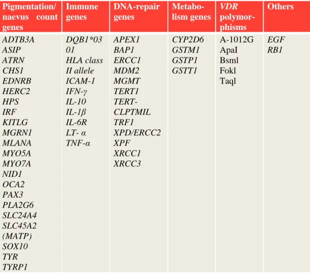
MM susceptible host factors such as nevi count, skin pigmentation, ability to tan and freckles are inherited with a polygenic trait. Some of these genes, together with other immune related-, DNA repair-, and metabolic genes together with vitamin D3 receptor polymorphisms are also linked to MM formation however by lesser strength (summarized in Table 2).
24
Table 2. Low penetrance MM susceptibility and prognostic genes (According to reviews Wiesner et al. 2011, Ward et al. 2012).
Pigmentation/
naevus count genes
Immune genes
DNA-repair genes
Metabo- lism genes
VDR polymor- phisms
Others
ADTB3A ASIP ATRN CHS1 EDNRB HERC2 HPS IRF KITLG MGRN1 MLANA MYO5A MYO7A NID1 OCA2 PAX3 PLA2G6 SLC24A4 SLC45A2 (MATP) SOX10 TYR TYRP1
DQB1*03 01
HLA class II allele ICAM-1 IFN-γ IL-10 IL-1β IL-6R LT- α TNF-α
APEX1 BAP1 ERCC1 MDM2 MGMT TERT1 TERT- CLPTMIL TRF1
XPD/ERCC2 XPF
XRCC1 XRCC3
CYP2D6 GSTM1 GSTP1 GSTT1
A-1012G ApaI Bsml Fokl Taql
EGF RB1
I.4.2. Somatic mutations and polymorphisms in MM
Somatic mutations may occur in the genomes of normal dividing cells during fetal development or in postnatal life. The rate and type of these mutations are determined by exogenous and endogenous exposures inducing DNA damage or altered DNA repair processes. Somatic mutations are distributed throughout the genome more or less randomly. In the cells that undergo clonal expansion to become a cancer, the original so called driver mutations disturb the control of cell proliferation, differentiation, apoptosis or homeostatic interactions with the tissue microenvironment. Driver mutations confer growth advantage of neoplastic clones therefore they can expand more than the normal cells, invade the healthy tissues or metastasize. Most mutations are passenger mutations,
25
which usually do not confer growth advantage (reviewed in Hanahan and Weinberg 2000, and in Stratton et al. 2011).
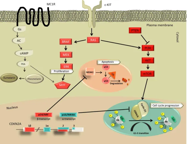
Somatic events during MM formations may occur along multiple signal pathways simultaneously (Figure 9), namely:
1) Activation of the MAPK pathway,
2) Inactivation of the p16/INK4A-CDK4/6-Rb senescence barrier, 3) Inactivation of p53/ARF barrier or
4) Activation of the PTEN/PI3K/AKT pathway.
Figure 9. Summary of signal transduction pathways in MM development.
To date BRAF, NRAS, c-KIT and MITF somatic mutations are considered as driver mutations during MM development and progression (Curtin et al. 2005). The first therapeutic experiences with BRAF inhibitors in MM showed that MAPK and PI3K/AKT concomitant pathway activations induce acquired resistance during the course of therapy (reviewed in Hocker and Tsao 2008 and Lo 2013).
26
A number of additional genes might harbor somatic driver mutations in MM (PPP6C, RAC1, SNX31, TACC1, STK19) (Hodis et al. 2012).
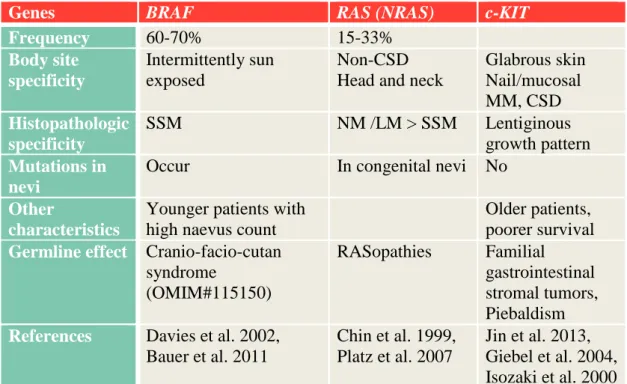
The most common genes affected at somatic level in MM development and their association with other tumor features is summarized in Table 3.
Table 3. Most common genes with somatic alterations in MM genesis and their associated characteristic clinical features.
Abbreviations: CSD: chronic sun damage, SSM: superficial spreading melanoma I.4.2.1. MAPK pathway activation in MM
MAPK pathway is a key regulator of cell proliferation, differentiation, survival and death, and also has a significant role in MM development. The most frequently mutated member of this pathway is BRAF, but almost every MM harbors mutations in any of this signal pathway’s proteins (Fecher et al. 2008). Type of mutations (BRAF, NRAS) varies upon body site and histology (Table 3).
I.4.2.1.1. BRAF in MM
BRAF is an oncogene, a protein kinase located in the cytosol. Somatic BRAF mutations are detected in 60-70% of MMs and the V600E substitution accounts for more than 90% of them (Davies et al. 2002). As many benign and dysplastic nevi (~80%) express also V600E (Pollock et al. 2003), it is questionable whether it is an early event in MM
Genes BRAF RAS (NRAS) c-KIT
Frequency 60-70% 15-33%
Body site specificity
Intermittently sun exposed
Non-CSD Head and neck
Glabrous skin Nail/mucosal MM, CSD Histopathologic
specificity
SSM NM /LM > SSM Lentiginous
growth pattern Mutations in
nevi
Occur In congenital nevi No
Other
characteristics
Younger patients with high naevus count
Older patients, poorer survival Germline effect Cranio-facio-cutan
syndrome
(OMIM#115150)
RASopathies Familial gastrointestinal stromal tumors, Piebaldism References Davies et al. 2002,
Bauer et al. 2011
Chin et al. 1999, Platz et al. 2007
Jin et al. 2013, Giebel et al. 2004, Isozaki et al. 2000
27
genesis (reviewed in Palmieri et al. 2009), or might be a marker from ‘in situ’ to invasive progression state (Greene et al. 2009). It is however evident now that this BRAF mutation alone is not sufficient for MM formation.
I.4.2.1.2. RAS in MM
RAS is one of the most studied oncogene in human cancers; it is a 21kDa protein with a GTPase activity regulating receptor tyrosine kinase-induced MAPK activation. It binds to and activates PI3K, and its downstream targets (AKT), and interacts with p16/INK4A and p53 too (summarized in Chin et al. 1999).
Activating mutations in NRAS are found in approximately half of congenital nevi, and one third of MMs (Chin et al. 2006). Besides the most prominent NRAS mutations in MM, other RAS family members, such as HRAS (1%) and KRAS (2%) are also altered in MM (Forbes et al. 2008). HRAS mutations were detected to be at a higher frequency (7.7%) in NM (Jafari et al. 1995).
Germline mutations of RAS proteins cause RASopathies, and these patients are not reported to have elevated risk to MM development.
I.4.2.2. p16/INK4A-CDK4/6-RB inactivation I.4.2.2.1. CDKN2A
Somatic loss of the 9p21 chromosome region encoding CDKN2A locus may manifests via deletion, point mutation or even by epigenetic changes and is a common event in many human cancer types such as MM, PaC, lung cancer (LC), high-grade glioma, colon adenocarcinoma, breast adenocarcinoma, biliary tract-, head and neck tumors and transitional cell carcinoma of the bladder (Dahl and Guldberg 2007, Bennett 2008). In MM, somatic CDKN2A alterations are detected in 30-70% of sporadic MMs (Bartkova et al. 1996, Walker et al. 1998).
I.4.2.2.2. CDK4, Rb1
Somatic mutations in CDK4 are common at the 24th (Wolfel et al. 1995), as well as at the 22nd amino acid position (Bennett 2008, Dahl and Guldberg 2007), while amplification of this oncogene is also observed in MMs (Muthusamy et al. 2006).
28
Rb1 protein is less commonly affected in MM and mainly by nonsense mutations at somatic level (Bartkova et al. 1996).
I.4.2.3. ARF/p53 inactivation in MM
ARF deletion occurs around 50% of MMs (Curtin et al. 2005), while protein p53, the common tumor suppressor gene of many cancer types, is altered only in 10-30%
(Albino et al. 1994, Zerp et al. 1999).
I.4.2.4. PTEN /PI3K/AKT pathway activation in MM
PTEN is altered in 30-40 % of cultured MM cell lines and approximately 10% of primary MMs (Guldberg et al. 1997, Tsao et al. 1998) by mutation, allelic loss, epigenetic silencing by methylation (Mirmohammadsadegh et al. 2006) or impaired subcellular localization (Trotman et al. 2007). It is still controversial whether PTEN loss is an early or late event in MM genesis (summarized in Palmieri et al. 2009). As PTEN downregulates AKT and MAPK pathways, it is possible that RAS activation and PTEN loss confers a reciprocal mutation status (Nogueira et al. 2010).
I.4.2.5. MC1R in MM at somatic level
Besides the well-known effects of MC1R germline variants on MM predisposition, no somatic genetic or copy number alterations have been proved so far in any type of MM.
(Valverde et al. 1996, Kim et al. 2008).
I.4.2.6. Novel signaling pathways in MM
Further genes with lesser significance might also undergo somatic mutations, deletions, amplifications, or epigenetic changes (promoter hypermethylation) (Table 4).
Table 4. List of further genes involved in MM genesis at somatic level (On the basis of Dahl and Guldberg 2007, with personal changes).
Gene type
Gene Altered
signaling pathway
Most frequent genetic changes
Frequ- ency %
Ref.
Oncogenes
CTNNB1 Mutation 2-23
CCND1/
Cyclin D
Amplification 6-44 Sauter et al.
2002 MITF Amplification 10-16
AKT3 PI3K Amplification 40-60
29 Gene
type
Gene Altered
signaling pathway
Most frequent genetic changes
Frequ- ency %
Ref.
ERBB4 AKT Mutation 19 Prickett et al.
2009 Tumor suppressor genes
MTAP SNP Bishop et al.
2009
Stevens et al.
2009 STK11/
LKB1
Mutation 10 Forbes et al.
2011 Others
Non- receptor tyrosine kinase (glutamate) receptors
GRIN2A Mutation 25 Wei et al.
2011
GRM3 Mutation 16.3 Prickett et al.
2011
Abbreviations: Ref: reference, SNP: single nucleotide polymorphism I.4.2.7. MicroRNAs (miRNA)
miRNAs are small noncoding RNAs with posttranscriptional regulatory function. They modulate the gene expression mainly by translational repression or mRNA degradation and might also contribute to MM genesis and/or progression. Dysregulation of miRNAs by genetic or epigenetic alterations and aberrant miRNA biogenesis may lead to down-, or upregulated miRNAs. More miRNAs regulate MITF expression (miR-137, miR-148 and others) promoting development and invasiveness of MM (reviewed by Bell and Levy 2011). miRNA expression profiles of primary and metastatic MMs are associated with somatic BRAF or NRAS mutational status and predict survival and sites of distant metastasis (Caramuta et al. 2010).
30
I.5. MM-associated other primary malignancies
MM survivors are at an increased risk of developing subsequent primary tumors with an incidence rate of 6.4-20% (Levi et al. 1997, Bhatia et al. 1999, Wolff et al. 2000, Schmid-Wendtner et al. 2001, Crocetti et al. 2008, Bradford et al. 2010, Spanogle et al.
2010). The most frequently observed high risk tumor is second and subsequent MM.
I.5.1. Multiple primary melanomas (MPM)
I.5.1.1. Incidence, etiology
Pack described firstly the phenomenon of MPM in 1952 (Pack et al. 1952). Among patients with a history of primary MM, second primary MM occurs in 3.4-8.3%
(Slingluff et al. 1993, Giles et al. 1995, Blackwood et al. 2002, Ferrone et al, 2005, Helsing et al, 2008, Savoia et al, 2012).
It is important to ensure that subsequent MMs are primary tumors and not cutaneous epidermotropic metastasis of previous MM (Heenan et al. 1991, Bengoechea- Beeby et al. 1993). A recent report on somatic mutation patterns of first and subsequent MMs excluded any common clonal origin, and proved that subsequent MMs are indeed clinically and histologically independent primary tumors (Orlow et al. 2009).
Estimating the real MPM incidence on large MM populations is limited by the difficulty of follow-up for a long period of time.
The majority (63-88%) of patients develop two primary MMs (Ferrone et al.
2005, Savoia et al. 2012), however higher number of primaries are also observed (Ferrone et al. 2005, Savoia et al. 2012, Hwa et al. 2012)
Most strongly associated risk factors include positive family history of MM (Giles et al. 1995, Blackwood et al. 2002) and personal history of dysplastic naevus (Blackwood et al. 2002, Titus-Ernstoff et al. 2006, de Giorgi et al. 2010) or a higher number of common nevi (de Giorgi et al. 2010), all suggesting an underlying genetic contribution factor. Additional risk factors are 1) high UV irradiance at birth and before 10 years of age, 2) lifetime recreational sun exposure, 3) beach and waterside activities and 4) vacations in sunnier climate (Kricker et al. 2007).
31 I.5.1.2. Clinicopathological characteristics
The mean age at diagnosis of first MM is lower if number of primary MMs rise (Helsing et al. 2008). Second MM develops often within the first 1-2 years from the initial MM diagnosis (Ferrone et al. 2005, Hwa et al. 2012, Savoia et al. 2012), and there is also a strong site concordance (42-56%) (Ferrone et al. 2005, Savoia et al. 2012, Slingluff et al. 1993, Giles et al. 1995, Bower et al. 2010, Manganoni et al. 2012, Hwa et al. 2012). The subsequent MMs are showing a trend of being thinner, the proportion of Mis rises and they exhibit histological features with more favorable prognostic value (less ulceration, more regression) (Brobeil et al. 1997, Ferrone et al. 2005, Murali et al.
2012a, Hwa et al. 2012). SSM is the most frequently presented histological subtype among MPM, while NM is far less frequent (Vecchiato et al. 2013).
MPM patients have a better survival than single primary melanoma (SPM) patients (Doubrowsky et al. 2003, Bower et al. 2010, Murali 2012b, Kricker et al.
2013). Two possible explanations are 1) the careful follow-ups of these patients, that have been proven to be a reason why the subsequent MMs are thinner; and 2) a distinct biological behavior of these tumors (summarized in Hwa et al. 2012). Comparing prognostic features such as mitotic rate, ulceration and pathologic stage of the primary MMs in SPM and MPM patients, no significant difference have been detected (Hwa et al. 2012).
I.5.1.3. Genetics of MPM
I.5.1.3.1. CDKN2A
Familial MM and MPM patients own a higher genetic predisposition, therefore are particularly good candidates exploring germline genetic background of MM susceptibility. Many studies have been conducted in order to determine predisposing gene mutation frequency among MPM patients. Mutation rate of CDKN2A among MPM patients is higher than among SPM patients, varying between 2.9-32.6%, from which sporadic MPMs exhibit 8-15% of mutation rate, while it can be much higher (even 63%) if family history is positive for MM (summarized in Table 5). Factors such as selection bias, geographical differences of MM incidences, overrepresentation of familial MM cases, founder effects and sensitivity differences in terms of mutation detection may explain this wide range of prevalence (Helsing et al. 2008). Risk of
32
germline CDKN2A mutation raises with the number of MMs (Puig et al. 2005, Helsing et al. 2008, Pastorino et al. 2008) irrespectively of positive family history. Type of CDKN2A mutations doesn’t differ between MPM, SPM or familial MM cases.
CDKN2A mutation carrier MPM patients develop their first MM at an earlier age than mutation negative patients (Helsing et al. 2008, Pastorino et al. 2008).
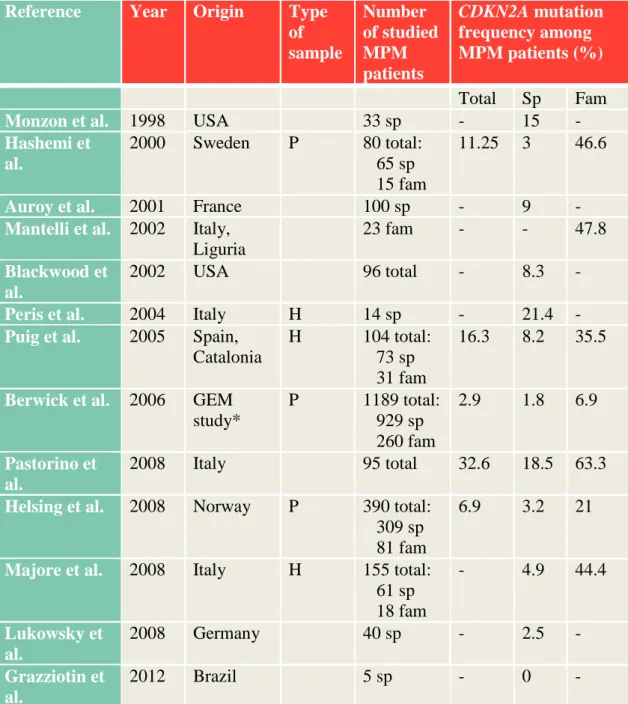
Table 5. Published frequencies of CDKN2A mutations among MPM patients.
Reference Year Origin Type of sample
Number of studied MPM patients
CDKN2A mutation frequency among MPM patients (%) Total Sp Fam
Monzon et al. 1998 USA 33 sp - 15 -
Hashemi et al.
2000 Sweden P 80 total:
65 sp 15 fam
11.25 3 46.6
Auroy et al. 2001 France 100 sp - 9 -
Mantelli et al. 2002 Italy, Liguria
23 fam - - 47.8
Blackwood et al.
2002 USA 96 total - 8.3 -
Peris et al. 2004 Italy H 14 sp - 21.4 - Puig et al. 2005 Spain,
Catalonia
H 104 total:
73 sp 31 fam
16.3 8.2 35.5
Berwick et al. 2006 GEM study*
P 1189 total:
929 sp 260 fam
2.9 1.8 6.9
Pastorino et al.
2008 Italy 95 total 32.6 18.5 63.3
Helsing et al. 2008 Norway P 390 total:
309 sp 81 fam
6.9 3.2 21
Majore et al. 2008 Italy H 155 total:
61 sp 18 fam
- 4.9 44.4
Lukowsky et al.
2008 Germany 40 sp - 2.5 -
Grazziotin et al.
2012 Brazil 5 sp - 0 -
*GEM (Genes, Environment and Melanoma) study population consists of 8 population- based cancer registries from nine geographic regions: Australia (New South Wales and
33
Tasmania), Canada (British Columbia and Ontario), Italy (Piemonte), and United States (Orange County and San Diego County from California, Michigan, New Jersey, North Carolina).
Abbreviations: P: population based; H: hospital or clinic-based; sp: sporadic, fam:
familial
Besides CDKN2A gene mutations, the c.442 G>A resulting p.A148T amino acid change located in exon 2, as the third most common polymorphism in the gene, seems to occur also statistically more frequently in MPM patients than in healthy controls: 13.5%
versus 5.45%; p=0.05 (Puig et al. 2005), and 15.7% versus 6.6 %; p=0.011 (Pastorino et al. 2008). Frequency differences between healthy compared to SPM patients are debated; Pastorino et al found no difference (Pastorino et al. 2008), while in Brazil this variant was more frequent in MM patients than in controls (12.6% versus 3.9%;
p=0.009), and was also more frequent if family history of cancers other than MM was positive (Bakos et al. 2011).
I.5.1.3.2. MC1R
Carrying multiple variant alleles of MC1R gene is in association with the development of subsequent primary MMs both in CDKN2A mutation positive (Goldstein et al. 2005, Fargnoli et al. 2010) and negative MM patients (Pastorino et al. 2008, de Torre et al.
2010). Kanetsky and co-workers found another, more qualitative link between MPM formation and carrier status of 1 or 2 ‘R’ alleles, or ‘R’/‘r’ alleles, while carrier status of 2 ‘r’ alleles did not follow this trend (Kanetsky et al. 2010). In CDKN2A mutation positive MPM patients, as number and type of variants increase, the median age of onset decrease significantly (Goldstein et al. 2007).
There are only a few studies on MC1R variants among pure MPM patients (Kanetsky et al. 2006, Helsing et al. 2008), therefore so far no pattern or variant specificity among this patient population have been observed.
Role of CDK4 and MITF E318K mutation in MPM are discussed above (I.4.1).
I.5.1.4. Other non-melanoma primary malignancies among MPM patients
Second primary cancer among MM survivors are commonly explored, however risk of developing subsequent non-melanoma primaries among MPM patients is rarely
34
discussed. Manganoni and co-workers reported recently, that non-melanoma primaries are more common among MPM than SPM patients (OR: 2.1; 95%CI 1.11-3.97) (Manganoni et al. 2012). Majority of these tumors were NMSC, while breast cancer and prostate cancer (PrC) were also observed among others. When comparing SPM and MPM patients regarding MM associated malignancies MPM patients have significantly more co-occurred malignant tumors (Pollio et al. 2013).
I.5.2. MM and co-aggregation of other cancers
Further tumors that are most consequently established with increased risk among MM survivors are NMSC, pancreas, non-Hodgkin lymphoma (NHL), chronic lymphocytic leukemia (CLL), female breast-, kidney-, bladder-, corpus uteri-, small intestine-, brain-, oropharynx-, salivary gland-, soft tissue-, and thyroid-, prostate-, and bone cancers (Table 6). Interestingly some malignant tumors are less frequently observed among MM survivors, namely LC (Crocetti et al. 2008, Balamurugan et al. 2011), and liver cancer (Crocetti et al. 2008).
Table 6. Summary of studies on MM associated subsequent primary malignancies.
Reference Type of sample
Patient
number SIR Incidence rate %
Overall risk %
Cancers with significantly increased risk Wassberg
et al. 1996
P 20,354 1.45 7.9 Nervous system
tumors, leukemia Colon cancer in men Wolff et al.
2000
H 554 - 11 MM, BCC
Schmid- Wendtner et al. 2001
H 4597 6.4 Kidney cancer in
men Wu et al.
2006
H 955 6.2 NHL and renal cell
cancer in men Crocetti et
al. 2008
P 14,560 1.27 - 27 NMSC, bone,
kidney Bradford
et al. 2010
P 89,515 12.1 28 Breast, PrC, NHL
Spanogle et al. 2010
P 16,591 11 32 Soft tissue, MM of
the eye and orbital, non-epithelial skin, salivary gland, bone and joint, thyroid, kidney, CLL, brain
35 Reference Type of
sample
Patient
number SIR Incidence rate %
Overall risk %
Cancers with significantly increased risk and nervous system, NHL, PrC, and female breast Balamu-
rugan et al. 2011
P 40,881
Mis 6041 MM
32% men, 35%
women 57% men 64%
women
CLL CLL
CLL, NHL thyroid CLL, NHL, thyroid
Abbreviations: SIR: standardized incidence ratio, NHL: non-Hodgkin lymphoma, CLL:
chronic lymphocytic leukemia, NMSC: non-melanoma skin cancer, Mis: melanoma in situ, P: population-based study, H: hospital-based study
In our previous study analyzing subsequent primary tumor development in 740 MM survivors showed an extremely elevated risk of subsequent MM (SIR men:160; women:
93), Mis (SIR men: 342, women: 77) and NMSC (SIR men: 17, women: 18), while regarding non-skin malignancies, significantly elevated risk was observed for CLL, colon-, and kidney cancers in both genders, NHL and cervical cancer in women only, and bladder cancer in men (Tóth V, Hatvani Z. et al. 2013).
Co-occurring tumor types may indicate common environmental and/or genetic background, while coincidental risk factor exposures must be also taken into account.
Early MM diagnosis is in most cases followed by only surgical therapy, therefore irradiation and systemic chemotherapy in the genesis of subsequent primaries may not play significant role. Socioeconomic status may also have an impact as it represents a pattern of environmental risk factors; and certain risk factors may coincidentally affect individuals with higher socioeconomic status (e.g.: late pregnancies and holiday/recreational sun exposure habits to elevated risk of breast cancer and MM) (summarized in Spanogle et al. 2010).
Common genetic and environmental factors of the most frequent tumors associated with MM are summarized in Table 7.