Original Article
Metabolic syndrome and other cardiovascular risk factors associated with the progression of IgA nephropathy
Tibor Kovács1,*, Tibor Vas1,*, Csaba P. Kovesdy2, István Késõi1, Balázs Sági1, István Wittmann1and Judit Nagy1
1Second Department of Medicine and Nephrological Center, Faculty of Medicine, University of Pécs, Pécs, Hungary and2Health Science Center, University of Tennessee, Memphis, TN, USA
Correspondence and offprint requests to:Judit Nagy; E-mail: judit.nagy@aok.pte.hu
*These authors contributed equally to this work.
Abstract
Background.The metabolic syndrome is associated with modest but independent and additive risk of new onset chronic kidney disease (CKD) in several studies. The purpose of our study was to determine whether metabolic syndrome and other cardiovascular risk factors (hyperuricaemia and smoking) are associated with the progression of IgA nephropathy (IgAN).
Methods. Two hundred and twenty three IgAN patients (107 with and 116 without metabolic syndrome) were examined. The primary renal end point was doubling of serum creatinine; sec- ondary end points were reaching eGFR of≤60 ml/min/1,73m2or eGFR of≤30 ml/min/1.73 m2, and end-stage renal disease, ESRD (the composite of serum creatinine≥500 µmol/l, initiation of dialysis treatment or transplantation). The association of metabolic syndrome with renal end points was examined using the Kaplan-Meier method and Cox models.
Results. Metabolic syndrome established at the diagnosis or during follow-up of IgAN patients was significantly associated with the primary renal end point (unadjusted hazard ratio of dou- bling of serum creatinine, 95% confidence interval: 1.96 (1.17–1.33, p = 0.011). The association remained significant after adjustment for confounders: 1.70 (1.02–3.83, p = 0.040). Results were similar for secondary end points except ESRD which was not associated with the presence of metabolic syndrome. Hyperuricaemia and smoking were independent risk factors of progression.
Survival curves stratified on metabolic syndrome status showed significant differences for the end points ( p = 0.017–0.001) except for ESRD.
Conclusions.Early diagnosis and treatment of metabolic syndrome, hyperuricaemia and smoking may be an additional cost-effective strategy for preventing the progression of IgAN.
Keywords:cardiovascular risk factors; chronic kidney disease progression; end-stage renal disease; IgA nephropathy;
metabolic syndrome
Introduction
The term metabolic syndrome is commonly used to de- scribe the clustering of cardiovascular risk factors, namely central obesity, hypertension, impaired glucose metabolism and dyslipidaemia. Metabolic syndrome is found in over 25% of adults in the USA and in several other industrialized countries, and there appears to be an increasing preva- lence in higher age groups [1]. Individuals with metabolic syndrome are at increased risk for cardiovascular diseases as well as cardiovascular and all-cause mortality [2].
The recent interest of nephrologists in metabolic syn- drome increased after the publication of Chen et al. [3], demonstrating in a cross-sectional study that metabolic syndrome was associated with a 2.26-fold higher risk (1.68–4.03) of chronic kidney disease (CKD) in a sample representative of the US population. Other cross-sectional studies have also demonstrated a link between metabolic
syndrome and CKD (see later). In the only longitudinal study, Kurella et al. [4] demonstrated a significantly in- creased risk of incident CKD in non-diabetic adults with metabolic syndrome. However, the effects of metabolic syndrome on the progression of CKD beyond the contri- bution of impaired glucose metabolism and hypertension are far from being established with certainty.
IgA nephropathy (IgAN) is the most common primary glomerulonephritis and is an important cause of end- stage renal disease (ESRD) worldwide [5]. Long-term observation in many countries has shown that IgAN causes ESRD in as many as 40% of patients within 20 years after diagnosis [6, 7]. Clinical presentation is usually with haematuria and with variable degrees of proteinuria. Pathologically, IgAN is characterized by the glomerular deposition of polymeric IgA1 mainly in the mesangium accompanied by mesangial hypercellularity, mesangial matrix expansion, and varying degrees of
© The Author 2012. Published by Oxford University Press on behalf of ERA-EDTA. All rights reserved.
For permissions, please email: journals.permissions@oup.com.
Clin Kidney J (2012) 0: 1–7 doi: 10.1093/ckj/sfs131
Clinical Kidney Journal Advance Access published November 2, 2012
by guest on January 18, 2013http://ckj.oxfordjournals.org/Downloaded from
glomerulosclerosis and interstitial fibrosis. Adverse prog- nostic indicators include the presence of heavy proteinur- ia and hypertension, a significant reduction in glomerular filtration rate (GFR) at the time of renal biopsy and the extent of glomerulosclerosis and tubulointerstitial fibrosis on renal pathology [7, 8]. In addition to these known risk factors, other cardiovascular risk factors, such as hypertriglyceridaemia, hyperuricaemia, excessive body weight or cigarette smoking have also been associated with the progression of IgAN in recent studies [9–11].
However, there are no data about the prevalence of metabolic syndrome in IgAN patients, and there have not been any reports of an association between metabolic syndrome and the progression of IgAN.
The purpose of the present study was to determine whether there are differences in the progression of IgAN according to the presence of metabolic syndrome and other cardiovascular risk factors at the time of diagnosis and during the course of IgAN. We emphasized that the clustering of cardiovascular risk factors is associated with a more severe progression of IgAN.
Materials and methods
Study population
We examined 240 biopsy-proven IgAN patients with normal or mild to moderately decreased renal function (CKD Stage 1–3) at the time of the diagnosis of IgAN. All of the patients were diagnosed in the Nephrology Center, Medical Faculty, University of Pécs, Hungary and fol- lowed-up in 3- to 6-month intervals by the same two ne- phrologists, TK and JN. Seventeen patients were not included in the statistical analyses of this study because of insufficient clinical data at the time of the diagnosis of IgAN. Further exclusion criteria were: secondary IgAN cases, rapidly progressive crescentic patients, patients with nephrotic syndrome and immunosuppressive treat- ment. The analysed cohort included 223 patients.
Definition of metabolic syndrome
All IgAN patients were analysed to determine whether criteria for metabolic syndrome were met by using a modified NCEP ATP III (National Cholesterol Education Programme—Adult Treatment Panel III) definition of metabolic syndrome [12]. Metabolic syndrome was defined as any three or more of the following criteria: (i) fasting plasma glucose level of 5.6 mmol/L or higher or impaired glucose tolerance; (ii) triglyceride level of 1.7 mmol/L or higher or lipid-lowering drug treatment; (iii) high-density lipoprotein (HDL) cholesterol level <1.0 mmol/L for men and <1.3 mmol/L for women or drug treatment; (iv) body mass index (BMI)≥30 kg/m2; (v) hy- pertension with blood pressure≥130/85 mmHg or antihy- pertensive treatment.
Study measurements
Demographic, anthropometric and laboratory data as well as information about lifestyle habits were collected on all participants at the time of diagnosis and at each follow-up visit. Blood was collected by venepuncture after an overnight fast of at least 10 h at all follow-up examin- ations. The Central Laboratory of Medical Faculty, Univer- sity of Pécs measured all serum chemistry levels in fresh
samples with commercially available reagents. The glo- merularfiltration rate (eGFR; mL/min/1.73 m2) was esti- mated with the Chronic Kidney Disease Epidemiology Collaboration equation [13]. BMI was calculated as weight in kilograms divided by the square of the height in metres.
Definition of progression of IgAN
The primary renal outcome was the doubling of serum creatinine, secondary renal outcomes were the decrease of eGFR to≤60 mL/min/1.73 m2, or to≤30 mL/min/1.73 m2 or reaching ESRD (defined as the composite of a serum creatinine≥500 µmol/L or the initiation of dialysis treatment or transplantation).
Statistical analysis
Data analysis was performed using the SPSS software program version 13.0 (SPSS Inc., Chicago, IL, USA). Con- tinuous variables with normal distribution were expressed as mean ± standard deviation and were compared by using Student’st-tests. Variables with non-normal distri- bution were compared by the Mann–WhitneyU-test, and categorical variables were expressed as percentage and compared by theχ2 test. The mean renal survival time until the selected end points was calculated using the Kaplan–Meier method. Differences between the calcu- lated mean renal survival times were compared using the log-rank test. The effect of confounders was assessed by Cox regression analysis.
Confounders were determineda prioribased on theor- etical considerations and by examining baseline covariate associations with metabolic syndrome and with the renal end points [14]. Multivariate models were constructed with sequential adjustments for age and gender as well as for age, gender, uric acid, eGFR, smoking and angioten- sin-converting enzyme inhibitors/angiotensin II receptor blocker (ACEis/ARB). A value of P < 0.05 was considered statistically significant.
Results
Baseline clinical data
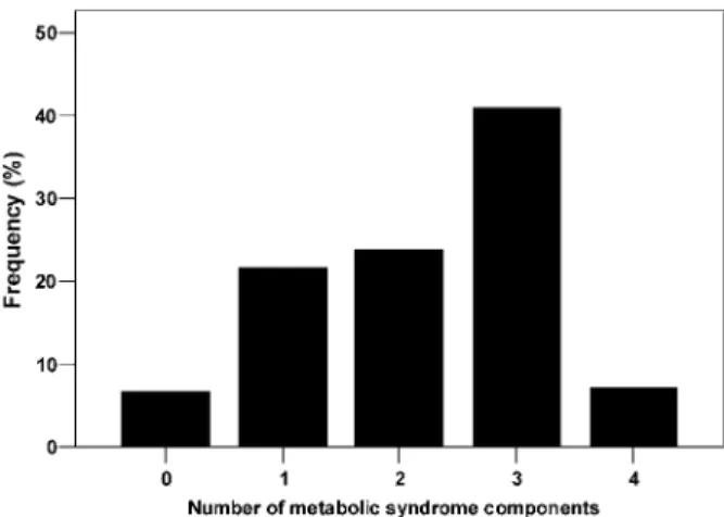
The mean age of the 223 participants included in the analytic cohort was 36.4 ± 13.0 years, 72% were male and 28% were female. Metabolic syndrome was already present in 17% of participants (38 patients) at the diag- nosis of IgAN and was diagnosed de novo in a further 31% of participants (69 patients) during the follow-up period. Analysing all (n= 223) IgAN patients together at the end of the follow-up period, the prevalence of zero, one, two, three and four parameters of the metabolic syndrome was 15 (7%), 48 (22%), 53 (24%), 91 (41%) and 16 (7%), respectively (Figure 2). Sixty-eight (31%) patients were obese and impaired carbohydrate metab- olism [impaired glucose tolerance (IGT), impaired fasting glucose (IFG) or diabetes mellitus] was present in 77 (35%) patients, HDL cholesterol was low in 108 (48%) patients, hypertriglyceridaemia was present in 133 (60%) patients and elevated blood pressure or antihypertensive treatment was present in 195 (87%) patients at the end of the follow-up period. Analysing together hypertrigly- ceridaemia, lower HDL cholesterol and statin treatment, 151 (68%) patients were dyslipidaemic.
by guest on January 18, 2013http://ckj.oxfordjournals.org/Downloaded from
Baseline characteristics of IgAN patients with and without metabolic syndrome are summarized in Table 1.
There were no significant differences in the age, sex, follow-up time or number of smokers between the two groups. Patients with metabolic syndrome had higher uric acid levels in addition to the expected higher values for BMI, triglyceride, glucose and lower HDL cholesterol level that define metabolic syndrome. At the time of the diagno- sis of IgAN, the eGFR of patients with metabolic syndrome was lower (73.6 ± 31.8 versus 82.2 ± 27.7 mL/min/1.73 m2 in patients without metabolic syndrome, P < 0.05).
However, at the end of the follow-up period, the difference between the eGFR of the two groups was more substantial (46.8 ± 31.6 versus 67.4 ± 35.3 mL/min/1.73 m2, P < 0.001).
The systolic and diastolic blood pressures of patients with metabolic syndrome were significantly higher (Table 1), and significantly more patients were treated with ACEi/ARB in the metabolic syndrome group (P < 0.001).
Association of metabolic syndrome with progression of IgAN
Survival curves stratified on metabolic syndrome status showed statistically significant differences in the time to reach three different end points for the renal outcome:
the doubling of serum creatinine (P = 0.009, Figure 1A), eGFR ≤60 mL/min/1.73 m2 (P = 0.001, Figure 1B) and eGFR ≤30 mL/min/1.73 m2 (P = 0.017, Figure 1C). The
difference in time to reach the composite end point of ESRD (defined as the composite of a serum creatinine
≥500 µmol/L or the initiation of dialysis treatment or transplantation) was not significant (P = 0.2).
Metabolic syndrome was significantly associated with the progression of IgAN in unadjusted Cox models for three different renal end points eGFR≤60 mL/min/1.73 m2, eGFR ≤30/mL/min/1.73 m2 and the doubling of serum creatinine (Table 2). The association remained sig- nificant after adjustment for confounders (Table 2). The association, however, was not significant for the end points of ESRD in neither unadjusted nor adjusted Cox models (Table 2). At the analysis of the effect of single traits of metabolic syndrome: obesity (yes/no, Y/N), hy- pertension (Y/N), dyslipidaemia (Y/N), dysglycaemia (Y/N) and complete metabolic syndrome on the prognosis of IgAN hypertension and complete metabolic syndrome were the two strongest influencing parameters (Table3).
In Cox regression analysis, higher uric acid level was also an independent risk factor for three different renal end points:
(i) eGFR≤60 mL/min/1.73 m2[hazard ratio (HR) associ- ated with a 1 mmol/L higher serum uric acid level:
1.003; 95% confidence interval (95% CI) 1.001– 1.005].
(ii) eGFR≤30 mL/min/1.73 m2(HR 1.002; 95% CI 1.001– 1.005) and
(iii) ESRD (HR 1.004; 95% CI 1.001–1.006).
Smoking was significantly associated with ESRD (HR 2.024; 95% CI 1.046–3.920).
Discussion
Our study showed that metabolic syndrome established at the time of diagnosis or during the follow-up of IgAN was significantly associated with the primary renal end point and remained significant after adjustment for con- founders. Results were similar for the secondary end points except for ESRD. Hyperuricaemia was also an inde- pendent risk factor for all secondary end points, and smoking was an independent risk factor for ESRD.
Survival curves stratified on metabolic syndrome status showed significant differences for the association with the various end points except for ESRD.
The criteria for metabolic syndrome diagnosis were set-up originally to identify those individuals most likely to develop cardiovascular diseases [15]. There is no doubt nowadays that the patients with metabolic syn- drome are at significantly higher risk for CKD too, as sum- marized by a number of review articles and by the recent meta-analysis of Thomas et al. [16–22]. Most of the observational studies discussed in these papers found a significant association between metabolic syndrome and CKD. The odds ratios for CKD in the different studies were:
2.60 in the National Health And Nutrition Examination Survey III (NHANES III) database [3], 1.43 in the Athero- sclerosis Risk in Communities study [4], 1.3 in American Indians [23], 1.54 in Japanese adults [24], 1.88 in the Tehran Lipid and Glucose Study [25], 1.64 in the Inter- Asia study [26], 1.31 in hypertensive African Americans [27], 1.31 in the Hong-Kong Diabetes Registry [28], 1.77 in Korean adults [29], 1.74 [30] and 1.42 [31] in Chinese adults as well as 1.30 in non-diabetic Taiwanese adults [32]. However, the majority of the studies were cross-sec- tional and, as such, unable to establish a cause-effect
Table 1.Baseline characteristics of patients with and without metabolic syndrome
Metabolic syndrome (n= 107)
No metabolic syndrome
(n= 116) P-value
Age 37.9 ± 13.8 34.9 ± 12.2 0.089
Follow-up (months) 146.6 ± 112.5 146.1 ± 99.4 0.589
Sex (M/F) 81 (76%)/26
(24%)
80 (69%)/36 (31%)
0.296 Parameters of metabolic syndrome
BMI (kg/m2) 30.8 ± 4.8 24.9 ± 3.4 <0.001
Systolic BP (Hgmm) 143 ± 20 136 ± 19 0.005
Diastolic BP (Hgmm) 89 ± 12 85 ± 12 0.014
Hypertension (Y/N) 105 (98%)/2 (2%)
90 (78%)/26 (22%)
<0.001 Triglyceride
(mmol/L)
2.18 ± 1.21 1.59 ± 1.55 <0.001 HDL (mmol/L) 1.21 ± 0.48 1.39 ± 0.39 <0.001 Blood sugar
(mmol/L)
6.32 ± 1.44 5.10 ± 0.81 <0.001
Uric acid (mmol/L) 390 ± 124 351 ± 118 0.006
Smoking (Y/N) 27 (25%)/79 (75%)
30 (26%)/83 (74%)
0.879 Renal function
eGFR at the diagnosis (mL/min/1.73 m2)
73.6 ± 31.8 82.2 ± 27.7 <0.05
eGFR at the end of follow-up (mL/min/
1.73 m2)
46.8 ± 31.6 67.4 ± 35.3 <0.001
Drugs
ACEi/ARB (Y/N) 92 (86%)/15 (14%)
73 (63%)/43 (37%)
<0.001 Statins (Y/N) 48 (45%)/59
(55%)
19 (16%)/97 (84%)
<0.001
Data expressed as mean ± standard deviations and number of participants (for categorical variables).
BMI, body mass index; BP, blood pressure; HDL, high-density lipoprotein;
eGFR, estimated glomerularfiltration rate; ACEi, angiotensin-converting enzyme inhibitor; ARB, angiotensin II receptor blocker. Comparisons were made byt-tests orχ2tests.
by guest on January 18, 2013http://ckj.oxfordjournals.org/Downloaded from
relationship between metabolic syndrome and the reduction in kidney function, and also unable to deter- mine if metabolic syndrome is associated with longitudi- nal changes in kidney function. In this study, we detected a significant association between metabolic syndrome and the progressive loss of kidney function except in ESRD, suggesting a potential effect of metabolic syndrome on the early progression of IgAN. The lower eGFR level of patients with metabolic syndrome seen at the time of diagnosis of IgAN suggests a potential effect of metabolic syndrome on the incidence of the renal disease too. Our observation that metabolic syndrome did not associate with the progression of IgAN in the end-stage of IgAN is in agreement with the results of the study of Lee et al. [33] on CKD patients participating in the CKD prevention programme regulated by the Public Health Bureau of Taiwan.
Among the components of metabolic syndrome, dia- betic and hypertensive injuries, the two major aetiologies of CKD worldwide, have been well studied and described.
Concerning the association between IgAN and diabetes, Fliseret al. [34] published that insulin resistance and hy- perinsulinaemia are already present in patients with inci- pient renal disease, among others in IgAN patients. In our present study, we found impaired glucose regulation (IGT, IFG or diabetes mellitus) in 77 (35%) patients. It is known that IgAN can also be superimposed on diabetic nephro- pathy or can be the only renal abnormality of diabetic patients. The association between IgAN and diabetes may not be coincidental, because the intraglomerular hy- pertension and hyperfiltration as well as biochemical
alterations in the glomeruli of diabetic patients may facili- tate the deposition of IgA1 immune complexes or aggre- gates. Furthermore, the abnormalities of the IgA immune system are common in Type 2 diabetes [35].
Hypertension is common in CKD patients and, similar to the general population, it predicts cardiovascular morbidity and mortality. Hypertensive CKD patients with metabolic syndrome have an excess cardiovascular risk. The relation- ship between blood pressure and mortality is U-shaped;
low mean and diastolic blood pressure predicts early mor- tality [36]. As we already mentioned in the introduction, the progression in IgAN is more severe in the presence of hypertension and strict blood-pressure control portents renal protection in IgAN [37]. In our study, among the par- ameters of metabolic syndrome, hypertension had the greatest influence on the prognosis of IgAN.
Obesity has been associated with an increased risk for the incidence of CKD and for ESRD in several epidemiologi- cal studies [38–41]. In the examined populations, obesity as indicated by the elevated BMI was associated with de- creased renal function and ESRD independent of the pres- ence of hypertension and diabetes. Weight loss has a protective effect against the progression of CKD [40]. Higher BMI shows a seemingly paradoxical association with better survival in advanced CKD and in ESRD, which could be related to a better nutritional status in patients with elev- ated BMI [42].Concerning IgAN patients, obesity was a pre- dictive factor not only for the development of hypertension, but also for chronic renal failure and obese patients have glomerular enlargement and ultrastructural modification of the glomerular basement membrane [9,43].
Dyslipidaemia, in particular atherogenic dyslipidaemia (high triglyceride and low HDL cholesterol), has been recognized as an independent risk factor for the develop- ment and progression of CKD in observational studies and in meta-analyses [44,45]. Furthermore, the progress- ive decline in renal function may engender inflammation and oxidative stress, which could in turn induce various metabolic alterations, such as insulin resistance and dia- betes, elevation of arterial blood pressure and hypertri- glyceridaemia, leading to potential vicious cycles. In the study of Syrjänen et al.[10], elevated triglyceride levels were associated with progressive IgAN.
Recent epidemiologic and experimental evidence suggests a role for hyperuricaemia not only as a marker of reduced kidney function but also as a causal risk factor for the development and progression of renal disease [46, 47]. Serum uric acid was a GFR-independent long- term predictor of acute and chronic renal insufficiency in the Jerusalem Lipid Research Clinic cohort study [48].
Fig. 1.Time to reach the different end points (Kaplan–Meier analysis). (A) Time to doubling of serum creatinine (months). (B) Time to reach eGFR≤60 mL/min/1.73 m2(months). (C) Time to reach eGFR≤30 mL/min/1.73 m2(months).
Fig. 2. Frequency of metabolic syndrome components at the end of follow-up.
by guest on January 18, 2013http://ckj.oxfordjournals.org/Downloaded from
There is a close connection between hypertension and hyperuricaemia and at the re-evaluation of metabolic syndrome; Reaven [15] suggested to include hyperuricae- mia among the criteria of metabolic syndrome. In the only previous study on IgAN patients, hyperuricaemia was found to be a predictor of poor prognosis similarly to our investigation [10].
An association between smoking and CKD has been found in various studies, including in lupus patients, poly- cystic kidney disease, primary glomerular diseases and diabetic nephropathy [49–52]. In the present study, smoking was significantly associated with the progression to ESRD, similar to the study by Yamamotoet al. [11] who, however, examined the smoking status only at time of di- agnosis of IgAN.
Our study is notable for the well-characterized nature of the study population and the long follow-up of the patients by the same two nephrologists. Our study also has a number of limitations that have to be considered when interpreting thefindings. This was a single-centre study hence the external validity of ourfindings may be limited. We lacked measurements of waist circumference as a better measure of abdominal obesity. Instead, we used BMI which is an acceptable alternative used in the World Health Organization classification of metabolic syndrome. This modification of the NCEP ATP III classifi- cation of metabolic syndrome we used was also used by Leaet al. examining the metabolic syndrome and the risk of progressive CKD in hypertensive African Americans [27]. Our study did not involve and discuss proteinuria as a predictor of the progression of CKD, because proteinuria could be a clinical characteristic of IgAN and also a consequence of metabolic syndrome.
This is the first study describing the frequent preva- lence of metabolic syndrome at the time of diagnosis
and during the follow-up of IgAN patients, and support- ing the role of metabolic syndrome in the progression of IgAN. Hyperuricaemia and smoking seem to also be important risk factors of progression in IgAN. In con- clusion, the early diagnosis and treatment of metabolic syndrome and hyperuricaemia, and cessation of smoking could be an added beneficial cost-effective strategy in the prevention of the progression of IgAN and in the prevention of the development of cardiovascular dis- eases, on the basis of the close connection between the progression of CKD and cardiovascular diseases.
Acknowledgements.Part of this material was presented at the American Society of Nephrology Kidney Week 2011 November 10–13, Philadelphia, PA, USA. Dr Kovesdy is an employee of the US Department of Veterans Affairs. The opinions expressed in this paper are those of the authors’and they do not necessarily reflect the opinions of the US Department of Veterans Affairs.
Funding. This study was supported by SROP-4.2.2/B-10/1/2010- 0029 Supporting Scientific Training of Talented Youth at the Uni- versity of Pécs.
Conflict of interest statement.None declared.
References
1. Expert Panel on Detection, Evaluation and Treatment of High Blood Cholesterol in Adults. Executive Summary of the Third Report of the National Cholesterol Education Program (NCEP) Expert Panel on Detection, Evaluation and Treatment of High Blood Cholesterol in Adults (Adult Treatment Panel III).JAMA 2001; 285: 2486–2497
2. Lakka HM, Laaksonen DE, Lakka TAet al. The metabolic syn- drome and total and cardiovascular disease mortality in middle-aged men.JAMA2002; 288: 2709–2716
Table 3.Crude hazard ratios (95% CI) of various end points associated with the presence of single metabolic syndrome components and metabolic syndrome itself
Doubling of serum creatinine
GFR
Composite of ESRD
≤60 mL/min/1.73 m2 ≤30 mL/min/1.73 m2
Obesity (Y/N) 0.95 (0.54–1.66) 1.52 (1.05–2.20) 1.29 (0.77–2.15) 1.25 (0.67–2.32)
P = 0.852 P = 0.026 P = 0.334 P = 0.484
Hypertension (Y/N) 5.73 (1.40–23.46) 4.50 (1.83–11.02) 5.93 (1.45–24,24) 3.89 (0.94–16.07)
P = 0.015 P = 0.001 P = 0.013 P = 0.061
Dyslipidaemia (Y/N) 1.65 (0.94–2.92) 1.44 (0.97–2.14) 1.64 (0.95–2.85) 1.32 (0.69–2.53)
P = 0.082 P = 0.073 P = 0.077 P = 0.395
Dysglicaemia (Y/N) 0.67 (0.41–1.10) 0.60 (0.42–0.86) 0.67 (0.42–1.09) 0.88 (0.49–1.60)
P = 0.116 P = 0.005 P = 0.110 P = 0.680
Metabolic syndrome (Y/N) 1.87 (1.12–3.12) 2.04 (1.42–2.93) 1.98 (1.20–3.25) 1.470 (0.81–2.66)
P = 0.016 P < 0.001 P = 0.007 P = 0.207
Table 2.Crude and adjusted hazard ratios (95% CIs) of various renal end points associated with the presence of metabolic syndrome
Doubling of serum creatinine
GFR
Composite of ESRD
≤60 mL/min/1.73 m2 ≤30 mL/min/1.73 m2
Unadjusted 1.95 (1.16–3.28) 2.04 (1.30–3.10) 1.90 (1.11–3.24) 1.46 (0.80–2.66)
P = 0.011 P = 0.002 P = 0.019 P = 0.207
Adjusted for age, gender 1.85 (1.10–3.11) 2.15 (1.37–3.37) 1.84 (1.07–3.14) 1.41 (0.78–2.57)
P = 0.019 P = 0.001 P = 0.025 P = 0.251
Adjusted for age, gender, uric acid, eGFR, smoking 1.81 (1.07–3.08) 2.04 (1.28–3.26) 1.81 (1.05–3.13) 1.36 (0.74–2.50)
P = 0.027 P = 0.003 P = 0.033 P = 0.320
Adjusted for age, gender, uric acid, eGFR, smoking, ACEi/ARB 1.70 (1.02–2.83) 2.11 (1.31–3.40) 1.64 (0.94–2.87) 1.29 (0.69–2.41)
P = 0.040 P = 0.002 P = 0.081 P = 0.419
by guest on January 18, 2013http://ckj.oxfordjournals.org/Downloaded from
3. Chen J, Muntner P, Hamm LLet al. The metabolic syndrome and chronic kidney disease in US adults. Ann Intern Med 2004; 140: 167–174
4. Kurella M, Lo JC, Chertow GM. Metabolic syndrome and the risk for chronic kidney disease among nondiabetic adults.J Am Soc Nephrol2005; 16: 2134–2140
5. Schena FP, Pesce F. Epidemiology and ancestral difference.
In: Lai KN (ed).Recent Advances in IgA Nephropathy. New Jersey: World Scientific Publishing Co., 2009, pp. 9–20 6. Berthoux CB, Mohey H. Clinical course of primary IgA nephro-
pathy. In: Lai KN (ed).Recent Advances in IgA Nephropathy.
New Jersey: World Scientific Publishing Co., 2009, pp.
107–120
7. Floege J, Feehally J. IgA nephropathy: recent developments.
J Am Soc Nephrol2000; 11: 2395–2403
8. Barrat J, Feehally J. IgA nephropathy. J Am Soc Nephrol 2005; 16: 2088–2097
9. Bonett F, Deplere C, Sassolas Aet al. Excessive body weight as a new independent risk factor for clinical and pathological progression in primary IgA nephritis.Am J Kid Dis2001; 37:
720–727
10. Syrjänen J, Mustonen J, Pasternack A. Hypertriglyceridaemia and hyperuricaemia are risk factors for progression of IgA nephropathy.Nephrol Dial Transplant2000; 15: 34–42 11. Yamamoto R, Nagasawa Y, Shoji Tet al. Cigarette smoking
and progression of IgA nephropathy.Am J Kidney Dis2010;
56: 313–324
12. Grundy SM, Cleeman JI, Daniels SRet al. Diagnosis and man- agement of the metabolic syndrome: American Heart Association/National Heart, Lung, and Blood Institute scien- tific statement.Circulation2005; 112: 2735–2752
13. Levey AS, Stevens LA, Schmid CH et al. A new equation to estimate glomerular filtration rate. Ann Intern Med 2009;
150: 604–612
14. Thadhani R, Tonelli M. Cohort studies: marching forward.Clin J Am Soc Nephrol2006; 1: 1117–1123
15. Reaven G. Metabolic syndrome: pathophysiology and impli- cations for management of cardiovascular disease.Circula- tion2002; 106: 286–292
16. Lastra G, Manrique C, McFarlane Set al. Cardiometabolic syn- drome and chronic kidney disease. Curr Diab Rep2006; 6:
207–212
17. Natali A, Pucci G, Boldrini Bet al. Metabolic syndrome: at the crossroads of cardiorenal risk.J Nephrol2009; 22: 29–38 18. Zoccali C. Overweight, obesity and metabolic alterations in
chronic kidney disease.Contributions Soc Biol Med Sci MASA 2009; 30: 17–31
19. Locatelli F, Pozzoni P, Del Vecchio L. Renal manifestations in the metabolic syndrome. J Am Soc Nephrol 2006; 17:
S81–S85
20. Ritz E. Metabolic syndrome and kidney disease. Blood Purif 2008; 26: 59–62
21. Mallamaci F, Leonardis D, Tripepi G. The metabolic syndrome and chronic kidney disease epidemics: severing the link.Port J Nephrol Hypert2007; 21: 71–76
22. Thomas G, Sehgal AR, Kashyap SRet al. Metabolic syndrome and kidney disease: a systematic review and meta-analysis.
Clin J Am Soc Nephrol2011; 6: 2364–2373
23. Lucove J, Vupputuri S, Heiss Get al. Metabolic syndrome and the development of CKD in American Indians: the Strong Heart Study.Am J Kidney Dis2008; 51: 21–28
24. Tanaka H, Shiohira Y, Uezu Yet al. Metabolic syndrome and chronic kidney disease in Okinawa, Japan.Kidney Int2006;
69: 369–374
25. Rashidi A, Ghanbarian A, Azizi F. Are patients who have metabolic syndrome without diabetes at risk for developing chronic kidney disease? Evidence based on data from a large cohort screening population.Clin J Am Soc Nephrol2007; 2:
976–983
26. Chen J, Gu D, Chen C-Set al. Association between the meta- bolic syndrome and chronic kidney disease in Chinese adults.Nephrol Dial Transplant2007; 22: 1100–1106 27. Lea J, Cheek D, Thornley-Brown D et al. Metabolic
syndrome, proteinuria, and the risk of progressive CKD in hy- pertensive African Americans. Am J Kidney Dis 2008; 51:
732–740
28. Luk AO, So WY, Ma RC et al. Metabolic syndrome predicts new onset of chronic kidney disease in 5,829 patients with type 2 diabetes: a 5-year prospective analysis of the Hong Kong Diabetes Registry.Diabetes Care2008; 31:
2357–2361
29. Jang SY, Kim IH, Ju EYet al. Chronic kidney disease and metabolic syndrome in a general Korean population: the Third Korea National Health and Nutrition Examination Survey (KNHANES III) Study. J Public Health 2010; 32:
538–546
30. Zhang LX, Zuo L, Wang F et al. Metabolic syndrome and chronic kidney disease in a Chinese population aged 40 years and older.Mayo Clin Proc2007; 82: 822–827
31. Yang T, Chu CH, Hsu CHet al. Impact of metabolic syndrome on the incidence of chronic kidney disease: a Chinese cohort study.Nephrology2012; 17: 532–538
32. Sun F, Tao Q, Zhan S. Metabolic syndrome and the develop- ment of chronic kidney disease among 118 924 non-diabetic Taiwanese in a retrospective cohort.Nephrology 2010; 15:
84–92
33. Lee CC, Sun CY, Wu IWet al. Metabolic syndrome loses its predictive power in late-stage chronic kidney disease pro- gression —a paradoxical phenomenon.Clin Nephrol 2011;
75: 141–149
34. Fliser D, Pacini G, Engelleiter Ret al. Insulin resistance and hyperinsulinemia are already present in patients with incipi- ent renal disease.Kidney Int1998; 53: 1343–1347
35. Nagy J, Kovacs T. Special clinical syndromes. In: Lai KN (ed).
Recent Advances in IgA Nephropathy. New Jersey: World Scientific Publishing Co., 2009, pp. 121–138
36. Kövesdy CP, Trivedi BK, Kalantar-Zadeh Ket al. Association of low blood pressure with increased mortality in patients with moderate to severe chronic kidney disease. Nephrol Dial Transplant2006; 21: 1257–1262
37. Nagy J, Kovács T, Wittmann I. Renal protection in IgA ne- phropathy requires strict blood pressure control.Nephrol Dial Transplant2005; 20: 1533–1539
38. Kwan BCH, Murtaugh MA, Beddhu S. Associations of body size with metabolic syndrome and mortality in moder- ate chronic kidney disease. Clin J Am Nephrol 2007; 2:
992–998
39. Kopple JD, Feroze U. The effect of obesity on chronic kidney disease.J Ren Nutr2011; 21: 66–71
40. Hsu C-Y, McCulloch CE, Iribarren Cet al. Body mass index and risk for end-stage renal disease.Ann Intern Med2006;
144: 21–28
41. Madero M, Sarnak MJ, Wang Yet al. Body mass index and mortality in CKD.Am J Kidney Dis2007; 3: 404–411
42. Kovesdy CP, Anderson JE, Kalantar-Zadeh K. Paradoxical association between body mass index and mortality in men with CKD not yet on dialysis. Am J Kid Dis2007; 49:
581–591
43. Tanaka M, Yamada S, Iwasaki Yet al. Impact of obesity on IgA nephropathy: comparative ultrastructural study between obese and non-obese patients.Nephron Clin Pract2009; 112:
71–78
44. Muntner J, Coresh J, Smith Jet al. Plasma lipids and risk of developing renal dysfunction: the atherosclerosis risk in community study.Kidney Int2000; 58: 293–301
45. Fried F, Orchard TJ, Kasiske BL. Effect of lipid reduction on the progression of renal disease: a metaanalysis.Kidney Int 2001; 59: 260–269
by guest on January 18, 2013http://ckj.oxfordjournals.org/Downloaded from
46. Feig ID. Uric acid: a novel mediator and marker of risk in chronic kidney disease. Curr Opin Nephrol Hypertens 2009;
18: 526–530
47. Obermayr RP, Temml C, Gutjahr Get al. Elevated uric acid in- creases the risk for kidney disease.J Am Soc Nephrol2008;
19: 2407–2413
48. Ben-Dov IZ, Kark JD. Serum uric acid is a GFR-independent long-term predictor of acute and chronic renal insufficiency:
the Jerusalem Lipid Research Clinic cohort study. Nephrol Dial Transplant2011; 26: 2558–2566
49. Hallan IS, Orth RS. Smoking is a risk factor in the progression to kidney failure.Kidney Int2011; 80: 516–523
50. Yacuob R, Habib H, Lahdo A et al. Association between smoking and chronic kidney disease: a case control study.
BMC Public Health2010; 10: 731–736
51. Stegmayr BG. A study of patients with diabetes mellitus (type 1) and end-stage renal failure: tobacco usage may increase risk of nephropathy and death.J Intern Med1990;
228: 121–124
52. Ward MM, Studenski S. Clinical prognostic factors in lupus nephritis: the importance of hypertension and smoking.Arch Intern Med1992; 152: 2082–2088
Received for publication: 27.6.12; Accepted in revised form: 18.8.12
by guest on January 18, 2013http://ckj.oxfordjournals.org/Downloaded from