Differentiating sepsis from similar groups of symptoms at triage level in emergency care
G. MOLN AR
1, V. A. GYARMATHY
1,2,3, J. TAK ACS
4, S. S ANDOR
1, B. KISS
1, J. FAZAKAS
1and P. L. KANIZSAI
1,5p1Faculty of Medicine, Semmelweis University, Budapest, Hungary
2EpiConsult Biomedical Consulting and Medical Communications Agency, Dover, DE, USA
3Johns Hopkins Bloomberg School of Public Health, Baltimore, MD, USA
4Faculty of Health Sciences, Semmelweis University, Budapest, Hungary
5Department of Emergency Medicine, Clinical Centre, University of Pecs, Pecs, Hungary
Received: February 24, 2020 • Accepted: September 8, 2020 Published online: March 24, 2021
© 2021 The Author(s)
ABSTRACT
Objectives:Conditions that have similar initial presentations as sepsis may make early recognition of sepsis in an emergency room (ER) difficult. We investigated whether selected physiologic and metabolic pa- rameters can be reliably used in the emergency department to differentiate sepsis from other disease states that mimic it, such as dehydration and stroke.Methods:Loess regression on retrospective follow-up chart data of patients with sepsis-like symptoms (N 5664) aged 18þ in a large ER in Hungary was used to visualize/identify cutoff points for sepsis risk. A multivariate logistic regression model based on standard triage data was constructed with its corresponding receiver operating characteristic (ROC) curve and compared with another model constructed based on current sepsis guidelines.Results:Age, bicarbonate, HR, lactate, pH, and body temperature had U, V, W, or reverse U-shaped associations with identifiable inflexion points, but the cutoff values we identified were slightly different from guideline cutoff values. In contrast to the guidelines, no inflexion points could be observed for the association of sepsis with SBP, DPB, MAP, and RR and therefore were treated as continuous variables. Compared to the guidelines-based model, the triage data-driven final model contained additional variables (age, pH, bicarbonate) and did not include lactate. The data-driven model identified about 85% of sepsis cases correctly, while the guidelines-based model identified only about 70% of sepsis cases correctly.Conclusion:Our findings contribute to the growing body of evidence for the necessity of finding improved tools to identify sepsis at early time points, such as in the ER.
pCorresponding author. Department of Emergency Medicine, Clinical Centre, University of Pecs, Ifjusag u. 13.
7624 Pecs, Hungary. Tel.:þ36 70 3957800. E-mail: kanizsai.peter@pte.hu
KEYWORDS
sepsis, SIRS, SOFA, clinical patient data, guidelines
INTRODUCTION
Sepsis is a life-threatening organ dysfunction that results every year in about six million deaths worldwide [1]. Most of sepsis deaths are preventable by early recognition, but it is frequently underdiagnosed at an early stage due to its undefined appearance [2]. Despite numerous scores and recommendations, there has been much incongruence in and a challenge for the future of finding the ultimate way for best diagnostic practices, especially in the emergency department, where the whole process of recognition and early stabilization begins [3–6]. Along with the basic parameters such as low blood pressure, high or low temperature, increased heart rate, unspecific signs such as altered mental state (AMS) might be a symptom of sepsis [7]; however, AMS can be associated with any kind of transient or permanent brain dysfunction either secondary to toxins or resulting from diminished bloodflow. AMS is often mimicked by cognitive disorders and sensomotory aphasia, which are factors frequently associated with ischemic stroke, thereby enabling stroke to show similarities with sepsis [7] especially in prehospital settings or at triage level. AMS might also be a symptom of dehydration, which–most frequently in the elderly– can also mimic sepsis/associated disorientation [7].
In the early 1990s, sepsis was diagnosed on the basis of systemic inflammatory response syndrome (SIRS) parameters [8, 9] such as body temperature, respiratory rate (RR), heart rate (HR), and the presence of leukocytosis or leukopenia. Due to the limitation of the SIRS criteria definitions, however, they were removed from the newest sepsis definition, although they are not replaced by the quick sequential organ failure assessment score (qSOFA) [10, 11], which is based on systolic blood pressure (SBP), altered mentation, and increased RR). In case of manifest organ dysfunction, the use of the SOFA score is recommended, which is calculated based on oxygenation disturbances (PaO2/FiO2), mental disturbances, decreased mean arterial pressure (MAP) and need for inotropic support, renal involvement, liver dysfunction, and abnormal coagulation [12, 13]. If there is a need for vasopressors to keep MAP above 65 mmHg and serum lactate is higher than 2 mM/l, then the patient is considered to be in septic shock [12]. The predisposition, infection (present or suspected), response (immune system activation) and organ dysfunction (PIRO) concept enables understanding sepsis/septic shock as a complex and multidimensional process, when it is diagnosed, but it does not approximate sepsis diagnosis [14]. Controversy surrounds the use of different sepsis scores and vital parameters along with laboratory results to diagnose sepsis [12, 15]. Therefore, there is a need to come up with a better diagnostic system, especially in the emergency department [15–18] triage where the time lim- itation does not allow to calculate SOFA score. In most units a blood gas analyzer is available that can present valuable data on the patient in a matter of minutes. One of the most easily measured parameters is lactate. Measurement of lactate levels is still recommended by the Sepsis Six approach, initially developed in 2009 but still applied in recent years [19, 20]. Sepsis Six is also an initial bundle that contributes to timely care including the administration of crystalloids, oxygen, antibiotics in thefirst hour along with microbiological sampling, measurement of urine
output and lactate levels. The authors are aware that this approach is being challenged, but no other recommendation is available yet.
Recently there has been some dispute about the usefulness of the otherwise generally recommended classic parameters and approach endorsed by the Surviving Sepsis Guidelines.
The reason for this dispute is the controversies regarding, for example, the time to initiation of empiric antibiotic therapy, distinguishing sepsis from noninfectious syndromes, blood cultures and intravenous access catheters, and the use of procalcitonin, as issued by the Infectious Diseases Society of America (IDSA) [21]. This and other arising issues in the early recognition of sepsis focusing on previously unmeasured cofounders [22] make the situation more complicated despite the fact that simple and straight guidelines would be desirable to be able to initiate timely sepsis management.
Little is known about whether basic physiologic and metabolic parameters, in addition to those that form part of prior sepsis diagnostic criteria, could be used for differentiating between sepsis and other conditions with similar initial presentations, such as dehydration and stroke.
Therefore, the aim of our study was to assess the relationship profile of basic physiologic and metabolic parameters to sepsis risk, and to simulate clinical decision-making by comparing a data-driven model to a model that is built based on currently existing sepsis guidelines un- derstanding that no classical approach apart from a blend of recommendations and scores are available in the emergency department triage, unlike in an intensive care unit, where laboratory results, invasive monitoring and clinical experience all assist in sepsis care.
METHODS
Study population
This analysis is based on a retrospective chart review of patients aged 18 and over of the Emergency Department, Clinical Centre, Semmelweis University, Budapest, Hungary. Patient data with a final discharge diagnosis of sepsis (ICD code A.4190), stroke (I.64H0), and dehydration (E.84H0) were collected and extracted from patients’ charts by three of the authors. Confirmation of diagnoses was based on the clinical picture and laboratory and imaging findings confirmed by the senior emergency department specialist on duty in each case. Data for septic and stroke patients were extracted for the period between 13.06.2016 and 12.06.2018, and dehydrated patients’ data were extracted for the period between 13.06.2016 and 16.12.2017.
Outcomes and definitions
Temperature, SBP and diastolic blood pressure (DBP), HR, RR were measured on admission.
MAP was calculated as: 1/3 SBP þ2/3 DBP. Point of care measurement of pH, lactate, and bicarbonate took place at triage level. The diagnosis of sepsis was based on suspected or confirmed infection along with life-threatening organ dysfunction and clinical picture [14]. The diagnosis of stroke was based on National Institutes of Health Stroke Scale and confirmed by CT scan and a structured examination by a neurologist. The diagnosis of dehydration was estab- lished by widely accepted diagnostic criteria and confirmed by measuring renal function and
blood electrolytes along with clinical signs offluid loss, such as capillary refill time, skin turgor, urine output.
Ethics approval and consent to participate
The study was approved by the Ethics Board of Semmelweis University (49/2018).
Data management and analysis
Data for only the first hospitalization were kept for re-admitted patients. Those who were hospitalized with more than one of the three groups of symptoms were removed.
We built two models. The first model was driven by our data. For this, we visualized the rela- tionship between sepsis and the continuous variables by creating loess local regression [23, 24]
smooth curvefit plots by means of the proc loess procedure (SAS V9.4). If there was a clear inflexion point in the loess curve (where the curve changes from an upward direction to a downward direction or vice versa), then variables were dichotomized: if the predicted probability was above 30% (sample prevalence of sepsis), then the transformed variable was given a value of 1 (indicating higher risk), otherwise it was given a value of 0 (indicating lower risk). If there was no inflexion point, then the original values of the continuous variables were preserved. Logistic regression diagnostics were used to check for influential outliers. Univariate logistic regression models were built for each of the finalized dependent variables (that were either dichotomized or kept original). Then, all variables were entered in a preliminary multivariate logistic regression model. Afinal multivariate logistic regression model was subsequently built using backwards elimination, where only variables that had statistically significant (P< 0.05) Wald chi-square values were kept.
The second model was driven by PoC variables and were dichotomized accordingly. As such, the model included body temperature (risk under 368C or above 388C), HR (risk above 90 bpm), RR (risk above 22 breaths per minute), lactate (risk above 2 mM/L), SBP/MAP (SBP risk under 100 mmHg or MAP risk under 70 mmHg). All these variables were included in the second model.
A Receiver Operating Characteristic (ROC) curve was built for both final logistic regression models to test their accuracies. The ROC curve is a plot that shows the true positive rate (sensitivity) against the false positive rate (1-specificity) for different cutpoints of a model or test [25], allowing for the tradeoff between sensitivity and specificity, since an increase in one is connected to a decrease in the other. The area under the ROC curve shows the accuracy: an area of 1 is considered a perfect model or test, whereas an area of 0.5 is considered a failed model or test (0.90–1 is excellent, 0.8–0.9 is good, 0.7–0.8 is fair, 0.6–0.7 is poor, and 0.5–0.6 is fail).
Frequencies and percentages for categorical variables and means calculations with their corresponding standard deviations (SDs) for continuous variables, univariate odds ratios (ORs) and multivariate adjusted odds ratios (aORs) with their corresponding 95% confidence intervals (95% CI) are reported. Data management and analysis were performed in SAS V9.4.
RESULTS
Sample description
Charts of 228 sepsis patients, 274 dehydration patients, and 228 stroke patients were extracted.
Altogether 40 observations were deleted because they were repeat visits, and altogether 26
observations (13 patients) were removed from the data because those patients had presented with more than one of the three diagnoses of interest. No influential outliers were identified.
Therefore, the final analysis data set included a total of 664 patients with one observation each:
205 (30.9%) were septic, 244 (36.7%) were dehydrated, and 215 (32.4%) were stroke patients;
about half (54.1%) were female and the mean age was 70.2 years (SD515.7).Table 1shows the mean (SD) values and the ranges for the vital parameters and the PoC results.
Inflexion points
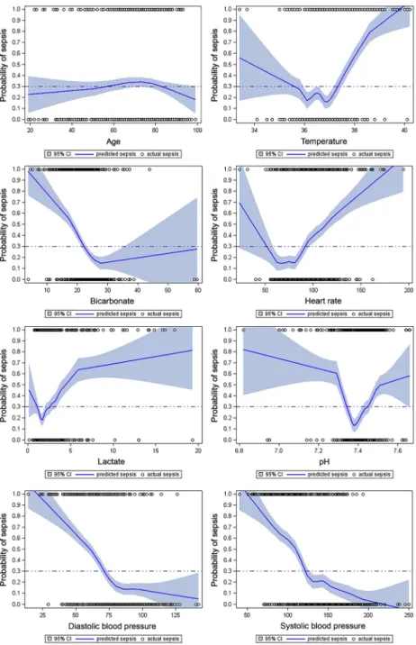
No inflexion points were observed for SBP, DBP, MAP, and RR, therefore these variables were kept as continuous (Fig. 1a–j). Age showed a reverse U-shaped curve: higher-risk cutoffs were identified for ages between 56 years and 83 years. Body temperature showed a W-shaped curve:
higher-risk cutoffs were identified for temperatures under 35.68C and above 37.3 8C. Bicar- bonate and HR showed U-shaped curves: higher-risk cutoffs were identified at under 22.3 mM/L for bicarbonate, and under 53 bpm and above 91 bpm for HR. Lactate and pH showed V-shaped curves: higher-risk cutoffs were identified at 1 mM/L or under and above 2.5 mM/L for lactate, and under 7.34 and above 7.45 for pH.
Univariate and multivariate analyses
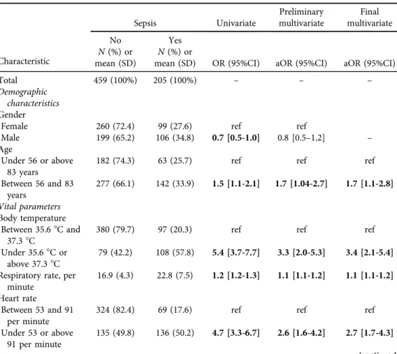
In the univariate analysis of the data-driven model, RR and higher-risk age, bicarbonate, HR, lactate, pH, and temperature were positively associated; and MAP was inversely associated with sepsis risk (Table 2). In thefinal multivariate analysis, RR and higher-risk age, bicarbonate, HR,
Table 1.Description of sample characteristics
Characteristic N(%) or mean (SD) Range
Demographic characteristics Gender
Female 359 (54.1) N/A
Male 305 (45.9) N/A
Age–years 70.2 (15.7) 19–99
Vital parameters
Body temperature–8C 36.9 (0.9) 32.3–40.2
Respiratory rate–per minute 18.7 (6.1) 10–50
Heart rate–per minute 91.0 (23.2) 23–194
Systolic blood pressure–mmHg 135.3 (36.2) 40–250
Diastolic blood pressure–mmHg 75.7 (19.9) 14–142
Mean arterial pressure–mmHg 95.6 (23.8) 26.3–176.7
Point of care test results
Lactate level–mM/L 2.6 (2.2) 0.1–19.3
pH 7.40 (0.09) 6.82–7.66
Bicarbonate level–mM/L 22.9 (5.3) 3.7–59.6
Diagnosis
Sepsis 205 (30.9) N/A
Stroke 215 (32.4) N/A
Dehydration 244 (36.7) N/A
Note:N/A–not applicable.
Fig. 1.Loess regression plots depicting the relationship between the predicted probability of sepsis and a.
age, b. body temperature, c. bicarbonate level, d. heart rate, e. lactate, f. pH, g. diastolic blood pressure, h.
systolic blood pressure, i. mean arterial pressure, and j. respiratory rate.
Note:The high-risk vs. low-risk cutoff level based on actual population prevalence is marked with a dotted line atP50.3
Fig. 1.Continued
Table 2.Univariate and multivariate associations with sepsis–data-driven model
Characteristic
Sepsis Univariate
Preliminary multivariate
Final multivariate
No Yes
N(%) or mean (SD)
N(%) or
mean (SD) OR (95%CI) aOR (95%CI) aOR (95%CI)
Total 459 (100%) 205 (100%) – – –
Demographic characteristics Gender
Female 260 (72.4) 99 (27.6) ref ref
Male 199 (65.2) 106 (34.8) 0.7 [0.5-1.0] 0.8 [0.5–1.2] –
Age
Under 56 or above 83 years
182 (74.3) 63 (25.7) ref ref ref
Between 56 and 83 years
277 (66.1) 142 (33.9) 1.5 [1.1-2.1] 1.7 [1.04-2.7] 1.7 [1.1-2.8]
Vital parameters Body temperature
Between 35.68C and
37.38C 380 (79.7) 97 (20.3) ref ref ref
Under 35.68C or
above 37.38C 79 (42.2) 108 (57.8) 5.4 [3.7-7.7] 3.3 [2.0-5.3] 3.4 [2.1-5.4]
Respiratory rate, per minute
16.9 (4.3) 22.8 (7.5) 1.2 [1.2-1.3] 1.1 [1.1-1.2] 1.1 [1.1-1.2]
Heart rate
Between 53 and 91 per minute
324 (82.4) 69 (17.6) ref ref ref
Under 53 or above 91 per minute
135 (49.8) 136 (50.2) 4.7 [3.3-6.7] 2.6 [1.6-4.2] 2.7 [1.7-4.3]
(continued)
Table 2.Continued
Characteristic
Sepsis Univariate
Preliminary multivariate
Final multivariate
No Yes
N(%) or mean (SD)
N(%) or
mean (SD) OR (95%CI) aOR (95%CI) aOR (95%CI) Mean arterial
pressure, mmHg
103.2 (20.4) 78.4 (21.8) 0.95 [0.94-
0.95] 0.95 [0.94-
0.96] 0.95 [0.94- 0.96]
Point of care test results Lactate level
Between 1.0 and 2.5 mM/L
329 (76.5) 101 (23.5) ref ref ref
Under 1.0 or above 2.5 mM/L
130 (55.6) 104 (44.4) 2.6 [1.8-3.6] 1.5 [0.9–2.4] – pH
Between 7.34 and 7.45 335 (78.8) 90 (21.2) ref ref ref
Under 7.34 or above 7.45
124 (51.9) 115 (48.1) 3.1 [2.2-4.4] 1.7 [1.1-2.7] 1.6 [1.04-2.6]
Bicarbonate level 22.3 mM/L and
above
315 (80.2) 78 (19.8) ref ref ref
Under 22.3 mM/L 144 (53.1) 127 (46.9) 3.6 [2.5-5.0] 2.0 [1.3-3.2] 2.1 [1.3-3.3]
Note:Statistically significant values are bolded.
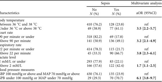
Table 3.Multivariate associations with sepsis–guidelines-based model
Characteristics
Sepsis Multivariate analysis
No Yes
aOR (95%CI) N(%) N(%)
Body temperature
Between 368C and 388C 410 (76.2) 128 (23.8) ref
Under 368C or above 388C 49 (38.9) 77 (61.1) 3.5 [2.2–5.7]
Heart rate
90 per minute or under 318 (82.2) 69 (17.8) ref
Above 90 per minute 141 (50.9) 136 (49.1) 3.2 [2.1–4.9]
Respiratory rate
22 per minute or under 414 (78.3) 115 (21.7) ref
Above 22 per minute 45 (33.3) 90 (66.7) 3.8 [2.3–6.1]
Lactate level
2 mM/L or under 293 (77.9) 83 (22.1) ref
Above 2 mM/L 166 (57.6) 122 (42.4) 1.7 [1.1–2.5]
Blood pressure measures
SBP 100 mmHg or above and MAP 70 mmHg or above 430 (76.1) 135 (23.9) ref SPB under 100 mmHg or MAP under 70 mmHg 29 (29.3) 70 (70.7) 6.1 [3.8–9.7]
Note:Statistically significant values are bolded.
pH, and temperature were positively associated; and MAP was inversely associated with sepsis risk–gender and lactate did not stay in thefinal model as significant correlates.Table 3shows the results of the guidelines-based model. In summary, compared to the guidelines-based model, the data-driven final model contained additional variables (age, pH, bicarbonate) and did not
Fig. 2.A Receiver Operating Characteristics (ROCs) curve showing the true positive rate against the false positive rate for the different possible cutoff points of a. the data-drivenfinal multivariate regression model (Area Under the Curve50.9021) and b. the guidelines-based multivariate model (Area Under the Curve50.8536)
Fig. 3.A curve showing the cumulative predicted probability against the predicted probability for both the data-driven model and the guidelines-based model.
Note:The dotted black line shows the actual probability ofP50.3. The solid blue and red lines depict the data-driven new model, and the dotted blue and red lines depict the guidelines-based model, showing about 85% true positives for the data-driven model and about 70% true positives for the guidelines-based model,
and showing about the same true negatives for both
include lactate. The area under the ROC curve was 0.9021 for the data-driven model, and 0.8536 for the guidelines-based model (Fig. 2).
Fig. 3shows the relationship between predicted probability and cumulative actual probability for both models. As seen in the section of the curves above the reference line aty530, when a cut-off for the predicted probability is set at the actual probability (30%–which is the reference line aty5 30), then the data-driven model correctly identifies about 85% of the cases (true positives: the blue curve above the dotted line) and incorrectly identifies 15% of non-cases (false positives: the red curve above the dotted line). In contrast, the guidelines-based model had a true positive rate of about 70% and a false positive rate of about 15%.
DISCUSSION
Our study provides further evidence that improved tools to identify sepsis at early time points, such as in the emergency room are much needed. We found that while some variables indeed have non-linear associations with sepsis risk and therefore require binarization, the binary cutoff values are slightly different from the cutoff values that are used in current sepsis guidelines.
Additionally, while guidelines have set cutoff values for other variables, we found sepsis risk for those variables linear, which makes their binarization inappropriate. Moreover, compared to the guidelines-based model, the data-driven final model contained additional variables such as age, pH, and bicarbonate (that are– to our knowledge–not present in any of the guidelines for sepsis diagnosis), and did not include lactate (an important predictor in current guidelines).
Finally, the data-driven model proved to be superior to the guidelines-based model in identi- fying sepsis cases.
SIRS postulates sepsis risk under 36 8C or above 388Cbody temperature [8, 9]. While we found a W curve that indicates both lower and higher body temperatures (under 35.68C and above 37.38C) as risk factors for sepsis, ourfindings indicate that the risk limits of infection- induced temperature change might be shifted towards higher temperatures. As such, we found that between 35.68C and 368C the risk is the same as between 368C and 37.38C, and therefore SIRS might over-diagnose the risk of sepsis at lower temperatures (between 35.68C and 368C) and under-diagnose at higher temperatures (between 37.38C and 388C). This might suggest that while accurate measurements of body temperature will play an important role in the diagnosis of sepsis, hypothermia and normal body temperature range still remain to be defined more clearly [26, 27]. Our results also indicate that no fever is needed to have elevated sepsis risk, but a febrile condition might already be a risk indication.
SIRS also predicates sepsis risk at aheart rateabove 90 bpm [8, 9]. While our results support this as an upper value, we also identified a lower limit (a HR under 53 bpm) under which there was increased sepsis risk. Currently, a lower HR value is not included in any sepsis guideline recommendation, although it is understood that bradycardia in sepsis might be associated with sepsis-induced myocardial dysfunction that might impair survival [28]. We would therefore recommend taking into consideration HR values below about 50 bpm as higher-risk – in addition to the currently used value of above 90 bpm–when considering the diagnosis of sepsis.
Respiratory rateabove 22 breaths per minute is another sepsis risk criterion [8, 9]. Given that we found no inflexion point for this variable, risk related to RR appears to be a sliding scale as opposed to a real cutoff: the higher the RR the higher the risk of patients having sepsis.
One of the criteria of severe sepsis is a serum lactate levelabove 2 mM/L [12]. While our results suggest that a 2 mM/L cutoff point might be somewhat low (compared to our cutoff point of 2.5 mM/L), we also identified a cutoff of 1 mM/L or under. Interestingly enough, though, lactate did not persist in the multivariate model as a correlate. There has been an ongoing debate on the use of lactate as an accurate biomarker of septic shock. Garcia-Alvarez et al. dispute that lactate is a precedent of sepsis and propose that it is rather its result [29], and Marik argues that lactate is not an accurate indicator of tissue hypoxia, because experimental models have failed to demonstrate cellular hypoxia in sepsis [30]. Our finding that in an ER setting sepsis was not associated with lactate levels but with pH instead, appears to confirm this proposition. Indeed, other researchers failed to demonstrate direct connections among oxygen carrying capacity, mixed venous oxygen saturation and levels of lactate, along with lacking evidence of direct tissue hypoxia in sepsis –it is therefore not surprising that we did notfind lactate levels to be associated with sepsis. Therefore, we provide further evidence that net lactate levels should be interpreted cautiously in septic patients, or that at least lactateper semay not be a pure indicator of the severity of circulatory derangement [31]. Considering that we found two cutoff points of lactate risk, further clarification is needed how lactate levels lower than 1 mM/L (or even lactate in general) are associated with sepsis.
With regard to blood pressure, the defined cut off values used by qSOFA for SBP ≤100 mmHg and by SOFA for MAP <70 mmHg might be easy and user-friendly values [12, 32]. We, however, found no inflexion point but rather a sliding scale. In addition, our findings suggest that the previously defined MAP of 65 mmHg might be too permissive in terms of perfusion pressure [33]. We understand that no direct correlation can be established between MAP/SBP and tissue perfusion [33]. However, our results suggest that higher target pressures might be set in terms offluid and vasoactive therapy [34].
Normal pH values are described as those between 7.35–7.45 [34], and indeed we found a sepsis risk outside this exact interval. Although pH is an easily measurable parameter that has not been explicitly pinpointed yet as a factor in diagnosing sepsis, it seems that our findings might give basis to pH measurement along with or instead of other metabolic parameters, such as lactate. Even though the extent of pH change is influenced by a variety of parameters, lactate per seis not likely to affect pH unless clear lactate acidosis is diagnosed [35]. This, however, is not a characteristic pathophysiological pathway in sepsis [36].
To our knowledge, bicarbonate has not been used in the diagnosis of sepsis. The normal levels for serum bicarbonate are postulated to be between 22 and 29 mM/L [37], and we indeed found an increased sepsis risk under 22.3 mM/L. We found no indication for an upper risk cutoff value.
A defined risk cutoff level for bicarbonate might help to differentiate sepsis from other disease states that mimic it, such as dehydration and stroke. Caution should be exercised, though, when using bicarbonate as a definitive parameter because of the commonly ill-defined origin of acidosis.
Additionally,agehas not been identified as a sepsis risk in any scoring system. In their study of 47,475 patients, Inada-Kim et al. found a gradual increase in the number of admissions with increasing age until age 85, when the number of admissions started to decline [16]. Ourfindings that the risk of sepsis was highest between the ages of 56 years and 83 years correspond with the results of the above study, suggesting this age range as a probability variable in assessing the risk of sepsis.
Some limitations of this study are noteworthy. First, this analysis is based on a retrospective chart review of mostly elderly adult patients, therefore our results might not be generalizable to other age groups. However, most septic patients are elderly, and since age–albeit as a binary variable–was included in the final multivariate model, this might somewhat counteract this limitation. Moreover, not all patients admitted to the emergency room (ER) were assessed for the parameters examined in this study, but only those where the physician in charge considered such evaluation necessary. Our goal was not to assess sepsis risk among all ER admissions, but to differentiate the diagnosis among those patients that present with conditions that mimic sepsis, such as stroke and dehydration. Additionally, the study is based on a single center retrospective dataset, and therefore our findings might not be representative of all patient populations, either in Hungary or in other countries. This study focused only on early recognition and not on survival, and therefore survival data are unavailable.
CONCLUSIONS
We can conclude that in addition to some SIRS and qSOFA parameters that are easy to measure at triage level, other easily measurable variables, such as pH, bicarbonate levels, and age might be useful in the diagnosis of sepsis in the ER and provide better accuracy and predictive power than the tools provided by current sepsis guidelines. Since the currently used sepsis criteria are rather unspecific, our results suggest that the model and its variables that we constructed in this analysis and that proved to have excellent predictability might be such a tool that would aid in a more specific identification of sepsis in the first line of care. Therefore, future studies should duplicate our analysis with these variables in order to confirm our findings. Ideally, a new tool might be developed that would help rapid and early identification of sepsis in the triage, enabling the physician to perform the necessary actions that contribute to lower mortality. Our findings contribute to the growing body of evidence in the quest of finding improved tools to identify sepsis at early time points, such as in the emergency room [15–18].
Authors’contribution:Gyula Molnar collected the data, was involved in the study design, and wrote the paper. V. Anna Gyarmathy designed the study, performed data management and analysis, designed and created the figures, designed the tables, and wrote the paper. Johanna Takacs performed data management and analysis, contributed to the creation of the figures, created the tables, and wrote the paper. Szilard Sandor and Balint Kiss collected the data and contributed to writing the paper. Janos Fazakas provided recommendations for the analysis and revised the manuscript. Peter L. Kanizsai was involved in the study design, supervised data collection, and wrote the paper. All authors provided final approval of the version submitted for publication.
ACKNOWLEDGEMENTS
The authors would like to thank the medical and nursing teams at the Emergency Department of Semmelweis University Clinical Center for their contribution to the database.
LIST OF ABBREVIATIONS
AMS Altered mental state aOR adjusted odds ratio CI confidence intervals DBP diastolic blood pressure ER emergency room HR heart rate
MAP mean arterial pressure OR odds ratio
PIRO predisposition, infection (present or suspected), response (immune system activation) and organ dysfunction
PoC point of care
qSOFA quick sequential organ failure assessment score ROC receiver operating characteristic
RR respiratory rate SBP systolic blood pressure SD standard deviations
SIRS systemic inflammatory response syndrome
REFERENCES
1. Fleischmann C, Scherag A, Adhikari NKJ, Hartog CS, Tsaganos T, Schlattmann P, et al. Assessment of global incidence and mortality of hospital-treated sepsis. Current estimates and limitations. Am J Respir Crit Care Med 2016; 193: 259–72.https://doi.org/10.1164/rccm.201504-0781oc.
2. Dellinger RP, Levy MM, Rhodes A, Annane D, Gerlach H, Opal SM, et al. Surviving sepsis campaign: in- ternational guidelines for management of severe sepsis and septic shock, 2012. Intensive Care Med 2013; 39:
165–228.https://doi.org/10.1007/s00134-012-2769-8.
3. Freund Y, Lemachatti N, Krastinova E, Van Laer M, Claessens Y-E, Avondo A, et al. Prognostic accuracy of sepsis-3 criteria for in-hospital mortality among patients with suspected infection presenting to the emer- gency department. JAMA 2017; 317: 301–08.https://doi.org/10.1001/jama.2016.20329.
4. Seymour CW, Liu VX, Iwashyna TJ, Brunkhorst FM, Rea TD, Scherag A, et al. Assessment of clinical criteria for sepsis. JAMA 2016; 315: 762–74.https://doi.org/10.1001/jama.2016.0288.
5. Song J-U, Sin CK, Park HK, Shim SR, Lee J. Performance of the quick Sequential (sepsis-related) Organ Failure Assessment score as a prognostic tool in infected patients outside the intensive care unit: a systematic review and meta-analysis. Crit Care 2018; 22: 28.https://doi.org/10.1186/s13054-018-1952-x.
6. Tusgul S, Carron P-N, Yersin B, Calandra T, Dami F. Low sensitivity of qSOFA, SIRS criteria and sepsis definition to identify infected patients at risk of complication in the prehospital setting and at the emergency department triage. Scand J Trauma Resusc Emerg Med 2017; 25: 108.https://doi.org/10.1186/s13049-017- 0449-y.
7. Golzari SEJ, Mahmoodpoor A. Sepsis-associated encephalopathy versus sepsis-induced encephalopathy.
Lancet Neurol 2014; 13: 967–68.https://doi.org/10.1016/s1474-4422(14)70205-4.
8. Balk RA. Systemic inflammatory response syndrome (SIRS). Virulence 2014; 5: 20–6.https://doi.org/10.4161/
viru.27135.
9. Bone RC, Balk RA, Cerra FB, Dellinger RP, Fein AM, Knaus WA, et al. Definitions for sepsis and organ failure and guidelines for the use of innovative therapies in sepsis. The ACCP/SCCM consensus conference com- mittee. American college of chest physicians/society of critical care medicine. Chest 1992; 101: 1644–55.
https://doi.org/10.1378/chest.101.6.1644.
10. Quinten VM, van Meurs M, Wolffensperger AE, ter Maaten JC, Ligtenberg JJM. Sepsis patients in the emergency department: stratification using the clinical impression score, predisposition, infection, response and organ dysfunction score or quick sequential organ failure assessment score? Eur J Emerg Med 2018; 25:
328–34.https://doi.org/10.1097/mej.0000000000000460.
11. Finkelsztein EJ, Jones DS, Ma KC, Pabon MA, Delgado T, Nakahira K, et al. Comparison of qSOFA and SIRS for predicting adverse outcomes of patients with suspicion of sepsis outside the intensive care unit. Crit Care 2017; 21: 73.https://doi.org/10.1186/s13054-017-1658-5.
12. Singer M, Deutschman CS, Seymour CW, Shankar-Hari M, Annane D, Bauer M, et al. The third international consensus definitions for sepsis and septic shock (Sepsis-3). JAMA 2016; 315: 801–10.https://doi.org/10.1001/
jama.2016.0287.
13. Williams JM, Greenslade JH, McKenzie JV, Chu K, Brown AFT, Lipman J. Systemic inflammatory response syndrome, quick sequential organ function assessment, and organ dysfunction: insights from a prospective database of ED patients with infection. Chest 2017; 151: 586–96.https://doi.org/10.1016/j.chest.2016.10.057.
14. Rathour S, Kumar S, Hadda V, Bhalla A, Sharma N, Varma S. PIRO concept: staging of sepsis. J Postgrad Med 2015; 61: 235–42.https://doi.org/10.4103/0022-3859.166511.
15. Yealy DM, Huang DT, Delaney A, Knight M, Randolph AG, Daniels R, et al. Recognizing and managing sepsis: what needs to be done? BMC Med 2015; 13: 98.https://doi.org/10.1186/s12916-015-0335-2.
16. Inada-Kim M, Page B, Maqsood I, Vincent C. Defining and measuring suspicion of sepsis: an analysis of routine data. BMJ Open 2017; 7: e014885.https://doi.org/10.1136/bmjopen-2016-014885.
17. Alsolamy S, Al Salamah M, Al ThagafiM, Al-Dorzi HM, Marini AM, Aljerian N, et al. Diagnostic accuracy of a screening electronic alert tool for severe sepsis and septic shock in the emergency department. BMC Med Inform Decis Mak 2014; 14: 105.https://doi.org/10.1186/s12911-014-0105-7.
18. Guerra WF, Mayfield TR, Meyers MS, Clouatre AE, Riccio JC. Early detection and treatment of patients with severe sepsis by prehospital personnel. J Emerg Med 2013; 44: 1116–25.https://doi.org/10.1016/j.jemermed.
2012.11.003.
19. Daniels R, Nutbeam T, McNamara G, Galvin C. The sepsis six and the severe sepsis resuscitation bundle: a prospective observational cohort study. Emerg Med J 2010; 28: 507–12. https://doi.org/10.1136/emj.2010.
095067.
20. Frankling C, Patel J, Sharif B, Melody T, Yeung J, Gao F, et al. A snapshot of compliance with the sepsis six care bundle in two acute hospitals in the west midlands, UK. Indian J Crit Care Med 2019; 23: 310–15.https://
doi.org/10.5005/jp-journals-10071-23204.
21. IDSA Sepsis Task Force. Infectious diseases society of America (IDSA) POSITION STATEMENT: why IDSA did not endorse the surviving sepsis campaign guidelines. Clin Infect Dis 2018; 66: 1631–35.https://doi.org/
10.1093/cid/cix997.
22. Filbin MR, Lynch J, Gillingham TD, Thorsen JE, Pasakarnis CL, Nepal S, et al. Presenting symptoms independently predict mortality in septic shock: importance of a previously unmeasured confounder. Crit Care Med 2018; 46: 1592–99.https://doi.org/10.1097/ccm.0000000000003260.
23. Cleveland WS, Devlin SJ. Locally weighted regression: an approach to regression analysis by localfitting. J Am Stat Assoc 1988; 83: 596–610.https://doi.org/10.1080/01621459.1988.10478639.
24. Gyarmathy VA, Caplinskiene I, Caplinskas S, Latkin CA. Social network structure and HIV infection among injecting drug users in Lithuania: gatekeepers as bridges of infection. AIDS Behav 2014; 18: 505–10.https://
doi.org/10.1007/s10461-014-0702-6.
25. Tape TG. ROC curves. In: Tape TG. Interpreting diagnostic tests. University of Nebraska Medical Center.
Available at:http://gim.unmc.edu/dxtests/(Accessed 08 April 2019).
26. Bakker J. Lost in Translation: on lactate, hypotension, sepsis-induced tissue hypoperfusion, quantitative resuscitation and Surviving Sepsis Campaign bundles. Crit Care Med 2015; 43: 705–06.https://doi.org/10.
1097/ccm.0000000000000870.
27. Parsons PE, Wiener-Kronish JP. Critical care secrets. 5th ed. St. Louis, MO: Elsevier Health Sciences; 2012.
28. Lv X, Wang H. Pathophysiology of sepsis-induced myocardial dysfunction. Mil Med Res 2016; 3: 30.https://
doi.org/10.1186/s40779-016-0099-9.
29. Garcia-Alvarez M, Marik P, Bellomo R. Sepsis-associated hyperlactatemia. Crit Care 2014; 18: 503.https://doi.
org/10.1186/s13054-014-0503-3.
30. Marik PE. SEP-1: the lactate myth and other fairytales. Crit Care Med 2018; 46: 1689–90.https://doi.org/10.
1097/ccm.0000000000003313.
31. Cecconi M, De Backer D, Antonelli M, Beale R, Bakker J, Hofer C, et al. Consensus on circulatory shock and hemodynamic monitoring. Task force of the European Society of Intensive Care Medicine. Intensive Care Med 2014; 40: 1795–815.https://doi.org/10.1007/s00134-014-3525-z.
32. Vincent J-L, Martin GS, Levy MM. qSOFA does not replace SIRS in the definition of sepsis. Crit Care 2016;
20: 210.https://doi.org/10.1186/s13054-016-1389-z.
33. Hasanin A, Mukhtar A, Nassar H. Perfusion indices revisited. J Intensive Care 2017; 5: 24.https://doi.org/10.
1186/s40560-017-0220-5.
34. Hall JE. Guyton and Hall textbook of medical physiology. 13th ed. Philadelphia, PA: Elsevier;2015.
35. Foucher CD, Tubben RE. Lactic acidosis. StatPearls Publishing. (2018)https://www.ncbi.nlm.nih.gov/books/
NBK470202/(Accessed 08 April 2019).
36. Suetrong B, Walley KR. Lactic acidosis in sepsis: it’s not all anaerobic: implications for diagnosis and management. Chest 2016; 149: 252–61.https://doi.org/10.1378/chest.15-1703.
37. Bronfenbrener R. Acid-base interpretation. https://emedicine.medscape.com/article/2058760-overview (Accessed 08 April 2019).
Open Access.This is an open-access article distributed under the terms of the Creative Commons Attribution 4.0 International License (https://creativecommons.org/licenses/by/4.0/), which permits unrestricted use, distribution, and reproduction in any medium, provided the original author and source are credited, a link to the CC License is provided, and changes–if any–are indicated. (SID_1)