ORIGINAL ARTICLE
Prone Positioning on a Belly Board Decreases Rectal and Bowel Doses in Pelvic Intensity-Modulated Radiation Therapy (IMRT) for Prostate Cancer
Renáta Kószó1 &Linda Varga1&Emese Fodor1&Zsuzsanna Kahán1&Adrienne Cserháti1&Katalin Hideghéty1&
Zsófia Együd1&Csilla Szabó1&Emőke Borzási1&Dorottya Szabó1,2&Kitti Müllner1&Zoltán Varga1&Anikó Maráz1
Received: 1 May 2018 / Accepted: 29 May 2018 / Published online: 7 June 2018
#Arányi Lajos Foundation 2018
Abstract
The presence of normal tissues in the irradiated volume limits dose escalation during pelvic radiotherapy (RT) for prostate cancer.
Supine and prone positions on a belly board were compared by analyzing the exposure of organs at risk (OARs) using intensity modulated RT (IMRT). The prospective trial included 55 high risk, localized or locally advanced prostate cancer patients, receiving definitive image-guided RT. Computed tomography scanning for irradiation planning was carried out in both positions.
Gross tumor volume, clinical and planning target volumes (PTV) and OARs were delineated, defining subprostatic and periprostatic rectal subsegments. At the height of the largest antero-posterior (AP) diameter of the prostate, rectal diameters and distance from the posterior prostate wall were measured. IMRT plans were generated. Normal tissue exposure and structure volumes were compared between supine and prone plans using paired t-test. In the volumes of the prostate, PTV, colon and small bowel, no significant differences were found. In prone position, all rectal volumes, diameters, and rectum–prostate distance were significantly higher, the irradiated colon and small bowel volume was lower in dose ranges of 20–40 Gy, and the exposure to all rectal segments was more favorable in 40–75 Gy dose ranges. No significant difference was found in the exposure of other OARs. Prone positioning on a belly board is an appropriate positioning method aiming rectum and bowel protection during pelvic IMRT of prostate cancer. The relative reduction in rectal exposure might be a consequence of the slight departure between the prostate and rectal wall.
Keywords Prostate cancer . IMRT . Prone . Belly board . Small bowel . Rectum
Introduction
Prostate cancer is the second most common malignancy worldwide [1]. Its prognosis has improved as a result of adjuvant androgen deprivation therapy and the escalated dose, and the efficacy of radiotherapy (RT) [2].
Therefore, pelvic irradiation including the prostate, sem- inal vesicles, and lymphatic regions is an integral com- ponent of high-risk [3], organ-confined, and locally ad- vanced prostate cancer management.
Although RT is getting more targeted, the tolerance of nor- mal tissues limits dose escalation and tumor control probability, and makes the incidence of acute and chronic gastrointestinal (GI) morbidity higher, aggravating the co-existing urological, sexual, and psychological problems of the increasing number of cancer survivors [4]. The phenomena of GI injury secondary to RT are described as pelvic radiation disease (PRD) [5]. Acute PRD, occurring during or shortly after RT, presents in abdom- inal–anorectal pain, lack of appetite, nausea, vomiting, bloating, diarrhea, and rectal bleeding. Chronic complications developing between 1.5 and 6 years after the completion of pelvic RT may manifest as anorexia, lactose intolerance, mal- absorption, fistula formation, bowel obstruction, perforation, and fecal incontinence [6]. The symptoms depend on the de- gree and extent of the tissue damage [7] and have a significant adverse effect on the patient’s quality of life [8]. The most important factors related to the probability of the complications are the total dose of RT delivered to the pelvic organs, the applied regime, the size of the treatment fields, the presence
* Renáta Kószó
koszorenata@gmail.com
1 Department of Oncotherapy, University of Szeged, Korányi Alley 12, Szeged H-6720, Hungary
2 Oncological Center, Ferenc Csolnoky Hospital, Kórház Str.1, Veszprém 8200, Hungary
https://doi.org/10.1007/s12253-018-0436-2
of radiation implants, concurrent chemotherapy, and the vol- ume of the bowel irradiated [7].
The irradiated bowel volume can be minimized by surgical and non-surgical methods [9]. Surgical means include pelvic reconstruction, reperitonealization of the pelvic floor, place- ment of an omental sling, and the inserting of a synthetic pros- thesis under the small intestine. Radiotherapeutic techniques embrace among others the use of intensity modulated (IM) and image-guided (IG) RT, adaptive irradiation, a shrinking field, modified fractionation schemes, endorectal balloons, tis- sue spacers, bladder distension, and optimal patient position.
The purpose of our study was to assess whether a supine or prone position on a belly board, applying IMRT technique, results in the reduction of the radiation dose to organs at risk (OARs), primarily the rectum, colon, and small intestines dur- ing pelvic RT of prostate cancer patients.
Materials and Methods Patient Population
The prospective analysis included patients with a histologically confirmed, high risk [10], localized or locally advanced (2009 TNM classification [11] stage T2–4 N0–1 M0) prostate cancer graded according to the Gleason score system [12], receiving a definitive pelvic RT at the Department of Oncotherapy, University of Szeged, Hungary. The tumor stage assessment was based on the findings of thoracic computed tomography (CT), abdominal and pelvic CT and magnetic resonance imag- ing (MRI), and whole-body bone scintigraphy. Clinical and pathological data were extracted from the patient files.
Patient Positioning and Computed Tomography Scanning
Patients were positioned on the supine and prone pelvis mod- ules of the All in One (AIO) Solution (ORFIT, Wijnegem, Belgium) system. In supine pose, the patient was positioned with bent knees, and the genitalia were distracted with extrud- ed polystyrene blocks. In prone position, a belly board was applied to allow the abdomen to extend into its aperture, and a polystyrene wedge was placed between the buttocks. For im- mobilization a six-point thermoplastic mask fixation (Pelvicast system, ORFIT, Wijnegem, Belgium) was employed. All patients underwent five-millimeter slice-incre- ment topometric CT scanning in both positions from the dia- phragm to the level of 10 cm below the femoral necks, using a Somatom Emotion 6 CT Simulator (Siemens, Erlangen, Germany). CT scanning was prepared with full bladder ac- cording to our internal protocol, and following an antiflatulent diet for at least 7 days prior and during RT delivery.
Target and Critical Structure Delineation
The gross tumor volume (GTV), clinical target volume (CTV), planning target volume (PTV), and OARs were delin- eated in the ARIA Oncology Information System (Varian Oncology Systems, Palo Alto, CA, USA) in both positions by radiation oncologists and reviewed by an experienced ra- diologist. The prostate was contoured as GTVp, the proximal thirds, or in case of involvement, the full extension of the seminal vesicles were contoured as GTVvs, and pathologic lymph nodes, if present, as GTVN, considering MRI records.
CTVNincluded the parailiac, upper subaortic presacral and obturator lymph nodes, contoured according to the RTOG GU Radiation Oncology Specialists Reach Consensus [13].
PTVp included GTVp with a 10 mm margin along the supero-inferior, left–right axis, in anterior direction and 7 mm in posterior direction. PTVpvswas defined as the com- bination of GTVpand GTVvswith a safety margin of 10 mm and 15 mm in posterior direction and any other directions, respectively. PTV was determined as PTVpvs, a 7 mm margin around CTVNand 10 mm around GTVN, if present. The rec- tum, large and small intestines, urinary bladder, femoral heads, and bony structures were outlined as OARs. The rec- tum was defined from the ischial tuberosities to the sigmoid flexure, but at least 2 cm above PTVpvs. Each rectal section, the whole rectum (R), the segment at the height of the prostate (R1), and R1 + 10 mm along the supero-inferior axis (R2) were individually delineated. Large and small bowel volumes contained all identifiable segments. The bladder was delineat- ed from the apex to the dome [14].
Rectal Extension and Rectum
–Prostate Distance Measurement
At the height of the largest antero-posterior (AP) diameter of the prostate, rectal diameters along the AP and left–right axis were defined, and perpendicular lines were created from the center and lateral edges of the back wall of the prostate to the outer anterior rectal wall in both supine and prone positions (Fig. 1). Two independent radiation oncologists performed rectum–prostate distance measurements, both of them twice.
Intensity-Modulated Radiotherapy Planning and Dosimetric Analysis
IMRT planning was performed using the Eclipse treatment planning system (Varian Oncology Systems, Palo Alto, CA, USA). The prescribed doses were 45 Gy to the center of the PTV (1.8 Gy/day, 5 days/week), 14 Gy of the PTVpvs and 18 Gy of PTVp, both delivered in daily 2 Gy fractions, 5 days per week. OAR dose constraints were determined as the fol- lowing [13]: V55Gy (bladder)≤50%, V70Gy (bladder)≤30%; V50Gy (rectum)≤50%, V70Gy (rectum)≤20%; V50Gy (colon)≤50%, V70Gy
(colon)≤20%; V52Gy (small intestine)= 0%; V50Gy (femoral heads)<
5%. For the coverage of the PTV sliding window IMRT plans were designed in both positions with a seven-field beam ar- rangement using 6 MV photon beam quality, consisting copla- nar beam directions as the following: in prone position 0°, 136.1°, 208.3°, 258.7°, 101.7°, 306.1° and 55.2°, in supine position 0°, 38.2°, 98°, 142°, 215.7°, 269.5° and 318.2°. For the PTVpvs and PTVp volumetric modulated arc therapy (VMAT) plans were generated in both positions using 6 MV photon beam quality, 181°–179° and 179°–181° gantry angles and 30° and 15° collimator angles, respectively. IMRT plans were created to obtain a 95% coverage of the PTV with the 95% isodose curve. The highest priority was PTV coverage, and the second one was the sparing of OARs. Planning assis- tant contours of the PTV, PTVpvs, and PTVpwere designed with uniform margins of 15 mm, 30 mm, 40 mm, and 50 mm in both positions. Dose-volume histograms were calculated for all defined volumes. Data of mean volumes of the contoured structures, mean absolute volumes of the small bowel and co- lon receiving 20–50 Gy, mean relative volumes of the rectal segments receiving 30–75 Gy and of the bladder receiving 30– 70 Gy doses and mean of doses regarding PTV D95, PTVpvs
D95, and PTVpD95 were collected.
Radiation Treatment and Image-Guidance
Irradiation was carried out by using a Varian TrueBeamSTx (Varian Oncology Systems, Palo Alto, CA, USA) in prone po- sition. Image-guidance was based on daily kV-cone beam CT (CBCT) scanning of the pelvis prior to treatment, using the standard mode settings: 125 kV, 80 mA, 13 ms, and half-fan bowtie filter. An automatic match algorithm was used to match the bony structures displayed on the planning CT and the CBCT.
Statistical Analysis
Data were reported as mean ± SD, mean ± SE or median values. The difference between the volumes and doses in
supine and prone position was analyzed with the paired sam- ples t-test. Intraobserver and interobserver variabilities were calculated from the mean of distances by using correlation analysis, given a correlation coefficient (r). SPSS 20.0 for Windows (SPSS Inc., Chicago, IL, USA) was used to perform the analysis. Apvalue <0.05 was considered significant.
Results
Patient Population
Between October 13, 2016 and October 11, 2017, 55 patients with high risk localized or locally advanced prostate cancer were administered definitive pelvic lymph node RT. Patients belonged to the elderly age group with a median [range] age of 65.60 [53.33–83.49] years, and they were mostly overweight showing a median [range] value of body mass index of 26.96 [19.37–41.62] kg/m2. More than three-quarters of them had a cardiovascular co-morbidity, and one-third of them were smokers. All the patients had stage T2–4 N0 M0 tumor with a Gleason score≥7 and a prostate specific antigen (PSA) level at the time of the diagnosis established >5 ng/ml. Most of the patients received a 6-month course of luteinizing hormone- releasing hormone analogue and antiandrogen (total androgen blockade, TAB) endocrine therapy, launched before the com- mencing of RT. The relevant patient and tumor characteristics are shown in Table1.
Structure Volumes and Rectal Extension
No significant differences were found between prone and su- pine positions in the volumes of the GTVp, PVS, PTV, colon, small bowel, and urinary bladder. All rectal volumes (R, R1 and R2) were significantly higher in prone position. The higher SD values of mean bladder volumes in the two positioning methods might be the consequence of pre-existing urinary symptoms, such as incontinence. At the height of the largest Fig. 1 Rectal extension and rectum–prostate distance measurement: At
the height of the largest antero-posterior diameter of the prostate perpen- diculars were created from the center and both lateral edges of the
posterior prostate wall to the anterior rectal wall in both prone (a) and supine (b) positions. Larger rectal diameters in prone, smaller in supine position in case of the same patient at the same time
AP level of the prostate, both the AP and the lateral rectal diameters were significantly higher in prone position (Table2).
Rectum
–Prostate Distance
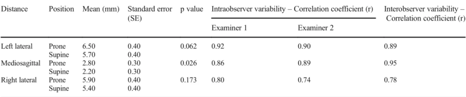
The rectum–prostate distance measured from the center of the rear prostate wall to the outer anterior rectal wall was significantly higher in prone position. No signifi- cant differences in the distance values measured from the left and right edges of the posterior prostate wall were found. Both intraobserver and interobserver vari- abilities showed close correlation (Table 3).
Normal Tissue Doses
A prone position with the additional use of a belly board led to a significant decrease in the absolute volumes receiving doses greater than 20 to 40 Gy in the small intestine and the colon;
however, the difference between the volumes receiving 50 Gy was not significant (Table4). In dose ranges of 40 to 75 Gy, the exposure of all rectal segments was more favorable in prone position. The relative volume receiving 30 Gy dose was lower in respect of R1 segment; nonetheless, the differ- ence was not significant. The relative exposed volume of the urinary bladder, femoral heads, and bony structures was in accordance with the dose constraints. No significant differ- ence was found between the positioning methods (Table5).
Planning Target Volume Coverage
PTV coverage did not differ significantly between the two positions (PTV D95 - mean of dose 43.01 vs. 43.00 Gy, SD 0.26 vs. 0.26 in prone vs. supine position, respectively, p= 0.782; PTVpvsD95 - mean of dose 13.36 vs. 13.35 Gy, SD 0.07 vs. 0.07 in prone vs. supine position, respectively, p= 0.591; PTVpD95 - mean of dose 17.16 vs. 17.15 Gy, SD 0.09 vs. 0.07 in pronevs.supine position, respectively,p= 0.435).
Discussion
Clinically localized high-risk prostate cancer frequently shows micrometastatic spreading to the pelvic lymph nodes; therefore, RT and three years of androgen suppress- ing endocrine treatment are the standard of care. Dose es- calation to the prostate even to 80–86.4 Gy reduces bio- chemical failure and the appearance of distant metastases [2]. However, survival data are controversial regarding field size [2]. There is no consensus recommendation for patient selection for pelvic RT in this population, considering the increased exposure of OARs and toxicity. 90% of patients treated with pelvic RT develop permanent alterations in bowel habits [8], 50% of them complain about adverse changes in life quality [15], and 20–40% of them assess this impact as moderate or severe [16]. The small intestine, the rectum, and to a lesser extent, the colon are dose- Table 1 Patient and tumor characteristics
Tumor and patient characteristics Number of patients (%)
Number of patients 55
Concurrent cardiovascular disease 44 (80.00)
History of smoking 18 (32.73)
Clinical stages
T2 41 (74.55)
T3 12 (21.82)
T4 2 (3.64)
Gleason scores
7 27 (48.21)
8 5 (9.09)
9 19 (33.93)
10 4 (7.14)
PSA levels on establishing the diagnosis (ng/ml)
10 > x > 5 13 (23.21)
20 > x≥10 9 (16.36)
≥20 33 (58.93)
Endocrine treatment 49 (89.09)
Table 2 Volumes of the delineated structures and rectal diameters in prone and supine positions
Structure Position Mean volume (cm3)
Standard deviation (SD)
p value
GTVp Prone 130.11 49.13 0.217
Supine 133.28 50.87
PVS Prone 188.77 58.19 0.748
Supine 190.23 58.20
PTV Prone 1123.54 138.90 0.282
Supine 1130.98 146.66
Whole rectum (R)
Prone 155.13 105.26 <0.001
Supine 95.61 45.89
Rectal subsegment R1
Prone 50.32 31.84 <0.001
Supine 34.76 23.64
Rectal subsegment R2
Prone 74.37 41.51 <0.001
Supine 50.78 27.64
Colon Prone 580.32 299.38 0.486
Supine 604.37 337.12
Small bowel Prone 812.93 354.25 0.373
Supine 772.71 353.21
Urinary bladder Prone 184.18 117.13 0.403
Supine 192.40 112.56
Rectal diameter Position Mean diameter (mm)
Standard error (SE)
p value
AP Prone 50.60 2.20 <0.001
Supine 36.70 1.50
Lateral Prone 43.80 2.60 0.003
Supine 35.90 1.80
limiting organs, tolerating a 50–60 Gy dose at conventional fractionation [17,18]. Normal tissue complication probability (NTCP) studies suggest that the small intestine volume receiv- ing 15 and 45 Gy (V15and V45) is a relevant parameter for GI morbidity [19,20]. According to the review of Fiorino et al.
[21], keeping V70and V75to <25 and 5%, respectively, results in a decrease in the development of late rectal bleeding.
Moderate dose volumes, such as V40and V50are predictive for chronic late incontinence [22] and are also important in developing rectal bleeding [21]. The dosimetric analysis [23]
of the anatomical subregions showed that rectal bleeding is associated with V70 of the anorectal region, fecal inconti- nence with V15 of external sphincter, and V55 of the iliococcygeal muscle, whereas stool frequency with V40 of the levator ani and V45 of the iliococcygeal muscle. In the prospective study of Dréan et al. [24], rectal subregions at risk have been delineated, and the authors have found that the exposure of the subprostatic anterior hemirectum and the up- per part of the anal canal was 4 Gy higher in patients devel- oping rectal bleeding.
Table 3 Rectum–prostate distance and intraobserver and interobserver variability correlation in prone and supine positions Distance Position Mean (mm) Standard error
(SE)
p value Intraobserver variability–Correlation coefficient (r) Interobserver variability– Correlation coefficient (r)
Examiner 1 Examiner 2
Left lateral Prone 6.50 0.40 0.062 0.92 0.90 0.89
Supine 5.70 0.40
Mediosagittal Prone 2.80 0.30 0.026 0.86 0.89 0.95
Supine 2.20 0.30
Right lateral Prone 5.90 0.40 0.173 0.80 0.74 0.78
Supine 5.40 0.40
Table 4 Small intestine and colon exposure in prone and supine position
Organ at risk
DVH parameter
Position Mean volume (cm3)
Standard deviation (SD)
p value
Small intestine
V20 Gy Prone 79.85 89.83 <0.001
Supine 170.34 103.62
V30 Gy Prone 36.74 51.24 <0.001
Supine 84.55 63.01
V40 Gy Prone 16.99 26.08 <0.001
Supine 32.91 31.35
V50Gy Prone 0.16 1.06 0.398
Supine 0.33 1.54
Colon V20 Gy Prone 122.43 74.52 <0.001
Supine 181.22 109.48
V30 Gy Prone 84.09 57.17 <0.001
Supine 121.21 73.36
V40 Gy Prone 53.23 44.20 0.043
Supine 63.19 44.89
V50 Gy Prone 2.06 4.02 0.627
Supine 1.81 3.62
Table 5 Exposure of rectal segments and urinary bladder in prone and supine positions
Organ at risk DVH parameter
Position Mean relative volume (%)
Standard deviation (SD)
p value
Whole rectum V30Gy Prone 106.40 118.98 0.296 Supine 89.60 7.46
V40Gy Prone 65.79 14.96 <0.001 Supine 78.58 10.14
V50Gy Prone 35.51 13.83 <0.001 Supine 48.38 12.29
V60Gy Prone 17.45 8.18 <0.001
Supine 24.04 9.11
V70Gy Prone 7.57 4.10 <0.001
Supine 10.43 4.97
V75Gy Prone 3.67 2.61 0.021
Supine 4.58 3.19
Rectal subsegment R1
V30 Gy Prone 99.78 0.75 0.735
Supine 99.80 0.61
V40Gy Prone 80.58 13.50 <0.001 Supine 94.95 5.74
V50Gy Prone 52.25 14.18 <0.001 Supine 68.55 10.90
V60Gy Prone 32.37 10.90 <0.001 Supine 40.49 10.13
V70Gy Prone 16.51 5.83 <0.001
Supine 20.74 7.14
V75Gy Prone 8.79 4.52 0.099
Supine 9.97 5.67
Rectal subsegment R2
V30Gy Prone 99.52 1.21 0.001
Supine 98.61 1.96
V40Gy Prone 78.55 12.66 <0.001 Supine 91.45 6.05
V50Gy Prone 49.40 13.14 <0.001 Supine 64.83 9.89
V60Gy Prone 28.95 9.04 <0.001
Supine 37.43 8.76
V70Gy Prone 13.52 4.75 <0.001
Supine 17.86 5.79
V75Gy Prone 6.82 3.59 0.051
Supine 7.86 4.43
Bladder V30Gy Prone 95.82 7.10 0.657
Supine 95.45 5.13
V40Gy Prone 67.99 18.89 0.687
Supine 68.78 16.13
V50Gy Prone 41.90 16.53 0.982
Supine 41.86 14.84
V60Gy Prone 26.73 11.77 0.235
Supine 25.36 10.62
V70Gy Prone 15.91 7.90 0.276
Supine 14.94 7.31
Technological advances allowing rectal sparing include endorectal balloons filled with air or water, reducing the expo- sure of the posterior rectal wall by moving away the prostate from it, depending on the volume of the balloons [25].
Bioabsorbable tissue spacers injected into the retroprostatic fascia also increase the distance between the prostate and the anterior rectal wall, resulting in significant reduction in both acute and late GI toxicities [26]. Regarding patient positioning, Zelefsky et al. [27] and McLaughlin et al. [28] have described significantly lower rectal doses in prone position, using 3DCRT technique. The results have also been confirmed in the phase II trial of O’Neil et al. [29] and by Bajon et al. using tomotherapy [30]. Nevertheless, Baylay et al. [31] have found supine position more favorable by using larger PTV margins in prone position, and Kato et al. [32] by applying IMRT in supine and 3DCRT in prone position. In prone position, the decreased rectal exposure is a result of the posterior retraction of the rectum and anterior displacement of the prostate; however, the accurate mechanism of it is unknown [27,28,32].
In the 3D-CRT of rectal malignancies, a prone treatment position without a belly board compared to a supine posture results in the reduction of the irradiated small intestine volume [33]. In case of pelvic malignancies, a larger decrease in the small intestine exposure can be obtained by the additional use of a belly board in comparison with both prone position alone [34,35] or supine position [36,37]. The use of IMRT tech- nique decreases bowel doses by 40–50%, as compared to 3D- CRT [38,39]. In case of gynecological and rectal tumors, a belly board assisted prone position using IMRT results in a further reduction in the irradiated volume of the small intes- tine, even in low dose areas [40,41]. The advantage of the use of a belly board is also confirmed in postoperatively irradiated patients [42,43], which might be the consequence of the sig- nificantly higher mobilization of the small intestine loops. The findings of Fu et al. [44] show that the gain of the use of a belly board is greater if the irradiated small intestine volume close to the target volume is larger. According to that study, a prone position on a belly board results in a remarkable de- crease in the small bowel volume in case of gynecological malignancies but not in rectal cancer patients. A full bladder also functions as a natural spacer, transposing the small intes- tine loops from the pelvis to the abdomen, resulting in a re- duction in the irradiated small intestine volume [42].
In rectal cancer patients treated with chemo-radiotherapy, Baglan et al. [19] have demonstrated an explicit relationship between the volume of the small bowel receiving at least 15 Gy and the degree of acute small intestinal toxicity.
Robertson et al. [45] have proved that a reduction in the small bowel volume receiving low dose results in a significant de- crease in the complication rate. Both authors have delineated the single small intestinal loops. In case of gynecological can- cer patients treated with pelvic IMRT, Roeske et al. [20] have detected that drawing the abdominal space, the risk of acute
GI toxicity is five times as little for small bowel volume of 100 cm3gaining the prescribed 45 Gy dose as of 200 cm3. According to Gunnlaugsson et al. [46], the former technique is the recommended contouring method instead of delineating the abdominal space. Gunnlaugsson et al. have observed strong correlation between the occurrence of early side effects and small intestinal loop exposure, and no significant connec- tion with the peritoneal cavity.
Our study was limited by the lack of delineating the penile bulb, and the relatively small number of patients involved, which however was double the number of patients previously reported. As most papers have described larger intrafraction prostate and respiratory motion in prone position [11] and lit- erature data [47] show that a 3 mm PTV margin allows for CTV to be covered for 99% of cases when daily CBCT is used, accurate patient repositioning, daily reconstruction of the rec- tum, prostate safety margins, early toxicity and life quality dur- ing and after RT were also evaluated, and found to be similar to literature data of patients treated in supine position. These promising results have recently been submitted. Late toxicities need further examination due to the short follow-up period.
In conclusion, in the pelvic IMRT for prostate cancer, a prone position on a belly board decreases the irradiated small bowel volumes even in low dose ranges and contributes to rectal sparing. The relative dose reduction in the rectal exposure might be a consequence of the slight departure between the prostate wall and the rectal wall, as consistent with the litera- ture, and the increasing volume and diameters of the rectum generated by the displacement of rectal gases. Considering the dosimetric advantages, prone position on a belly board could be recommended for the pelvic IMRT of prostate cancer.
Compliance with Ethical Standards
Conflict of Interest The authors declare that they have no conflict of interest.
Ethical Approval All procedures performed in studies involving human participants were in accordance with the ethical standards of the institu- tional and/or national research committee and with the 1964 Helsinki declaration and its later amendments or comparable ethical standards.
The study was registered on September 19, 2016 by the Human Investigation Review Board, Regional Human Biomedical Research Ethics Committee, Albert Szent-Györgyi Health Centre, University of Szeged, Hungary, registration number: WHO 3856/2016.
Informed Consent Informed consent was obtained from all individual participants included in the study.
References
1. Ferlay J, Shin HR, Bray F, Forman D, Mathers C, Parkin DM (2010) Estimates of worldwide burden of cancer in 2008:
Globocan 2008. Int J Cancer 127:2893–2917
2. Juloori A, Shah C, Stephans K, Vassil A, Tendulkar R (2016) Evolving paradigm of radiotherapy for high-risk prostate cancer:
current consensus and continuing controversies. Prostate Cancer 2016:2420786
3. Lukka H, Warde P, Pickles T, Morton G, Brundage M, Souhami L, Canadian GU Radiation Oncologist Group (2001) Controversies in prostate cancer radiotherapy: consensus development. Canadian GU Radiation Oncologist Group. Can J Urol 8:1314–1322 4. Andreyev HJ (2007) Gastrointestinal problems after pelvic radio-
therapy: the past, the present and the future. Clin Oncol (R Coll Radiol) 19:790–799
5. Stacey R, Green JT (2014) Radiation-induced small bowel disease:
latest developments and clinical guidance. Ther Adv Chronic Dis 5:
15–29
6. Theis V, Sripadam R, Ramani V, Lal S (2010) Chronic radiation enteritis. Clin Oncol 22:70–83
7. Kennedy G, Heise C (2005) Radiation colitis and proctitis. Clin Colon Rectal Surg 20:64–72
8. Olopade F, Norman A, Blake P, Dearnaley DP, Harrington KJ, Khoo V et al (2005) A modified inflammatory bowel disease ques- tionnaire and the Vaizey incontinence questionnaire are simple ways to identify patients with significant gastrointestinal symptoms after pelvic radiotherapy. Br J Cancer 92:1663–1670
9. Wiesendanger-Wittmer E, Sijtsema N, Muijs C, Beukema JC (2012) Systematic review of the role of a belly board device in radiotherapy delivery in patients with pelvic malignancies.
Radiother Oncol 102:325–334
10. Zelefsky MJ, Cowen D, Fuks Z, Shike M, Burman C, Jackson A et al (1999) Long term tolerance of high dose three-dimensional con- formal radiotherapy in patients with localized prostate carcinoma.
Cancer 85:2460–2468
11. Sobin LH, Gospodarowicz MK, Wittekind C (2009) TNM classifi- cation of malignant tumors, 7th edn. Wiley-Blackwell, London 12. Mellinger GT, Gleason D, Bailar J 3rd (1967) The histology and
prognosis of prostatic cancer. J Urol 97:331–337
13. Lawton CAF, Michalski J, El-Naga I, Buyyounouski MK, Lee WR, Menard C et al (2009) RTOG GU radiation oncology specialists reach consensus on pelvic lymph node volumes for high-risk pros- tate cancer. Int J Radiat Oncol Biol Phys 74:383–387
14. Viswanathan AN, Yorke ED, Marks LB, Eifel PJ, Shipley WU (2010) Radiation dose-volume effects of the urinary bladder. Int J Radiat Oncol Biol Phys 76(Suppl 3):S116–S122
15. Gami B, Harrington K, Blake P, Dearnaley D, Tait D, Davies J, Norman AR, Andreyev HJN (2003) How patients manage gastro- intestinal symptoms after pelvic radiotherapy. Aliment Pharmacol Therapeut 18:987–994
16. Andreyev H (2007) Gastrointestinal symptoms after pelvic radio- therapy: a new understanding to improve management of symptom- atic patients. Lancet Oncol 8:1007–1017
17. Letschert JGJ (1995) The prevention of radiation-induced small bowel complications. Eur J Cancer 31:1361–1365
18. Michalski JM, Gay H, Jackson A, Tucker SL, Deasy JO (2010) Radiation dose-volume effects in radiation-induced rectal injury.
Int J Radiat Oncol Biol Phys 76(Suppl 3):S123–S129
19. Baglan KL, Frazier RC, Yan D, Huang RR, Martinez AA, Robertson JM (2002) The dose–volume relationship of acute small bowel toxicity from concurrent 5-FU-based chemotherapy and ra- diation therapy for rectal cancer. Int J Radiat Oncol Biol Phys 52:
176–183
20. Roeske JC, Bonta D, Mell LK, Lujan AE, Mundt AJ (2003) A dosimetric analysis of acute gastrointestinal toxicity in women re- ceiving intensity-modulated whole-pelvic radiation therapy.
Radiother Oncol 69:201–207
21. Fiorino C, Valdagni R, Rancati T, Sanguineti G (2009) Dose- volume effects for normal tissues in external radiotherapy: pelvis.
Radiother Oncol 93:153–167
22. Peeters ST, Hoogeman MS, Heemsbergen WD, Hart AA, Koper PC, Lebesque JV (2006) Rectal bleeding, fecal incontinence, and high stool frequency after conformal radiotherapy for prostate can- cer: normal tissue complication probability modeling. Int J Radiat Oncol Biol Phys 66:11–19
23. Schaake W, van der Schaaf A, van Dijk LV, Bongaerts AH, van den Bergh AC, Langendijk JA (2016) Normal tissue complication prob- ability (NTCP) models for late rectal bleeding, stool frequency and fecal incontinence after radiotherapy in prostate cancer patients.
Radiother Oncol 119:381–387
24. Dréan G, Acosta O, Ospina JD, Fargeas A, Lafond C, Corrégé G et al (2016) Identification of a rectal subregion highly predictive of rectal bleeding in prostate cancer IMRT. Radiother Oncol 119:388– 397
25. van Lin EN, Hoffmann AL, van Kollenburg P, Leer JW, Visser AG (2005) Rectal wall sparing effect of three different endorectal bal- loons in 3D conformal and IMRT prostate radiotherapy. Int J Radiat Oncol Biol Phys 63:565–576
26. Serrano N, Kalman NS, Anscher MS (2017) Reducing rectal injury in men receiving prostate cancer radiation therapy: current perspec- tives. Cancer Manag Res 9:339–350
27. Zelefsky MJ, Happersett L, Leibel SA, Burman CM, Schwartz L, Dicker AP et al (1997) The effect of treatment positioning on nor- mal tissue dose in patients with prostate cancer treated with three- dimensional conformal radiotherapy. Int J Radiat Oncol Biol Phys 37:13–19
28. McLaughlin PW, Wygoda A, Sahijdak W, Sandler HM, Marsh L, Roberson P et al (1999) The effect of patient position and treatment technique in conformal treatment of prostate cancer. Int J Radiat Oncol Biol Phys 45:407–413
29. O’Neil L, Armstrong J, Buckney S, Assiri M, Cannon M, Holmberg O (2008) A phase II trial for the optimisation of treat- ment position in the radiation therapy of prostate cancer. Radiother Oncol 88:61–66
30. Bajon T, Piotrowski T, Antczak A, Bąk B, Błasiak B, Kaźmierskaa J (2011) Comparison of dose-volume histograms for supine and prone position in patients irradiated for prostate cancer–a prelim- inary study. Rep Pract Oncol Radiother 16:65–70
31. Bayley AJ, Catton CN, Haycocks T, Kelly V, Alasti H, Bristow R et al (2003) A randomized trial of supine vs. prone positioning in patients undergoing escalated dose conformal radiotherapy for prostate cancer. Radiother Oncol 70:37–44
32. Kato T, Obata Y, Kadoya N, Fuwa N (2009) A comparison of prone three-dimensional conformal radiotherapy with supine intensity- modulated radiotherapy for prostate cancer: which technique is more effective for rectal sparing? Br J Radiol 82:654–661 33. Drzymala M, Hawkins MA, Henrys AJ, Bedford J, Norman A, Tait
DM (2009) The effect of treatment position, prone or supine, on dose–volume histograms for pelvic radiotherapy in patients with rectal cancer. Br J Radiol 82:321–327
34. Kim TH, Chie EK, Kim DY, Park SY, Cho KH, Jung KH et al (2005) Comparison of the belly board device method and the distended bladder method for reducing irradiated small bowel vol- umes in preoperative radiotherapy of rectal cancer patients. Int J Radiat Oncol Biol Phys 62:769–775
35. Huh SJ, Park W, Ju SG, Lee JE, Han Y (2004) Small-bowel dis- placement system for the sparing of small bowel in three- dimensional conformal radiotherapy for cervical cancer. Clin Oncol 16:467–473
36. Martin J, Fitzpatrick K, Horan G, McCloy R, Buckney S, O'Neill L et al (2005) Treatment with a belly-board device significantly re- duces the volume of small bowel irradiated and results in low acute toxicity in adjuvant radiotherapy for gynecologic cancer: results of a prospective study. Radiother Oncol 74:267–274
37. Pinkawa M, Gagel B, Demirel C, Schmachtenberg A, Asadpour B, Eble MJ (2003) Dose–volume histogram evaluation of prone and
supine patient position in external beam radiotherapy for cervical and endometrial cancer. Radiother Oncol 69:99–105
38. Mundt A, Roeske J, Lujan A (2002) Intensity-modulated radiation therapy in gynecologic malignancies. Med Dosimetry 27:131–136 39. Portelance L, Chao K, Grigsby P, Bennet H, Low D (2001) Intensity-modulated radiation therapy (IMRT) reduces small bow- el, rectum, and bladder doses in patients with cervical cancer re- ceiving pelvic and Para-aortic irradiation. Int J Radiat Oncol Biol Phys 51:261–266
40. Stromberger C, Kom Y, Kawgan-Kagan M, Mensing T, Jahn U, Schneider A et al (2010) Intensity-modulated radiotherapy in pa- tients with cervical cancer. An intra-individual comparison of prone and supine positioning. Radiat Oncol 5:63–68
41. Beriwal S, Jain SK, Heron DE, de Andrade RS, Lin CJ, Kim H (2007) Dosimetric and toxicity comparison between prone and su- pine position IMRT for endometrial cancer. Int J Radiat Oncol Biol Phys 67:485–489
42. Kim TH, Kim DY, Cho KH, Kim YH, Jung KH, Ahn JB et al (2005) Comparative analysis of the effects of belly board and blad- der distension in postoperative radiotherapy of rectal cancer pa- tients. Strahlenther Onkol 181(9):601–605
43. Saynak M, Kucucuk S, Aslay I (2008) Abdominal pillow for the sparing of small bowel in four-field conventional pelvic radiotherapy. Eur J Gynaecol Oncol 29:643–648
44. Fu YT, Lam JC, Tze JM (1995) Measurement of irradiated small bowel volume in pelvic irradiation and the effect of a belly board.
Clin Oncol (R Coll Radiol) 7:188–192
45. Robertson JM, Lockman D, Yan D, Wallace M (2008) The dose-volume relationship of small bowel irradiation and acute grade 3 diarrhea during chemoradiotherapy for rectal cancer. Int J Radiat Oncol Biol Phys 70:413–418
46. Gunnlaugsson A, Kjellen E, Nilsson P, Bendahl PO, Willner J, Johnsson A (2007) Dose–volume relationships between enteritis and irradiated bowel volumes during 5-fluorouracil and oxaliplatin based chemoradiotherapy in locally advanced rectal cancer. Acta Oncol 46:937–944
47. Gill SK, Reddy K, Campbell N, Chen C, Pearson D (2015) Determination of optimal PTV margin for patients receiving CBCT-guided prostate IMRT: comparative analysis based on CBCT dose calculation with four different margins. J Appl Clin Med Phys 16:252–262