DOI: 10.1002/med.21592
R E V I E W A R T I C L E
The mechanism(s) of action of antioxidants: From scavenging reactive oxygen/nitrogen species to redox signaling and the generation of bioactive secondary metabolites
Attila Hunyadi
1,21Institute of Pharmacognosy, Interdisciplinary Excellence Centre, University of Szeged, Eötvös str. 6, H‐6720, Szeged, Hungary
2Interdisciplinary Centre for Natural Products, University of Szeged, Eötvös str. 6, H‐6720, Szeged, Hungary
Correspondence
Attila Hunyadi, Institute of Pharmacognosy, Interdisciplinary Excellence Centre, University of Szeged, Eötvös str. 6, H‐6720 Szeged, Hungary.
Email: hunyadi.a@pharm.u-szeged.hu
Funding information
National Research, Development and Innovation Office, Hungary, Grant/Award Number: K119770; Ministry of Human Capacities, Hungary, Grant/Award Number:
20391‐3/2018/FEKUSTRAT; Hungarian Academy of Sciences; UNKP‐18‐4 New National Excellence Program of the Ministry of Human Capacities
Abstract
Small molecule, dietary antioxidants exert a remarkably broad range of bioactivities, and many of these can be explained by the influence of antioxidants on the redox homeostasis. Such compounds help to modulate the levels of harmful reactive oxygen/nitrogen species, and therefore participate in the regulation of various redox signaling pathways. However, upon ingestion, antioxidants usually undergo extensive metabolism that can generate a wide range of bioactive metabolites. This makes it difficult, but otherwise a need, to identify the ones responsible for the different activities of antioxidants. By better understanding their ways of action, the use of antioxidants in therapy can be improved.
This review provides a summary on the role of the
in vivometabolic changes and the oxidized metabolites on the mechanisms behind the bioactivity of antioxidants. A special attention is given to metabolites described as products of biomimetic oxidative chemical reactions, which can be considered as models of free radical scavenging. During such reactions a wide variety of metabolites are formed, and they can exert completely different specific bioactivities as compared to their parent antioxidants. This implies that
© 2019 The Authors.Medicinal Research ReviewsPublished by Wiley Periodicals, Inc.
Med Res Rev. 2019;1-29. wileyonlinelibrary.com/journal/med
|
1- - - - This is an open access article under the terms of the Creative Commons Attribution License, which permits use, distribution and reproduction in any medium, provided the original work is properly cited.
exploring the free radical scavenging
‐related metabolite fingerprint of each antioxidant molecule, collectively defined here as the scavengome, will lead to a deeper understanding of the bioactivity of these compounds.
Furthermore, this paper aims to be a working tool for systematic studies on oxidized metabolic fingerprints of antioxidants, which will certainly reveal an often
‐neglected segment of chemical space that is a treasury of bioactive compounds.
K E Y W O R D S
antioxidant mechanism of action, bioactive metabolite, free radical scavenging, oxidative stress, scavengome
1 | I N T R O D U C T I O N
The field of antioxidants undoubtedly lives its renaissance, largely due to the fact that oxidative stress is closely connected to the development of many chronic diseases, including diabetes, cancer, various aging related and central nerve system (CNS) disorders, etc.1Oxidative stress, a term coined by Helmut Sies,2has recently been redefined as“a disruption of redox signaling and control”,3emphasizing the importance of a dynamic but fine‐tuned redox balance in the maintenance of cellular homeostasis.4-7Moreover, an intensive reconceptualization of the chemical, biological and pharmacological aspects of oxidative stress and antioxidant defense is ongoing in the field;
such perspectives were recently reviewed8-10and will not be covered in detail here.
Small‐molecule antioxidants have long been considered as compounds able to decrease or, under certain circumstances, contribute to oxidative stress. While their chemical structure makes them able to directly scavenge reactive oxygen and nitrogen species (ROS and RNS, respectively; collectively referred to as RONS),11thein vivo antioxidant action of these compounds seems to be linked to their potential to interact with various redox signaling pathways by modulation of the activity of redox enzymes.9,10 Moreover, many dietary antioxidants undergo extensive metabolism; those that are not absorbed in the small intestines can also suffer fragmentation into smaller compounds by gut microbiota. This certainly leads to a situation where the activity observedin vivois necessarily a superposition of the effects of these metabolites on several possible targets.12,13
This paper provides a brief summary on the various mechanisms influenced by the activity of small‐molecule antioxidants. A set of dietary phenolic compounds including curcuminoids, a stilbene, hydroxycinnamic acid derivatives, a lignan, and a flavonoid are discussed in detail. These are: curcumin (1), demetoxycurcumin (2), bisdemetoxycurcumin (3), resveratrol (4), methyl‐p‐coumarate (5), methyl‐caffeate (6), methyl‐ferulate (7), secoisolariciresinol (8) and luteolin (9). Selection of these compounds took into consideration their chemical diversity, as well as the fact that they represent classes of natural products whose antioxidant activity attracts particularly high scientific and popular interest. The mechanisms to be discussed include free radical scavenging, modulation of antioxidant/pro‐oxidant enzymes and/or transcription factors, and the formation of bioactive metabolites. Mechanisms extensively reviewed elsewhere are touched upon lightly. It is the primary aim of this paper to shed light on the potential biomedical importance of a largely neglected part of the metabolism of antioxidants, the formation of new, bioactive chemical entities upon scavenging ROS or RNS. With this idea in mind, RONS scavenging‐related formation of bioactive species from ascorbic acid and estrone are also briefly discussed.
Literature search was conducted using online databases including SciFinder, PubMed, ScienceDirect and Google Scholar. Whenever possible, publications not older than 10 years were favored. However, survey on RONS
scavenging‐related oxidative transformation by antioxidants was not limited to a specific time frame. This part of the search focused on biomimetic oxidative transformations of compounds1‐9, and references were included only if a direct comparison between the bioactivity of the antioxidant and its oxidized analog(s) was available in the same pharmacological model.
2 | F R E E R A D I C A L S C A V E N G I N G A N D R E D O X M O D U L A T I O N O F E N Z Y M E S A N D T R A N S C R I P T I O N F A C T O R S
Decrease of RONS levels as a result of their scavenging by small‐molecule antioxidants had been used as a simplified description of the bioactivity of these molecules, but this point of view has long been outdated. There are many easy‐to‐performin vitrotechniques available for assessing the free radical scavenging capacity of a dietary antioxidant,11,14-16 and thanks to the simplicity of such bioassays, an abundance of related reports has been published over the last several decades. A large number of studies reported the free radical scavenging activity of the compounds selected for discussion here, and these include curcuminoids,17 resveratrol,18-21 cinnamic acids,11,22,23lignans,24and flavonoids.11,25,26Despite the popularity of such antioxidant assays, these have little if anyin vivorelevance in terms of decreasing RONS levels. Due to the reactivity and short half‐life of damaging RONS, it is now generally accepted that dietary antioxidants cannot overcome kinetic limitations in free radical scavenging to make a relevant difference in RONS levelsin vivo.9A possible sole exception is the case of vitamin E that is present in large enough amounts in biological membranes to act efficiently against peroxyl radicals.27 Nevertheless, it must be noted that, regardless of of the low efficiency of this mechanism in preventing cellular damage, RONS scavenging by dietary antioxidants (and/or their metabolites) should take placein vivo, and this might still deserve attention from another important perspective, namely the formation of additional metabolites, not as yet studied in detail (see Section 4).
Major ROS with large physiological/pathophysiological importance include hydrogen peroxide (H2O2),28 superoxide anion radical (O2•–),29 hypochlorous acid (HOCl),30 singlet oxygen (1O2),31 hydroxyl radical (•OH), alkoxyl radical (RO•), and peroxyl radical (ROO•).32Major RNS are nitric oxide (•NO), nitrogen dioxide (•NO2), and peroxynitrite (ONOO–).11Primary RONS, ie H2O2, O2•–, and•NO, have relatively low damaging potential, and their levels are under specific enzymatic control.32,33Structural damage of macromolecules, associated with oxidative stress and related pathologies, is connected to the more reactive and less regulated, toxic secondary species, mainly
•OH radical, ONOO–and HOCl.33
It is generally well accepted that i) the production of RONS is in principle due to enzymatic processes; and ii) the primary counterbalance of the resulting oxidative stress is in fact the pool of antioxidant enzymes and not small‐ molecule free radical scavengers.34 Accordingly,in vivorelevant antioxidant activity, ie a decrease of oxidative stress by dietary compounds, is a result of the enzymatic modulation of redox equilibrium. ROS are produced primarily by mitochondria (ETC complexes I and II), NADPH oxidases, lipoxygenases (LOXs), cyclooxygenases (COXs), xanthine oxidoreductase (XOR) and cytochrome P (CYP) monooxygenases.32Among RNS, the nitrogen‐ centered radical nitric oxide is produced by nitric oxide synthases (NOSs), and its reaction with O2•–leads to the formation of ONOO–.32,33Peroxynitrite can then react with CO2to yield nitrosoperoxycarboxylate (ONOOCOO−) that further decomposes into•NO2and carbonate radicals. Alternatively, protonation of peroxynitrite yields its acid form (ONOOH), which can undergo homolysis to yield•NO2and•OH radicals.35Though this latter reaction is less efficient than that between peroxynitrite and CO2, it has been shown to occur and have biological significance if peroxynitrite is formed and may affect proton pumps of the cell membrane.36
Antioxidant enzymes counterbalance the potentially harmful effects of RONS. These can exert their function directly, such as for example superoxide dismutases (SODs), glutathione peroxidase (GPx) and catalase (CAT), or indirectly, such as for example glutathione‐S‐transferases (GSTs), UDP‐glucuronosyl transferases, and NADP
F I G U R E 1 (A) Metabolites of curcuminoids (1‐3) identified fromin vivo,ex vivoand microbial fermentation studies.
Bioactivity of these metabolites is discussed in Section 3.1. Glu: glucuronidyl. (B) Some oxidized derivatives of curcuminoids (1‐3) obtained from autoxidation or biomimetic oxidative chemistry that had bioactivity profile different from that of their parent compounds, see Section 4.1 and Table 1. These compounds may likely form through free radical scavenging
quinone oxidoreductase. The role of these enzyme systems in oxidative stress has extensively been reviewed by others.34,37,38
It is now also well known that certain types of ROS (eg H2O2) and RNS (eg•NO and ONOO–) play a pivotal signaling role. This might occur through the reversible oxidation of proteins on the SH groups of cysteine and/or methionine, and in particular, S‐sulfenylation, S‐glutathionylation, S‐nitrosylation, S‐polysulfidation or disulfide bond formation, and/or nitration of tyrosine and tryptophan residues. MAPK‐mediated pathways including ERK1/2, p38 and JNK, PI3K/Akt signaling, and several transcription factors such as Nrf2, AP‐1, NF‐κB, HIF‐1α, p53, Wnt/β‐catenin, that control the expression of a large number of genes, are known to be under redox regulation. For more details on this subject and most recent developments in the field, see the review by Moldogazieva et al.7
Accordingly, it is not surprising that many dietary antioxidants have been reported to interfere with a variety of the above‐mentioned pathways/transcription factors, and such interactions appear to play an important role in the polypharmacology of these compounds.39Briefly, it seems an important common feature of polyphenol compounds to induce an adaptive response to oxidative stress through the activation of Nrf2.9This leads to the upregulation of antioxidant enzymes including CAT, GPx, GST, paraoxonases (PONs), glutathione reductase (GR), andγ‐glutamyl cysteine synthetase (γ‐GCS).39Many plant polyphenols also share an ability to down‐regulate PI3K/Akt,40activate AMPK41-43and sirtuins,43-46and suppress NF‐κB signaling.47 These redox‐regulated pathways have also been implicated as underlying mechanisms for the health‐promoting and lifespan‐increasing effects of dietary polyphenols.48
3 | M E T A B O L I S M O F A N T I O X I D A N T S I N V I E W O F T H E I R B I O A C T I V I T Y
While there is an abundance ofin vitropharmacological studies on dietary antioxidants, promising results obtained in such experimental systems are difficult to translate intoin vivoapplications. A major reason for this is the poor oral bioavailability of such compounds, which is largely due to their metabolism by gut microbiota and/or the F I G U R E 2 (A) Metabolites of resveratrol (4) identified fromin vitroandin vivostudies. Bioactivity of these metabolites is discussed in Section 3.2. Glu: glucuronidyl. (B) Some oxidized derivatives of resveratrol obtained from biomimetic oxidative chemistry that had bioactivity profile different from that of their parent compound, see Section 4.2 and Table 1. These compounds may likely form through free radical scavenging
intestinal mucosa, and the subsequent extensive metabolism of the absorbed fraction mostly within the liver and kidneys. The microbial metabolism of ingested polyphenols involves enzymatic hydrolysis ofβ‐glycosidic bonds liberating aglycons that are absorbed better than their glycosides. Microbial metabolism frequently leads to a variety of smaller fragments, including simple phenolic acids, phloroglucinol derivatives, etc. Detailed reviews on F I G U R E 3 (A) Metabolites of methyl hydroxycinnamates (5‐7) identified fromin vitro,in vivoandex vivostudies.
Bioactivity of these metabolites is discussed in Section 3.3. Glu: glucuronidyl. Due to the methylation of6by catechol‐O‐methyltransferase (COMT), overlaps are observed for compounds6and7, i.e.6band7aare metabolites of7and6, respectively. Metabolites6h‐6kwere obtained through cleavage of chlorogenic acid to6a by gut microbiota. (B) Some oxidized derivatives of5‐7obtained from biomimetic oxidative chemistry that had bioactivity profiles different from that of their parent compounds, see Section 4.3 and Table 1. These compounds may likely form through free radical scavenging
this subject have recently been published by others.13,49-52 The following sub‐sections attempt to provide an overview on the known Phase I and Phase II metabolic transformations of curcuminoids (1‐3), resveratrol (4), a set of methyl‐hydroxycinnamates (5‐7), secoisolariciresinol (8), and luteolin (9), and on related pharmacological consequences.
To facilitate comparison between metabolites described in Sections 3 and 4, the corresponding Figures 1A‐5A will present previously identified phase I and II metabolites of compounds1‐9, and Figures 1B‐5B metabolites obtained by biomimetic oxidative chemistry discussed in Section 4.
3.1 | Metabolism of curcuminoids
The diarylheptanoid antioxidants from Curcuma species (turmeric:C. longa, wild turmeric:C. aromatica, Javanese turmeric:C. xanthorrhiza), curcumin (1), demethoxycurcumin (2) and bisdemethoxycurcumin (3) (Figure 1), have achieved important scientific and clinical interest. At the time of this writing, 57 current clinical studies on curcumin (1) at various stages are registered at the National Institutes of Health (NIH) database. A wide range ofin vitroand in vivo studies have claimed beneficial bioactivities attributed to curcumin (1), and these bioactivities include chemo‐preventive, neuroprotective, antitumor and anti‐mutagenic, antimetastatic, Antiangiogenic, immunomodu- latory, anti‐inflammatory, etc., and are extensively reviewed by others.17,53-57
F I G U R E 4 (A) Metabolites of secoisolariciresinol (8) identified fromin vitroandin vivostudies. Bioactivity of these metabolites is discussed in Section 3.4. Glu: glucuronidyl, SDG: diglucoside of8. In case of asymmetric conjugates of8band8c, identification was based on mass spectrometry, hence the differently substituted phenolic rings may be interchangeable.106,107(B) Some oxidized derivatives of8obtained from biomimetic oxidative chemistry that had bioactivity profiles that differed from that of their parent compound, see Section 4.4 and Table 1. These compounds may likely form through free radical scavenging. Lariciresinol (8g) is also a naturally occurring lignan that may be metabolized by gut microbiota to8, and subsequently to8a‐8c
It is well understood that curcumin has very low bioavailability and low chemical stability.58For example, oral administration of an amount as high as 500 mg/kg of curcumin to streptozotocin‐induced diabetic rats results in a maximum of only 0.06 µg/mL plasma concentration, which represents an oral bioavailability of ca. 0.5%.59 Therefore, metabolites and decomposition products of curcumin and its analogs are thought to play a central role in their remarkable polypharmacology.56,60,61
Structures of the known metabolites of curcuminoids (1‐3) identified fromin vivo(mice, rats, or humans) andex vivo(tissue slices) studies, as well as those from fermentation with gut microbiota, are presented in Figure 1A as compounds1a‐1r,2a‐2r, and3a‐3k. The biotransformation of curcuminoids is known to involve reduction (a major metabolic step by gut microbiota 62,63 and by the liver 64,65), demethylation and/or further hydroxylation, cyclization (by gut microbiota62), and the formation of glucuronide and/or sulfate conjugates primarily at phenolic OH groups (accordingly,1d is a minor metabolite).64-68Sulfate conjugation of curcumin (1) was recently also demonstrated to take place in breast cancer cells, and rapid excretion of its conjugates took place through a yet unidentified efflux transporter, possibly ABCG2.69As previously mentioned, free, unchanged curcumin appears to be scarcely present in the peripheral blood and tissues after oral ingestion.66When administered intravenously in mice, it has a half‐life of around 30 minutes.70The main hepatic metabolite of curcumin (1) in humans and rats was found to be hexahydrocurcumin (1a) that can further be reduced to octahydrocurcumin, also referred to as curcuminol (1b).64Moreover, at least in rats, phase I metabolism can involve further reduction and methoxylation of the heptyl chain (forming2land2n), as evidenced by the isolation of demethoxycurcumin metabolites (2k‐2q) from rat feces and urine.71
The reduction and/or conjugation certainly influences the bioactivity of curcumin. In terms of affecting COX‐2 expression, these metabolic changes may represent bio‐inactivation.64The reduced metabolites tetra‐, hexa‐, and F I G U R E 5 (A) Metabolites of luteolin (9) and its glycosides identified fromin vitroandin vivostudies. Bioactivity of these metabolites is discussed in Section 3.5. COMT: catechol‐O‐methyltransferase, Glu: glucuronidyl, UDP‐GluT: UDP‐glucuronosyltransferase. The formation of phloroglucinol (9o) has been suggested based on the presence of dihydrocaffeic acid (9p). (B) Specific oxidized derivatives of9that were identified from biomimetic oxidative chemistry and are well‐known to form through free radical scavenging. Glutathione‐S‐transferase inhibitory activity9qand9rin comparison with that of9is discussed in Section 4.5 and summarized in Table1
octahydrocurcumin (1m, 1a, and 1b, respectively) are also less active than curcumin as inhibitors of lipopolysaccharide (LPS)‐induced expression of inducible nitric oxide synthase (iNOS) and NF‐κB activation in RAW 264.7 macrophages.72 However, Lee et al found that hexahydrocurcumin (1a) exerts potent anti‐ inflammatory activity by inhibiting LPS‐induced, COX‐2 derived prostaglandin E2production in RAW264.7 cells with an IC50value of 0.7 µM.73Although in this study curcumin (1) was unfortunately not used as a control, Mohd Aluwi et al found it to act with an IC50value of ca. 16 µM.74Therefore, it seems that hexahydrocurcumin (1a) is more potent in this regard than its parent compound, although no direct comparison was made in this case. A broad range of further bioactivities of hexahydrocurcumin (1a) shows that this compound is a potent bioactive metabolite of curcumin (1) and this has been recently reviewed by Huang et al.75A number of studies demonstrated that thein vitroantioxidant activity of1ais at least comparable to that of curcumin as scavenger of DPPH,•NO, and•OH radicals, and as inhibitor of AAPH‐induced linoleic oxidation and hemolysis.76-78Hexahydrocurcumin (1a) was also reported as a potent antioxidantin vivo such that a single intraperitoneal injection of 10 to 40 mg/kg dose significantly decreased the MDA and•NO levels and the expression of NF‐κB and COX‐2 in a rat stroke model. This same study found1ato protect the rats' brain from ischaemia/reperfusion injury, and to reduce inflammatory response.79Curcumin was also studied and found effective in a similar rat model, however at a much larger dose (300 mg/kg).80 Unfortunately, this makes it difficult to draw any comparison between the efficacies of the two compounds. Nevertheless, there is no doubt of the importance of the activity of metabolite1a in vivo. Most recently, this compound was also found to efficiently inhibit corneal neovascularization, an important reason for corneal blindness, induced by the p‐bFGF‐SAINT‐18 & p‐VEGF‐SAINT‐18 complex in anin vivorat model, and it exerted ca. 50% inhibition after 6 days at a dose of 1 µg.81
3.2 | Metabolism of resveratrol
Resveratrol (4; Figure 2) is probably the most popular dietary antioxidant. This stilbene is best known as the
“magic”constituent of blue grape skin and red wine, and it occurs in lower amounts in several foods including a variety of berries (eg cranberry, mulberry), tomato skin, peanuts, pistachios, cocoa, etc.82Resveratrol (4) offers a broad spectrum of health benefits including its widely accepted cardiovascular protective effect and its ability to reduce cancer risk, even though the related literature is very diverse and not fully conclusive as to its real value.83 The pharmacokinetics and metabolism of resveratrol (4) has most recently been reviewed by Wang & Sang.84 Despite its rapid absorption, the extensive and fast metabolism makes the bioavailability of resveratrol extremely low. Oral ingestion of as high as 5 g of resveratrol by humans was found to lead to peak plasma concentrations of ca. 2.4 µM, while resveratrol‐3‐O‐sulfate (4c; Figure 2A) could reach 8 times higher concentration and over 20 times higher AUC value.85Major metabolic transformations of resveratrol involve phase II and include glucuronide and/or sulfate conjugation, leading to metabolites4ato4i, and reduction by gut microbiota to form compounds4j to4l.84Conjugation takes place in the intestinal mucosa and liver, and the sulfate conjugate4cwas also identified to form in adipocytes.86 Relative importance of the different conjugation routes shows dose‐dependency.
Administration of smaller, 5 to 50 mg doses of resveratrol (4) led to the appearance of glucuronides as main metabolites in the plasma of human volunteers. In contrast, when larger,≥250 mg doses were administered, the sulfate conjugate4cwas observed as the major metabolite.84
Conjugation significantly alters the bioactivity profile of resveratrol in a way that some activities decrease or disappear, while others are at least partially retained.87,88The Na+salts of compounds4iand4hare much weaker inhibitors of NF‐κB, COX‐1, and COX‐2, as well as of the •NO production of LPS‐stimulated RAW 264.7 macrophages as compared to resveratrol. However, the Na+salt of compound4cand the K+salt of compound4d showed COX‐1 inhibition (IC50= 3.60 and 7.53 µM, respectively) comparable to that of their parent compound (IC50= 6.65 µM). Both of these metabolites exerted a similarly strong inhibition of COX‐2, i.e. they lost the selectivity of resveratrol towards COX‐2 that is about 10 times (IC50= 0.75 µM).88As an interesting change in bioactivity due to metabolism, compound 4c was found to exert a selective antiestrogen effect on estrogen
receptorαin a transformed yeast model and also in MCF‐7 cells. Resveratrol acted as a partial agonist in this study.89 Resveratrol 3‐glucuronide (4b) and 3‐sulfate (4c) conjugates also play an important role in tissue accumulation. Following a 6 weeks‐long intraperitoneal daily treatment of streptozoticin‐induced diabetic rats with 5 mg/kg of resveratrol (4), conjugates4band4cwere found to accumulate in the heart but not in the pancreas tissue. Importantly, concentrations of 4c (reaching a maximum of 20 nM) positively correlated with the improvement of cardiac function and haemodynamic performance.90It is worth noting that since deconjugation can also take place, the circulating conjugates can as well be considered as a pool of resveratrol to release in certain tissues.83,84
3.3 | Metabolism of hydroxycinnamates
Hydroxycinnamic acids are among the most abundant dietary antioxidants, present in many vegetables, fruits, cereals and beverages.91These compounds can occur in plants either in free form, or as conjugates (eg as esters in chlorogenic acids, rosmarinic acid, etc.), and they are also well known as structural elements in the biosynthesis of many plant phenolic compounds such as lignans or flavonoids.23,91-93
After oral ingestion, enteral metabolism of methyl esters ofp‐coumaric, caffeic and ferulic acids (compounds5, 6, and7, respectively; Figure 3) involves de‐esterification94and primarily sulfate or, to a lesser extent, glucuronide conjugation, leading to the formation of compounds5a‐5d,6a‐6g, and7a‐7c, respectively (Figure 3A).95In case of the catechol methyl caffeate (6) and its metabolite caffeic acid (6a), methylation by catechol‐O‐methyltransferase (COMT) also takes place, with subsequent formation of the sulfate conjugates of ferulic acid (6b) and isoferulic acid (6d) as main products.94-96Following absorption, glucuronidation, methylation and/or sulfation takes place in the liver and the formed metabolites participate in enterohepatic circulation. Methylation by COMT was also observed in the kidneys.92Extrapolating from results obtained for chlorogenic acid (5‐O‐caffeoylquinic acid), it is also clear that bacterial metabolism of caffeic acid involves side‐chain saturation as a major step (forming6h), and that it can also be dehydroxylated and undergo gradual side‐chain shortening all the way down to benzoic acid (forming6g and6i‐6k).97,98Similar metabolic transformations are expected for compounds5and7, as well.
Unsurprisingly, the bioactivity profiles of methylated, glucuronidated and sulphated metabolites of hydroxycinnamic acids show significant differences as compared with those of their parent compounds.92Thein vitroantioxidant activities such as ferric reducing activity and 2,2′‐azinobis‐(3‐ethylbenzothiazoline‐6‐sulfonic acid) (ABTS) radical scavenging, as well as the antibacterial activity of the methylated and/or conjugated metabolites typically decrease as compared to their corresponding parent compounds. However, some activities are at least partially retained. Heleno et al found that the acyl glucuronidated derivatives of compound5aand those of its expectable metabolitep‐hydroxybenzoic acid exert stronger cytotoxicity on some human cancer cell lines than their parent antioxidant.99It should be noted that these compounds were of synthetic origin and the metabolism‐ related formation of acyl glucuronides has not been reported for compounds5‐7nor for their demethylated forms.
3.4 | Metabolism of secoisolariciresinol
Secoisolariciresinol (8; Figure 4) is a lignan present in largest amounts in its diglycoside (SDG) form in flax (Linum usitatissimumL.) seed, and it is widely acknowledged for its health benefits in preventing many lifestyle‐ associated problems, including cardiovascular disease and metabolic syndrome, and it also appears to have chemo‐ preventive activity against cancer, particularly colorectal cancer.100-102
Secoisolariciresinol (8) is a classic example for a compound where transformation by gut microbiota is crucial for the formation of major bioactive metabolites, so called mammalian lignans or enterolignans, particularly enterodiol (8b) and enterolactone (8c); for chemical structures, see Figure 4A. These compounds are efficiently absorbed in humans103and seem to play a significant role in the health‐preserving effect attributed to flax seed consumption.104In healthy postmenopausal women, 86 or 172 mg single oral dose of an extract containing 43%
SDG (ie 37 or 74 mg SDG) resulted in peak plasma concentrations of ca. 250 or 540 ng/mL (8), 35 or 75 (8b) and 40 or 50 ng/mL (8c).103Subsequent to bacterial deglycosilation, demethylation, and dehydroxylation of SDG (and lactone ring formation in case of8c),1058b and 8cundergo phase II metabolism during absorption by colon epithelial cells to form sulfate and glucuronide conjugates (8ba‐8beand8cg‐8ck).106,107The free, nonconjugated fractions of both4band4cundergo microsomal oxidative metabolism in the liver, leading to hydroxylated products at the aromatic rings (8bf‐8bhand8ca‐8cf) or in the aliphatic region.108,109Structures of the latter metabolites, however, have not been unequivocally identified, hence they are not presented here.
The enterolignans8band8care well‐known for their bioactivity.110Briefly, they are antioxidants themselves, and act as phytoestrogens both at the receptor level and through competing with endogenous steroid hormones for plasma protein binding. In addition,8c also inhibits key enzymes of the human steroid metabolism including aromatase,111 5α‐reductase,112 cholesterol‐7α‐hydroxylase 110 and 17β‐hydroxysteroid dehydrogenase.113 Epidemiological evidence suggests the preventive role of8band8cin breast and prostate cancer,110and several recent in vitro and in vivo studies imply their positive effect in a variety of cancers including ovarian,114 colorectal,115and lung cancer.116However, while8band8chave extensively been studied, little is known on the bioactivity of their further metabolites.
3.5 | Metabolism of luteolin
Luteolin (9; Figure 5) is a catechol‐type B‐ring containing flavone, present in many fruits, vegetables and medicinal plants, and valued for a wide range of health benefits including anti‐inflammatory, neuro‐protective and chemo‐ preventive effects as detailed in the reviews by Kwon117and Aziz et al,118and the recent book by Dwight.119
In healthy human subjects, a single‐dose oral intake of 20 mg/kg of luteolin resulted in a peak plasma concentration of ca. 330 ng/mL at 1 hour after administration,120suggesting that bioactivities observedin vitroat around 0.1‐1 µM concentrations might have in vivorelevance. As to its metabolic fate, several recent studies addressed the metabolism of luteolin (9) in cell cultures and/or after oral ingestion in rodents.121-123Luteolin (9) is absorbed faster in the small than in the large intestine, and undergoes a significant intestinal first‐pass metabolism through conjugation to mono‐ and diglucuronides (9a‐9d,9k‐9m) by UDP‐glucuronosyltransferases,121and, in parallel to this,O‐methylation by COMT forms chrysoeriol (9e) and diosmetin (9 f) that are then glucuronidated as is the parent compound123(chemical structures are presented in Figure 5A). Administering a single 20 µM/kg oral dose in rats, tissue distribution of luteolin (9) and its metabolites9b‐9g,9iand9jwas found to be rapid into the organs, with the exception of9eand9jwhose presence was not detectable in the brain at any time probably due to the blood‐brain barrier. In most tissues, the 3′‐glucuronide conjugate (9c) was the predominant metabolite reaching peak concentrations ofca.2.5 nM/g in the small intestine,ca.0.7 nM/g in the lungs, andca.0.5 nM/g in the stomach, liver and kidney, and the peak plasma concentration wasca.1.5 nM/mL. In contrast, luteolin (9) itself reached only a F I G U R E 6 Cyclic antioxidant mechanism for estrone and estradiol, involving a chemically stable,p‐quinol intermediate formed through•OH radical scavenging. DHED was successfully appliedin vivoas a brain‐targeted neuroprotective pro‐drug of estradiol147-149
0.6 nM/mL peak plasma concentration in that study, and its concentration in the organs was also typically below that of9c.124Unlike luteolin, its glycosides reach the large intestine where the sugar moieties can efficiently be cleaved by gut bacteria. Subsequent bacterial metabolism of luteolin involves the reduction of the 2,3‐double bond to form the flavanone eriodictyol (9n) whose cleavage to phloroglucinol (9o) and 3‐(3,4‐dihydroxyphenyl)propionic acid (9p) can also take place.125
Concerning the bioactivity of luteolin metabolites, the anti‐inflammatory activity of three major mono‐ glucuronide conjugates (9b‐9d) was recently studied in comparison with that of their parent compound in LPS‐ treated RAW264.7 cells. At 25 µM, luteolin (9) could completely prevent the LPS‐induced increase in the mRNA expression ofIL‐6,IL‐1β,NF‐κB1,Ccl2,Ccl3 and Ccl5. While the main metabolite 9cwas inactive at the same concentration,9aand particularly9bwere found to partially retain the activity of their parent compound, with9b acting ca. half as strong as luteolin (9).126Considering the concentrations that can be achievedin vivo, compounds acting weak at 25 µM concentration might seem to have low chance to exert a relevantin vivoactivity. However, evidence suggests that a microenvironment‐dependent deconjugation can also take place: an unspecified mono‐ glucuronide of9, most likely9c, was found to be hydrolyzed to free luteolin by humanβ‐glucuronidase released by neutrophils.127This can largely influence the bioactivity of circulating luteolin conjugates during inflammation processes.128
4 | B I O L O G I C A L S I G N I F I C A N C E O F R O N S S C A V E N G I N G ‐ R E L A T E D A N T I O X I D A N T M E T A B O L I T E S
The structure and function of antioxidants changes upon RONS scavenging. It needs to be stressed that the sensitive chemical structures of these compounds make them subjects not only to reversible redox circles, but their oxidation can clearly result in chemically stable metabolites as well. Moreover, this can take place not only due to well‐controlled enzymatic oxidation for example through the CYP450 system, but it is also possible through RONS scavenging. For example, the relatively stable RNS peroxynitrite (ONOO‐; first half‐life at pH = 7.2 is ca. 2.2 seconds
129) can not only oxidize but also hydroxylate and nitrate phenolic rings130,131leading to stable products that cannot be changed back into their parent compounds through a simple reduction. While the scavenging itself has little chance to significantly decrease RONS levels, the forming oxidized species need to be considered as part of the locally‐forming metabolite‐fingerprint in the biological compartment where this takes place.
Studies reviewed in this Section suggest that due to their altered 3D chemical structure as compared to that of the parent antioxidant, it is reasonable that any such metabolites have the potential to interfere with other signaling pathways, enzymes, receptors or cell machinery, i.e. their ROS/RNS‐dependent formation will have an impact on the observed bioactivity. Moreover, the ensuing bioactivity must not be limited to redox processes and related signaling pathways, but any druggable target has the chance of being involved. It is also worth noting that since the commonly observed polypharmacology of antioxidants is a result of a superposition of a wide variety of mechanisms including those affected by the bioactive metabolites, any stable, individual metabolite should likely act in a more specific way than its parent compound. The below‐discussed vitamin C and estrone provide good examples to demonstrate the biological relevance of this phenomenon.
It is of particular interest that the oxidized form of vitamin C, dehydroascorbic acid (DHA) was identified as a specific inhibitor of IκBαKinaseβ(IKKβ) and IKKα, regulators of the transcription factor NF‐κB.132NF‐κB plays an important and complex role in stress, immune response, cell death and survival, and ROS have an activating effect on IKKβ.133As such, vitamin C plays a dual role in modulating these pathways, both pointing in the same direction.
On the one hand, ascorbic acid acts as an antioxidant decreasing ROS levels (hence decreasing their potential to activate IKKβ), while DHA, formed through ROS scavenging, specifically inhibits the above‐mentioned kinase.132 The activation pathways of NF‐κB, including the IKK kinases, are considered as emerging antitumor targets,134but the very high number of pro‐or antiapoptotic genes135controlled by NF‐κB makes the overall picture complex.
TABLE1Comparisonofthebioactivitiesofantioxidants(AO)1‐9andtheiroxidizedmetabolites(OM)obtainedfrombiomimeticoxidativechemistry.Chk1/Chk2: Checkpointkinases1and2,CR:cross‐resistance,dox:doxorubicin,L5178B1:L5178cellstransfectedwiththehumanABCB1transporter,NCI‐H460Dox:NCI‐H460cells adaptedtodox,TopoII:topoisomeraseII,XO:xanthineoxidase. Bioactivityof A- OOxidizingagentOMBioactivitytargetAOOM 1K3Fe(CN)6Reactive intermediates TopoIIInactiveat50μM164RelativeDNAcleavageat5μM3‐fold(Topo IIα)or2‐fold(IIβ)164 O2(autoxidation)1z,1za,1zb,1zc, 1ze
XOExperimental:conflicting170,171Insilico:1z,1za,1zb,1zc,and1ze:Kd=4.57, 70.8,24.0,91.2,and97.7μM, respectively170Insilico:Kd=141(keto)or186μM(enol)166 O2(autoxidation)1z,1za,1zb,1zd, 1ze CYP450enzymesCYP1A2,CYP3A4,CYP2D6,CYP2C9, CYP2B6IC50=40.0,16.3,50.3,4.3,and 24.5μM169
1zbCYP1A2andCYP2B6:IC50=228and 260μM,allothers>300μM169 O2(autoxidation)Reactive intermediates
NF‐κBIC50=18μM;dependsonautoxidation rate167Stableoxidationproductslesspotent; intermediatescovalentlybindtoIKKβat Cys179167 2K3Fe(CN)6Reactive intermediates
TopoIIαInactiveat25μM,2‐foldrelativeDNA cleavageat50μM1652‐FoldrelativeDNAcleavageat25μM165 3K3Fe(CN)6Inactiveat100μM1651.5‐FoldrelativeDNAcleavageat10μM165 4FeCl3Mixtureat33% conversion Hydroperoxidaseactivityof LOX
Inactiveat100μM18098%Inh.at50μM180 FeCl3orCuCl24mIC50=17μM180 FeCl3orDPPH4nIC50=62μM180 5PIFA5fCancercellviabilityVeryweakonadiversecelllinepanel;strong CRwithDox;
WeakCRwithDox;L5178,L5178B1IC50=5 and12μM;NCI‐H460,NCI‐H460Dox IC50=3.8and7μM183 L5178IC50=125μM PIFAorFeSO4+H2O25eL5178B1IC50>150μMNoCRwithDox;L5178,L5178B1IC50=0.46 and0.62μM;NCI‐H460,NCI‐H460Dox IC50=1.85and2.06μM183NCI‐H460IC50=120μM NCI‐H460DoxIC50=1043μM183 (Continues)
TABLE1(Continued) Bioactivityof A- OOxidizingagentOMBioactivitytargetAOOM PIFAorFeSO4+H2O25eChk1/Chk2Inactiveat10µM183Chk1‐S345p↓,Chk2‐T68p↑at10µM183 6Ag2O6lCancercellviabilityContradictoryonMCF‐7:IC50ca.100μM184 or0.62μM185MCF‐7:IC50=29nM;MDA‐MB‐435,MDA‐N, BT‐549IC50<10nM186 Ag2O6lAngiogenesisInactiveinzebrafish190(2R,3R)Antiangiogenicat5µgperpelletin chorioallantoicmembraneassay188 7Ag2OorTrametes versicolorlaccase 7dAngiogenesisInECV304cellsproangiogenic:CyclinD1and VEGF↑at0.1‐10µg/mL191;or Antiangiogenic:•NO↓,VEGF↓at20‐ 80µM192
Antiangiogeniconaorticendothelialcells,and VEGF↓inswinegranulosacells,bothat 1µM190 8AIBNinethyllinoleate/ ACN1938gNOproductionofLPS‐ activatedBV‐2cells
IC50=87.62µM196IC50=51.80µM196 8gAdipocytedifferentiation, fataccumulation
InhibitionatIC50>100nM197InhibitionatIC50<10nM197 9Tyrosinase,withor withoutthepresenceof GSH
9qHumanplacentaGSTπInactiveat25µM,90%inh.at100µM;mixed modeinh.(Ki=53µM,Ki'=97µM)b19985%‐90%Inh.at10‐20µM;irreversiblemixed modeinh.(Ki=20nM,Ki'=50nM)199 9r88%‐95%Inh.at5‐25µM;reversible competitiveinh.(Ki=0.74µM)199 aEstimatedvalues. bGSTπinhibitionwasdeterminedbytheCDNBmethod;datainthetablerefertoresultsobtainedwithrespecttoGSH,KiandKi':inhibitionconstantsfortheinhibitor‐enzymeand inhibitor‐(enzyme‐substratecomplex)bindings,respectively.
For example, vitamin C was found to exert an antagonistic effect on the cytotoxic activity of several antineoplastic drugs, and the antagonism was much stronger than that expected from the weak decrease in ROS levels.136On the other hand, large doses of vitamin C were recently found to selectively killKRASorBRAFmutant colorectal cancer cells, and this activity was rather connected to DHA, not ascorbic acid.137-139This is an important finding since KRASandBRAFare major oncoproteins in the gastrointestinal tract, and their mutation is closely connected with the progression and therapy resistance of colon cancer.140Whether or not, and if yes, to what extent IKK inhibition by DHA plays a role in the above, remains unknown. Another interesting example to the possible pharmacological importance of DHA is the antiviral activity of vitamin C. DHA was reported to have a much stronger antiviral activity as compared to that of ascorbic acid against Herpes simplex virus 1 (HSV‐1).141Based on the weaker cytotoxicity observed with DHA than with ascorbic acid, Furuya et al concluded that the antiviral effect of DHA is unlikely to arise due to an effect on the host cells.141However, HSV‐1 replication is highly NF‐κB dependent and IKK inhibition can greatly reduce virus yield through inhibition at the transcription level,142,143exactly as found in the above‐mentioned study.141As an important consequence of this possible mechanism of action for the antiviral activity of DHA, one could conclude that IKK inhibition by DHA matters enough to make relevant the pharmacological differences between the two forms of vitamin C. In other words, it would mean that oxidative stress directly modulates the specific bioactivity of this antioxidant through its metabolite.
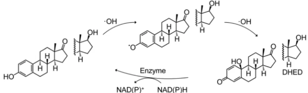
Another good example for the biological relevance of a compound emerging from RONS scavenging by an antioxidant is the Prokai cycle of estrone and estradiol that explains their neuroprotective activity. Prokai et al found that•OH radical scavenging by estrone yields ap‐quinol A‐ring‐containing intermediate that subsequently undergoes a NADP‐dependent enzymatic reduction back to estrone, without contributing to oxidative stress on its own but becoming able to scavenge further radicals.144-146 In vivo administration of the analogous p‐quinol derivative of estradiol (DHED) demonstrated no estrogenic activity, and it was rapidly taken up into the brain where it initiated the above‐mentioned redox cycle; therefore it acted as a targeted neuroprotective agent without any apparent systemic side‐effects.147-150On one hand, this provides an attractive strategy to deliver estrogens to the brain through their pro‐drugs. On the other hand, it also represents a good example for a phenolic antioxidant (estradiol or estrone) to be transformed to a metabolite (eg DHED) on a free radical scavenging‐related manner.
Certainly, such a nonaromatic metabolite will have a significantly different bioactivity profile than its parent compound (ie no estrogenic effect in this case; other specific bioactivities were not studied). The reaction mechanism for the above‐mentioned redox cycle is shown in Figure 6.
Concerning possible chemical approaches to explore the RONS scavenging‐related metabolic fingerprint of antioxidants, biomimetic oxidative chemistry offers a readily available toolkit. Development and application of biomimetic or bio‐inspired chemical reactions represent a rapidly emerging approach in today's chemistry. Such reactions use transition metals as catalysts either in free forms or in their complexes, for example as metallo‐ porphyrins,151to perform various oxidative transformations similar to those observed in biological systems. A common feature of these types of reactions is that the oxidizing agent is an activated (ie reactive) oxygen or nitrogen species formed by electron transfer from the transition metal, aiming to mimic the way oxidase enzymes work.152,153
Accordingly, it is unsurprising that biomimetic approaches are extensively used in the total synthesis of natural products,154 including various functionalization or cross‐coupling reactions of phenolic/polyphenolic com- pounds.155-157
Nevertheless, more than providing a remarkable toolkit for solving synthetic chemical challenges, biomimetic oxidative chemistry also offers a platform to study the oxidative metabolism of drugs.158Such approaches require the best possible models of selected metabolizing oxidase enzymes. From a different perspective, however, biomimetic oxidative reactions can also be considered as reasonable chemical models for studying the potential impact of RONS on compounds including for example dietary antioxidants. Apparently, no related systematic studies have been performed so far, but sporadic reports can be found where one or more bioactivities of an
antioxidant were directly compared in the same pharmacological model to those of its oxidized metabolites obtained by biomimetic oxidative chemistry.
In the following sub‐sections, chemically oxidized derivatives of compounds1‐9will be presented from this perspective. For each compound, selected examples are discussed, in which the bioactivity of the parent antioxidant on specific pharmacological models could directly be compared to that of its chemically stable metabolite(s). An overview on these comparisons is provided in Table 1.
Since the field is extremely rich in reports on antioxidant natural products, it is almost certain that the below examples do not provide a complete coverage. Still, their chemical and pharmacological diversity might provide an overview suggesting that oxidative stress‐related metabolites may represent an important piece of the antioxidant puzzle, which is frequently overlooked.
4.1 | Curcuminoids
In the case of curcumin (1; Figure 1) and its derivatives, oxidative changes have been extensively studied over the years as a likely explanation for the polypharmacology of these compounds.56,159Spontaneous aerobic oxidation of curcumin leads to the formation of a bicyclopentadione derivative (1u) as a major product160; for its chemical structure, see Figure 1B. A most recent, in‐depth mechanistic study identified several intermediates (eg1tand1v) and by‐products (1s,1w,1xand 1y) of this metabolite, arising from a course of multiple chemical rearrangements initiated by the phenoxyl radical.161This radical is obviously among the primary products when curcumin participates in ROS scavenging. The broad chemical diversity of the subsequently forming intermediates and end‐products raises many exciting questions concerning their bioactivities in relation to their parent compound1. Unfortunately, as of today, little is known about them. Topoisomerase II poisoning activity of curcumin162,163is clearly associated with its transformation: its activity could be observed only in oxidative conditions.164 Moreover, the same oxidative activation applies for poisoning topoisomerase IIα by demethoxycurcumin (2) and bisdemethoxycurcumin (3): the activity of both curcuminoids significantly increased when combined with K3Fe(CN)6, a biomimetic oxidative reagent.165Transformation route of both2and3was found to be related to that of curcumin, and2also yielded a bicyclopentadione derivative (2r) as major metabolite. Both2and3were, however, more stable to resist autoxidation, and3even required the addition of K3Fe(CN)6or H2O2and horseradish peroxidase (forming ROS) to transform to a bisdemetoxy‐spiroepoxide (3k), and stop at that phase without converting to a bicyclopentadione.165In view of most recent studies, however, it seems that a great deal of the bioactivity of curcuminoids is connected rather to the intermediates of the autoxidation and not the stable bicyclopentadiones.166,167Sanidad et al found that, while curcumin (1) had potent antiproliferative and proapoptotic activity against colon cancer cells, and exerted anti‐inflammatory activity through inhibition of the NF‐κB signaling, both the mixture of the degradation products and1rwere weaker or rather inactive in these bioassays.166However, by using an experimental setup that allowedin situ formation of the autoxidized products, Edwards et al found that NF‐κB inhibition by curcumin depends on oxidative activation, and that this is due to the reactive electrophile intermediates covalently binding to signaling proteins, such as for example IKKβ. Very interestingly, and in line with the perspective discussed here, they could clearly connect this activity to the oxidative status of the cells, in other words, they demonstrated that oxidative stress modulates curcumin's bioactivity through thein situformation of oxidized intermediates.167
Curcumin is also known to rapidly decompose in cell culture medium under aerobic, physiological conditions, that yield a set of cleaved products, such as trans‐6‐(4′‐hydroxy‐3′‐methoxyphenyl)‐2,4‐dioxo‐5‐hexenal (1z) as major product, and ferulic acid (1za), ferulic aldehyde (1zb), feruloyl methane (1zc), vanillic acid (1zd), and vanillin (1ze) as minor metabolites.168,169These compounds have received attention in numerous studies with curcumin, leading to an intense discussion on their potential role in some previous controversial findings.159For example, they were suggested as potentially responsible for the xanthine oxidase (XO) inhibition by curcumin,170which was previously reported both with positive171and negative172results. Based onin silicopredictions validated by experimental XO inhibitory activities of quercetin and luteolin, the side‐chain cleaved metabolite1zwas suggested as a strong inhibitor of the enzyme, while curcumin itself was too large to bind to the active center of XO.170In
contrast to this, the testing of curcumin and its cleaved metabolites1z,1za,1zb,1zd, and1zeagainst human recombinant cytochrome P (CYP) enzymes CYP1A2, CYP3A4, CYP2D6, CYP2C9 and CYP2B6 showed that while curcumin (1) can inhibit all of them, none of the metabolites were significantly active in this regard.169
In a recent debate initiated by comments on the results of Dhillon et al on the potential therapeutic use of large doses of orally administered curcumin in cancer patients,173the same degradation products were hypothesized to be responsible for its beneficial effect.174In their response, the authors disapproved this theory and presented evidence for the inactivity of1zaand1zcfor suppressing the activation of NF‐κB, in contrast with the case of curcumin.175While the clinical relevance of the above‐mentioned recent findings by Edwards et al167on this pathway is unclear, one could hypothesize a similar oxidative stress‐related formation of reactive intermediates.
The theory suggesting high importance of the cleaved metabolites (1z‐1zc) in the bioactivity of curcumin159has also been challenged; based on the fact that under biologically more relevant conditions they form in smaller amounts than the bicyclopentadione compound1u.176However, when encapsulated curcumin was administered to healthy human subjects through enriched bread, to at least partly protect it from phase I and II metabolism, vanillic and ferulic acids (1zdand1za, respectively) were detected as major metabolites.177While it is unknown how this would correlate to the amount of1u, which should possibly formin vivodepending on the oxidative status, these cleaved products may also play their role in curcumin's bioactivities, particularly concerning the XO and CYP enzyme inhibition.178
Altogether, curcumin (1) represents an excellent example for an antioxidant that can undergo many different types of oxidative transformations, apparently leading to an oxidative status‐related change in its bioactivity profile. Considering that much of the related dispute appears to originate from different applied oxidative conditions, one must assume that the metabolite pattern arising in a biological system will greatly depend on the local microenvironment, for the types and levels of RONS to be scavenged.
4.2 | Resveratrol
In view of the possible biological role of oxidized metabolites of resveratrol (4; Figure 2), its bioactivity on lipoxygenase (LOX) enzyme demonstrates an interesting example. Resveratrol was reported as a potent competitive inhibitor (IC50= 13 µM) of the dioxygenase but not the hydroperoxidase activity of LOX. At the same time, LOX gradually oxidized resveratrol to an in situ, non‐characterized derivative that was suggested to be similarly active on the dioxygenase activity of LOX as resveratrol itself.179More recently, resveratrol was involved in a study where 30 polyphenols were subjected to an Fe3+ catalyzed biomimetic aerobic oxidation and the resulting mixtures were screened for specific LOX inhibitory activity through the xylenol orange (FOX) assay that measures the hydroperoxidase activity.180As expected, resveratrol itself was inactive at a concentration as high as 100 µM, while its oxidized mixture exerted 98% inhibition at a concentration equivalent to 50 µM of resveratrol, despite the rather low (33%) conversion.
Isolation of the predominant metabolites led to the identification of two active dihydrobenzofurane dimers, compound 4mandδ‐viniferin (4n); for the relevant structures, see Figure 2B. Further investigations revealed that the metabolite profile is highly dependent on the reaction conditions: by using excess CuCl2in ethanol, compound4mwas obtained at relatively higher yields, while oxidation with DPPH radical made compounds4nand4o(the latter inactive as LOX inhibitor) as preferred metabolites.180Besides the low conversion of resveratrol (4), it should be highlighted that more than 20 products were visible on the HPLC fingerprint of the active mixture. This mixture exerted a complete inhibition of LOX at a concentration equivalent to 50μM of resveratrol (4), implying that the activities reported for the individual metabolites4nand4m(IC50values ca. 17 and 62μM, respectively) cannot explain the activity observed for the mixture that contained these compounds in a few percent amount only.180This suggests either the presence of further active metabolites exerting stronger LOX inhibitory activities than compounds4mand4n, or strong synergistic interactions between the minor compounds of the reaction mixture.
Considering the very low (free) resveratrol concentrations achievablein vivo(see Section 3.2), the biological relevance for the RONS‐dependent formation of such dimers and/or oligomers might seem to be at least questionable. Certainly, it seems there can be only a very low chance that the intermediates formed upon RONS
scavenging would react with each other instead of for example GSH or other, endogenous compounds present in excess amounts. This is, however, not necessarily the case. NMR studies181and high‐levelin silicocalculations182 provided evidence for a very strong self‐association of stilbenes stabilized by π‐π interactions in aqueous environment, which could explain the inconsistencies in the regio‐and stereoselectivity observed in oxidative coupling reactions similar to the case above.182 In addition to these implications, this also means that fully π‐conjugated stilbenes (eg resveratrol) preferably stick together in the aqueous environment provided by a living organism. As such, there might be a reasonably high chance for the formation of dimer/oligomer metabolites of such antioxidants through cross‐coupling upon RONS scavenging, and for related modulation of their bioactivity.
4.3 | Hydroxycinnamates
The oxidized metabolites of hydroxycinnamates may provide interesting and valuable insights into the antitumor activity of their parent compounds.
In a most recent, proof‐of‐concept study of our research group, methyl‐p‐coumarate (5; Figure 3) was studied for its potential to form bioactive metabolites upon free radical scavenging.183A hypervalent iodine reagent (PIFA) was used to obtain oxidized metabolite mixtures of compound5. PIFA can oxidize phenolic compounds through single‐electron transfer (SET) whose intermediate can transform into the same phenoxyl radical upon deprotonation as that forming in a hydrogen‐atom transfer (HAT) reaction. Therefore, it was expected that the use of this reagent would result in metabolites like those forming through free radical scavenging. Two metabolites (5e and 5f; see Figure 3B) were identified with antitumor activities much stronger than that of compound5.
Graviquinone (5e) had over 2 orders of magnitude stronger cytotoxicity on certain resistant cancer cell lines as compared to compound5. Ten µM of graviquinone (5e) induced DNA damage in NCI‐H460 and NCI‐H661 lung carcinoma cells while it exerted DNA protective activity in normal HaCaT cells as determined by the relative expression of Histone 2A.X. At the same concentration it modulated DNA damage response through the inhibition of Checkpoint kinase‐1 (Chk1) phosphorylation and the induction of Checkpoint kinase‐2 (Chk2) phosphorylation in MCF‐7 cells. It was also demonstrated that graviquinone (5e) can be formed as the result of the Fenton reaction of compound5.In silicostudies suggested that5eis a kinetic metabolite when methyl‐p‐coumarate (5) scavenges•OH radicals. The fact that cytotoxic activity of methyl‐p‐coumarate (5) was also potentiated by H2O2 is indirect evidence for the oxidative stress‐relatedin situformation of antitumor metabolites;for examplecompound5e.183It is also worth noting the structural similarity of compound5eto the above‐mentionedp‐quinol estrone derivative DHED (see Figure 6). This suggests thatp‐phenols in general can be transformed into theirp‐quinol analogs upon free radical scavenging. Moreover, suchp‐quinol derivatives, including DHED itself, might be worthy of further study for activity on checkpoint kinases Chk1/Chk2, two of the key enzymes involved in response to DNA damage.
These kinases are important in cancer development and, possibly, treatment.184
Concerning other hydroxycinnamates, methyl caffeate (6) was reported as a weak‐moderate antitumor agent against various cell lines within vitroantiproliferative IC50values at the medium‐low micromolar range against MCF‐7 breast cancer cell line (ca. 110μM).185Still, there are some studies showing much stronger antitumor effects for this compound, for example Balachandran et al observed cytotoxic activity of6against MCF‐7 cells with an IC50value as low as 0.62μM, and the involvement of several pro‐and antiapoptotic genes in apoptotic cell death.186
Ag2O catalyzed biomimetic oxidation of a set of cinnamic acid methyl esters including compound6yield metabolites with highly potent antitumor activity, and compound 6m, an oxidized dimer of 6, exerts a particularly strong antiproliferative activity (IC50< 10 nM) against breast cancer cell lines MDA‐MB‐435, MDA‐N and BT‐549, and sub‐ micromolar activity against several other cell lines. Moreover, the 2R,3R enantiomer of compound 6l (but not the inactive 2S,3S) was identified as an antitubulin agent as strong (IC50= 6.0 µM) as combretastatin A‐ 4,187a well‐known natural inhibitor of tubulin polymerization. The same compound, the 2R,3Renantiomer of6l, was also found to exert potent antiangiogenic activity apparently without interfering with fibroblast growth factor‐2 (FGF‐2) or vascular endothelial growth factor (VEGF).188Moreover, in a recent, in‐depth pharmacological study usingin vivoand
3D cell culture models, Yin et al found that compound6lhas a significant potential for further development of a clinically applicable antimetastatic agent.189Of significant pharmacological interest, compound6lexerts its activity mainly on the microenvironment of the tumor through inducing the IL‐25 secretion of tumor‐associated fibroblasts, and this effect could be achieved in mice at doses as low as 20 to 100μg kg−1body weight.189The activity of compound6 was not investigated, and it is hard to imagine that its activity would be like that of its oxidized metabolite6l. As for the antiangiogenic effect, compound6was reported to be inactive in zebrafish.190
In another related study, the racemic mixture of compound7d, obtained enzymatically from methyl ferulate (7), exerted a similarly strongin vitro antiangiogenic effect to that of6l(2R,3R). A significant decrease in the VEGF production was found in swine granulosa cells at 1μM concentration of this compound, and it also retained the antioxidant activity of its parent compound, methyl ferulate (7).191Contradictory results have been reported with ferulic acid and its effects on angiogenesis.192,193Ferulic acid was found to up‐regulate cyclin D1 and VEGF in endothelial cells, leading to a proangiogenic effect,192while in the other study, antiangiogenic effect was found through downregulation of•NO, which, depending on the cell type, can lead to VEGF downregulation.193This contradiction may be connected to the perspective presented here: namely, results that depend on the oxidative status of the studied system. The effect of ferulic acid would be tuned in either direction through possible variations in its metabolite profile likely including compounds similar to those obtained from methyl ferulate.
4.4 | Secoisolariciresinol
The oxidation of secoisolariciresinol (8; Figure 4) was previously studied by employing 2,2′‐azobis(isobutyronitrile) (AIBN), a radical oxidant initiator and ethyl linoleate dissolved in acetonitrile producing a lipophilic environment F I G U R E 7 Contextualization of the chemical perspectives discussed in Section 4, in connection with the canonic view on antioxidant activity. (A) Role of antioxidants in interfering with the redox balance and redox‐modulated biochemical pathways. The antioxidant may enter a redox cycle and exert its bioactivities through both its reduced and oxidized form. (B) Potential importance of minor, chemically stable oxidized metabolites that may form due to free radical scavenging. Depending on the antioxidant's chemical properties, the type of ROS/RNS scavenged, re‐arrangements of emerging reactive intermediates and/or other secondary reactions, etc., a complex mixture of oxidized metabolites is expected to be present and interact with various cellular mechanisms involved in the regulation of many biochemical processes. This localized metabolite pattern may be characteristic to the microenvironment, implying a crosstalk between redox signaling and a co‐existent“antioxidant‐metabolite signaling”. Since individual oxidized metabolites tend to show higher chemical complexity than their parent antioxidant, they are also expectable to act in a more targeted way