I N T R O D U C T O R Y L E C T U R E
That melanin functions in man primarily as a solar screen is almost indisputable. Melanized epidermis acts as a cloak that shields the underlying dermis by effectively blocking the passage of injurious u.v.
rays. In man, melanin-forming cells, melanocytes, are present in small numbers in vestigial loci in the meningeal sheaths of the central nervous system and in abundance in the eyes and skin, where they form a front line of defence against the effects of solar radiation
( Fi t z p a t r i c k, Se i j i and McGu g a n , 1961). The remarkable resistance of pigmented skin to u.v. radiant energy is attributable to the melanin produced by these cells. What evidence is there to justify these state- ments ? What evidence have we that melanin granules protect sub- epidermal tissues against the harmful effects of u.v. ? By what mechanism do the melanin granules of the epidermis fulfil their protective function ? What factors determine the colour, the amount and the distribution of melanin granules in the epidermis? The answers to these questions are of both clinical and theoretical interest.
Let me discuss them in order.
I. What evidence is there that melanin granules protect sub-epidermal tissues against the harmful effects of u.v. ?
A comparison of clinical findings in the skin of normal and albino Negroes living in the tropics provides quite striking evidence. Albino Negroes in Africa ( Sh a p i r o, Ke e n, Co h e n and Mu r r a y , 1953) and albino Cuna Indians on the San Bias Islands off the coast of Panama are peculiarly susceptible to the effects of sunlight. Degenerative changes and carcinomas of exposed areas of skin develop in very young albino Negroes ( Sh a p i r o et aly 1953) who live in the tropics. Actinic keratoses are found in almost all albino Cuna Indian children;
Ke e l e r (1962) has observed them in a majority of these children who
had reached the age of 2 years. The only known difference between
365
T . B . FI T Z P A T R I C K
Harvard University and Massachusetts General Hospital, U.S.A.
the albino and the normal members of these dark-skinned groups is the amount of melanin present in the skin.
Exposure to sunlight is the principal cause of skin carcinoma in man.
The incidence of skin cancer is relatively low in the skin of Negroes, Asiatics, East Indians and American Indians in spite of chronic exposure to sunlight, but in light-skinned Caucasians carcinoma are found frequently on the face and back of the hands, and are limited almost exclusively to areas of skin that have been repeatedly subjected to the action of sunlight. In a thorough study, El e a n o r Ma c d o n a l d
(1959) has shown that the incidence of carcinoma is very high in the exposed skin of the head, neck and hands of light-skinned individuals who live in such regions as El Paso County, Texas, where there is prolonged exposure to high-intensity solar radiation.
In addition to these clinical evidences of the protective role of melanin, there is abundant experimental evidence that melanin protects mammalian skin against the harmful effects of u.v. light. With sheets of stratum corneum separated from Negro and Caucasian skin,
Th o m s o n (1955) has demonstrated the greater efficacy of Negro skin
as a screen against u.v. radiant energy. He found that the stratum corneum of Negroes and Caucasians does not differ significantly in thickness, and attributed the greater screening-capacity of stratum corneum from the Negro to its high melanin content.
There is a striking difference in the transmission of u.v. through equal thicknesses of stratum corneum and epidermis from Negro and from Caucasian skin. Pa t h a k has found that much less u.v. passes through the epidermis and stratum corneum of Negro skin than through the epidermis and stratum corneum of Caucasian skin. With other investigators, Pa t h a k (1959) has also shown that in pigmented mice exposed to the carcinogenic effect of u.V., tumours either do not develop at all or develop more slowly than in albino mice subjected to identical conditions.
Im b r i e and his co-workers (1959) noted that 6 weeks after a single exposure to sunlight, the erythemal threshold rose approximately 100 per cent in areas of skin that had been exposed to light 2 h after ingestion of 8-methoxypsoralen. The degree of pigmentation in these areas was greater than that seen in subjects who had received no psoralen before exposure. Presumably this rise in the erythemal threshold was at least partially related to the increase in pigmentation, although the possibility that the stratum corneum had been altered in psoralen-treated subjects was not ruled out.
The erythemal response to u.v. differs markedly in the hypomelano- tic lesions of vitiligo and in the immediately adjacent pigmented areas.
Following exposure to solar radiation for 60 min, marked sunburn erythema develops in vitiliginous macules, while in pigmented areas only a few microns away there is little or no erythema. The only obvious difference between the vitiliginous areas and the adjacent, normally pigmented areas is a difference in melanin-granule content.
The striking contrast in the response of these areas is well illustrated by a recent investigation in which Dr Pa t h a k and I exposed a Negro subject with vitiligo to noon sunlight in Boston during June for periods of 45 and 90 min. Twenty hours after a single 90-min exposure there was bright red, markedly tender erythema in the vitiliginous areas while only barely perceptible erythema could be seen in adjacent pigmented areas.
T o assess the relative roles of the stratum corneum and melanin granules, the stratum corneum was removed from certain areas by stripping (23 three times) with adhesive cellulose tape. Twenty hours after exposure to sunlight for 90 min there was fiery erythema with vesiculation in the stripped vitiliginous areas and non-tender, pink erythema in adjacent stripped pigmented areas. This experiment clearly illustrates the effectiveness of melanin granules as agents that protect against the deleterious effects of u.v.
In very careful, early experiments, which seem to have been forgotten, Mi e s c h e r (1931) studied the role of pigment in the protec- tion of skin against u.v. He showed that the ratio of the thresholds for surface damage in white and Negro skin is 1:8, while the ratio of the thresholds for deep damage is 1:120. This led him to conclude that these differences in the sensitivity of white and Negro skin can only be explained by differences in the absorption of light. He stated that Negro skin absorbed more u.v. than white skin because of the presence of pigment in all layers of the epidermis, including the stratum cor- neum. These experiments, plus others on pigmented and albino guinea pigs, led Mi e s c h e r to conclude that melanin provides important protection against short-wave u.v.
It cannot be denied that the stratum corneum filters out a certain amount of the u.v. radiant energy, especially when, after repeated exposure, the stratum corneum has become two or three times thicker than normal. This can be clearly demonstrated in albinos and patients with vitiligo, whose tolerance to sunlight is increased, presumably as the result of such thickening, by carefully graded exposure of amelanic
skin to solar radiation. The incredible resistance of normal palmar skin to the development of erythema is further evidence of the protective role of the stratum corneum. Exposure of the palms to sunlight three times more intense than that needed to induce marked erythema in the skin of the back of white Caucasians evokes not even barely perceptible erythema.
In estimating the relative effectiveness of stratum corneum and melanin granules as barriers to u.v. light, it should be kept in mind that unmelanized (albino) skin with normally thick stratum corneum has poor tolerance to sunlight. Thus, of the two protective barriers, stratum corneum and melanin granules, the latter appears to be far more efficient.
II. By what mechanism do melanin granules fulfil their protective function ?
Before this question can be answered it will be necessary to discuss the chemical nature of the melanins and some of their physical characteris- tics. Melanins are black, brown or yellow pigments of high molecular weight that occur naturally and are formed by the enzymatic oxidation of phenols. Those found in mammals, birds, reptiles, amphibians and certain other lower animals are derived from tyrosine and are therefore called tyrosine-melanins ( Fi t z p a t r i c k, Se i j i and McGu g a n , 1961).
In human skin, melanin does not exist as such, but is always bound to a protein with which it forms particles (melanin granules) that vary in size from ο·ι χ 0-4 μ to ο·ι8 χ ο·6 μ. In man, the enzyme, tyrosinase, which is attached to specialized organelles (melanosomes) catalyses the oxidation of tyrosine to indole 5,6-quinone ( Se i j i, Sh i m a o, Bi r b e c k
and Fi t z p a t r i c k , 1963). This quinone polymerizes to dimers, trimers and huge polymers to form a highly conjugated system. The very large melanin polymer becomes strongly bound to protein, probably through sulphydryl linkages. Therefore, the chemical unit present in human skin should be called ' melano-protein ' not ' melanin pigment ' and one should be aware that the so-called 'melanin granule' is composed of this substance. The melanin polymer shows general absorption in the u.v. and visible spectrum. It is one of the most dense substances observed in tissue by electron microscopy. X-ray dif- fraction studies show that it is without crystalline form. The melanin polymers are extremely insoluble and thus far have resisted all attempts to degrade them into identifiable fragments.
The protective capacity of melanin may be related to the fact that
it can act as a stable free radical (i.e., as a one-dimensional semi- conductor) that can function as a repository of photoactively produced electrons ( Co m m o n e r, To w n s e n d and Pa k e , 1954; Da n i e l s , 1959;
Lo n g u e t- Hi g g i n s , i960; Ma s o n, In g r a m and Al l e n , i960). For example, the number of free radicals generated in non-white human skin by u.v. differs from the number generated in white skin; the free radical concentration is much lower in non-white than in white skin
( Pa t h a k and St r a t t o n , 1964). Melanin may capture the free radicals generated by the action of u.v. on tissue and thus protect the skin from their harmful metabolic effects. Ke r k u t (1962) has noted that hair which contains melanin produces free-radical signals, the darker the hair the stronger the signal produced. Grey hair does not produce any free-radical signal.
In summary, then, the defensive role of melanin granules may be accomplished by the universal optical absorbency of the melanin granules that are distributed throughout the Malpighian layer and the stratum corneum. In these locations, melanin granules prevent the passage of light or, in other words, constitute a biological optical filter.
In addition they are a repository of the free radicals generated by the action of u.v. on the epidermis. Melanin granules therefore render the skin uniquely resistive to the effects of the u.v. region.
III. What factors determine the colour, the amount and the distribution of melanin granules in the epidermis ?
Since melanin plays the most important single role in screening body tissues against u.V., it will be well to review some of the recent concepts of melanin biology.
The intensity of clinically visible pigmentation of the skin is related not only to the number, but also to the location, of melanin granules in the epidermis. Even when melanocytes are engorged with melanin granules, the skin will not appear clinically to be pigmented unless melanin granules have entered the Malpighian cells. In a histologic study of skin from individuals with different degrees of skin coloura- tion, Ga t e s and Zi m m e r m a n n (1953) were able to show that the various degrees of skin colour are directly related not only to the relative number of melanin granules in the epidermis, but also to their location within this tissue. In clinically less pigmented skin they found great variation in both the number and location of granules within the epidermis ; there were only scanty melanin deposits in the Malpighian layer and none in the stratum corneum. In the most lightly pigmented
24
specimen in their series, there were only traces of melanin granules in the basal cells. It is apparent from these studies that clinically evident skin colour is determined by the melanin granules present in the stratum malpighii and the stratum corneum.
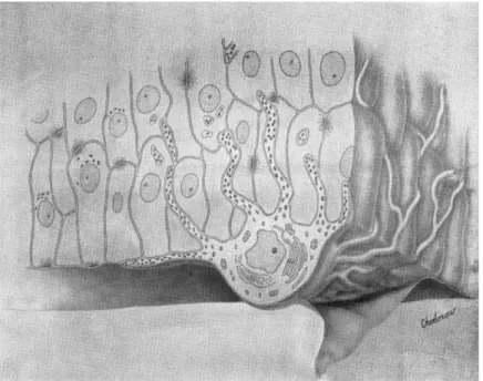
Present knowledge indicates that melanin pigmentation takes place within the 'epidermal melanin unit' (Fig. i) which may be defined as a melanocyte surrounded by a constellation of Malpighian cells ( Fi t z -
p a t r i c k and Br e a t h n a c h , 1963). This unit is a structural and functional entity comparable to the nephron. There is strong evidence that the melanocytes and Malpighian cells that constitute this unit are functionally interdependent and that Malpighian cells participate actively in the process of melanin pigmentation by providing clues to melanocytes. Melanin pigmentation involves: first, the formation within the melanocyte of structures known as premelanosomes ; second, transformation of these structures by melanization into melanosomes and eventually melanin granules ( Se i j i, Sh i m a o, Bi r b e c k and Fi t z - p a t r i c k , 1963); and third, transference of mature melanin granules from the melanocytes in which they are formed to the Malpighian cells in which they are transported.
Melanogenesis, which is the production of melanin granules, should be clearly distinguished from melanin pigmentation, which is a dual process involving both the production of melanin granules and their delivery to the Malpighian cells. Clinically visible pigmentation of the skin depends upon both the production and the transference of melanin granules. Basic skin colour and pigmentary response to u.v.
are controlled primarily by genetic factors. It is very unlikely that pituitary hormones or estrogens exert any major effect on pigmentary responses to u.v.
Qu e v e d o and Sm i t h (1963) have recently made important observa- tions that support the view that the presence of melanin granules in Malpighian cells is necessary for the production of clinically visible skin colour. These investigators irradiated the plantar skin of various mouse genotypes with u.v. and found that in two genotypes with brown coats no grossly observable tanning of irradiated areas devel- oped, although on histologic examination the melanocytes of this irradiated skin were found to contain a large number of melanin granules. The engorged melanocytes had 'short' dendrites and, probably because of this, had been unable to transfer melanin granules to the Malpighian cells with which they were associated.
Genes regulate both melanogenesis and melanin pigmentation. They
FIG. Ι. The epidermal melanin unit.
facing p. 370
determine the fine structure of premelanosomes, which are the pre- cursors of melanin granules, and they also determine the length and size of the melanocytic dendrites that deliver melanin granules to the Malpighian cells. In heavily pigmented skin, u.v. might be expected to evoke little response, because the cloak of melanin is habitually adequate for protection; in other words, without special exposure the epidermal units of such skin normally produce a good supply of melanin granules and deliver them to the Malpighian cells that eventually form the stratum corneum. In lightly pigmented skin that habitually lacks a heavy protective cloak, failure to tan after exposure to u.v. could be related to poor development of melanocytic dendrites.
As Qu e v e d o has pointed out in the mouse (1963), the response to u.v.
depends upon the genetically determined capacity of the epidermal melanin unit to react to this type of radiant energy.
Dr Ru t h Mi t c h e l l, of Australia, has described some striking changes that take place in human skin that fails to tan after prolonged exposure to solar radiation (1963). She reports that the melanocytes in such skin are essentially round and contain abnormally few melanin granules. The Malpighian cells associated with these deformed melanocytes suggest that the transfer of melanin granules has been blocked in some way.
Failure of the skin to tan may result from pathologic changes in the Malpighian cells as well as from abnormality of the melanocytes ( Fi t z p a t r i c k, unpublished). Such changes are clearly seen in the skin of patients with eczematous dermatitis, in which melanocytes with long dendrites are associated with edematous Malpighian cells that contain little or no melano-protein. An inadequate pigmentary response to solar radiation, that is, failure to tan, may also result from abnormally rapid proliferation of Malpighian cells in the epidermis, such as occurs in psoriasis. When this takes place, Malpighian cells appear to proliferate so rapidly that they are unable to phagocytize melanin granules.
In summary, melanin pigmentation depends upon the responsive- ness and activity of the epidermal melanin unit. The degree of tanning achieved at any given time is determined by the functional balance established between the component parts of this unit, that is, the melanocytes and the Malpighian cells. Increases in clinically visible pigmentation are impossible when melanocytes are absent ; when the premelanosomes formed within the melanocytes fail to become pigmented, as in albinism; when delivery of melanin granules to the
Malpighian cells is blocked by pathologic conditions in either melano- cytes or Malpighian cells, as in intracellular oedema, eczematous dermatitis, and skin chronically exposed to the sun; and when the proliferation of Malpighian cells is abnormally rapid, as in psoriasis.
The presence of melanin granules is essential for protection of skin against the deleterious effects of u.v. The effectiveness of the protection that exists at any moment is directly related to the number of melanin granules in the Malpighian cells and the stratum corneum.
C O D A
In mammals, the body surface is covered with a dense, usually pig- mented coat of hair which is practically impenetrable to u.v. In most mammals, therefore, the epidermal melanin unit is not highly devel- oped and most of the pigment is located in the hair. In man, who is practically hairless, nature has provided means of defence against the deleterious effects of u.v. This spectral region sets into action an integrated mechanism for the formation of dense packets of chromo- protein and the delivery of these packets (melanin granules) to Mal- pighian cells. Within the Malpighian cells, these granules scatter and absorb u.v. ; they also constitute a repository for the damaging free radicals that are generated in skin after exposure to u.v. This mechan- ism is probably triggered by the Malpighian cells which respond to u.v. injury by mitosis, thus creating a new demand for melanin granules. The epidermis then behaves functionally like hair in the anagen phase, and reacts with continuous bursts of mitosis. When this occurs, melanin granules are transferred into Malpighian cells through the dendrites of the melanocytes into which they are secreted.
Actinic injury of the epidermis and dermis by u.v. is thus directly related to the number of melanin granules present and/or the melano- genic potential of the epidermal melanin unit.
In Fi n s e n ' s (1900) well known investigations, melanin pigment is considered to be the principal means of protection against u.v. During the last 64 years, only Mi e s c h e r (1931) and, more recently, Th o m s o n
(1955) have emphasized this role of melanin pigment. The very title of this symposium,' Light and Melanin Pigment ' indicates that a growing number of investigators believe that melanin plays a major, if not the dominant, role in the defence of skin against actinic injury.
INTRODUCTORY LECTURE REFERENCES
Co m m o n e r B . , To w n s e n d J. and Pa k e G. E . (1954) Nature, Lond. 1 7 4 , 689.
Da n i e l s F . Jr. (1959)^. Invest. Derm. 3 2 , 147.
Fi n s e n N . R . (1900) Mitt. Finsens med. Lysinst. 1 , 8.
Fi t z p a t r i c k T . B . and Br e a t h n a c h A . S . (1963) Derm. Wschr. 1 4 7 , 481.
Fi t z p a t r i c k T . B . , Se i j i M. and McGu g a n A . D . (1961) New Engl.J. Med.
2 6 5 , 328, 374, 430.
Ga t e s H.R. and Zi m m e r m a n n A . A . (1953)^. Invest. Derm. 2 1 , 339.
Im b r i e J. D . , Da n i e l s F . Jr., Be r g e r o n L., Ho p k i n s C.E. and Fi t z p a t r i c k
T . B . (1959)^7. Invest. Derm. 3 2 (Part II), 331.
Ke e l e r C. (1962) National Cancer Institute Monograph No. 10.
Ke r k u t G . A . , Ed w a r d s M.L. and Mu n d a y K . A . (1962) Life Sciences, No. 4,
p. 129, Pergamon Press Ltd., London.
Lo n g u e t- Hi g g i n s H . C . (i960) Arch. Biochem. Biophys. 8 6 , 231.
Ma c d o n a l d E.J. (1959) J. Invest. Derm. 3 2 , 379.
Ma s o n H.S., In g r a m D.J.E. and Al l e n B . (i960) Arch. Biochem. Biophys.
8 6 , 225.
Mi e s c h e r G . (193 ι ) Strahlentherapie 3 9 , 601.
Mi t c h e l l R . E . (1963) J. Invest. Derm. 4 1 , 199.
Pa t h a k M. A . , Da n i e l s F . Jr., Ho p k i n s C. E . and Fi t z p a t r i c k T . B . (1959)
Nature, Lond. 1 8 3 , 728.
Pa t h a k M.A. and St r a t t o n K. (1964) Proc. \th International Photobiology Congress, Oxford, England, July 26-30, 1964.
Qu e v e d o W . C . Jr. (1963)^. Soc. Cosmetic Chemists 1 4 , 609.
Qu e v e d o W . C . Jr. and Sm i t h J.A. (1963) Ann. Ν. Y. Acad. Sei. 1 0 0 (Part I), 364.
Se i j i M., Sh i m a o K . , Bi r b e c k M. S . C . and Fi t z p a t r i c k T . B . (1963) Ann.
Ν. Y. Acad. Sei. 1 0 0 (Part II), 497.
Sh a p i r o M.P., Ke e n P., Co h e n L. and Mu r r a y J. F . (1953) B n t. J . Cancer 7, 45.
Th o m s o n M.L. (1955)^. Physiol. 1 2 7 , 236.