Research Article
Hydrogen Sulfide Abrogates Hemoglobin-Lipid Interaction in Atherosclerotic Lesion
László Potor,1,2Péter Nagy,3Gábor Méhes,4Zoltán Hendrik ,1,4Viktória Jeney ,5 Dávid Pethő,5Anita Vasas,3,6Zoltán Pálinkás,3EnikőBalogh ,5Ágnes Gyetvai,1
Matthew Whiteman,7Roberta Torregrossa,7Mark E. Wood,8Sándor Olvasztó,9Péter Nagy,9 György Balla,1,2and József Balla 5
1HAS-UD Vascular Biology and Myocardial Pathophysiology Research Group, Hungarian Academy of Sciences, Debrecen 4012, Hungary
2Department of Pediatrics, Faculty of Medicine, University of Debrecen, Debrecen 4012, Hungary
3Department of Molecular Immunology and Toxicology, National Institute of Oncology, Budapest 1122, Hungary
4Department of Pathology, Faculty of Medicine, University of Debrecen, Debrecen 4012, Hungary
5Division of Nephrology, Department of Medicine, Faculty of Medicine, University of Debrecen, Debrecen 4012, Hungary
6Department of Inorganic and Analytical Chemistry, University of Debrecen, Debrecen 4032, Hungary
7University of Exeter Medical School, Exeter, UK
8College of Life and Environmental Sciences, University of Exeter, Exeter, UK
9Division of Vascular Surgery, Department of Surgery, Faculty of Medicine, University of Debrecen, Debrecen 4012, Hungary
Correspondence should be addressed to József Balla; balla@belklinika.com
Received 2 August 2017; Revised 6 November 2017; Accepted 14 November 2017; Published 21 January 2018
Academic Editor: Kota V. Ramana
Copyright © 2018 László Potor et al. This is an open access article distributed under the Creative Commons Attribution License, which permits unrestricted use, distribution, and reproduction in any medium, provided the original work is properly cited.
The infiltration of red blood cells into atheromatous plaques is implicated in atherogenesis. Inside the lesion, hemoglobin (Hb) is oxidized to ferri- and ferrylHb which exhibit prooxidant and proinflammatory activities. Cystathione gamma-lyase- (CSE-) derived H2S has been suggested to possess various antiatherogenic actions. Expression of CSE was upregulated predominantly in macrophages, foam cells, and myofibroblasts of human atherosclerotic lesions derived from carotid artery specimens of patients.
A similar pattern was observed in aortic lesions of apolipoprotein E-deficient mice on high-fat diet. We identified several triggers for inducing CSE expression in macrophages and vascular smooth muscle cells including heme, ferrylHb, plaque lipids, oxidized low-density lipoprotein, tumor necrosis factor-α, and interleukin-1β. In the interplay between hemoglobin and atheroma lipids, H2S significantly mitigated oxidation of Hb preventing the formation of ferrylHb derivatives, therefore providing a novel function as a heme-redox-intermediate-scavenging antioxidant. By inhibiting Hb-lipid interactions, sulfide lowered oxidized Hb-mediated induction of adhesion molecules in endothelium and disruption of endothelial integrity.
Exogenous H2S inhibited heme and Hb-mediated lipid oxidation of human atheroma-derived lipid and human complicated lesion. Our study suggests that the CSE/H2S system represents an atheroprotective pathway for removing or limiting the formation of oxidized Hb and lipid derivatives in the atherosclerotic plaque.
1. Introduction
Atherosclerosis-related morbidity and mortality are closely associated with the presence of vulnerable plaques and com- plicated lesions [1–3]. These lesions contain products of lipid peroxidation such as lipid hydroperoxides, aldehydes and
carbonyls, calcium, and redox active iron [4]. Vascular lesions are called complicated where due to a disruption of the atheromatous plaque, infiltration of red blood cells (RBCs) are visualized inside the lesion [2]. It was recently shown by Michel et al. that the neovascularization could be observed from the adventitia through the media into the
Volume 2018, Article ID 3812568, 16 pages https://doi.org/10.1155/2018/3812568
plaque [5]. Often, RBCs infiltrate these lesions as a con- sequence of leaky neovessels or intraplaque hemorrhage [3, 6]. It was shown that part of the damaged RBCs are taken up by macrophages via erythrophagocytosis and degraded in the lysosome. The generated iron could be exocytosed from the macrophages to the extracellular space and induce oxida- tion of the LDL and its uptake by macrophages [7–9]. It was shown that invading RBCs lyse and release hemoglobin (Hb) inside the plaque and react with the surrounding plaque lipids [10–12]. In the reactions between Hb and plaque lipids, different oxidized Hb derivatives are formed including metHb (Fe3+) and ferrylHb (Fe4+=O2−) species [10–12].
Furthermore, the ferryl form is unstable and triggers an elec- tron transfer from proximal amino acids of the globin chain to iron, resulting in globin radical formation. Termination reactions of globin radicals yield covalently cross-linked Hb multimers which are present in human complicated athero- sclerotic lesions [10, 12].
Oxidized Hb species exert different prooxidant and pro- inflammatory effects. Both metHb and ferrylHb (we herein use the term ferrylHb to refer to the sum of all those oxidized Hb species in which production the unstable Fe4+oxidation state has been involved) sensitize vascular endothelial cells to oxidant-mediated killing [11, 13] and induce lipid per- oxidation via the release of heme and redox active iron [11, 14]. Oxidized Hb has been recently shown to provoke the rearrangement of F-actin cytoskeleton and subsequently the formation of intercellular gaps in endotheliumin vitro and facilitate the adherence of monocytes to the endothelium through the induction of adhesion molecules: vascular cell adhesion molecule-1 (VCAM-1), intercellular adhesion molecule-1 (ICAM-1), and E-selectin [11, 15].
Hydrogen sulfide (H2S), recently proposed as the newest member of the gaseous mediators’family, has been shown to possess antiatherogenic effects in different animal models (reviewed in [16]). Wang et al. previously reported that atherosclerotic lesion formation is inhibited by sodium hydrosulfide (NaSH, an inorganic source of sulfide) in apoli- poprotein E knockout mice (ApoE−/−), whereas pharmaco- logical inhibition of cystathionine gamma-lyase (CSE), a key enzyme of H2S generation in the vasculature, resulted in accelerated plaque formation [17]. A more recent and robust study by Mani et al. using CSE knockout mice showed decreased vascular production of H2S accompanied by accelerated atherosclerotic plaque progression compared to wild-type animal highlighting the central importance of CSE and H2S as endogenous vasoprotective mediators [18].
Based on these observations, we aimed to investigate whether hydrogen sulfide inhibits hemoglobin-lipid interac- tions in atherosclerotic lesions and alter subsequent endothe- lial cell reactions. We also determined how atherogenesis influenced the vascular expression of CSE and identified pathophysiological modulators of CSE expression.
2. Materials and Methods
2.1. Materials. All chemicals were analytical reagent grade or better and purchased from Sigma-Aldrich (St. Louis, MO, USA). The sulfide donor molecules used in this
study—GYY4137 (P-(4-methoxyphenyl)-P-4-morpholinyl- phosphinodithioic acid morpholine salt), AP67 (4-methox- yphenyl)(pyrrolidin-1-yl)phosphinodithioc acid), and AP72 (4-methoxyphenyl)(piperidin-1-yl)phosphinodithioc acid)—
were synthesized in-house [19, 20]. Sulfide stock solutions were prepared fresh daily in water and used immediately.
2.2. Human Tissue Samples.For the study, we used 54 carotid artery specimens collected from 54 human patients who underwent carotid endarterectomy surgery. 15 samples were used for immunohistochemistry analysis, and 39 carotid arteries were used for the in vitro experiments. Written informed consent was received from the participants accord- ing to the Declaration of Helsinki. A pathologist examined the samples and classified them according to AHA guide- lines. Type I (healthy), IV (atheromatous), and VI (compli- cated) lesions were selected for the study.
2.3. Mice. All the animal experiments were approved by the guidelines from Directive 2010/63/EU of the European Parliament on the protection of animals used for scientific purposes. Animal experiments performed in this study were approved by the Scientific and Research Ethics Committee of the Scientific Council of Health of the Hungarian Govern- ment under the registration number DE MÁB/157-5/2010 and are reported in accordance with the ARRIVE guidelines.
C57BL/6 ApoE−/−mice were maintained at the University of Debrecen under specific pathogen-free conditions in accor- dance with guidelines from Institutional Ethical Committee.
To induce atherosclerotic plaque formation, standard chow diet was changed to atherogenic diet (15% fat, 1.25% cho- lesterol, ssniff-Spezialdiäten GmbH, Soest, Germany) at the age of 8 weeks. Mice were randomly divided into three groups, and parallel with the atherogenic diet, mice were injected intraperitoneally with NaSH (56μmol/kg body weight; N= 9), PPG (50 mg/kg, N= 5), or vehicle (PBS;
N= 21) every other day as previously described [17]. Aortas were harvested after 8 weeks of treatment. All mice were euthanized by predictable and controllable administering slow-fill compressed CO2asphyxiation.
2.4. Cell Culture. Human aortic endothelial cells (HAoECs) (PromoCell, Heidelberg, Germany) were cultured in medium 199 containing 15% FBS, antibiotics, L-glutamine, sodium pyruvate, and EC growth factor as described previously.
HAoECs were used at passages 2 and 3 within 2 days post- confluence. Human aortic smooth muscle cells (HASMCs) (PromoCell, Heidelberg, Germany) were cultured in DMEM supplemented with 10% FBS, L-glutamine, sodium pyruvate, and antibiotics. RAW264.7 murine macrophages (ATCC) were grown in RPMI supplemented with L-glutamine, sodium pyruvate, and antibiotics.
2.5. Immunohistochemistry. Immunohistochemistry from the carotid arteries was performed on formalin-fixed, paraffin-embedded tissue sections. 4μm slides were then deparaffinated using xylol and ethanol.
Samples were incubated with the anti-CSE primary monoclonal antibody (12217-1-AP Proteintech, Chicago, IL, USA) at a dilution of 1 : 200. Other slides of the same
samples were incubated with antihemoglobin (clone: goat polyclonal HRP (ab19362), Proteintech Group, Rosemont, IL 60018, USA) primary monoclonal antibody at a dilution of 1 : 100; with HO-1 (clone: rabbit polyclonal (10701-1- AP), Proteintech Group, Rosemont, IL 60018, USA) primary monoclonal antibody at a dilution of 1 : 200; and with HO-2 (clone: rabbit polyclonal (14817-1-AP), Proteintech Group, Rosemont, IL 60018, USA) primary monoclonal antibody at a dilution of 1 : 400. Specific antibody binding was visual- ized by the Dako EnVision FLEX/HRP and FLEX DAB3 Chromogen detection system (Dako, Glostrup, Danmark) followed by hematoxylin counterstaining and coverage.
The intensity and distribution of protein immunostaining were assessed by light microscopy (Leica DM2500 micro- scope, DFC 420 camera and Leica Application Suite V3 software, Leica).
2.6. Dual Immunohistochemistry. Dual IHC was done as follows. The CSE enzyme was specifically labeled with a polyclonal antibody clone (12217-1-AP, Proteintech, Manchester, UK; dilution 1 : 100) and detected by the EnVision Flex HRP/DAB+ (brown color) procedure. This was followed by a second incubation procedure with either of the primary antibodies (CD4, CD34, or SMA) which were detected by the Flex HRP system using the violet VIP chromogen (Vector Laboratories, Burlingame, CA) as a substrate. This setting clearly differentiated the DAB- related brown staining and the VIP-related violet staining even within the same cells by light microscopy. Methyl- green solution (Vector Laboratories, Burlingame, CA) was used to counterstain unlabeled cell nuclei of any of the tissue constituents.
2.7. Hemoglobin Preparation. Hb of different redox states, that is, (Fe2+) oxyHb, (Fe3+) metHb, and ferrylHb, were pre- pared as described [15]. Briefly, Hb was isolated from fresh blood drawn from healthy volunteers using ion-exchange chromatography on a DEAE Sepharose CL-6B column.
MetHb was generated by incubation (30 min, 25°C) of puri- fied Hb with a 1.5-fold molar excess of K3Fe(CN)6over heme.
FerrylHb was obtained by incubation (1 h, 37°C) of Hb with a 10 : 1 ratio of H2O2 to heme. After oxidation, both metHb and ferrylHb were dialyzed against saline (3 times for 3 hours at 4°C) and concentrated using Amicon Ultra centrifugalfil- ter tubes (10,000 MWCO, Millipore Corp., Billerica, MA, USA). Aliquots were snap-frozen in liquid nitrogen, and stored at−80°C until use. The purity of each Hb preparation was evaluated by SDS-PAGE followed by silver staining. The purity of Hb preparations was above 99.9%. Hb concentra- tions were calculated as described by Winterbourn [21].
2.8. Isolation and Oxidation of LDL.LDL was isolated from the plasma of EDTA-anticoagulated venous blood of healthy volunteers by gradient ultracentrifugation (Beckman Coulter Inc., Brea, CA, USA). The density of plasma was adjusted to 1.3 g/mL with KBr and a two-layer gradient was made in a Quick-Seal ultracentrifuge tube by layering saline on 10 mL plasma. Ultracentrifugation was performed at 302,000g for 2 hours at 4°C (VTi 50.2 rotor). LDL samples were kept at
−70°C until use, and the protein concentration was deter- mined by Pierce BCA protein assay kit (Pierce Biotechnol- ogy, Rockford, IL, USA). LDL oxidation was carried out at 37°C in a reaction mixture containing LDL (200μg/mL), heme (5μmol/L), and H2O2(75μmol/L).
2.9. Oxidation of LDL.LDL (200μg/mL) was oxidized with heme (5μmol/L) and H2O2 (75μmol/L) in the presence or absence of the sulfide donors NaSH, GYY4137, AP67, and AP72 at concentrations of 20 and 200μmol/L at 37°C. Con- jugated diene formation was monitored continuously for 1 hour at 234 nm. Delta OD234 nm was calculated by sub- tracting optical density measured at the 0 time point from optical density measured at 1 hour. The formations of lipid hydroperoxides (LOOH) and thiobarbituric acid reactive substances (TBARS) were measured at 60 minutes follow- ing the initiation of lipid peroxidation. The method of Wolf was used to evaluate LOOH content in the LDL samples [14]. For the TBARS measurement, 50μL of a 200μg pro- tein/mL LDL sample was mixed with 100μL of thiobarbitu- ric acid reagent (0.375 g 2-thiobarbituric acid, 2.08 mL HCl, and 15 mL 10% trichloroacetic acid to a final volume of 100 mL). After heating at 90°C for 20 minutes, the samples were cooled and extracted with 200μL n-butanol. The upper phase was measured spectrophotometrically at 532 nm.
Results were calculated using a molar extinction coeffi- cient of 1.56×10⁵M−1·cm−1 and are expressed as nmol TBARs/mg protein.
2.10. Plaque Lipid Oxidation. Lipids were extracted from human carotid artery plaques as described previously [13]. Plaque lipids (0.5 mg/mL) were incubated with Hb (100μmol/L) in the presence or absence of the sulfide donors NaSH, GYY4137, AP67, and AP72 at 200μmol/L concentration for 4 days at 37°C. In other cases, complicated lesions containing intraplaque hemorrhage were homoge- nized in saline. These samples (0.5 mg/mL) were incubated at 37°C for 3 days in the presence or absence of the sulfide donors NaSH, GYY4137, AP67, and AP72. Lipid peroxida- tion was assessed by measuring LOOH and TBARs.
2.11. Hb Oxidation and Detection of Covalently Cross-Linked Hb Species.Purified Hb (5μmol/L heme) was incubated with H2O2(25μmol/L) or oxidized LDL (50μg protein/mL) in the presence or absence of the sulfide donors NaSH, GYY4137, AP67, and AP72 at 37°C for 1 hour. For the detection of the covalently cross-linked Hb species, 0.5μg of Hb sam- ples were applied to 12.5% SDS-PAGE gels. After electro- phoresis, proteins were transferred to a nitrocellulose membrane (Amersham Biosciences Corp., Piscataway, NJ, USA) and Hb was identified using an HRP-conjugated goat anti-human Hb polyclonal antibody (ab19362-1 Abcam, Cambridge, UK) at a dilution of 1 : 15,000.
2.12. Stopped-Flow Spectrophotometry. Kinetic measure- ments were performed with a sequential stopped-flow appa- ratus (DX-18 MV, Applied Photophysics Ltd., Leatherhead, UK) using a 150 W Xe arc lamp. The reactions were followed at λ= 406, 425, 570, and 620 nm. All kinetic traces were collected in 20 mmol/L phosphate buffer at pH 7.40 at
25°C. At least 3 kinetic runs were made and averaged at each concentration to establish the corresponding kinetic traces.
2.13. Endothelial Cell Cytotoxicity Assay. LDL (200μg/mL) was oxidized with heme (5μmol/L) and H2O2 (75μmol/L).
Oxidized LDL was incubated at 37°C overnight with the sul- fide donors NaSH, GYY4137, AP67, and AP72 at concentra- tions of 20 and 200μmol/L. Confluent HAoECs grown in 96- well tissue culture plates were washed twice with PBS and exposed to oxLDL samples for 6 hours. Cell viability was assessed by MTT assay as described previously [22].
2.14. Endothelial Cell Monolayer Integrity Assay.Electric cell substrate impedance sensing method was used to measure endothelial monolayer integrity. HAoECs were cultured on 8-well electrode arrays (8W10E, Applied BioPhysics Inc., Troy, NY, USA). Upon confluence, cells were challenged with Hb (10μmol/L) oxidized with H2O2 (50μmol/L) in the presence of sulfide donor molecules at the concentration of 200μmol/L. The complex impedance spectrum was mon- itored with an ECIS Zᴓinstrument (Applied BioPhysics Inc., Troy, NY, USA) for 3 hours in every minute. Intercellular gap formation was calculated based on the difference between monolayer resistance at 4000 Hz at the 0 time point and 3 hours. In other experiments, HAoECs were treated with Hb (20μmol/L) oxidized with plaque lipids (400μg/mL) in the presence of sulfide donor molecules (200μmol/L) and impedance spectrum was monitored for 12 hours. Intercellu- lar gap formation was calculated based on the difference between monolayer resistance at 4000 Hz at the 0 time point and 12 hours.
2.15. Western Blot.The cells were cultured in 6-well plates, and upon reaching the confluence, the cells were treated with different triggers. After 8 hours of treatment, the cells were solubilized in protein lysis buffer containing 10 mmol/L Tris-HCl, 5 mmol/L EDTA, 150 mmol/L NaCl (pH 7.2), 1%
Triton X-100, 0.5% Nonidet P-40, and protease inhibitors (Complete Mini, F. Hoffmann-La Roche Ltd., Basel, Switzer- land). In other experiments, tissue samples were homoge- nized under liquid nitrogen and solubilized in protein lysis buffer. Proteins (10–20μg) were applied to 12.5% SDS- PAGE gels. After electrophoresis, proteins were transferred to a nitrocellulose membrane (Amersham Biosciences Corp., Piscataway, NJ, USA). Proteins were identified using the following antibodies: mouse anti-human HO-1 antibody (Calbiochem, San Diego, CA, USA, 374087, dilution 1 : 2500), rabbit anti-human HO-2 antibody (Proteintech, Chicago, IL, USA, 14817-1-AP), rabbit anti-human CSE antibody (Proteintech, Chicago, IL, USA, 12217-1-AP, dilution 1 : 1000), rabbit anti-human VCAM-1 (Santa Cruz Biotechnology Inc., Dallas, TX, USA, sc8304, dilution 1 : 200), mouse anti-human GAPDH (Novus Biologicals, Littleton, CO, USA NB-300-221, dilution 1 : 1000), anti- rabbit IgG HRP-conjugate (GE Healthcare Life Sciences, Piscataway, NJ, USA, NA934, dilution 1 : 15,000), and anti-mouse IgG HRP-conjugate (GE Healthcare Life Sci- ences, Piscataway, NJ, USA, NA931, dilution 1 : 15,000).
Antigen-antibody complex was detected by a horseradish
peroxidase chemiluminescence system according to the manufacturer’s instructions (GE Healthcare Life Sciences, Piscataway, NJ, USA). Quantification was performed using video densitometry (AlphaDigiDoc RT, Alpha Innotech Corp., San Leandro, CA, USA).
2.16. Quantitative Real-Time PCR (qRT-PCR).ApoE−/−mice were intraperitoneally injected with NaSH (56μmol/kg body weight) or vehicle (PBS) in every other day over 8 weeks.
Parallel with the treatment, the mice were fed with athero- genic diet. Control mice were fed with standard chow diet.
After, the mice were sacrificed and aortas were harvested.
Total RNA was isolated using RNAzol STAT-60 according to the manufacturer’s instructions (cat. number Tl-4120, TEL-TEST Inc., Friendswood, TX, USA). RNA concentra- tion was measured with NanoDropTM 2000c spectropho- tometer (cat. number S06497c, Thermo Scientific Inc., Waltham, MA, USA). After that, cDNA synthesis was per- formed using a high-capacity cDNA kit (cat. number 43- 688-13, Applied Biosystems, Foster City, CA). We used real-time PCR technique for quantification of mRNA levels of HO-1 (Mm00516005_m1, Thermo Fisher Scientific Inc.) and beta-actin (Mm02619580_g1, Thermo Fisher Scientific Inc.). TaqMan Universal PCR Master Mix was purchased from Applied Biosystems (cat. number 4269510, Applied Biosystems, Foster City, CA). Finally, we performed TaqMan quantitative PCR (40 cycles at 95°C for 15 sec. and 60°C for 1 min.) in 96-well plates with the Bio-Rad CFX96 (Bio-Rad Laboratories Inc., Hercules, California, USA) detection sys- tem. Results were expressed as mRNA expression normalized to beta-actin.
2.17. Determination of Sulfide Level from Tissue with Zinc Precipitation Assay. Sulfide levels were measured with zinc precipitation method based on a method developed by Gilboa-Garber [23] and modified by Ang et al. [24]. The human carotid artery was homogenized under liquid nitro- gen in 7.4 pH PBS and was sonicated. After that, the sample was centrifuged at 12,000gfor 15 min and the lipid-free clear supernatant was collected. 200μL sample was mixed with 350μL 1% zinc acetate and 50μL 1.5 mol/L sodium hydrox- ide and incubated for 60 minutes on a shaker. Incubation step was followed by centrifugation at 2000gfor 5 minutes to pellet the generated zinc sulfide. The supernatant was then removed, and the pellet washed with 1 mL of distilled water by vortexing extensively, followed by centrifugation at 2000gfor 5 minutes. The supernatant was then aspirated offand the pellet reconstituted with 160μL of distilled water and mixed with 40μL of premixed dye (20μL of 20 mmol/L dimethyl-p-phenylenediamine dihydrochloride (NNDP) in 7.2 mol/L hydrochloric acid (HCl) and 20μL of 30 mmol/L iron(III) chloride (FeCl3) in 1.2 mol/L HCl). After 10 min, the absorbance of the generated methylene blue (MB) was measured with spectrophotometer at 667 nm. Since during the reaction 1 mol/L MB formed from 1 mol/L sulfide, the concentration was determined by the MB’s extinction coefficient (30,200 M−1·cm−1). Samples were normalized for protein concentration.
2.18. Experimental Units.“N”represents the number of tissue samples used in each group.“n”denotes the number of rep- lications of the independent results.
2.19. Study Approval. Collection of carotid artery plaques from patients who underwent carotid endarterectomy surgery was approved by the Scientific and Research Ethics Committee of the Scientific Council of Health of the Hungarian Government under the registration number of DE OEC RKEB/IKEB 3712-2012.
2.20. Statistics.Data were analyzed by GraphPad Prism 5.02 software (GraphPad Software Inc., 7825 Fay Avenue, Suite 230 La Jolla, CA 92037). All statistics data are expressed as mean±sem. Differences in means were analyzed by Student’s t-test or one-way ANOVA with Dunnett’s post test as appro- priate.p< 0 05was considered significant.
3. Results
3.1. CSE Is Upregulated in Human and Mouse Atheromatous Plaques.To investigate whether CSE expression changes dur- ing atherogenesis, we performed both immunostainings of human carotid artery specimens and Western blotting.
Western blot analysis of human vessel samples revealed that atheromatous lesions exhibit the higher content of CSE as compared to the healthy artery, or complicated lesions (Figure 1(a), lower panels). Localization of CSE by immuno- staining (Figure 1(a), upper panels) demonstrated that CSE was highly expressed in macrophages, and foam cells of ath- eromatous plaque as well as in macrophages and foam cells of complicated lesions showing visible evidence of intraplaque hemorrhage (Figure 1(a), upper panel). For better visibility, we present same histological pictures with higher magnifica- tion as Supplementary Figure 1. We found that CSE-positive cells were more common in the atheroma compared to the complicated lesions. To evaluate CSE expression more accu- rately in different cell types of human samples, we performed double staining with cellular markers for macrophages, endothelial cells, and smooth muscle cells (Figure 2). Double immunohistochemistry revealed tissue macrophages (violet) presenting CSE expression (brown color) in complicated lesions (upper panels). Endothelium (middle panels, violet) was also stained for CSE (middle panels, brown color).
Importantly, we identified myofibroblasts in complicated lesions expressing high CSE level (Figure 3). SMA-positive myofibroblasts (violet) coexpressing CSE (brown color) in their cytoplasm were abundant.
To further support this observation, we compared CSE expression in the aorta of ApoE−/− mice fed with normal or atherogenic diet for 8 weeks. Mice fed on the athero- genic diet provoked extensive atherosclerotic plaque for- mation which was associated with elevated expression of CSE as demonstrated by Western blot (Figure 1(b), lower panels). CSE was localized in endothelium, macrophages, and foam cells as revealed by immunohistochemistry (Figure 1(b), upper panel). For improved visibility, we show the same histological pictures with higher magnification as Supplementary Figure 2.
Furthermore, we investigated the endogenous sulfide levels in different types of the carotid arteries and found increased sulfide levels in atheroma compared to healthy vessel and complicated lesion (Figure 1(c)).
3.2. Sulfide Inhibited Lipid Peroxidation In Vitro and In Vivo.
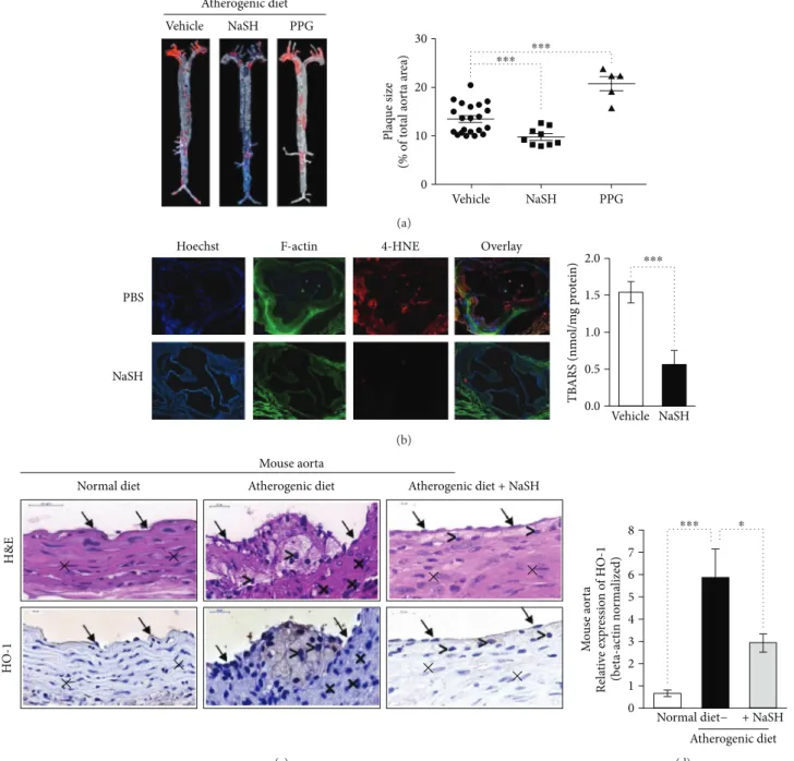
In accordance with previousfindings [17], we observed that administration of NaSH solution reduced, whereas inhibition of CSE activity by DL-propargylglycine (PPG) increased ath- erogenic diet-induced atherosclerotic plaque formation in ApoE−/−mice (Figure 4(a), left and right panels). Therefore, we examined the extent of oxidative injury by performing immunofluorescence staining for the cytotoxic lipid peroxi- dation product 4-hydroxynonenal (4-HNE) in aortic root in a vehicle or NaSH-treated ApoE−/−mice. Strong positive 4-HNE immunostaining was observed in aorta from vehicle-treated mice, whereas 4-HNE level was markedly lower in aorta from NaSH-treated mice (Figure 4(b), left panel). To further confirm that lipid peroxidation was inhib- ited by sulfide, we measured thiobarbituric acid reactive sub- stance (TBARS) content of the aorta derived from a vehicle or NaSH-treated ApoE−/−mice. TBARS content of sulfide- treated aorta was approximately one-third of the vehicle- treated control samples (Figure 4(b), right panel). Since heme oxygenase-1 (CSE) was shown to be induced by lipid hydro- peroxide [25], we also tested whether sulfide alters HO-1 expression in an atherosclerotic mouse model. HO-1 was strongly upregulated in the aorta of ApoE−/−mice fed with atherogenic diet and it was mainly expressed by endothelial cell and macrophages (Figure 4(c)). We found that HO-1 mRNA level was reduced in NaSH-treated mice, presumably due to the attenuated oxidative stress (Figure 4(d)). Next, we examined whether sulfide releasing molecules could prevent lipid peroxidation of lipids derived from human atheromatous lesionsin vitro. We provoked lipid peroxida- tionin vitrowith heme or hemoglobin (Hb) to model intra- plaque hemorrhage. Heme induced a robust increase in lipid peroxides (LOOH) and TBARS content of plaque lipids which was inhibited by all sulfide donors (Suppl. Figures 3A and 3B). Similarly, sulfide-releasing molecules attenuated Hb-mediated formation of both LOOH and TBARS (Suppl.
Figures 3C and 3D). We also checked whether polysulfides and decomposed sulfide donors alter lipid peroxidation in vitro. Neither polysulfide nor decomposed sulfide donors were able to block lipid peroxidation (Suppl. Figure 5).
3.3. Sulfide Inhibited the Formation of Lipid-Peroxidation Products in Human Hemorrhaged Lesions. Next, we exam- ined whether the different H2S-releasing compounds could prevent hemorrhage-mediated lipid-peroxidation upon intraplaque hemorrhage. Human carotid endarterectomy specimens with obvious macroscopic evidence of intraplaque hemorrhage (Suppl. Figure 4A) were homogenized and incu- bated at 37°C in the presence or absence of the sulfide donors (200μmol/L). We determined LOOH and TBARS content of the samples at day 0 and day 4. During the four-day incubation, LOOH content increased about 2.7-fold, whereas the level of TBARS elevated by 2.4-fold (Suppl.
Figures 4B and 4C). The formation of both LOOH and
TBARS were attenuated by the sulfide donors (Suppl.
Figures 4B and 4C). Moreover, AP67 and AP72 were able to decrease LOOH content below the basal LOOH level measured at day 0 (Suppl. Figure 4B).
3.4. Sulfide Donor Molecules Inhibit Oxidative Cross-Linking of Hb Subunits. Covalent cross-linking of Hb occurs in advanced atherosclerotic lesions. To model the oxidative environment present in the atherosclerotic plaque in vitro,
Human carotid artery
Healthy Atheroma Complicated
CSE−
GAPDH−
1.6 1.2 0.8 0.4 0.0
Human carotid artery
Healthy Atheroma Complicated
Human carotid artery
Healthy Atheroma Complicated
⁎⁎⁎
CSEH&EMacroscopic CSE expression (GAPDH normalized density)
⁎
(a)
CSE−
Mouse aorta
Normal diet Atherogenic diet
Mouse aorta
Normal diet Atherogenic diet
Mouse aorta Normal diet Atherogenic diet GAPDH−
1.5
1.0
0.5
0.0
⁎⁎⁎
CSEH&E CSE expression (GAPDH normalized density)
(b) 3
2
1
0
Human carotid artery
Healthy Atheroma Complicated
⁎⁎⁎
ns
Endogenous H2S levels (휇mol/L)
(c)
Figure1: CSE is markedly upregulated in atheromatous lesions. (a, upper panels) Immunohistochemistry detection of the CSE expression in human carotid artery specimens. Macroscopic pictures, H&E, and CSE staining are shown. Magnification of the human histology samples was 150x. Arrows indicate endothelial cells, crosses show smooth muscle cells, and comparison signs mark macrophages. (a, lower panels) Pieces of carotid artery representing the healthy vessel; atheromatous and complicated lesions were selected by macroscopic examination.
Representative Western blot showing CSE expression in carotid artery tissue lysates (20μg/lane) (N= 3, three human carotid artery specimens from three patients in each group). Immunoblots were reprobed with GAPDH. The bar graph shows CSE expression normalized to GAPDH.∗∗∗p< 0 005and ∗p< 0 05compared to healthy samples. (b) ApoE−/−mice were kept on normal chow diet or atherogenic diet for 8 weeks. (b, upper panels) H&E and CSE staining are shown. Magnification of the mice aortas was 700x. Arrows indicate endothelial cells, crosses show smooth muscle cells, and comparison signs mark macrophages. (b, lower panels) Representative Western blot showing CSE expressions of carotid artery samples (10μg/lane) (n= 3, each performed in triplicate). Immunoblots were reprobed with GAPDH. The bar graph shows CSE expression normalized to GAPDH. (c) Endogenous H2S levels in different types of the carotid arteries (N= 6).
we used H2O2(25μmol/L) or oxLDL (50μg/mL) to trigger such oxidative cross-linking of Hb (5μmol/L) in the presence or absence of sulfide donors. Covalently cross-linked Hb spe- cies were subsequently detected by Western blot analysis.
The formations of dimeric, tetrameric, and multimeric Hb species were detected in both H2O2 and oxLDL- treated Hb samples. At a concentration of 200μmol/L, all the sulfide-generating molecules significantly inhibited the formation of covalently cross-linked Hb multimers triggered by either H2O2 or oxLDL (Figures 5(a)–5(d)).
AP67 and AP72 provided complete inhibition against H2O2-induced Hb oxidation, and these donors were also the most effective at inhibiting oxLDL-mediated Hb oxida- tion (Figures 5(a)–5(d)).
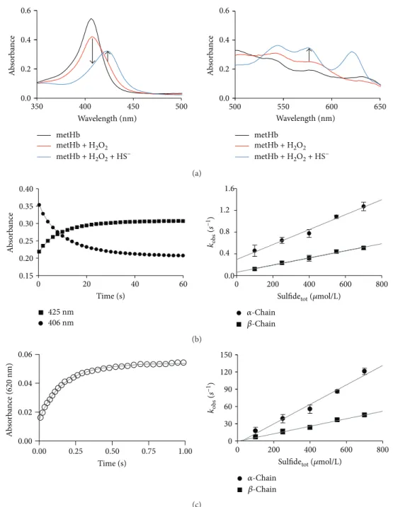
3.5. Reactions of FerrylHb with Sulfide.To test the possibility that the observed protective effects of sulfide donors were due to the reduction of ferrylHb, we next investigated the kinetics of the reaction of ferrylHb species with sulfide in a cell- free system using stopped-flow spectrophotometry. Ini- tially, ferrylHb was prepared in situ by the reaction of metHb with 5 molar equivalents of H2O2 in the first mix- ing cycle of a sequential stopped-flow experiment. Under
our experimental conditions, the formation of ferrylHb was complete in 400 s and resulted in spectral transitions (see the 350–500 and 500–650 nm ranges on Figure 5(a), left and right panels) that were characteristic to the formation of ferrylHb. Therefore, sulfide was reacted with ferrylHb in the second mixing cycle using a delay time of 400 s. Rapid changes in the UV-vis spectra predicted a favorable reaction between sulfide and ferrylHb under these conditions. The appearance of a new peak at 620 nm together with the shift of the Soret band at 400 nm and the absorbance increases at 530 and 580 nm were indicative of the formation of sulfhe- moglobin (Figure 6(a), left and right panels). Kinetic traces were measured at a>10-fold excess of sulfide over ferrylHb to maintain pseudo-first-order conditions and recorded ini- tially at 406, 425, and 570 nm (Figure 6(b), left and right panels). Under these conditions, they exhibited a biexponen- tial character, which most likely corresponded to separated reactions of the alpha and beta chain ferryl heme centers (fas- ter and slower phase, resp.) as shown before. The obtained pseudo-first-order rate constants from the double exponen- tial fits of the kinetic traces at 406, 425, and 570 nm show similar linear correlations with the applied sulfide concentra- tions, indicatingfirst-order dependencies of the rate law on both ferrylHb and sulfide concentrations (Figure 6(b), right panel). Therefore, the apparent second-order rate constants at pH 7.4 were calculated from the slopes of the lines on Figure 6(b), right panel, to be (1.43±0.06)×103M−1·s−1 and (6.5±0.2)×102M−1·s−1 for the alpha and beta chains, respectively. The previous report proposed that related fer- rylHb species are generated in the reactions of oxyHb with H2O2as with metHb [26]. Indeed, we observed similar kinet- ics with sulfide for the ferrylHb species that were produced in the reaction of oxyHb with H2O2as the ones described above for metHb (data not shown).
However, kinetic traces that were recorded at 620 nm (representing the formation of sulfheme Hb) (Figure 6(c), left panel) indicated two orders of magnitude faster rates (also exhibiting double-exponential behaviors with second- order rate constants of (1.7±0.1)×105 and (6.5±0.3)× 104M−1·s−1) (Figure 6(c), right panel). We obtained prelimi- nary kinetic evidence that the relatively slow reactions recorded at 425 nm and the 2 orders of magnitude faster reactions at 620 nm were parallel and not consecutive reac- tions. As mentioned above, ferrylHb represents a mixture of oxidized Hb species, where an unpaired electron is located at different sites of the protein [27]. Therefore, we propose that the observed kinetically isolated reactions were due to different ferrylHb species reacting with sulfide. We are cur- rently investigating the chemical nature of these ferrylHb species, but their characterization is outside the scope of the present study.
3.6. Hb-Lipid Interactions and Subsequent Endothelial Responses Are Attenuated by Sulfide. Interactions between Hb and lipids upon intraplaque hemorrhage result in the formation of reactive lipid mediators as well as oxidized Hb species. These molecules trigger oxidative stress and cell acti- vation, respectively. To investigate whether sulfide could attenuate these harmful reactions, we treated oxidized LDL
Atherosclerotic plaques
Early Complicated
⁎ ⁎ ⁎
⁎ CSE + SMACSE + CD34CSE + CD4
20 휇m 20 휇m
20 휇m 20 휇m
20 휇m 20 휇m
Figure2: Double immunohistochemistry is demonstrating CSE expression in different cellular compartments of early (left) and complicated (right) atherosclerotic plaques of the carotid artery. Upper panels: CSE (DAB, brown color) and CD4 (VIP, violet) highlighting tissue macrophages (arrows); middle panels:
CSE (DAB, brown color) and CD34 (VIP, violet) presenting endothelial cells (arrows); and lower panels: CSE (DAB, brown color) and smooth-muscle actin (VIP, violet) referring to smooth-muscle cells (arrows) and myofibroblasts (asterisk) (×400 magnification).
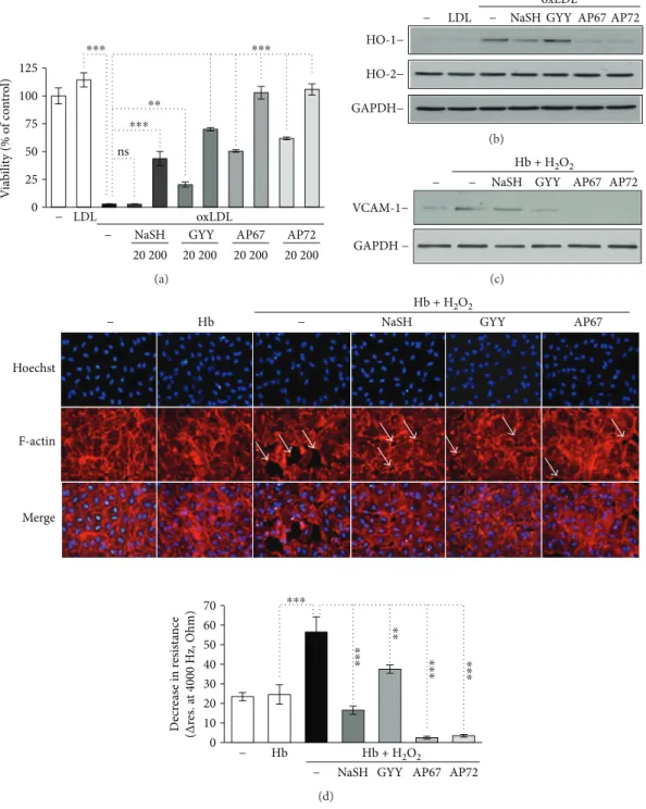
with sulfide donors for 4 hours. Then, HAoECs were exposed to the LDL samples for 6 hours. Oxidized LDL decreased cell viability by about 97% (Figure 7(a)). Pretreatment of oxLDL with sulfide donors at the concentration of 200μmol/L significantly attenuated oxLDL induced to HAoECs (Figure 7(a)). AP67 and AP72 were more potent at inhibiting LDL-induced cytotoxicity presumably because they generate H2S more efficiently than GYY4137 [19] (Figure 7(a)). Suble- thal concentrations of oxLDL induced the expression of heme oxygenase-1 (HO-1), a stress-responsive protein in HAoECs (Figure 7(b)). We found that HO-1 expression provoked by a sublethal dose of oxLDL was suppressed by AP67 and AP72 but not by NaSH and GYY4137 in HAoECs (Figure 7(b)). We also tested the constitutive heme oxygenase-2 (HO-2) expression in these samples and we found that HO-2 level remained unchanged (Figure 7(b)). H2S donors at concentrations we used did not enhance the level of HO-1 and HO-2 in HAoECs (data not shown). Thus, in our system, heme oxygenases were not involved in the cellular responses provided by H2S. Oxi- dized Hb species in particular ferrylHb trigger endothelial cell activation, characterized by elevated expression of adhe- sion molecules and increased intercellular permeability.
Therefore, we next examined whether sulfide donor mole- cules could modulate endothelial responses via inhibiting fer- rylHb formation. Hb was treated with H2O2in the absence or presence of the sulfide releasing molecules. Confluent HAoECs were then treated with the obtained samples, and VCAM-1 expression as a marker of endothelial cell activa- tion, endothelial monolayer resistance, and intercellular gap formation was investigated.
We found that Hb samples treated with H2O2in the pres- ence of slow sulfide-releasing molecules were able to attenu- ate VCAM-1 expression, while NaSH could not diminish it (Figure 7(c)). Then, we investigated the integrity of HAoECs monolayers. Native Hb did not change HAoECs monolayer resistance compared to control cells during the 3-hour treat- ment (Figure 7(d), lower panel), but when HAoECs mono- layers were exposed to oxidized Hb, monolayer resistance
was significantly decreased (Figure 7(d), lower panel), sug- gesting that oxidized Hb provoked intercellular gap forma- tion in accordance with previous observations [11, 15].
Importantly, each sulfide-releasing molecule attenuated the decline of monolayer resistance (Figure 7(d), lower panel) presumably via the inhibition of ferrylHb formation.
Moreover, AP67 and AP72 not only inhibited the oxHb- mediated decrease in monolayer resistance but also increased resistance significantly above the nontreated HAoECs monolayer’s resistance (Figure 7(d), lower panel).
To confirm that the observed decrease in monolayer resis- tance upon treatment of HAoECs with oxidized Hb was due to intercellular gap formation, we performed immuno- staining on cells after oxidized Hb exposure (Figure 7(d), upper panel). Accordingly, in the present studies, endothe- lium exposed to oxHb exhibited gaps between the cells visu- alized by F-actin staining (Figure 7(d), upper panel). As demonstrated in panel E, rearrangement of F-actin cytoskel- eton and subsequently the formation of intercellular gaps were prevented by NaSH, GYY4137, and AP67. We also tested if polysulfides and decomposed sulfide donors could affect endothelial responses. Interestingly, we found that polysulfides, but not decomposed sulfide donors, were able to inhibit oxLDL-induced cell death (Suppl. Figures 6A and 6B). Furthermore, increased expression of HO-1 and VCAM-1 provoked by oxLDL or oxHb at a sublethal dose was not altered by polysulfides and decomposed sulfide donors (Suppl. Figures 6C and 6D).
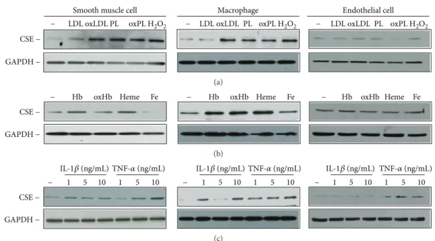
3.7. Atherogenic Lipids and Proinflammatory Cytokines Increased the Expression of CSE in Diverse Cells of the Atherosclerotic Vessel Wall.To identify triggers and cell types responsible for altered CSE expression in the atherosclerotic vessel wall, we next exposed HASMCs, macrophages, and HAoECs to different agonists considered relevant in athero- genesis. First, we examined the effect of lipids, for example, LDL and plaque lipids extracted from human aorta speci- mens with lipid-rich atheromatous plaques. We challenged the cells with native and oxidized forms of these lipids and
Complicated sclerotic plaque 20 휇m
(a)
Myofibroblast 20 휇m
(b)
Figure3: CSE expression in activated myofibroblasts in a cellular area of a complicated sclerotic plaque. (a) SMA+ myofibroblasts (VIP, violet) partially coexpress CSE (DAB, brown staining) in their cytoplasm. Additional CSE labeling can be observed in tissue macrophages (brown only) (×400 magnification). (b) A single enlarged SMA+ fibroblast-type cell with cytoplasmic brown CSE-related colabeling (×1000 magnification).
applied H2O2as a positive control (Figure 8(a)). We found that all the three cell types expressed CSE and that H2O2 exposure upregulated the expression of this H2S synthesizing enzyme in HASMCs and macrophages. Both HASMCs and macrophages responded to oxLDL, native, and oxidized pla- que lipids by upregulating the expression of CSE (Figure 8(a),
left and middle panels). Next, we challenged the cells with different forms of Hb and Hb breakdown products such as heme and iron which are known to accumulate in the athero- sclerotic lesions upon intraplaque hemorrhage [10]. When exposed to Hb, oxidized Hb, or heme, HASMCs and macro- phages upregulated the expression of CSE, whereas iron
Atherogenic diet Vehicle NaSH PPG
30
20
10
0
Vehicle NaSH PPG
⁎⁎⁎ ⁎⁎⁎
Plaque size (% of total aorta area)
(a)
PBS
Hoechst F-actin 4-HNE Overlay
2.0 1.5 1.0
⁎⁎⁎
NaSH 0.5
0.0 Vehicle
TBARS (nmol/mg protein)
NaSH (b)
Mouse aorta
Normal diet Atherogenic diet Atherogenic diet + NaSH
× ×
× ×
×
× ×
HO-1H&E ×
(c)
Mouse aorta Relative expression of HO-1 (beta-actin normalized) 8 ⁎⁎⁎
7 6 5 4 3 2 1
0 Normal diet− + NaSH Atherogenic diet
⁎
(d)
Figure4: Sulfide inhibits lipid-peroxidationin vivoandin vitro. (a, left panel) Representative oil red O staining of aorta derived from ApoE−/−
mice fed with atherogenic diet and treated with NaSH (N= 9), PPG (N= 5), or vehicle (PBS, N= 21) for 8 weeks. (a, right panel) Quantification of atherosclerotic plaque size was performed with ImageJ software. (b, left panel) Representative aortic sections (6μm) stained for F-actin (phalloidin-FITC, green), 4-HNE (red), and DNA (Hoechst, blue) (N= 9). (b, right panel) TBARS content of aorta derived from ApoE−/−mice fed with atherogenic diet and treated with vehicle (N= 9) or NaSH for 8 weeks (N= 9). (c) ApoE−/−mice were fed with regular diet (N= 9) or atherogenic diet for 8 weeks. Parallel with the atherogenic diet, the mice were kept intraperitoneally vehicle (N= 9) or NaSH (N= 9). Immunohistology was showing hematoxylin and eosin and HO-1 staining of mice aorta samples.
Endothelial cells are indicated by arrows, smooth muscle cells were marked by cross, and macrophages were marked by comparison signs.
The magnification of the mice aortas was 700x. (d) Real-time qPCR was showing HO-1 relative expression in mouse aorta. ∗ indicates statistical significance (p< 0 05).∗∗∗ indicates statistical significance (p< 0 0001).
treatment caused a marked decrease of CSE expression in HASMCs, but did not modulate CSE expression in macro- phages (Figure 8(b), left and middle panels). We also examined the upregulation of CSE by various forms of hemoglobin, lipids, and proinflammatory cytokines in HAoECs. CSE expression was not altered in HAoECs exposed to oxHb, oxLDL, and oxidized lipids derived from atheromatous pla- que (Figures 8(a) and 8(b), right panels). To examine the effect of proinflammatory mediators, we next exposed the cells to IL-1β and TNF-α. Figures 8(c) shows that CSE expression was induced in HASMCs and macrophages after cytokine stimulation. On the contrary, CSE level was signif- icantly increased in cells treated with TNF-α as it was observed in HAoECs (Figure 8(c), right panels).
4. Discussion
The oxidative environment in the atherosclerotic lesion triggers lysis of invading erythrocytes and the subsequent release of Hb upon intraplaque hemorrhage [10]. A com- plex interplay exists between atheroma lipids and cell- free Hb that leads to further lipid-peroxidation and Hb oxidation [10–12, 22].
In the reactions between Hb and plaque lipids, different oxidized Hb derivatives are formed (metHb (Fe3+), ferrylHb (Fe4+=O2−)) [10–12], resulting in the covalently cross- linked species. In our current study, we demonstrated that sulfide inhibited oxidative cross-linking of Hb subunits, the hallmark of the ferrylHb formation. These effects could be attributed to the ability of sulfide to reduce LOOH content of oxLDL because LOOH was identified previously as the most cytotoxic lipid-peroxidation product in oxLDL toward vascular endothelium [22]. However, as we argued previ- ously, the antioxidant properties of sulfide in biological
systems are most likely not due to direct scavenging of oxi- dant species, because protein thiols and GSH are present in much larger concentrations compared to sulfide. Hb cross- linking proceeds via ferrylHb intermediate species, those we found to be scavenged rapidly by sulfide, hence providing an alternative molecular model for sulfide-mediated inhibi- tion of Hb oligomerization. The measured rate constants for the reactions of ferrylHb species with sulfide represent at least 1–3 orders of magnitude faster reactions (for the slower reactions recorded at 425 nm and the faster ones at 620 nm, resp.) compared to those with ascorbate or urate [28] that are the previously proposed primary heme- redox-intermediate-scavenging antioxidants (because thiols in most cases have no access to the metal center at the active site). Also, the relatively high stability of the ferrylHb species implies that Hb oligomers must form in much slower reac- tions. Furthermore, the fact that Hb oligomerization was associated with intraplaque hemorrhage suggests a role for ferrylHb-initiated oxidative stress in the underlying molecular mechanisms of the pathophysiology of compli- cated atherosclerotic lesions [10]. Hence, the demonstration of sulfide-mediated reduction of ferrylHb species is a novel observation which could explain at least some of the observed cytoprotective effects of sulfide in our current model and adds to the rapidly growing body of literature detailing H2S-mediated cytoprotection.
Additionally, oxidized Hb species, in particular ferrylHb, induce proinflammatory signaling targeted to the vascular endothelium. By inhibiting Hb oxidation, sulfide donors also attenuated Hb oxidation-associated decrease in endothelial monolayer integrity and intercellular gap formation, as well as the induction of the adhesion molecule, VCAM-1.
Oxidized Hb species release heme, a potent trigger of lipid peroxidation [14, 25]. In our current study, sulfide
100 kDa − Hb
37 kDa − 25 kDa − 15 kDa −
Hb
(a) (c)
(b) (d)
Hb + H2O2
multimers Hb monomer
Hb Hb + oxLDL
120 90 60 30
− NaSH GYY AP67 AP72 NaSH GYY AP67
− NaSH GYY AP67 AP72
120 90 60 30
− LDL − NaSH GYY AP67 AP72
0 Hb 0
Hb Hb + oxLDL
− LDL − AP72
⁎⁎⁎ ⁎⁎⁎
⁎⁎⁎
Multimer formation (% of control) Multimer formation (% of control)
Hb + H2O2
⁎⁎⁎
⁎⁎⁎
⁎⁎⁎
⁎⁎⁎ ⁎⁎⁎
⁎⁎⁎
⁎⁎⁎
⁎⁎⁎
Figure5: Sulfide inhibits the formation of covalently cross-linked Hb multimers. (a) Hb (5μmol/L) was incubated with H2O2(75μmol/L) in the presence or absence of the H2S donors NaSH, GYY4137, AP67, and AP72 (200μmol/L) at 37°C for 90 minutes. Samples (0.5μg Hb) were subjected to SDS-PAGE and Hb species were detected by Western blotting. Representative experiment,n= 3. (b and d) Quantification of the multimer formation was showed. (c) Hb (5μmol/L) was incubated with oxLDL (50μg/ml) in the presence or absence of the sulfide donors NaSH, GYY4137, AP67, and AP72 (200μmol/L) at 37°C for 4 hours. Samples (0.5μg Hb) were subjected to SDS-PAGE and Hb species were detected by Western blotting. Representative experiment,n= 3. ∗∗∗indicates statistical significance (p< 0 0001).
metHb metHb + H2O2 metHb + H2O2 + HS−
metHb metHb + H2O2 metHb + H2O2 + HS− 0.6
0.4 0.2 0.0
350 400 450 500
Wavelength (nm) Wavelength (nm)
500 550 600 650
Absorbance
0.6 0.4 0.2 0.0
Absorbance
(a) 0.40
0.35 0.30 0.25 0.20 0.15
0 20 40 60
Time (s)
1.6 1.2 0.8 0.4 0.0
0 200 400 600 800
Sulfidetot (휇mol/L) 훼-Chain
훽-Chain 425 nm
406 nm
Absorbance kobs (s−1)
(b) 0.06
0.04 0.02 0.00
150 120 90 60 30 0
0.00 0.25 0.50 0.75 1.00
Time (s)
0 200 400 600 800
Sulfidetot (휇mol/L) 훼-Chain
훽-Chain
Absorbance (620 nm) kobs (s−1)
(c)
Figure6: Reactions of ferrylHb with sulfide. (a, left and right panels) Representative UV-vis spectra of 4μmol/L metHb (black line), reaction of 4μmol/L metHb with 8μmol/L H2O2after 400 s (red line) and reaction of 4μmol/L ferrylHb with 100μmol/L sulfide after an additional 60 s (blue line). Arrows indicate wavelength values where the kinetic runs were recorded. (b, left panel) Representative stopped-flow kinetics traces for the reaction of 4μmol/L ferrylHb with 100μmol/L sulfide at 406 nm (circles) and 425 nm (squares) and the corresponding double exponentialfits (solid lines). (b, right panel) Sulfide concentration dependencies of the obtained pseudo-first-order rate constants from the double exponentialfits of the stopped-flow kinetic traces (λ= 425 nm). MetHb (4μmol/L) was reacted with H2O2(8 or 20μmol/L) in the presence of sulfide (100–700μmol/L). The faster (circles) and slower (squares) reactions represent reactions of the ferryl moieties of the alpha and beta chains, respectively. Data points and error bars represent the average and standard deviations of 4 independent experiments. (c, left panel) Representative stopped-flow kinetics traces for the reaction of 4μmol/L ferrylHb with 100μmol/L sulfide at 620 nm (blank circles) and the corresponding double exponentialfit (solid lines). (c, right panel) Sulfide concentration dependence of the pseudo-first-order rate constant for the reaction of ferrylHb with sulfide from the double exponential fits of the kinetic traces, where ferrylHb was generated in the reaction of 8μmol/L metHb with 40μmol/L H2O2in thefirst mixing cycle of a sequential stopped-flow experiment, followed by mixing with sulfide solutions in a 1 : 1 ratio in the second mixing cycle after a 240 s delay time. The faster (circles) and slower (squares) reactions represent the reactions of the ferryl moieties of the alpha and the beta chains, respectively. Data points and error bars represent the average and standard deviations of 3 independent experiments.
ns 125
100
⁎⁎⁎ ⁎⁎⁎
⁎⁎
⁎⁎⁎
75 50 25
0 − LDL oxLDL
− NaSH GYY AP67 AP72
20 200 20 200 20 200 20 200
Viability (% of control)
(a)
oxLDL HO-1−
HO-2−
GAPDH−
− LDL − NaSH GYY AP67 AP72
(b)
Hb + H2O2
− − NaSH GYY AP72
VCAM-1−
GAPDH −
AP67
(c)
⁎⁎⁎ ⁎⁎⁎ ⁎⁎⁎
⁎⁎
NaSH GYY AP67 Hb + H2O2
Hb + H2O2
− Hb − NaSH GYY AP67
Hoechst
F-actin
Merge
70 60 50 40 30 20 10
0 − Hb
− AP72
Decrease in resistance (Δres. at 4000 Hz, Ohm)
⁎⁎⁎
(d)
Figure7: Endothelial responses provoked by Hb-lipid interactions are attenuated by sulfide donors. (a) OxLDL (200μg/mL) was incubated with sulfide donors NaSH, GYY4137, AP67, and AP72 at the concentrations of 20 and 200μmol/L at 37°C for 24 hours. Confluence HAoECs were exposed to the LDL samples for 4 hours then cell viability was determined by MTT assay. Representative experiment,n= 3, each performed in 8 wells in parallel. (b) OxLDL (200μg/mL) was incubated with sulfide donors NaSH, GYY4137, AP67, and AP72 at the concentration of 20 and 200μmol/L at 37°C for 24 hours. Confluence HAoECs were exposed to LDL samples (50μg/mL) for 8 hours.
HO-1 and HO-2 protein expressions were determined by Western blotting. Representative experiment,n= 3. (c–e) Hb (10μmol/L) was incubated with H2O2(50μmol/L) in the presence or absence of sulfide donors NaSH, GYY4137, AP67, and AP72 (200μmol/L) at 37°C for 90 minutes. (c) Confluence HAoECs were exposed to the obtained Hb samples for 8 hours and VCAM-1 expression was determined by Western blotting. Representative experiment,n= 3. (d, lower panel) Confluence HAoECs cultured in ECIS plates were exposed to the obtained Hb samples and transendothelial electrical resistance was monitored by ECIS instrument for 3 hours. Representative experiment, n= 3, each performed in triplicate (d, upper panel). HAoECs grown on coverslips, upon reaching confluence cells were exposed to the Hb samples for 8 hours. Cells were stained for F-actin (phalloidin-TRITC, red) and DNA (Hoechst, blue). White arrows show intercellular gaps. Representative image,n= 5.∗∗ indicates statistical significance (p< 0 001). ∗∗∗indicates statistical significance (p< 0 0001).