0
Differentiation-associated Downregulation of Poly(ADP-Ribose) Polymerase-1 (PARP-1)
Expression in Skeletal Muscle Increases Its Resistance to Oxidative Stress
Gabor Olah
Doctoral School of Sport Sciences University of Physical Education
Consultants: Dr. Zsolt Radak, PhD, DSc Dr. Csaba Szabo, MD, PhD, DSc Official reviewers: Dr. Attila Bacsi, PhD
Dr. Erika Koltai PhD Head of the Final Examination Committee:
Dr. Gabor Pavlik, MD, PhD, DSc Members of the Final Examination Committee:
Dr. Marta Szmodis, PhD Dr. Attila Bacsi, PhD
Galveston, TX, USA 2016
DOI: 10.17624/TF.2016.03
1
Table of Contents
Table of Contents 1
Figures and tables 3
1. Introduction 6
1.1. PARP-1, Poly(ADP-ribose) polymerase 1 6
1.2. Other PARPs 7
1.3. Metabolism of poly(ADP-ribose) 12
1.3.1. Poly(ADP-ribose), pADPr Synthesis 12
1.3.2. Poly(ADP-Ribose) Catabolism: Poly(ADP-Ribose) Glycohydrolase and
others degrades PAR 14
1.4. Biological function of PARP-1 16
1.4.1. Role of PARP-1 in the differentiation and gene expression 16
1.4.2. Role of PARP-1 in DNA repair 19
1.4.2.1. The base excision repair/single-strand break repair process (BER/SSBR) 19 1.4.2.2. Double-strand breaks (DSBs) repair by HR and NHEJ 20 1.4.2.3. PARP-1 in nucleotide excision repair (NER) 23
1.4.3. PARP-1 in mitochondria 25
1.4.4. PARP-1 in cell death 25
1.4.5. Role of PARP-1 in pathophysiology 34
1.5. Oxidative stress, as a trigger of PARP activation 36 1.5.1. The effect of the oxidative/nitrosative stress in the context of PARP 40
1.5.2. Endogenous antioxidant systems 41
1.6. The biology of skeletal muscle 42
1.6.1 Molecular machinery of the contractile force 43
1.6.2. Origin of skeletal muscle 44
1.6.3. The cellular basis of myogenesis in adults 44
1.6.4. Skeletal muscle cells as model system 46
2. Aims 47
3. Materials and Methods 48
3.1. Reagents 48
3.2. Cell culture 48
2
3.3. Preparation of whole-cell extracts and Western blots 48
3.4. MTT viability assay 49
3.5. LDH citotoxicity assay 49
3.6. Measurement of NAD+ levels 49
3.7. Annexin V-phycoerythrin (Annexin V-PE) -7-aminoactinomycin D (7-AAD) staining for apoptosis/necrosis detection by flow cytometry 50 3.8. Bioenergetic analysis in isolated mitochondria 50
3.9. Mitochondrial membrane potential assay 51
3.10. Fluorescence microscopy 51
3.11. PARP-1 silencing by small-interfering RNA and bioenergetic analysis in
PARP-1 silenced cells 52
3.12. Transient transfection of myoblasts with PARP1 52
3.13. Proximity Ligation Assay 53
3.14. Collection of muscle samples from children with severe burn injury 53
3.15. Propranolol treatment 54
3.16. Western blotting for poly(ADP-ribose) (PAR) 55
3.17. Immunohistochemical analysis 55
3.18. PAR immunostaining procedure 56
3.19. Double immunostaining procedure 56
3.20. Statistical analysis 57
4. Results 58
4.1. Myoblast differentiation is associated with downregulation of PARP-1
expression 58
4.2. Differentiated myotubes develop resistance to oxidative stress 61 4.3. Myotubes preserve mitochondrial functions during oxidative stress 68 4.4. Overexpression of PARP1 increases the oxidant sensitivity of myotubes 72 4.5. Oxidative stress leads to an early PARP-1 activation in U937 cells 74 4.6. β-adrenoceptor signaling is involved in PARP-1 activation during H2O2
challenge 74
4.7. The positive effect of PARP inhibition in skeletal muscle biopsies from burn
patients 76
5. Discussion 80
3
5.1. Differentiation-associated downregulation of PARP-1 expression increases
the resistance to oxidative stress 80
5.2. The localization of PARP-1 during oxidative challenge and the effect of propranolol on the PARP-1 activation in U937 and C2C12 cell 83 5.3. Poly(ADP-ribosyl)ation in skeletal muscle tissue of pediatric patients with
burn injury: the positive effect of propranolol 83
6. Future direction 85
7. Conclusion 86
8. Summary 87
9. Összefoglalás 89
10. References 91
11. List of publication related to the dissertation 119 12. List of publication not related to the dissertation 120
13. Acknowledgement 124
Figures and Tables
Figures
Figure 1: Domain organization of the human PARP-1 7
Figure 2: Domain Architecture of Human PARPs 11
Figure 3: Structure of PAR 13
Figure 4: Metabolism of poly(ADP-ribose) 15
Figure 5: Simplified model for the recruitment of repair factors to SSB 20 Figure 6: ATM- and PAR-dependent DNA break recognition 22 Figure 7: Schematic diagram of NHEJ and A-NHEJ after DSBs 23 Figure 8: A model of DDB2- and PARP1-dependent regulation of NER 24
Figure 9: Schematic diagram of necroptosis 28
Figure 10: Hallmark of parthanatos 30
Figure 11: PARP-1 modulates starvation-induced autophagy 32 Figure 12: Similarities and differences in apoptosis, necrosis, autophagy and
parthanatos 33
Figure 13: The mitochondrial electron transport chain produces ROS 38
4
Figure 14: ROS and RNS production in the peroxisome 39
Figure 15: Longitudinal section of frog sartorius muscle showing the overlapping thick and thin filaments 43
Figure 16: Mammalian skeletal myogenesis 44
Figure 17: PARP1 level is reduced in myotubes 59
Figure 18: Expression of PARP-1 is reduced in differentiated L6 and U937 cells 60 Figure 19: Lack of the contact inhibitory effect on PARP-1 expression in myoblasts 61 Figure 20: Effect of PARP inhibitor PJ34 on cell viability 62 Figure 21: PARylation induced by oxidative stress is reduced in myotubes 63 Figure 22: Myotubes are resistant to oxidant-induced loss of cell viability 66 Figure 23: Inhibition of PARP-1 reduces subpopulations of apoptotic and necrotic cells
induced by H2O2 67
Figure 24: Cellular bioenergetics of mitochondria isolated from myoblasts and
myotubes 69
Figure 25: PARP-1 protein levels in mitochondrial and nuclear fractions of myoblasts
and myotubes 70
Figure 26: Silencing PARP-1 increases both oxidative phosphorylation and glycolytic
activity of C2C12 myoblasts 71
Figure 27: PARP-1 overexpression sensitizes C2C12 myotubes to oxidative stress 73 Figure 28: Auto-PARylation of PARP1 in the extranuclear and nuclear compartments in
U937 cells during oxidative stimuli 74
Figure 29: Propranolol regulates PARP activation in U937 and C2C12 cells during
oxidative stress 75
Figure 30: Representative PAR Western blots are shown from homogenates of skeletal
muscle biopsies of control patients 76
Figure 31: PAR localized primarily to vascular endothelial cells and mononuclear cells 77 Figure 32: Time course of PARP activation in skeletal muscle biopsies of pediatric burn
patients and reduction of PARP activation in skeletal muscle biopsies of pediatric burn patients treated with propranolol 78
5 Tables
Table 1.: Reactive oxygen and nitrogen species 37
Table 2.: Demographic Characteristics of the Study Groups 54
6
1. Introduction
1. 1. PARP-1, Poly(ADP-ribose) polymerase 1
PARP-1, Poly(ADP-ribose) polymerase 1, is the major isoform of the poly (ADP- ribose) polymerase family, consisting of 17 proteins in humans and 16 in mice. It is a constitutive nuclear and mitochondrial protein with well-recognized roles in various essential cellular functions, such as deoxynucleic acid (DNA) repair, signal transduction, different cell death pathways, and a variety of pathophysiological conditions including burn, sepsis, diabetes and cancer. Most of the functions mediated by PARP are related to oxidative stress. Activation of PARP-1 in response to oxidative stress catalyzes the covalent attachment of the poly (ADP-ribose) (PAR) groups on itself and other acceptor proteins, utilizing nicotinamide adenine dinucleotide (NAD+) as a substrate. Overactivation of PARP-1 depletes intracellular NAD+, influencing mitochondrial electron transport, cellular adenosine triphosphate (ATP) generation and, if persistent, can result in necrotic cell death.
PARP-1 has three functionally distinct domains: the N-terminal DNA-binding domain (DBD) responsible for PARP-1’s interaction with DNA breaks, which are also found in DNA ligase III. The automodification domain (AD) contains the BRCT motif, which is common in DNA repair and cell-cycle proteins, constituting the major protein interface with various nuclear partners, and the C-terminal with the catalytic domain (CAT) showing common structural features with the active site of bacterial (ADP-ribosylating) toxins (Diphteria toxin or Pertussis toxin), notably the NAD-fold (PARP signature) (Langelier et al., 2008) (Figure 1). The primary structure of the enzyme is highly conserved in eukaryotes (human and mouse enzyme have 92% homology at the level of amino acid sequence) with the catalytic domain showing the highest degree of homology between different species; the catalytic domain contains or so-called PARP signature is a 50-amino acid-containing block showing 100% homology between vertebrates.
7
Figure 1. Domain organization of the human PARP-1 (Langelier et al., 2008). PARP-1
composed of 3 main domains, DNA-binding (DBD, residues 1-374), Modification (AD, residues 375-525) and Catalytic (CAT, residues 526-1014) domains. DBD contains three Zn-fingers (Zn1, Zn2 and Zn3), a nuclear localization signal (NLS) and a caspase cleavage site (C3). Modification domain has the BRCT fold (BRCA1 C terminus) with auto-modification property. Catalytic domain has the conserved PARP signature responsible for NAD+ binding..
1. 2. Other PARPs
Based on the comparative analysis of their catalytic domain homology, 17 mammalian proteins were named PARPs (Ame et al, 2004), although the nomenclature has been changing in recent years, and new classifications arise based on structural and enzymatic characterization (Hottiger et al, 2010).
Three-dimensional structures of the catalytic domain of chicken PARP-1 and mouse PARP-2 showed structural homology with the active site of the bacterial ADP ribosylating toxin from Corynebacterium diphtheria (Collier, 2001). Recently, the β−α- loop-β−α NAD-fold (which is the most conserved region in PARP-1 orthologous, and is considered the PARP signature) was used to search the non-redundant protein database from the National Center for Biotechnology Information (NCBI) for human PARP homologues. Novel putative PARPs were identified, and this increased the number of PARP-family members to 17.
PARP-2 was discovered as a residual PARP activity in embryonic fibroblast derived from PARP-1 deficient mice (Shieh et al., 1998; Ame et al., 1999). It has a molecular weight of 65kDa and it contains 570 amino acids. Crystal structure is very similar to the PARP-1, except for differences in the vicinity of the acceptor site that reflect differences in terms of substrates. It is a nuclear, DNA-dependent member of the PARP
8
family and interacts with PARP-1 and shares common partners involved in Single Strand Brake Repair (SSBR) and Base Excision Repair (BER). PARP-2 deficient mice show a delay in DNA-strand breaks resealing similar to that observed in PARP-1 deficient cells. (de Murcia et al. 2003; Schreiber, 2002). Knockout animals for both genes die in the early stage of embryogenesis, which shows the importance of these two genes.
PARP-3 is a mono-ADP ribosylase, a core component of the centrosome located at the daughter centriole throughout the cell cycle (Augustin et al., 2003). Its E and F domains share a 61% homology with PARP-1. It has two splice variants, 533 and 540 amino acid long, respectively, with a molecular weight of 67kDa. It was reported that PARP-3 is not activated upon binding to DNA (Loseva et al., 2010). PARP-3 interacts with PARP-1 suggesting a link between the DNA-damage surveillance network and the mitotic fidelity checkpoint.
VPARP (PARP-4), the largest of the PARP family with a molecular weight of ~200 kDa, possesses a mono-ADP-ribosylation feature. It is a part of the Vault particles, which are ribonucleoprotein (RNP) complexes containing small RNAs and proteins.
One of the Vaults proteins (identified by the molecular tool of yeast two-hybrid system) showed homology with the PARP family members. It also localizes in the nuclei, is associated with the mitotic spindle, and is able to carry out an ADP-ribosylation reaction (Kickhoefer et al., 1999). VPARP4-/- mice show higher sensitivity to carcinogen-induced tumorigenesis, suggesting a possible role in drug-induced abnormal tissue grow (Raval-Fernandes et al., 2005).
PARP5a or Tankyrase-1 is a 142 kDa molecular weight protein containing 1327 amino acids. It was named by Smith and colleagues as TRF-interacting, ankyrin-related ADP- ribose polymerase (Smith et al., 1998). It has a nuclear origin, but also can be found in the cytosol and its activity is modified by mitogen-activated protein kinases (MAPK).
Tankyrase-1 binds to TRF-1, a telomere-specific DNA binding protein, and was found to have PARP activity, which raises the question whether or not telomers are under the control of ADP-ribosylation. It was shown that is required for the polymerization of mitotic spindle-associated PAR localizing to mitotic spindle poles. Tankyrase-1 ribonucleic acid interference (RNAi) in HeLa cells showed spindle defect in a PAR concentration dependent manner (Chang et al., 2005).
9
PARP-5a or Tankyrase-2 is a 127 kDa protein structurally similar to tankyrase-1, except that it misses the N-terminal HPS domain (His, Pro, Ser residues). It possesses PARP activity, binds to TRF-1 and IRAP, as TNK-1 shares overlapping functions (given that TNK-1 and TNK-2 proteins associate and share most of their protein partners) (Sbodio et al. 2002). Knockdown experiments for TNK-2 showed no changes in telomerase capping and length, but the smaller size of the animals indicate a metabolic disturbance, proving the enzyme role in metabolism (Hsiao et al., 2006).
TiPARP or PARP-7 was found when analyzing the expression patterns of messenger RNAs (mRNAs) after 2,3,7,8-tetrachlorodibenzo-p-dioxin treatment. It contains 657 amino acids and has a ~75kDa molecular weight, showing similarity to PARP-1 catalytic domain with catalytic activity on histones. It is a mono-ADP-ribosylase and co-localized in the nucleus with AHR (aryl hydrocarbon receptor), a transcription factor activated by synthetic hydrocarbons and some endogenous ligands. MacPherson and colleagues found that TiPARP is a transcriptional repressor of AHR (MacPherson et al., 2013). It was also found to have a role in regulating gluconeogenesis through AHR, ADP-ribosylating PEPCK (phosphoenolpyruvate carboxykinase) with NAD+ consumption, and decreased SIRT1 activity of PGC1α (Diani-Moore et al., 2013).
Treatment with dioxin caused lethality in TiPARP -/- mice by day 5, with increased level of steatosis and toxicity in liver, while TiPARP +/+ mice survived the 30 day treatment period (Ahmed et al., 2015).
Other members of the PARP family not yet mentioned above can be categorized as CCCH-type PARPs, Macro PARPs and other PARPs. The group of CCCH-type PARPs contains PARP-12 and -13, and the already reviewed TiPARP above. They share a common CCCH-like zing finger, WWE (Tryptophan: W; Glutamate: E amino acids) and the catalytic domain. One of the members, PARP-13, has two isoforms. The shorter one in terms of size, which lacks the PARP domain, was identified as a binding protein to viral ribonucleic acid (RNA) and called ZAP. Screening mammalian cDNA libraries revealed CCCH-like zing finger protein inhibiting viral replication (Gao et al., 2002).
Later, in another study reported by the same group that analyzed the role of zinc fingers of PARP-13 in viral activity, the 2nd and 4th zing finger motif were shown to have major decreasing activity of viral infection in the cytoplasm, while the 1st and 3rd have minor (Guo et al., 2004).
10
Macro domain PARPs or BAL PARPs, PARP-9,-14,-15 (BAL 1, 2, 3), belong to this group, having a so-called macrodomain attached to their PARP domain, 2, 3, and 1 respectively. Macrodomains are highly conserved domains throughout the animal kingdoms, as they can be found at the C-terminus of macro-H2A histone proteins and in several non-structure proteins of ssRNA viruses. They are also found in the family of proteins in bacteria, archaebacteria, and eukaryotes, suggesting a ubiquitous cellular role. They have the capability of binding PAR. PARP-9 was described using a differential display method from diffuse large B-cell lymphoma (DLB-CL) patients. It is a ~88kDa protein that is highly expressed in fatal high-risk DLB-CLs, compared to the cured, low-risk group. The high-risk group cells also show higher rates of migration capability, indicating a PARP-9 role in the disease's malignancy (Aguiar et al., 2000).
Apoptosis inhibition is an important step in B-lymphoid oncogenesis, and elevated macro PARP corresponds to hostility. PARP-14 is a binding partner of Stat6, the IL-4- induced (interleukin-4) transcription factor, and mediates protection against apoptosis in IL-4 treated cells (Cho et al 2009.). The signaling pathway goes through AMPK (AMP- activated protein kinase), a cellular energy sensor, as enhancement restored the glycolytic activity and the survival signaling in PARP 14-/- B cells (Cho et al., 2011). It possesses a cell motility and focal adhesion function, namely that PARP-14 has a role in focal adhesion turnover, and is localized at the end of actin stress fibers (Vyas et al., 2013).
Other PARPs include PARP-6, PARP-8, PARP-10, PARP-11, PARP-16. Their function is still relatively unknown. PARP-6 is a mono-ADP-ribosyl-transferase found in colorectal cancer specimens. It is thought to be a tumor suppressor (Tuncel et al., 2012).
PARP-10 inhibits c-myc, a transcriptional regulator deregulated in cancers, and has a role in cell proliferation (Yu et al. 2005). Its activity depends on phosphorylation by CDK-2-cyclin E in vitro (Chou et al., 2006). It was shown to be a mono-ADP- ribosylase and able to regulate IL-1β- and TNF-α-dependent NFκB signaling (Verheugd et al., 2013). It interacts with PCNA, and is responsible for DNA damage sensitivity (Nicolae et al. 2013). PARP-16's crystal structure was revealed by Karlberg (Karlberg et al., 2012) and showed auto-mono-ADP-ribosylating activity. The enzyme is required during endoplasmatic reticulum stress activating stress sensors, PERK and IRE1α by ADP-ribosylating, these sensors, and itself (Jwa M et al., 2012).
11
The domain structure of PARPs is summarized in Figure 2, representing the new ARTD (ADP-ribosyltransferase diphteria toxin-like) nomenclature proposed by Hottiger and colleagues (Hottiger et al., 2010) according to the International Union of Biochemistry and Molecular Biology (IUBMB).
Based on the domain structure of the PARP family, different interactions are possible in different cell compartments. Moreover, PARP-1 interacts with PARP-2 or PARP-3 and Tankyrase 1 and 2 (Cook et al., 2002), widening the possibilities of their biological functions.
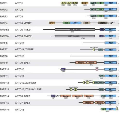
Figure 2. Domain Architecture of Human PARPs (Barkauskaite et al., 2015). PARP1–
16 with alternative names listed on the side domain structures. ARTD, ADP-ribosyl transferase diphtheria type; vPARP, vault PARP; TNKS, Tankyrase; TIPARP, TCDD inducible PARP; BAL, B cell aggressive lymphoma; ZC3HDC1, zinc finger CCCH- type domain containing 1; ZC3HAV1, zinc finger CCCH-type antiviral 1; ZAP, zinc finger antiviral protein. Structural information availability of specific domains is indicated by PDB code under the specific domains. The number of amino acids
12
composing the protein is indicated on the right. The following domain names were used: ZnF, zinc finger; BRCT, BRCA1 C-terminal; WGR, conserved Trp-Gly-Arg motif domain; HD, helical domain; ART, ADP-ribosyl transferase; VIT, vault protein inter-alpha-trypsin; vWFA, von Willebrand type A; ANK, ankyrin; SAM, sterile alpha motif; CCCH ZnF, CCCH type zinc finger; WWE, three conserved residues W-W-E motif domain; Macro, macrodomain; RRM, RNA recognition motif; UIM, ubiquitin interaction motif; TM, transmembrane motif.
1. 3. Metabolism of poly(ADP-ribose)
1. 3. 1. Poly(ADP-ribose), pADPr Synthesis
Poly(ADP-ribosylation) is a covalent, reversible modification by PARPs attaching poly- ADP-ribose on a variety of target proteins, using NAD+ as the substrate (Figure 3). It is a conserved process among metazoa.
In 1963, historically speaking, Chambon published that there is an enzymatic activity responsible for the synthesis of polymers (PAR) that requires NAD+. Namely, it was shown that 14C-adenine-labeled ATP incorporated into an acid-insoluble fraction in a nuclear extract (Chambon et al., 1963). That was the pioneer work that sparked interest from different laboratories and outgrew a competitive field of research.
13 Figure 3. Structure of PAR (Kraus and Lis, 2003).
The synthesis of PAR polymers consists ot the following steps: (a) initiation, (b) elongation, and (c) branching. PAR is attached to the glutamate residues of proteins on the position labeled 1. The covalent bonds between ADP-ribose units are made via glycosidic ribose-ribose 1″ → 2′ bonds at the positions labeled 1 and. The red and blue arrows represent the cleavage sites for PARG and ADP-ribosyl protein lyase, respectively.
pADPr is a homopolymer of ADP-ribose, and is linked by glycosidic 1"-2"bonds reaching up to 200 units. The first step is the initiation -- attaching mono-ADP-ribose to acceptor proteins, including PARP-1. In the elongation step, the mono-ADP-ribose units are added to the chain, reaching up to 20-50 units. Further, ADP-ribosylation may occur by branching of the polymer showing similar structure to ribonucleic acid (RNA) or DNA which was proven by the fact that antibody against pADPr recognized RNA and DNA.
In resting cells, the acceptor proteins are mono- or oligo--ADP-ribosylated, while in stress conditions, such as under nuclear genotoxic stimuli, proteins are poly-ADP- ribosylated. The degradation also depends on the stimuli, as stressors can trigger the degradation in a significantly quicker manner than in resting cells.
There are numbers of proteins as targets for PARylation, such as RNA/DNA polymerases, Topoisomerase II., p53, PCNA just to name a few. Adding ADP ribose
14
units to a protein changes the protein net charge, which results in modified function.
Usually this prevents the interaction of a DNA-binding capability, or if it occurs near the catalytic site of an enzyme, may result activity-modifying property (D'Amours et al.
1999).
Looking at the PARP structure, there are elements responsible for the PARylation. The N-terminal DNA binding domain (DBD) contains two zinc fingers (F1, F2) that underlie the structural basis for DNA strand break detection. Upon a single strand break, both zinc fingers participate in the PARP activation, although PARP activation by double-stranded breaks depends upon the F1 domain closer to the N-terminus (Ikejima et al., 1990, Gradwohl et al., 1990, Langelier and Pascal 2013). There is a third zinc finger that can be found within the DBD, and is necessary for the coordination of interaction with the catalytic domain upon DNA binding, which forms a head-to-tail structure of the homodimer (Langelier et al., 2008).
A discovery was made regarding PAR-binding proteins that revealed 20 amino acid motifs with basic and hydrophobic/basic residues. The proteins identified share a common function, as they participate in maintaining genomic stability. The proteins are as follows: p53, p21CIP1/WAF1, xeroderma pigmentosum group A complementing protein, MSH6, DNA ligase III, XRCC1, DNA polymerase ε, DNA-PKCS, Ku70, NF- κB, inducible nitric oxide synthase, caspase-activated DNase, and telomerase (Pleschke et al., 2000). Another approach, MALDI-TOF MS technology (matrix-assisted laser desorption-ionization time-of-flight mass spectrometry) was utilized for identifying pADPr binding proteins, resulting in 30 nuclear proteins as well-- mostly hnRNPs (heterogeneous nuclear ribonucleoproteins), proteins that bind RNA in the nucleus and have a role in RNA maturation and translocation (Gagne et al., 2003).
1. 3. 2. Poly(ADP-Ribose) Catabolism: Poly(ADP-Ribose) glycohydrolase and others degrades PAR
Mono-and poly-ADP-ribose units can be removed by PAR-removing enzymes (Figure 4). A prediction was made that the lifetime of the PAR polymer is less than 1 minute, which indicates a strictly controlled regulation of the synthesis and degradation (Virag and Szabo, 2002). The principal member of the enzymes is Poly(ADP-Ribose)
15
glycohydrolase (PARG), which was discovered in the 70s (Miwa and Sugimura, 1971;
Miwa et al., 1974). PARG is a product of a single gene on chromosome 10. There are the full-length nuclear PARG-111, PARG-102 and the cytosolic PARG-99. There is a PARG-60 with nuclear and mitochondrial localization. PARG-55 is an alternative splice variant of PARG-60 localized in mitochondria (Meyer-Ficca et al., 2004 and Meyer et al., 2007).
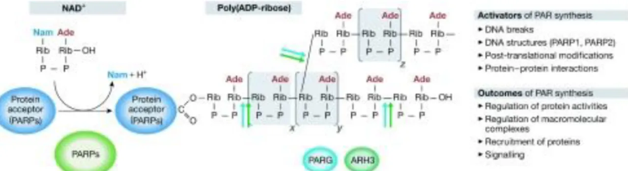
Figure 4. Metabolism of poly(ADP-ribose) (Hakmé et al., 2008). PARPs hydrolyse NAD+ and catalyse adding ADP-ribose units to glutamate residues of either acceptor proteins (heteromodification) or themselves (automodification). x, y and z labels represent values from 0 to >200. PARG and ARH3 can both hydrolyse PAR at shown positions; activators and outcomes of PAR synthesis are also noted on the right. Ade, adenine; ARH3, ADP-ribosyl hydrolase-3; Nam, nicotinamide; PAR, poly(ADP- ribose); PARG, poly(ADP-ribose) glycohydrolase; PARP, poly(ADP-ribose) polymerase; Rib, ribose.
Primarily, the degradation of poly(ADP-ribose) is carried out by PARG, and has a central role of the catabolism. Inactivating the gene causes lethality in early embryonic age mice (Koh et al., 2004). Using the lentiviral system elegantly presented by Erdelyi et al., it has been shown that PARG serves as an apoptosis to necrosis switch during severe oxidative stress. (Erdelyi et al., 2009).
A study reported by Oka showed an alternative enzymatic activity for the degradation of poly(ADP-ribose), with a similar reaction to PARG by a 39kDa enzyme called poly(ADP-ribose) hydrolase or ARH3, which shares amino acid identity with the catalytic domain of PARG (Oka et al., 2006).
16
1. 4. Biological function of PARP-1
1. 4. 1. Role of PARP-1 in the differentiation and gene expression
Differentiation is the process by which a cell changes from one cell type to another.
Usually it occurs when a less specialized type becomes more specialized to fulfill specific tasks, so they gain new functions and lose others. This strictly controlled process requires concerted gene activation and repression, and results in differentiation into specialized cells. For example, myosatellite cells or satellite cells are precursors of skeletal muscle cells and upon activation re-enter the cell cycle, start proliferation and differentiate to myotubes. Furthermore, many fully differentiated cell types such as lymphocytes, fibroblasts, and hepatocytes retain the ability to proliferate, such as in the course of immune response, wound healing, or liver regeneration, respectively.
Experimental data show the differentiation-modifying effect of PARP, but it is important to note that the effect is cell-specific. Inhibition of PARP has been shown to interfere with the differentiation of human granulocyte-macrophage progenitor cells to the macrophage cells (Francis et al., 1983). Overexpression of PARP arrested MB4 cells and blocked all trans-retinoic acid-induced neutrophilic differentiation (Bhatia et al., 1995). Benzamide PARP inhibitors induced melanogenesis and differentiation of melanoma cells (Durkacz et al., 1992). Moreover, PARP-1 has a role in angiogenesis (Caldini et al., 2011; Pyriochou et al., 2008) and adipocyte differentiation (Erener et al., 2012). Also, after DNA damage, it is of primary importance to stop replications at certain checkpoints to allow for the repair of DNA damage.
Co-purification techniques revealed the interaction of PARP-1 with replication factors such as DNA polymerase, and DNA primase, DNA helicase, DNA ligase, topoisomerases I and II and key components of multiprotein replication complex (Simbulan-Rosenthal et al., 1996; Dantzer et al., 1998; Bauer et al., 2001).
PARP-1 has been shown to function in various aspects of the transcription process through a variety of mechanisms, including roles as a modulator of chromatin, a coregulator for DNA-binding transcription factors, and a regulator of DNA methylation.
Chromatin is a protein–DNA complex, the structural base of information coded, that comprises genomic DNA, core histones (i.e., H2A, H2B, H3, and H4), linker histones
17
(e.g., H1), and other chromatin-associated proteins. The basic repeating units of chromatin are the nucleosome (i.e., 146 base pairs of DNA wrapped around a core histone octamer with two copies of each core histone) and the chromatosome (i.e., nucleosome, plus 20–40 base pairs of linker DNA and linker DNA-binding proteins, such as H1) (Widom, 1998; Wolffe and Guschin, 2000). One possible way PARP-1 regulates chromatin structure and transcription is the PARylation of proteins bind to chromatin and the auto-PARylation of PARP-1, which affects the structure of chromatin. Earlier studies showed that PARP-1 promotes decondensation, and the PARylation of H1 was proposed as the underlying mechanism (Poirier et al., 1982). In 2004, Kim and colleagues showed (Kim et al., 2004) that PARP-1 and H1 compete for binding to nucleosomes. PARP-1 depletion by RNAi increases the binding of H1 at many sites of the genome (Krishnakumar et al., 2008; Krishnakumar and Kraus, 2010), suggesting contribution to the dynamic regulation of gene expression. A considerable body of evidence suggests that PARP1 directly interacts with core histones (D’Amours et al., 1999; Pinnola et al., 2007). Histones H3 and H4 are preferential targets for PARP1 binding in vitro, and they control the enzymatic activity of PARP1 (Pinnola et al., 2007; Kotova et al., 2011). There are a number of studies showing that histone variants provide the physical link between PARP-1 and chromatin. H2Ax has been shown to be phosphorylated at the site of DNA damage, which pADPribosylated by genotoxic treatments. Chemical inhibition of PARP-1 triggers the accumulation of phosphorylated H2Ax (Bryant et al., 2005). Also, PARP-1 and H2Ax interaction, outside of DNA repair, was shown during rat spermatogenesis with high levels of pADPr (Meyer-Ficca et al., 2005). H2Az, another histone variant highly homologous to H2Ax, is considered as an interacting partner of PARP-1.
Histone variant H2Av is a functional homologue of H2Ax in Drosophila, and combines the function of H2Ax and H2Az proteins (Leach et al., 2000 and Madigan et al., 2002).
Effective repair of double strand breaks by homologous recombination requires phosphorylation of Drosophila H2Av (acetylated by Tip60), which is followed by chromatin remodeling involving un-phosphorylated and phosphorylated H2Av exchange (Kusch et al., 2004). In this model, (1) a complex of H2Av and PARP1 is activated by DNA break formation; (2) phosphorylation of H2Av triggers PARP1 enzymatic activity; (3) activated PARP1 modifies H1 histone with pADPr, which
18
removes it from damaged loci; (4) the Tip60 complex remodels local chromatin, and facilitates entry of DNA repair complexes; and, finally, (5) Tip60 removes Ser137- phospho-H2Av from repaired chromatin (Thomas and Tulin, 2013).
It is known that transcription induced during steroid response or the response of stress- activated genes, such as hsp70, is accompanied by a local loosening of the chromatin structure that manifests in polytene chromosomes as ‘‘puffs’’ at the site of transcription.
After hormone treatment, infection, and heat shock stress, pADPr polymers accumulate at the target loci. As a result of PARP-1 action, chromatin loosens, and gene transcription takes place. But how is gene expression induced? What is the role of PARP-1 in the transcription machinery?
The mode of action by PARP-1 is the modification of protein by: a) the covalent attachment of pADPr polymers; or b) non-covalent interaction. Gagne represents a proteome-wide identification of pADPr binding proteins, which are classified into 6 groups: DNA repair, DNA replication, cell cycle, chromosome organization/biogenesis, protein synthesis, and mRNA metabolism (Gagne et al., 2008). Evidence suggests that certain proteins possess pADPr-binding domains, specifically a 190 amino acid module known as macrodomain (Karras et al., 2005) and a novel zinc finger motif (PBZ), which present in many DNA damage and checkpoint proteins (Ahel et al., 2008).
Protein shuttling by PARP-1 and pADPr is also part of the process of loosening the chromatin structure. The process of shuttling, or pulling proteins from nucleic acid, is a paramount mechanical process in the dynamic, complex environment of the nucleus.
The role PARP1 plays in numerous nuclear activities stems from this fundamental event by which PARP1 covalently or noncovalently modifies proteins to dissociate from nucleic acid. It is believed that PARP’s broad role in shuttling proteins from nucleic acid is proceeded by two non-mutually exclusive events: electrorepulsion between the highly anionic poly-ADP-ribose and DNA/RNA, and the stealing/masking of DNA/RNA binding domains (D’Amours et al., 1999). Electrorepulsive shuttling occurs based on the repulsive force of similar charges, namely that increasing auto(ADP)ribosylation increases the repulsion between PARP and DNA. Removing PAR units by PARG increases the affinity of PARP to DNA (Ferro and Olivera, 1982;
Zahradka and Ebisuzaki, 1982).
19
1. 4. 2. Role of PARP-1 in DNA repair
PARP-1 is a key mechanism in maintaining the stability and integrity of the cell DNA.
There are constant intracellular (normal metabolism byproducts) and extracellular damaging sources (radiation, chemical agents, heat shock etc.), which might modulate the chemical status of a cell effecting its DNA. These could subsequently cause mutations, chromosomal aberrations, or cell death. Based on its DNA binding ability, it is presumed that PARP-1 is involved in DNA repair. Inhibitors led to some conclusions that PARP-1 is involved in the repair processes. PARP-1 inhibition sensitizes cells to genotoxic stimuli (Küpper et al., 1995; Ding and Smulson, 1994), although PARP inhibitors have limitations due to specificity. PARP knockout mice gave a better insight generated by the de Murcia’s laboratory. PARP -/- mice are viable and fertile with normal phenotype, but are highly sensitive to ionizing radiation and alkylating agents (de Murcia et al., 1997).
1. 4. 2. 1. The base excision repair/single-strand break repair process (BER/SSBR)
DNA single-strand break repair is a primary cellular pathway for repairing damaged bases or single-strand breaks. In this mechanism DNA glycosylases, AP (apuric/apyrimidic) endonucleases, DNA polymerases, DNA ligases (I., III.), XRCC1 are recruited to the damaged sites, which are repaired by either short patch (one nucleotide) or long-patch repair mechanisms.
Investigating wild type and PARP-1 deficient cell extracts gave insight into the involvement of PARP-1 in repair. PARP-1 interacted with DNA polymerase β giving a striking consequence. Cell extract from PARP-1 deficient cells failed in the Long Patch Repair and showed moderate effect in Short Patch Repair (Dantzer et al., 2000).
Another study showed the involvement of PARP-1 in connection with aprataxin-1 in single strand break, a repair protein that was found to be defective in a neuro degenerative disorder (ataxia oculomotor aprataxin-1). The two proteins showed a concerted mechanism in the recruitment of the repair process in SSB (Harris et al., 2009). Interaction of condensin, another performer of the chromatin structure with
20
PARP-1 along XRCC1 was identified as an SSB-specific response during DNA damage (Heale et al., 2006) (Figure 5).
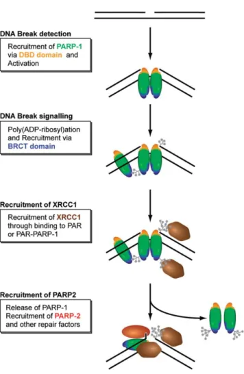
Figure 5. Simplified model for the recruitment of repair factors to SSB (Mortusewitz et
al., 2007). DNA binding domain of PARP-1 detects single-strand breaks and poly(ADP-ribosyl)ation occurs leading to chromatin relaxation. Further poly(ADP- ribosyl)ation at DNA lesions releases PARP-1 enabling other factors to be recruited such as XRCC1.
1. 4. 2. 2. Double-strand breaks (DSBs) repair by HR and NHEJ
Double-strand breaks (DSB) have the most serious consequences. DSB can be provoked by ionizing radiation, anti-cancer drugs or even by reactive oxygen/nitrogen species.
Nature evolved two mechanisms that step in when this type of damage develops. The
21
two major mechanisms involved are homologous recombination (HR) and non- homologous end joining (NHEJ). HR is utilized during S and G2 phase; it uses the sister chromatid as a template. It requires MRN complex (Mre 11-Rad50-Nbs1), Exo1, RPA, Rad 51, 54. NHEJ (which are referred to as "toxic" or mutagenic), can be divided into classical/canonical-NHEJ and alternative-NHEJ depending on the involvement of KU70-KU80 complex.
HR takes place when the NHEJ pathways are suppressed; HR requires PARP-1 proved by a study where chicken DT40 cells lacking PARP-1 have reduced level of HR capability (Hochegger et al., 2006). Ataxia telangiectasia mutated (ATM) is a DNA damage-responding kinase induced by the DNA damage. ATM phosphorylates the proteins required for DNA damage response and repair, including proteins of MRN (Mre11/Rad50/NBS1) complex, p53, SMC1 and histone variant H2Ax upon homologous recombination. ATM was shown to physically interact with PARP-1 (Haince JF et al., 2007) (Figure 6).
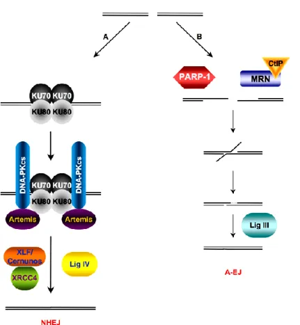
NHEJ can be divided into classical-NHEJ and alternative-NHEJ, depending on the active participation of KU70-KU80. Mostly, C-NHEJ occurs in cells, but A-NHEJ takes place to a lesser degree when C-NHEJ fails or factors are not available. In the C-NHEJ, KU70-KU80 complex is formed after DSB, which recruits DNA-PK phosphorylating subsequent proteins participating in the repair. It was shown that PARylation has effect on the kinase activity in vitro (Ruscetti et al., 1998). XRCC4-Lig IV catalyzes the ligation of DNA ends.
When C-NHEJ fails, A-NHEJ (A-EJ) takes over the repair. There is a competition between PARP-1 and KU proteins for the DNA. If KU is not available, PARP-1 is recruited and plays an active role in the A-NHEJ. KU80-/- cells highly depend on PARP-activity for the repair process, proving PARP-1 has a critical role in this alternative mechanism. Ligation needs the presence of XRCC1-LigIII (Figure 7).
22
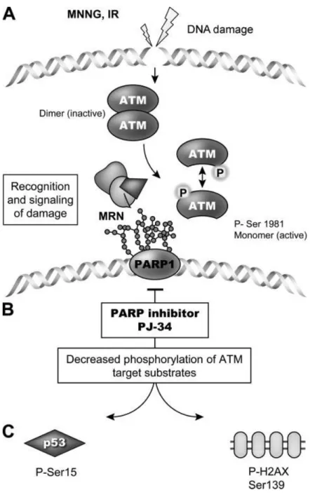
Figure 6. ATM- and PAR-dependent DNA break recognition (Haince et al., 2007).
Activation of ATM and PARP-1 by the DNA strand breaks (A), then PAR accumulation contributes to the recruitment of ATM providing phosphorylation of ATM downstream targets (B). PARP inhibition changes the phosphorylation of ATM- dependent substrates (C).
23
Figure 7. Schematic diagram of NHEJ and A-NHEJ after DSBs (Grabarz et al., 2012).
A. The heterodimer Ku70/Ku80 contacts with the damaged DNA and recruits DNA- PKcs and Artemis. The latter interacts with the DNA ends and prepares it for enzymatic ligation by the Cernunnos-XLF/XRCC4/Ligase IV complex. B. A-EJ (for Alternative end-joining) is always mutagenic. Damaged DNA, which is not processed by Ku70/Ku80, is degraded. A-EJ is independent on Xrcc4, Ligase IV, and is dependent on PARP-1, Ligase III.
1. 4. 2. 3. PARP-1 in nucleotide excision repair (NER)
The main repair mechanism in prokaryotes and eukaryotes against UV-induced DNA lesions and damages is the nucleotide excision repair (NER) that includes the production of thymine dimers (T-T) and other cyclobutane pyrimidine dimers (CPD), as well as 6–4 photoproducts (6-4PP). Mutations in the repair machinery members are associated with xeroderma pigmentosum, Cockayne syndrome and trichothiodystrophy.
There are two classes of NER, the global genomic (GG-NER) and transcription coupled
24
(TC-NER). The role of PARP-1 in NER is not fully understood.
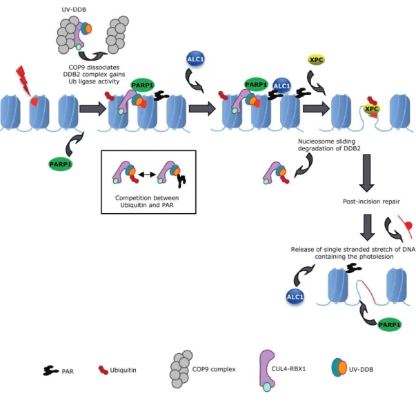
Robu and colleagues showed that PARP-1 works collaboratively with DDB2 (DNA- binding protein 2) and XPC (xeroderma pigmentosum group C) at UV-damaged lesions in GG-NER (Robu et al., 2013; Luijsterburg et al., 2012; Pines et al., 2012) (Figure 8).
Figure 8. A model of DDB2- and PARP1-dependent regulation of NER (Pines et al., 2012). UV-DDB is recruited to the UV damage site as a part of CUL4A-RBX1 complex. This binds to the DNA and DDB1 and DDB2 that have a role in binding PARP-1. CUL4A-RBX and PARP-1 regulate DDB2 by ubiquitylation and PARylation.
The PARYlation of chromatin recruits ALC1, which has a chromatin structure modifying effect mobilizing XPC.
25
1. 4. 3. PARP-1 in mitochondria
Although the scientific field is divided regarding the presence of mitochondrial PARP-1 (mtPARP-1) there are bodies of evidence that support this idea and the role of PARP-1 in mitochondrial DNA repair, bioenergetics, and mitochondrial cell death signaling (Masmoudi et al., 1988; Du et al., 2003; Rossi et al., 2009; Brunyanszki et al., 2016).
Depletion of PARP-1 by siRNA in A549 lung epithelial cells or using tissue from PARP-1 KO mice showed enhanced mitochondrial biogenesis and mitochondrial repair (Szczesny et al., 2014). Mitochondrial PARP-1 activation was showed during H2O2 treatment in U939 by our laboratory. Early mitochondrial DNA damage occurred in response to H2O2 challenge, which was also associated with increased PARylation reactions within the mitochondria (Brunyanszki et al., 2014). These processes were blocked by β-adrenoreceptor blockade with propranolol, the drug that used among burn patients after severe burn injury.
1. 4. 4. PARP-1 in Cell Death
PARP-1 has been implicated in different modes of cell death, apoptosis and necrosis, parthanatos and autophagy. The first correlation between cell death and PARP-1 was made by Berger (Berger et al., 1983). DNA strand breaks activate PARP-1 which plays in the repair process. Inhibition of PARP-1 caused the repair process to be failed while the persistent activation of the enzyme producing excessive poly(ADP-ribose) account for the rapid cell death due to decreased NAD+ pool and ATP synthesis. In the latter, PARP-1 inhibition proved to be beneficial maintaining the cellular NAD+ and ATP pools.
Cells that are no longer needed or are a threat to the organism are destroyed by a well- regulated cell suicide process known as programmed cell death or apoptosis. Regulated necrosis or necroptosis as recently used (Van den Berghe et al., 2014) was accepted as new phenomena after showing that tumor necrosis factor alpha (TNFα) can trigger necrosis mediated by Receptor-Interacting Proteins (RIPs) (Galluzzi et al., 2012). An other form of a caspase-independent cell death is parthanatos (Wang Y et al., 2009) which is accompanied with PARP-1 activation, poly(ADP-ribosyl)ation and AIF
26
(apoptosis-inducing-factor) translocation including a nuclear-mitochondrial-nuclear communication axis.
Apoptosis is a tightly regulated process is accompanied by cell shrinkage, chromatin condensation and DNA fragmentation, blebbing of cell membrane, cellular organelles degradation. The apoptotic process can be divided by two subgroups: a, extrinsic initiated by extracellular ligands and b, intrinsic activated by intracellular stressors, DNA damage, oncogenic factors converging on mitochondria. One of the hallmark of apoptosis is the generation of the cleaved PARP-1 by caspase-3 and caspase-7. These two caspases cleave PARP-1 for a 24 kDa N-terminal DNA-binding domain (DBD), which still maintain a strong DNA binding capability, and a 89 kDa C-terminal catalytic fragment which loses its catalytic activity upon cleavage (Tewari et al., 1995). This results the inactivation of PARP-1 promoting apoptosis by ensuing DNA fragmentation and preserving cellular energy for ATP-sensitive steps opposing the energy depletion- induced necrosis (Kim et al., 2000; D’ Amours et al., 2001). Herceg and Wang proved the above observation using caspase-uncleavable version of PARP in TNF-α-treated PARP-1 fibroblasts leading these cells to NAD depletion and necrosis (Herceg and Wang 1999).
An intensive line of research focused on whether or not poly(ADP-ribosy)lation by PARP-1 makes any differences in the apoptotic process. If there is PARylation involved, that must be taken place at the early phase of apoptosis since PARP-1 is cleaved at that stage and not active catalytically. Inhibition of PARP gave contradict results in terms of the effect on apoptosis. Inhibition of PARP also inhibited (Shiokawa et al., 1997), stimulated (Ray et al., 1992; Payne et al., 1998) or did not change the outcome of apoptosis (Watson et al., 1995) depending on cell type, stimuli, conditions applied.
Studies using PARP-1 knockout mice derived cells showed that PARP-1 mostly can be dispensable for apoptosis. Hepatocytes, thymocytes and neurons from knock out animals compared to their wild-type counterpart did not show differences in the apoptotic process after Fas, TNFα, γ-irradiation, or dexamethasone treatment; however PARP-1 cleavage is still a key step in the apoptotic machinery.
Necrosis or necroptosis as a new nomenclature was implemented based on the fact that it is a well-regulated process which was considered before as a passive, unregulated
27
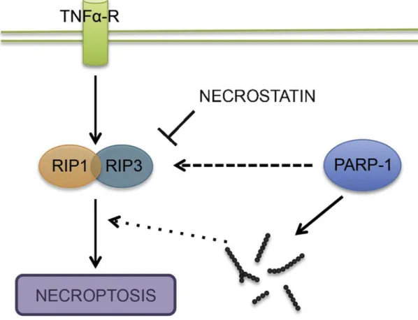
way of dying. It is activated by different extracellular stimuli and the most obvious feature of the necrosis is the disintegration of the plasma membrane and cell content release to the extracellular space. It has been implicated in the pathology of muscular dystrophy, myocardial ischemia-reperfusion injury, diabetes, Alzheimer’s, Huntington’s and Parkinson’s disease. The most crucial members of necroptosis are Receptor- Interacting-Proteins1 and 3 (RIPK1 and RIPK3) which are death domain-containing Ser/Thr kinases and activate nuclear factor kappa B (NF-kB) pathway. That can be inhibited by necrostatin-1 causing no effect on the apoptotic pathway (Van den Berghe et al., 2014). PARP-1 overactivation-induced necrosis was implicated in several pathophysiological conditions. Mouse embryonic fibroblast treated with DNA- alkylating agent N-methyl-N-nitro-N-nitrosoguanidine (MNNG) showed mitochondrial dysfunction, apoptosis inducing factor (AIF) -translocation and cell death in which c- Jun N-terminal kinase (JNK) is required for the PARP-1-induced consequences. Also, these effect were attenuated in RIP1 and TRAF2 (Tumor necrosis factor receptor- associated factor 2) knock out cells which are upstream of JNK which concludes the PARP-1 and necrosis close relation (Xu et al., 2006). High concentration of reactive oxygen and nitrogen species (ROS/RNS) caused overactivation of PARP-1 leading to necrotic cell death in thymocytes. Cells from PARP-1 deficient mice were protected against oxidant-induced damage. Same was true for the pharmacological intervention by 3-aminobenzamide or 5-iodo-6-amino-1,2-benzopyrone against the oxidative stimuli proving the important role of PARP-1 in the necrotic death (Virag et al., 1998 a, b). In a research paper published by Adam’s Laboratory the question was raised whether or not necrosis takes place as a single pathway or different signaling systems contributes to the overall outcome. DNA-alkylating agents, MNNG and methyl methanesulfonate (MMS), rapidly activates PARP-1, while in TNF-induced necrosis this is only a late step of the process. Also, PARP-1 inhibitor, 3 amino-benzamide (3-AB), attenuated the effect of MNNG, but failed to do so in TNF-treated cells. Finally, interfering with RIP1 and Rip3 or blocking ceramide (imipramine) generation was unable to protect necrosis through the PARP pathway (Sosna et al., 2013). This work proves the complexity of the necrosis which can not be concluded as a simplified and uncontrolled way of death (Figure 9).
28
Figure 9. Schematic diagram of necroptosis (Aredia and Scovassi 2014). TNF induces the association of RIP1 and RIP3 (necrostatin is inhibiting it) in which process, PARP-1 directly can regulate necroptosis or indirectly, producing Poly(ADP-ribose). RIP:
Receptor-Interacting Protein.
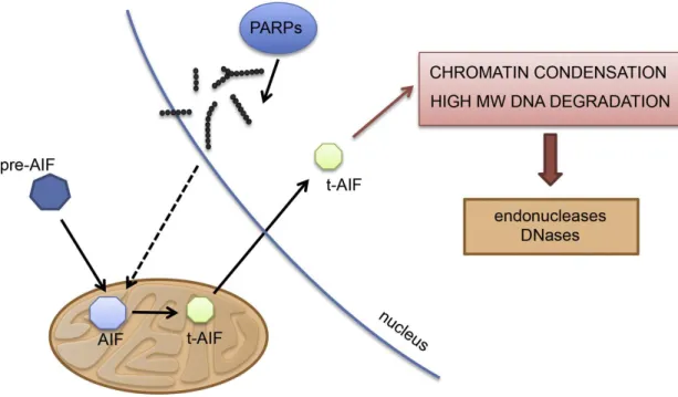
Parthanatos, which refers to PAR and the Greek word of death, is a recognized cell death by the Nomenclature Committee of Cell Death and it classifies as a form of regulated necrosis (Galluzzi et al., 2012). It is a PARP-1 activation driven upon nuclear damage through AIF translocation from mitochondria to nucleus showing phosphatidylserine externalization, loss of mitochondrial membrane potential, chromatin condensation, shrinkage of the cell typically seen also in apoptosis, but the distinction of loss of cell membrane integrity, the lack of dependence on caspase activation and on energy with the appearance of large 50kb DNA fragments (Figure 10). AIF is a 62kDa transmembrane flavoprotein found in the inner mitochondrial membrane anchoraged which is processed to a 57kDa soluble form before its release (Otera et al., 2005). After different stimuli with staurosporin, c-Myc, etoposide, or
29
ceramide, in Rat-1 cells induced AIF release and it was caspase independence showed by using caspase inhibitor Z-VAD.fmk which failed to prevent the translocation of AIF and the condensation of chromatin (Daugas et al., 2000). Immortalized mouse fibroblast showed AIF release after N-methyl-N′-nitro-N-nitrosoguanidine, a DNA-alkylating agent that potently activates PARP-1, exposure which was prevented by PARP-1 inhibitors, 1,5-dihydroxyisoquinoline (DHIQ) (300 μM) and 3,4-dihydro-5-[4-(1- piperidinyl)butoxy]-1(2H)-isoquinolinone (DPQ) (30 μM). The translocation and the cytotoxic effect was also abolished in PARP-1-KO fibroblasts. The broad range of caspase inhibitors failed to prevent the MNNG-induced cytotoxicity proving the caspase-independent mechanism (Yu et al., 2002). The above mentioned observation was proven in a cell free system where HeLa cells nuclei activated by MNNG were incubated with isolated brain mitochondria showed while nuclei non treated with MNNG did not show AIF release. The MNNG activated nuclei treated with PARG or phosphodiesterase markedly reduce the AIF from the mitochondria (Yu et al., 2006).
The release of AIF from the mitochondria is promoted by Bax (Bcl-2-associated X protein). It contains a nuclear localization signal (NLS) which helps the translocation from the cytoplasm to nucleus where it forms a complex with cyclophyllin-A and H2Ax resulting chromatin condensation and DNA degradation (Moubarak et al., 2007;
Delavalle et al., 2011).
30
Figure 10. Hallmark of parthanatos: AIF translocation (Aredia and Scovassi, 2014).
AIF, synthesized in the cytosol as a precursor (pre-AIF), in its mature form is imported into the mitochondria. During parthanatos, AIF is converted into its soluble form tAIF and translocates to the nucleus where it promotes chromatin condensation and high molecular weight DNA degradation through the recruitment of endonucleases and DNases. AIF relocalization is promoted by the accumulation of polymers of ADP- ribose synthesized by PARPs. AIF: Apoptosis Inducing Factor; tAIF: truncated AIF.
Autophagy is thought to be a self-defense mechanism helping cells with the maintenance of energy homeostasis degrading damaged compounds. There are different inducers such as oxidative stress, starvation, DNA-damage. It is mainly regulated by Ser/Thr kinases, mTOR (mammalian Target Of Rapamycin) and AMPK (AMP- activated Protein Kinase). They sense metabolic changes and work on the contrary. Akt is also plays a crucial role since it directly phosphorylates mTOR. mTOR is the negative regulator and active when energy sources are high or abundant keeping autophagy in an
“off” status. AMPK is active when nutrient is scarce, during starvation (Aredia and Scovassi, 2014). A constant level of autophagy can be seen in neurons and malfunction of autophagy may associate with Alzheimer's disease-, Parkinson's disease and Huntington disease. During cancer cachexia (muscle mass wasting) where myostatin
31
expression increased, mTOR/Akt pathway is inhibited (Gallot et al., 2014). Duchenne muscular dystrophy (DMD) is a X-linked degenerative muscle disorder affects male infants. In the cellular level, the condition goes hand in hand with elevated ROS (reactive oxygen species) production, altered Ca2+ homeostasis and impaired autophagy.
Mdx-Knock-out mice, a model of DMD lacking dystrophin, an important muscle protein, have elevated Nox2 activity and Src kinase expression which might be responsible for the elevated oxidant production and decreased autophagy through PI3K/Akt/mTOR pathway (Pal et al., 2014). On the other hand, by sport-physiological mean, during ultra-endurance running as an acute exercise, when energy demands are high, autophagy activity is increased via dephosphorylation of forkhead box O3 (FoxO3) (Jamart et al., 2012). Chronic aerobic exercise results different outcome of the autophagy process depending on the type of exercise, dietary restriction, oxidant production or type of muscles involved (Tam BT and Siu PM, 2014). PARP-1, as discussed above, is activated during oxidative stress and DNA damage which depending on the intensity of the stimuli could also activate the autophagy machinery. Embryonic fibroblasts from PARP-1+/+ and PARP-1−/− mouse (MEF) showed different delays in autophagic pattern after starvation, namely the number of autophagosomes were higher in PARP-1+/+ compared to PARP-1−/−. The same pattern was seen after induction of autophagy by rapamycin. Similar results were obtained by pharmacological inhibition and siRNA-based depletion of PARP-1 suggesting the active role of PARP-1 in autophagy upon starvation. ROS production following starvation was also higher in MEF from parp-1+/+ . The diffusion of ROS into nucleus caused DNA damage and it was more pronounced in the PARP-1+/+ cells. Cells lacking PARP-1 showed reduced level of ATP depletion which was corresponded with the significantly less activation of AMPK. In PARP-1+/+ cells mTOR was completely inhibited suggesting the commitment of these cells to autophagy and also that PARP-1 might control mTOR activity during starvation (Rodriguez-Vargas et al., 2012). Chemotherapy often induces autophagy in cancer cells, moreover PARP-1 inhibition potentiating chemotherapy-induced cell death. MEF from PARP-1+/+ and PARP-1−/− mouse were treated with doxorubicin, a DNA damaging agent, inducing a rapid, direct effect on PARP activation. NAD+ and ATP depletion was observed in PARP-1+/+ but in PARP-1−/− without going toward necrotic or apoptotic pathways. Cells from PARP-1+/+ underwent doxorubicin-induced
32
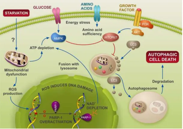
autophagy as seen by electron microscopy, but this was almost absent in PARP-1−/− cells. Pharmacological inhibition of PARP-1 also protected against the autophagic vacuole production after doxorubicin treatment. These results suggest whether or not cells undergo autophagy depends on the PARP-1 activation (Munoz-Gamez et al., 2009). In a subcutaneous tumor model, PARP-1 activation is required in the TNFSF10- induced ADP-ribosylation of high mobility group protein B1 (HMGB1) which causes cancer cell death. Pharmacological inhibition or knockdown of PARP-1 decreased autophagy for the good of apoptosis increasing the anticancer activity (Yang et al., 2015). Starvation and hypoxia can trigger metabolic stress, ROS production, DNA damage and PARP-1 activation leading to autophagy as a defensive mechanism to counterbalance the energy needs for these cells and to avoid cell death. Inhibiting PARP-1 might open new ways in therapeutic applications, probably with combination with other drugs, to inhibit autophagy, which in this case is an escape from death, and reactivate apoptosis to eliminate those cells (Figure 11).
Figure 11. PARP-1 modulates starvation-induced autophagy (Virag et al., 2013). After deprivation of nutrients, cells undergo autophagy through the activation of
33
AMPK/inhibition of mTOR, allowing the formation of the autophagosome core. Events upstream of AMPK involve energy depletion, ROS production and DNA damage. In this scenario, PARP-1 over-activation leads to ATP depletion, acting as a feedback loop to reactivate autophagy. This stress signal when maintained leads eventually to cell death through autophagy. PARP inactivation delays autophagy and favors apoptosis. In the absence of PARP-1 or after PARP inactivation, ROS levels decrease and ATP drop is reduced. As a consequence, the feedback loop reactivated by PARP-1 is cut-down, and apoptosis is triggered.
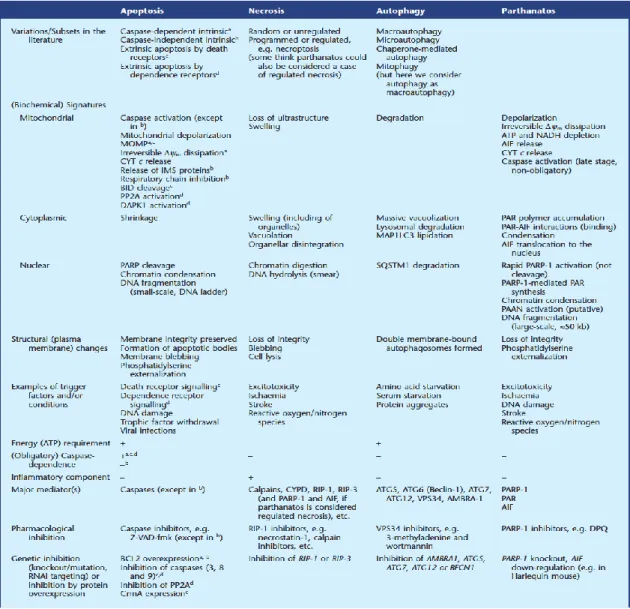
Figure 12. Similarities and differences in apoptosis, necrosis, autophagy and parthanatos (Fatokun et al., 2014). While parthanatos is generally considered to be
34
separate and distinct from necrosis, some investigators consider it to be a specific case of regulated necrosis, just as is necroptosis. Δψm: mitochondrial transmembrane potential. AIF, apoptosis-inducing factor; AMBRA1, activating molecule in Beclin-1- regulated autophagy protein 1; ATG, autophagy; BCL2, B-cell lymphoma 2; BECN1, Beclin-1; CrmA, cytokine response modifier A; CYPD, cyclophilin D; CYT, cytochrome; DAPK1, death-associated protein kinase 1; DPQ, 3,4-dihydro-5-[4-(1- piperidinyl)butoxyl]-1(2H)-isoquinolinone; IMS, intermembrane space; MAP1LC3, microtubule-associated protein 1 light chain 3; MOMP, mitochondrial outer membrane permeabilization; PAAN, parthanatos AIF-associated nuclease; PAR, poly (ADP- ribose); PP2A, protein phosphatase 2A; RIP, receptor-interacting protein; SQSTM1, sequestosome 1; VPS, vacuolar protein sorting; Z-VAD-fmk, N-benzyloxycarbonyl- Val-Ala-Asp-fluoromethylketone.
The role of PARP-1 in different forms of cell death such as apoptosis, necrosis, parthanatos or autophagy was described above. It is mainly associated with DNA damage in some degree but it is thought to have DNA-independent function as well which needs more and deeper investigation.
These 4 types of death can be distinguished based on morphological, biochemical phenotype or the changes to pharmacological inhibition, as outlined in Figure 12.
1. 4. 5. Role of PARP-1 in pathophysiology
An unbalanced cell cycle regulation, cell proliferation or DNA-repair are hallmarks of tumorigenesis. Deletion of one PARP-1, -2, -3 enzymes or pharmacological inhibition under non-stress condition does not lead to major physiological issues or subsequently cancer since the abundance of other repair enzymes step in for this role in the cell. But when they are challenged instability occurs. PARP-1-/- mice challenged with whole body γ-irradiation or N-methyl-N-nitroso urea (MNU) showed genomic instability.
Cells derived from PARP-1-/- mice also displayed high sensitivity to MNU. (de Murcia et al., 1997, Rouleau et al., 2007). Other repair proteins and PARP simultaneous deletion led to embryonic lethality or tumor formation (De Vos et al., 2012).
Reactive oxygen/nitrogen species (ROS/RNS) damage the cells and the components of
35
it such as proteins, lipids, nucleic acids. Generally speaking, under oxidative/nitrosative stimuli PARP-1 is activated and contributes to the DNA repair function. Depending on the severity of the oxidative stress signal, it might induce apoptosis, necrosis or autophagy. This is associated with a wide spectrum of diseases including cardiovascular, neurological, immunological and diabetic complications (for review, see Virag and Szabo 2002). In the last 20 years peroxynitrite received a lot of attention, which also activates PARP-1 and causes PARP-1 related cell injury potentiating NFκB activation turning on inflammatory pathways. The other side of the coin is that PARP-1 and PARP-2 takes part of the normal development of immune cells playing role in T and B-cells differentiation and function in the adaptive immunity. Inhibitors of PARP-1 improve immune-mediated diseases such as arthritis, colitis or allergy (Rosado et al., 2013).
PARP-1 also has a role in energy metabolism serving as a metabolic regulator. Muscles from PARP-1-/- mice showed higher mitochondrial activity, in connection with SIRT-1 (another NAD+ consuming enzyme), improved glucose removal and insulin sensitivity (Bai and Canto 2012). Long term pharmacological inhibition of PARP improves fitness in mice by increasing the number of mitochondrial complexes and enhancing mitochondrial respiratory capacity, which in terms of a pharmacological approach opens new possibilities for treatment for muscle dysfunction linked to mitochondrial function (Pirinen et al., 2012). In young exercised mice SIRT-1 deacetylates PARP-1 decreasing its activity. This effect is suppressed in aged mice, but was reversible with PARP-1 inhibitor suggesting that inhibition of PARP-1 may serve a pharmacological tool in muscle-related pathophysiological condition such as sarcopenia or disuse-induced atrophy by aging (Mohamed et al., 2014).
The importance of PARP-1 activity has been described in several other pathological condition such as ischemic-reperfusion where PARP-1 is an active participant of the neutrophil-mediated myocardial damage upon ischemia and reperfusion and genetic depletion or pharmacological intervention using inhibitors of the enzyme improved the outcome of the injury (Zingarelli et al., 1998). In a hemorrhagic shock model, PARP-1 was activated parallel with metabolic acidosis, lactate production which was reduced by PARP-1 inhibitor 3-AB in the ileum and liver (Watts et al., 2001). ROS and RNS are the two major contributor to endothelial dysfunction in diabetes in which PARP-1 is
36
responsible for the metabolic changes, specially since ROS/RNS can cause DNA damage activating the enzyme. Inhibition of the process by PARP-1 inhibitor could reverse the endothelial dysfunction (Soriano et al., 2001). PARP-1 activity also mediates inflammation in mouse model of contact hypersensitivity promoting leukocyte migration and expression inflammatory mediators, cytokines, chemokines and MMP (Matrix metalloproteinase) (Bai et al., 2009).
Targeting repair deficient tumor cells to promote their genomic instability, improving muscle function in aged individuals, reducing inflammatory cytokines expression and leukocyte migration have a common interest: PARP inhibition. Some of the inhibitors already have advanced to clinical trials and give hopes to fight against these pathological complications.
1. 5. Oxidative stress, as a trigger of PARP activation
As it was discussed earlier, the best characterized activator of PARP-1 is DNA-damage induced by free radicals. Prolonged ROS/RNS (Reactive oxygen/nitrogen species) production results in an excessive PARP-1 activation reducing the cell NAD+ and ATP stores, blocking apoptosis and resulting necrosis. The better understand the role of PARP-1, below an overview is given about the origin of reactive oxygen/nitrogen species.
Reactive oxygen and nitrogen species are known to contribute to wide variety of diseases such as cancer, atherosclerosis, diabetes, neurodegenerative disorders, or aging.
As a nomenclature point of view, reactive species, either ROS or RNS, are a collective terms and can be divided into two subgroups, free radicals and non-radicals (Table 1).