Review
Interplay of oxidative, nitrosative/nitrative stress, in fl ammation, cell death and autophagy in diabetic cardiomyopathy☆
Zoltán V. Varga
a,b, Zoltán Giricz
b, Lucas Liaudet
c, György Haskó
d, Peter Ferdinandy
b,e, Pál Pacher
a,⁎
aLaboratory of Physiological Studies, National Institutes of Health/NIAAA, Bethesda, MD, USA
bCardiometabolic Research Group, Department of Pharmacology and Pharmacotherapy, Semmelweis University, Budapest, Hungary
cDepartment of Intensive Care Medicine BH 08-621-University Hospital Medical Center 1011 LAUSANNE Switzerland
dDepartment of Surgery and Center for Immunity and Inflammation, Rutgers NJ Medical School, USA
ePharmahungary Group, Szeged, Hungary
a b s t r a c t a r t i c l e i n f o
Article history:
Received 6 May 2014
Received in revised form 11 June 2014 Accepted 24 June 2014
Available online 2 July 2014
Keywords:
Diabetic cardiomyopathy Protein oxidation Autophagy Oxidative stress
Diabetes is a recognized risk factor for cardiovascular diseases and heart failure. Diabetic cardiovascular dysfunction also underscores the development of diabetic retinopathy, nephropathy and neuropathy. Despite the broad availability of antidiabetic therapy, glycemic control still remains a major challenge in the management of diabetic patients. Hyperglycemia triggers formation of advanced glycosylation end products (AGEs), activates protein kinase C, enhances polyol pathway, glucose autoxidation, which coupled with elevated levels of free fatty acids, and leptin have been implicated in increased generation of superoxide anion by mitochondria, NADPH oxidases and xanthine oxidoreductase in diabetic vasculature and myocardium. Superoxide anion interacts with nitric oxide forming the potent toxin peroxynitrite via diffusion limited reaction, which in concert with other oxidants triggers activation of stress kinases, endoplasmic reticulum stress, mitochondrial and poly(ADP-ribose) polymerase 1-dependent cell death, dysregulates autophagy/mitophagy, inactivates key proteins involved in myocardial calcium handling/contractility and antioxidant defense, activates matrix metalloproteinases and redox-dependent pro-inflammatory transcription factors (e.g. nuclear factor kappaB) promoting inflammation, AGEs formation, eventually culminating in myocardial dysfunction, remodeling and heart failure. Understanding the complex interplay of oxidative/nitrosative stress with pro-inflammatory, metabolic and cell death pathways is critical to devise novel targeted therapies for diabetic cardiomyopathy, which will be overviewed in this brief synopsis. This article is part of a Special Issue entitled: Autophagy and protein quality control in cardiometabolic diseases.
Published by Elsevier B.V.
1. Introduction
Incidence of metabolic diseases, especially of obesity and type II dia- betes is constantly growing worldwide. Diabetes is a well-recognized risk factor for cardiovascular diseases and heart failure
[1]. The term ofdiabetic cardiomyopathy has been introduced by Rubler et al. back in 1972
[2]. Later the Framingham Heart Study has also demonstratedthat the occurrence of heart failure in male and female diabetics is twice and
five times more common, respectively, compared to age- matched control subjects
[3]. Similar cross-sectional studies confirmed increased risk for heart failure development in diabetics, even when corrected for other confounding variables such as coronary artery dis- ease, hypercholesterolemia, hypertension, and obesity
[4–7]. Notably,the higher occurrence of biventricular dysfunction and cardiomyopathy in diabetics is also suggestive of the fact that diabetes is an independent risk factor or cause of cardiomyopathy
[7–9].Although it is established that multiple factors may collectively con- tribute to the development and progression of diabetic cardiomyopathy (e.g. hyperglycemia, insulin resistance, increased fatty acid metabolism,
Abbreviations:AGEs, advanced glycosylation end products; AMPK, AMP-activated pro-tein kinase; AT1R receptor, angiotensine II receptor type 1; CaMKII, Ca2 +/calmodulin- dependent protein kinase II; CB1/2receptor, cannabinoid 1 and 2 receptors; eNOS, endothelial nitric oxide synthase; ER stress, endoplasmatic reticulum stress; ICAM-1, intercellular adhesion molecule 1, also known as CD54; iNOS, inducible nitric oxide synthase; MAPKs, mitogen activated protein kinases; MCP-1, monocyte chemoatractic protein-1; MMPs, matrix metalloproteinases; mTOR, mammalian target of rapamycin;
NADPH oxidase/NOX, nicotinamide adenine dinucleotide phosphate-oxidase; NFkB, nuclear factor kappaB; NO, nitric oxide; NOS, nitric oxide synthases; Nrf2, NFE2L2 nu- clear factor, erythroid 2-like 2; PARP-1, poly(ADP)ribose polymerase 1; PARP-1, poly(ADP-ribose) polymerase 1; RAGE, receptor for advanced glycation end product;
ROS/RNS, reactive oxygen and nitrogen species; SERCA2a, sarco/endoplasmic reticu- lum Ca2 +-ATPase; STZ, streptozotocin; VCAM-1, vascular cell adhesion molecule 1, also known as CD106; XO, xanthine oxidase/oxidoreductase.
☆ This article is part of a Special Issue entitled: Autophagy and protein quality control in cardiometabolic diseases.
⁎ Corresponding author at: Laboratory Physiological Studies, Section Oxidative Stress Tissue Injury, 5625 Fishers Lane, Room 2N-17, Bethesda, MD 20892-9413, USA.
Tel.: +1 301 443 4830.
E-mail address:pacher@mail.nih.gov(P. Pacher).
http://dx.doi.org/10.1016/j.bbadis.2014.06.030 0925-4439/Published by Elsevier B.V.
Contents lists available atScienceDirect
Biochimica et Biophysica Acta
j o u r n a l h o m e p a g e : w w w . e l s e v i e r . c o m / l o c a t e / b b a d i s
microcirculatory changes, sympathetic dysfunction, myocardial in
flam- mation, remodelling and
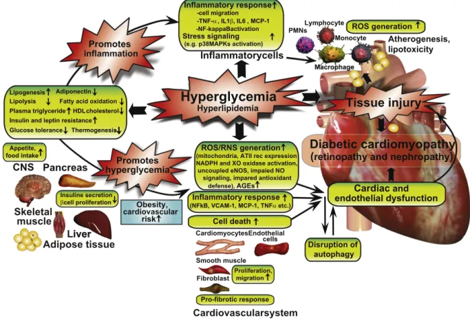
fibrosis), the exact molecular mechanisms that trigger and fuel these major pathological processes are not entirely clear (Fig. 1).
Accumulating recent evidence suggests that the complex interplay of oxidative, nitrosative and nitrative stress
[10–12]with major proin
flam- matory, metabolic
[13]and cell death pathways[14–17]play an essential role in the development of complex biochemical
[18,19], mechanical,and structural alterations associated with diabetic cardiomyopathy
[8,20,21], which will be overviewed in this brief synopsis.2. Oxidative, nitrosative and nitrative stress in diabetic cardiomyopathy
Oxidative and subsequent nitrosative damage of the myocardium and vasculature have been described as major primary mechanisms leading to pathologic alterations associated with diabetic cardiovascular complications
[22–29]. Factors such as hyperglycemia, glucose autoxi-dation, accumulation of advanced glycosylation end products (AGEs), enhanced receptor for advanced glycation end product (RAGE) and angiotensin II receptor type 1 (AT
1R) signaling, elevated levels of free fatty acids and leptin, have been reported to contribute to increased pro- duction of reactive oxygen and nitrogen species production (ROS/RNS) in diabetic vessels and myocardium
[22,24], among others. In spite ofthe broad availability of antidiabetic therapy, glycemic control still remains a major challenge in the management of diabetic patients
[30].Inadequate glycemic control likely accounts for the high prevalence of diabetic complications, since high glucose or
fluctuating glucose levels
[31]can induce acute oxidative stress that might be a critical factor in the initiation of pathologic alterations eventually leading to development of diabetic cardiomyopathy
[16,24]. Oxidative protein modifications play
a pivotal role in the development of diabetic myocardial dysfunction.
Oxidation of proteins involved in contractility, excitation
–contraction coupling, protein folding, antioxidant defence, metabolism (mainly fatty acid and glucose), and Ca
2+handling have been reported in diabet- ic hearts (see
Table 1). Oxidative modification may result in potentially harmful events including dissociation of catalytic subunits of enzymes, local or global unfolding, aggregation or fragmentation. Proteins can be oxidized directly by ROS, or by products of secondary oxidation reactions formed during lipid peroxidation (e.g., malondialdehyde or 4-hydroxynonenal). Moreover, oxidative protein modi
fications may also occur by reactive sugars in glycation or glycoxidation reactions
[32].Depending on the amount of modi
fied proteins and the extent of oxida- tion, different degradation mechanisms are activated. Mildly oxidized proteins are broken down primarily by the proteasome, while heavily oxidized proteins (potentially forming aggregates) are degraded by the endosomal
–lysosomal pathway thereafter
[33]. Thus, an increasein oxidative stress might produce substrates that fuel autophagic pro- tein degradation, at least in the
first stages of oxidative injury
[34]. Inaddition, certain proteins involved in autophagosome formation and maturation (Atg4, LC3-II) require oxidative posttranslational modi
fica- tions[35,36], thus, activity of these proteins are also strongly in
fluenced by their redox status
[35]. However, the accumulation of oxidized andnitrated proteins and lipids suggest abrogated autophagic processes in diabetic cardiomyopathy that may signi
ficantly contribute to myocardi- al dysfunction.
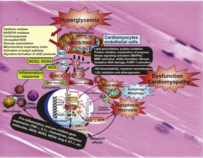
Several cellular and subcellular sources were described that may account for enhanced ROS production in diabetic cardiovascular system and at other sites of diabetic complications (e.g. in the retina and kidneys) (Fig. 2). This list includes nicotinamide adenine dinucleotide phosphate oxidases (NADPH oxidases)
[37–39], xanthine oxidase/oxidoreductase (XO)
[40,41], enzymes of the arachidonic acid cascadeLiver Skeletal
muscle
Cardiovascularsystem Adipose tissue
Appetite,
CNS
food intake
Inflammatorycells
Lymphocyte Monocyte -TNF-α
-cell migration , IL1β, IL6 , MCP-1 -NF-kappaBactivation
o oc
Liver
Stress signaling (e.g. p38MAPKs activation)
PMNs
Fibroblast
CardiomyocytesEndothelial cells
Smooth muscle
Pancreas
Pro-fibrotic response Inflammatory response
Insuline secretion βcell proliferation
y
Lipogenesis
Glucose tolerance
HDL cholesterol Plasma triglyceride
Insulin and leptin resistance Thermogenesis Adiponectin Lipolysis Fatty acid oxidation
n s
Promotes hyperglycemia
ROS generation
Obesity, cardiovascular
risk
inflammation
Promotes
Inflammatory response
(NFkB, VCAM-1, MCP-1, TNFα etc.)
Cell death ROS/RNS generation
(mitochondria, ATII rec expression NADPH and XO oxidase activation, uncoupled eNOS, impaied NO signaling, impared antioxidant defense), AGEs
Proliferation, migration
Diabetic cardiomyopathy ( retinopathy and nephropathy )
Cardiac and endothelial dysfunction
Hyperglycemia
Hyperlipidemia Tissue injury
Atherogenesis, lipotoxicity
Disruption of autophagy
Fig. 1.Interplay of hyperglycemia and peripheral metabolism in cardiometabolic syndrome in mediating diabetic cardiovascular complications. Hyperglycemia may indirectly (via its metabolic consequences) or directly enhance diabetes-associated inflammation and ROS generation, promoting tissue injury and the development of diabetic cardiovascular and other complications. AT II rec, angiotensin II receptor type 1; CNS, central nervous system; PMNs, polymorphonuclear leukocytes; XO, xanthine oxidase.
(cyclooxygenase and lypoxygenase enzymes), microsomal enzymes, the uncoupled nitric oxide synthases (NOS)
[10], and the mitochondrialrespiratory chain
[24]. Among these, increased mitochondrial ROSgeneration initially was considered to represent the primary source of high glucose-induced oxidative stress in several tissues and cell types, including cardiomyocytes
[24,37,42]and endothelial cells
[43,44]. Luoet al. recently showed, that mitochondrial oxidants signi
ficantly contribute to the oxidation of the multifunctional enzyme, Ca
2+/ calmodulin-dependent protein kinase II (CaMKII)
[45]. Diabetic micetreated with a mitochondria-targeted antioxidant, MitoTEMPO, showed reduced levels of oxidized-CaMKII, subsequently with preserved heart rates, and improved survival after myocardial infarction
[45].Mitoquinone, a mitochondrially targeted ubiquinone that can act as an antioxidant, also improves cardiac dysfunction
[46]and activates autophagy in an Nrf2-dependent manner in a non-cardiac system
[47], suggesting an interaction between mitochondrial redox balanceand protein quality control mechanisms, such as autophagy.
There is also evidence for increased production of ROS from non- mitochondrial sources. NADPH oxidases (NOX) are unique enzymes that may be responsible for large amounts of superoxide and hydrogen peroxide (H
2O
2) production under various pathological conditions.
Activity or expression of various NOX isoenzymes (that are involved in superoxide or H
2O
2generation), has been found to be signi
ficantly higher in the heart with metabolic derangements such as diabetes
[37–39,48,49]and hypercholesterolemia
[50]. The increased activityand expression of NOX4 in the diabetic heart
[49]is therapeutically tar- getable, and NOX4 inhibitors are indeed in preclinical development for various cardiovascular indications
[51]. Diabetes induced increase inNADPH activity is further accentuated by the increased production of NADPH by glucose-6-phosphate dehydrogenase, as it was described in the heart and aorta of Zucker diabetic fatty rats
[52]. Elevated concen-tration of hydrogen peroxide has been shown to modulate autophagy by several mechanisms. For example, H
2O
2, activates the LKB1/AMPK pathway which leads to inhibition of the mTORC1 complex and induced autophagy
[53]. NOX4-mediated production of hydrogen peroxide alsoinduces autophagy in human umbilical vein endothelial cells
[54]and in
cardiomyocytes
[55]after induction of endoplasmic reticulum stress.
Although, it is plausible that excessive amounts of ROS might incapaci- tate certain players of autophagy. Vascular NADPH oxidase is also an im- portant downstream target of angiotensin II, which has been proposed to play a pivotal pathological role in the development and progression of diabetic cardiovascular
[56–59]and other diabetic complications.
This is supported by convincing evidence obtained from preclinical rodent models of STZ (streptozotocin)-induced type 1 diabetes, as well as from human myocardial biopsy specimens, suggesting that the renin
–angiotensin system is up-regulated in diabetes and angiotensin II locally through AT
1R, which is overexpressed in diabetic hearts or in cardiomyocytes exposed to high glucose, importantly contributes to the development of diabetic cardiomyopathy
[38,57–60]. The beneficial effects of AT
1R blockade in diabetic hearts involve, but are not limited to, the attenuation of myocardial NADPH oxidase-dependent (such as p47phox) ROS generation, in
flammation, cell death,
fibrosis, and con- tractile dysfunction
[57–60]. AT1 receptor blockade also preventsglucose-induced cardiac dysfunction directly in ventricular myocytes via attenuating the AT1-NADPH oxidase signaling
[61]. Consistentlywith the important pathological role of NADPH oxidases-derived ROS generation in diabetic hearts, apocynin (a putative non-speci
fic NADPH oxidase inhibitor) not only attenuated the enhanced superoxide generation in diabetic hearts, but also improved the diabetes-associated cardiac dysfunction in type I diabetic mice
[39].Increased activation of another ROS generating enzyme, the XO, has also been shown in different organs
[62]of diabetic rats or mice, in- cluding in the heart
[41]. Conversely, inhibition of XO by allopurinolsigni
ficantly attenuated most pathological features of diabetic cardio- myopathy (e.g. myocardial ROS/RNS generation, iNOS expression, cell death, and
fibrosis) and improved both systolic and diastolic dysfunc- tions in type I diabetic mouse
[41]or rat
[63]hearts. Importantly, there is also current evidence, coupled with numerous previous studies
[40], for the beneficial cardiac effects of allopurinol in humans
[64].Rekhraj at al. recently described, that in patient with ischemic heart disease, high dose off allopurinol was capable to reduce left ventricular muscle mass and improve the symptoms of the disease
[65].Table 1
Oxidative and nitrative modifications of key myocardial proteins in diabetic cardiomyopathy.
Modified protein Modification Function Reference
CaMK-II Oxidation Singalling [45]
Protein disulfide isomerase Oxidation Protein folding [140]
Complexes I, III, and V Oxidation ATP synthesis [141]
mtHSP70, mitofilin Oxidation Mitochondrial protein import [141]
Alpha-enolase Oxidation/nitration Glycolysis [142]
Succinyl-CoA:3-oxoacid CoA transferase Nitration Ketone body metabolism [143–145]
Creatine kinase Nitration Muscle contraction [144]
Peroxiredoxin 3 Nitration Antioxidant defense [144]
Complex I (24 kDa subunit) Nitration ATP synthesis [144]
Acyl coenzyme A thioester hydrolase Oxidation Fatty acid metabolism [146]
Acetyl-CoA acetyl transferase Oxidation Fatty acid metabolism [146]
Acyl CoA dehydrogenase Oxidation Fatty acid metabolism [146]
3-ketoacyl-CoA thiolase Oxidation Fatty acid metabolism [146]
Enoyl-CoA hydratase Oxidation Fatty acid metabolism [146]
Heart-type fatty acid-binding protein Oxidation Fatty acid metabolism [146]
Glutathione Peroxidase-1 Oxidation Antioxidant defense [146]
Glutathione-S-Transferase Oxidation Antioxidant defense [146]
Superoxide Dismutase-2 Oxidation Antioxidant defense [146]
Peroxiredoxin-1 Oxidation Antioxidant defense [146]
Peroxiredoxin-2 Oxidation Antioxidant defense [146]
Peroxiredoxin-3 Oxidation Antioxidant defense [146]
Peroxiredoxin-6 Oxidation Antioxidant defense [146]
Ryanodine receptors (RyR2) Oxidation Excitation–contraction [147]
SERCA2a Oxidation Ca2+ handling [148]
Myosin heavy chain alpha and beta Oxidation Contraction [149]
GAPDH Nitrosylation Glycolysis [150]
Caspase-3 Nitrosylation Apoptosis [150]
Connexin43 Nitration Cell–cell communication [151]
ATP synthase subunits Nitration ATP synthesis [84]
thioredoxin-1 Nitration Antioxidant defense [152,153]
A substantial amount of experimental evidence suggests that there is reduced nitric oxide (NO) availability in diabetic tissues. Different mechanisms have been proposed to be responsible for the diabetes- induced dysfunction of NO production, bioavailability and/or signaling.
A generally accepted mechanism is the alterations in nitric oxide synthases (NOS), particularly in its endothelial isoform (eNOS), function
[10,66]. The composition of the eNOS complex is critical for the relativeformation of NO or superoxide. The mechanisms by which eNOS dys- function develops remain elusive, however, a decrease in the dimer to monomer eNOS ratio within the myocardium of diabetic animals has been reported
[67]. Monomerization i.e. uncoupling of NOS results inincreased oxidative stress and decreased NO bioavailability (since uncoupled NOS generates superoxide anion instead of NO), which has been implicated in the pathophysiology of numerous cardiovascular diseases. Accordingly, eNOS uncoupling has also been reported in diabetic hearts, while administration of the tetrahydrobiopterin precur- sor, sepiapterin inhibited uncoupling of NOS and improved LV function
[68]. Similarly, ascorbic acid or N-acetyl-cystein is also capable to
increase BH4/BH2 ratio and prevent NOS uncoupling in the diabetic heart
[69].In addition to uncoupling the major cardiac NOS isoforms, iNOS and eNOS expression has been shown to be increased (particularly iNOS) in the diabetic hearts
[37,38,70,71]. The increase in NOS expression inthe diabetic heart is associated with an increase in lipid peroxidation and 3-nitrotyrosine formation (marker of peroxynitrite generation and nitrative stress), which might be related to the uncoupled and monomer state of the enzyme, which most likely produces superoxide instead of NO, as well as triggers the formation of peroxynitrite. Inter- estingly, peroxynitrite itself also causes NOS un-coupling by selectively targeting and disrupting caveolae
–NOS interaction
[72].The rapid diffusion-limited reaction of superoxide with NO indeed forms peroxynitrite, a very strong cytotoxic oxidant, which attacks and damages various biomolecules via multiple mechanisms
[10]. In agreement with these, inhibition of NOS activity (byL-NAME
+
SODPRAG
AIF ΔΨ
↓
Damage
Nucleus NAD+
Autophagy?
Mitophagy?
Cytoplasm
Free PAR polymer
Apoptosis
DNA fragmentation DNA
breaks
Necrosis
(inactive)
PARP-1 (active)
cyt c
NAD+↓ ATP↓
PARP-1
Caspase activation Repair
Excessive damage Mild damage
ONOO _
endothelial cells Cardiomyocytes
iNOS
r
NF κ B
NO O _
2
NOX2, NOX4 .
H
2O
2response
Inflammatory
Mitochondrion
ROS/RNS
ROS/RNS
PMNs Monocyte Xanthine oxidase NAD(P)H oxidases Cyclooxygenase Uncoupled NOS Glucose autoxidation Mitochondrial respiratory chain Activation of polyol pathway Glycation-formation of AGE products
Hyperglycemia
NO O
2. _
SIRT-1
↓ NO bioavailability, impaired vasorelaxation,
↑ LDL oxidation and atherogenesis
activity↓
Metabolic effects
Lipid peroxidation, protein oxidation Protein nitration, inactivation of enzymes Stress signaling activation (MAPKs) MMP activation, AGEs formation, fibrosis Oxidative DNA damage, PARP-1 activation
Pro-inflammatory
and pro-oxidant gene expression (TNF
α, IL1
β, chemokines, adhesion molecules, iNOS, NOX2, NOX4, Ang II, ET-1, etc.
Dysfunction Cardiomyopathy
Fig. 2.Interplay of oxidative and nitrosative/nitrative stress with cell death pathways in diabetic cardiomyopathy. Hyperglycemia via activation of various pathways shown in the large yellow box increased superoxide anion (O2•−) production in cardiovascular cell types. NO and superoxide (O2•−) rapidly react to form peroxynitrite (ONOO−) which induces cell injury via enhanced lipid peroxidation, inactivation of enzymes and other proteins by oxidation and nitration, and also activation of stress signaling, matrix metalloproteinases (MMPs) among others. Peroxynitrite also triggers the release of proapoptotic factors such as cytochromecand apoptosis-inducing factor (AIF) from the mitochondria, which mediate caspase-dependent and -independent apoptotic cell demise pathways. Autophagy may be beneficial in diabetic cardiomyopathy in removal of injured cells, but additional support is required to prove its exact role. Peroxynitrite, in concert with other reactive oxidants, causes stand breaks in DNA, activating the nuclear enzyme poly(ADP-ribose) polymerase-1 (PARP-1). Mild damage of DNA activates the DNA repair machinery, but once excessive oxidative/nitrosative stress-induced DNA damage occurs, like in diabetes, overactivated PARP initiates an energy-consuming cycle by transferring ADP-ribose units from nicotinamide adenine dinucleotide (NAD+) to nuclear proteins, resulting in rapid depletion of the intracellular NAD+and ATP pools, slowing the rate of glycolysis and mitochondrial respiration, eventually leading to cellular dysfunction and demise. Poly(ADP-ribose) glycohydrolase (PARG) degrades poly(ADP-ribose) (PAR) polymers, generating free PAR polymer and ADP-ribose. Overactivated PARP also facilitates the activation of NFkB and expression of a variety of pro-inflammatory genes leading to increased inflammation and associated oxidative stress, thus facilitating the progression of cardiovascular dysfunction and heart failure. Via attenuation of the cellular NAD+levels PARP activation may also promote metabolic dysfunction via decreased activity of SIRT-1 in various tissues. In addition to these adverse consequences the NO bioavailability and signaling is also impaired in diabetic hearts promoting impaired vasorelaxation and enhanced atherogenesis eventually facilitating increased cardiovascular inflammation, and lipid deposition in vessels and myocardium, functional ischemia and enhanced cardiac injury.
or
L-NMMA) improves myocardial performance in diabetic hearts, sug- gesting that the increased production of superoxide and peroxynitrite rather than NO is a major contributor of suppressed contractile perfor- mance in diabetes
[39,66,73,74]. NO and peroxynitrite has a very com-plex relation to autophagy. Nitric oxide donors have been shown to decreased autophagy in HeLa cells, which has been con
firmed by the overexpression of eNOS
[75]. Although, LPS-induced up-regulation ofNOS izoenzymes were followed by an accelerated autophagy in HL-1 cells and in mice
[76]. Since LPS induces the production of not only NO,but peroxynitrite, these seemingly opposing results can be explained. It is further evidenced by other studies, where peroxynitrite exerted inhib- itory effects on autophagy. For example, peroxynitrite induced autopha- gy in endothelial cells and increased LC3-II protein
[77]. These data welldemonstrate that sub-physiological NO levels and nitrosative/nitrative stress which are characteristic to diabetes, might facilitate autophagic processes in the heart, however, direct observations are yet to be made. Peroxynitrite formation and consequent protein nitration (nitrative stress) have been suggested to play a central role in the devel- opment of cardiovascular dysfunction associated with diabetes, as well as in the development of diabetic retinopathy, nephropathy and neurop- athy. Peroxynitrite in hearts not only promotes apoptotic or necrotic cell death in cardiomyocytes and endothelial cells via activation of stress signaling pathways (e.g. mitogen activated protein kinases (MAPKs) and poly ADP ribose polymerase 1 (PARP-1)-dependent pathways), but also induces nitration and consequent inactivation of key proteins in- volved in intracellular calcium cycling, energy homeostasis, antioxidant defense, and myocardial contractility (see
Table 2.)[10]. Peroxynitrite,in concert with other oxidant may also activate matrix metalloprotein- ases (MMPs) in the failing hearts to promote pathological remodeling
[23,37,78]and may impair the NO-soluble guanylate cyclase signaling pathway rendering NO to unable to activate its primary protective
signaling machinery
[79,80]. Furthermore, peroxynitrite decompositioncatalysts have been shown to restore the normal contractile phenotype of high glucose treated cardiomyocytes and attenuate cardiac dysfunc- tion in diabetic hearts
[74]. The importance of peroxynitrite has alsobeen shown in diabetic patients. Increased serum and/or vascular 3-nitrotyrosine levels were positively correlated with increased blood pressure, and or endothelial dysfunction in diabetic patients
[29,81].The persistently increased myocardial oxidative and nitrative stress in diabetic hearts eventually also leads to dysfunction of important an- tioxidant defense mechanisms
[10](e.g. inactivation of important anti- oxidant enzymes such as superoxide dismutases and catalase, depletion of endogenous antioxidants (e.g. metallothionein, glutathione)
[37, 82–84]) and dysregulation of important redox-dependent transcriptionfactors (e.g. NFE2L2 nuclear factor, erythroid 2-like 2 (Nrf2))
[85,86],among others. Increased oxidative and nitrative stress in diabetes may also promote oxidation and/or nitration of various insulin receptors in peripheral tissues, which may contribute to the development of insulin resistance
[87].Hyperglycemia and/or hyperglycemia-induced ROS has been report- ed to increase accumulation of products of nonenzymatic glycation/
oxidation of proteins/lipids (AGE) and enhanced the expression of their receptor (RAGE) in cardiovascular tissues, which thought to play a key role in the development and progression of cardiovascular compli- cations of diabetes
[88,89]. Accumulation of AGEs and increased expres-sion of RAGE have been associated with diabetes-induced dysfunction and structural alterations in hearts of type 1 diabetic rodents
[88,89].Furthermore, in diabetic heart failure patients with reduced LV ejection fraction, the accumulation of AGEs and
fibrosis appear to contribute to the increased diastolic stiffness, but not in patients with normal LV ejection fraction, where the increased cardiomyocyte resting tension is responsible for this phenomenon
[90].Table 2
Main therapeutic approaches and targets in diabetic cardiomyopathy.
Therapeutic target Approach Reference Potential effect on autophagy
Oxidative, nitrosative, nitrative stress
NADPH oxidases Enzyme inhibition
Apocynin, (VAS2870 or GKT137831) [39,154] Potential inhibitors of autophagy[55,155]
AT-1 receptor antagonist [61] Potential inhibitors of autophagy[156]
Xanthine oxidase Enzyme inhibition
Allopurinol [40,41,63,64] No information
eNOS Uncoupling inhibitors
L-Arginine [157] Potentially inhibits autophagy by NO[75,158]
Tetrahydrobiopterin [68] Potentially inhibits autophagy by NO[159]
IGF-1 [160] Potential inhibitor of autophagy[161]
iNOS Enzyme inhibition
L-NIL [162,163] No information in the literature.
Peroxynitrite Decomposition catalysts
MnTMPyP [28] Potential inhibitors of autophagy[77]
FP15 [164]
Mitochondria oxidative stress MitoTEMPO [45] No information in the literature.
NRF2 Activators
Sulforafan [165] Potential autophagy inducer[166]
DH404 [167]
ROCK Enzyme inhibition
Fasudil [168] Potential autophagy inducer[169]
Cell death pathways—Autophagy/Mitophagy, Apoptosis, PARP-dependent cell death
p38 MAPK Enzyme inhibition
SB 239063 [170] Potential inhibitors of autophagy[76]
PARP Enzyme inhibition
PJ34 [118]
3-aminobenzamide [171] Potential inhibitors of autophagy[172]
AMPK activation Metformin [104] Potential autophagy inducer[99]
GLP-1 GLP-1 agonists, DPP-4 inhibitors [173–177] Potential autophagy inducers[178]
Inflammation
Cytokine expression (TNFα, IL1β, TGFβ) p38 MAPK inhibition [122] Potential inhibitors of autophagy[76]
AT-1 receptor antagonist [57] Potential inhibitors of autophagy[156]
Endocannabinoid system CB1 antagonist [38] Potential inhibitors of autophagy[139]
3. Autophagy and cell death mechanisms in diabetic cardiomyopathy
It has extensively been documented that increased oxidative and nitrative stress in diabetic hearts may trigger activation of stress signal- ling pathways facilitating apoptosis in cardiomyocytes and endothelial cells
[14]. However, recent evidence also suggests that in addition toapoptosis, other processes such as autophagy, mitophagy and PARP- dependent cell death pathways may also play an important role in controlling the cell demise during the course of cardiovascular disease
[91]and diabetic cardiomyopathy
[17], which will be discussed in thefollowing part.
Autophagy is a cellular housekeeping process that is essential for removal of damaged or unwanted organelles, protein and lipid aggregates. It is a dynamic process which is tightly regulated by the availability of nutrients and the metabolic balance of the cell, and func- tional autophagy is indispensable for cellular survival in low-energy conditions
[92]. Since in diabetes, where oxidative stress, protein- andlipid oxidation are elevated and cellular energy balance is disturbed, functional autophagy might have even higher importance in the main- tenance of cardiac cellular integrity. In the heart, constitutive autophagy is a homeostatic mechanism for maintaining cardiac structure and function
[93], while disruption of autophagy leads to heart failure[94].Interestingly, in healthy animals, autophagy seems to be higher in males than in females suggesting that the male heart has a major consti- tutive autophagy
[95]. This gender difference may elucidate the obser-vation of the Framingham Heart Study showing markedly increased occurrence of diabetic cardiomyopathy in females
[3,96]. Autophagy issuppressed at high glucose concentrations, such as present in diabetes, which might be associated with the development of diabetic cardiomy- opathy
[97]. In addition, the diabetic heart is characterized by excessivefatty acid uptake and oxidation, resulting in accumulation of toxic lipid intermediates (long-chain acyl-CoA, diacylglycerol, ceramides), known as lipotoxicity. Cardiac lipotoxicity contributes to insulin resistance, impairment of mitochondrial function, cardiomyocyte hypertrophy, and apoptosis, which may ultimately lead to left ventricular remodelling of the heart
[98].Elevated oxidative stress, apoptosis, in
flammation, and ER stress by increased production of monocyte chemoatractic protein-1 (MCP-1) is suspected to contribute to the disruption of cardiac autophagy in diabe- tes
[97,99–101]Several upstream signalling pathways of autophagy are altered in diabetes or metabolic syndrome as well[102]. The mammalian target of rapamycin (mTOR) pathway, a negative regulator of autophagy is blunted in obese, hyperlipidemic rats fed a high cholesterol-high fructose diet showing most characteristics of type II diabetes
[103].Also, activity of AMP-activated protein kinase (AMPK) is reduced in high glucose conditions, leading to disturbed autophagy
[104]. Althoughthe majority of reports agree on the attenuation of the autophagy in diabetes, not all studies are equivocal (possibly due to the different ex- perimental models used). For example, in a fructose-fed mouse model, induction of autophagy has been described
[105], and milk fat-basedchow blunted cardiac autophagy and facilitated cardiomyopathy more ef
ficiently than a lard-based chow
[106]. Further complicating the pic-ture is the discrepancy that oxidative stress is elevated in diabetes, which is supposed to induce autophagy, however, the majority of studies evidence quite the opposite. This phenomenon and the lack of any direct evidence from diabetic patients warrant further investigations on the status of cardiac autophagy in disease models relevant to human pathological conditions.
Aiming to restore pathological cardiac consequences of diabetes modulation of autophagy has been attempted by different tools. Chronic activation of AMPK (therefore autophagy) by metformin or overexpres- sion of ATG7 reduced cardiomyocyte apoptosis, cardiac
fibrosis, and im- proved myocardial functions in streptozotocin (STZ)-induced diabetic mice
[104]. Overexpression of hem oxygenase-1 in STZ-treated micealso induced autophagy, reduced oxidative stress and in
flammation
and prevented cardiac dysfunction
[97]. In a recent report, inhibitionof mTOR signalling by the overexpression of PRAS40 prevented the development of cardiomyopathy in high-fat diet-fed mice
[107].Furthermore, it was also demonstrated that disturbed autophagy undermine the bene
fit of exercise in protection against high-fat-diet- induced glucose intolerance
[108]. However, a few papers disagreewith this conclusion. Suppression of autophagy prevented apoptosis in neonatal and adult cardiomyocytes in extremely high glucose condi- tions resembling to uncontrolled diabetes
[109]. Furthermore, overex-pression of beclin-1, an inducer of autophagy, worsened cardiac function and enhanced apoptosis in STZ-treated mice, while cardiac damage by STZ-induced diabetes was blunted in
beclin-1+/−mice
[110]. In summary, although the usefulness of induction of autophagyin diabetes is still a controversial issue, the majority of reports implicate that the restoration of cellular energy household, induction of antioxi- dant systems and reduction of the detrimental protein synthesis by the modulation of autophagy-related pathways might serve as thera- peutic targets in the treatment or prevention of diabetes and its cardiac complications, once we better understand these processes.
ROS and RNS under pathological conditions may also lead to oxida- tive DNA injury and overactivation of the nuclear enzyme poly (ADP- ribose) polymerase 1 (PARP-1), the predominant isoform of the PARP enzyme family, which normally participates in the regulation of DNA repair, cell death, metabolism, and in
flammatory responses
[111].Following binding to damaged DNA, PARP-1 forms homodimers and catalyzes the cleavage of its substrate NAD
+into nicotinamide and ADP-ribose resulting in formation of longbranches of ADP-ribose poly- mers on target proteins such as histones and PARP-1 itself, which results in cellular energetic depletion, mitochondrial dysfunction, and ultimate- ly necrosis. Numerous transcription factors involved in controlling in
flammation such as NFkB, and various signaling molecules have also been shown to become poly(ADP-ribosylated) by PARP-1
[112]. Thus,overactivation of PARP-1 due to ROS/RNS formation not only promotes cell death by ATP and NAD
+depletion, but also stimulates proin
flamma- tory mediator production. PARP inhibitors exerted marked tissue protective and antiin
flammatory effects in preclinical models of ische- mia
–reperfusion injury, endothelial and cardiac dysfunction, circulatory shock, heart failure and diabetic complications
[16,112]. More excitinglyseveral recent studies have also suggested that PARP-1 and PARP-2 (a minor isoform of the PARP enzyme family) are involved in regulation of mitochondrial function/biogenesis, and adipogenesis in various organ systems
[113,114], including in the liver, via modulation of NAD+levels and consequently sirtuin 1 activity
[111,115]. Specifically, PARP inhibi- tors in rodent models of type I diabetes were very effective in improving endothelial
[116,117]and cardiac function
[118], and the PARP activationfrom skin biopsies in microvessels positively correlated with the degree of endothelial dysfunction in prediabetic and diabetic human subjects
[29]. PARP inhibition also prevented the hyperglycemia-induced patho-logical activation of PKC isoforms, hexosaminase pathway
flux, and AGE formation in vitro, suggesting its key role in regulating pathological processes promoting the development of all major diabetic complica- tions
[16].4. Inflammation in diabetic cardiomyopathy
Tschope et al. found using a STZ-induced rat model of type I diabetes
that diabetic cardiomyopathy was characterized by signi
ficant increases
in myocardial intercellular adhesion molecule 1 and vascular cell adhe-
sion molecule 1 (ICAM-1 and VCAM-1) expressions, as well as of
beta2-leukocyte-integrins + (CD18 +, CD11a+, CD11b+) and cyto-
kine tumor necrosis factor alpha (TNF-
α) and interleukin 1 beta (IL-
1
β)-expressing in
filtrating immune cells. These pro-in
flammatory
changes were positively correlated with oxidative stress and decline of
left ventricular function
[119]. Interestingly, transgenic activation ofthe kallikrein
–kinin system inhibited intramyocardial in
flammation, en-
dothelial dysfunction and oxidative stress in diabetic hearts
[119]. Theyfound that the AT-1 receptor antagonists irbesartan attenuated cardiac failure by decreasing cardiac in
flammation (IL1
β, TNF
α, and TGF
βlevels) and normalizing MMP activity, leading to attenuated cardiac
fi- brosis in STZ-induced diabetic cardiomyopathy
[57]. Using mouse orrat models of type I diabetes they also demonstrated that neutralization of TNF
α[120]or genetic deletion of kinin receptor B
[121]attenuated the development of experimental diabetic cardiomyopathy associated with a reduction of intramyocardial in
flammation and cardiac
fibrosis.
They also showed that inhibition of p38 mitogen-activated protein ki- nase attenuated left ventricular dysfunction by decreasing the level of myocardial pro-in
flammatory cardiac cytokines in a mouse model of di- abetes mellitus
[122], and more recently they suggested that the STZ-induced diabetic cardiomyopathy is a robust model for investigating cardiac immune response and LV remodeling processes under diabetic conditions
[123]. Several studies from other groups have also foundsimilar pro-in
flammatory changes in the myocardium of diabetic ro- dent hearts
[37,38,41]. Recently, using type I and II models of diabetesin mice several studies have also shown activation of the major pro- in
flammatory transcription factor nuclear factor kappa B in diabetic hearts
[37,124]or in human cardiomyocytes or coronary artery endo- thelial cells exposed to high glucose concentrations which triggered en- hanced ROS/RNS generation
[37]. Both studies demonstrated that theanti-in
flammatory compounds used for the treatments by attenuating in
flammation also decreased oxidative/nitrative stress, myocardial cell death, remodeling, and improved diabetes-induced contractile dysfunc- tion in rodent hearts
[37,124]. On the other hand, it has been also re-ported that the clinically effective drugs (statins
[125], RAAS inhibitors [57], metformin[126], and thiazolidinediones[127]) used in the treat-ment of metabolic syndrome attenuate the in
flammatory response in diabetes.
Cytokines can attenuate myocyte contractility and viability by various mechanisms. For example by formation of peroxynitrite, which was shown to be a major contributor to cytokine-induced myo- cardial contractile failure
[128]. Peroxynitrite is also a major trigger ofapoptosis in cardiomyocytes exposed to various insults in vitro or in vivo
[129,130]. Other proposed mechanisms may involve directaction on sarcoplasmic reticulum function and on the regulation on the SR calcium ATPase expression and activity by oxidation/nitration
[10,131]. It is also known, that inflammatory cytokines may regulate the extracellular matrix composition and dynamics in the heart, which is a known important determinant of myocardial function
[132]. Inagreement with this, inhibition of TNF
αsignalling with an antibody for 6 weeks attenuated myocardial in
flammation and
fibrosis in exper- imental diabetes
[120].Load
Energy Hemodynamic
overload
-Hypertension -Cardiomyopathy
Cardiac insult -Hyperglycemia
Oxidative, nitrosative and nitrative stress (mitochondria, NADPH and xanthine oxidases,
uncoupled NOS, increased peroxynitrite, ROS and RNS)
Neurohumoral activation (NE, AII, ET) Inflammation (cytokines,
chemokines: IL-6, TNF α , IL1 β , MCP-1; adhesion molecules;
transcription factor NFkB)
-Increased peripheral resistace -Peripheral alterations (kidney, lung, muscle, edema)
-Increased atherogenesis
Myocardial dysfunction
-Decreased contractility, abnormal calcium handling -Inactivation of antioxidant defense
-Cell death (apoptosis, PARP-1, autophagy, mitophagy) -Extracellular matrix and sarcomeric protein proteolysis (e.g. MMP activation, increased AGE formation) -Increased inflammation and fibrosis
FAILING HEART Endothelial dysfunction
AGEs
Fig. 3.Progression of diabetic cardiomyopathy: role of oxidative/nitrosative stress, inflammation, cell death and remodeling. The mechanisms leading to diabetic cardiomyopathy and fail- ure are complex. Eventually the pathological alterations will result in mismatch between the load applied to the heart and the energy needed for contraction, leading to mechanoenergic uncoupling. After initial insults (episodes of hyperglycemia), secondary mediators such as angiotensin II (AII), norepinephrine (NE), endothelin (ET), proinflammatory cytokines [e.g., tumor necrosis factor-α(TNF-α) and interleukin 6 and IL1β(IL-6 and IL1β), in concert with oxidative stress and peroxynitrite, activate downstream effectors (e.g., PARP-1 or MMPs)], act directly on the myocardium or indirectly via changes in hemodynamic loading conditions to cause endothelial and myocardial dysfunction, cardiac and vascular remodeling with hypertrophy,fibrosis, cardiac dilation, and myocardial necrosis, leading eventually to heart failure. The adverse remodeling and increased peripheral resistance further aggravate heart failure. MMPs, matrix metalloproteinases; PARP-1, poly(ADP-ribose) polymerase.
The role of myocardial cytokine signaling in the regulation of autophagy and protein quality control is unknown. In other cell types, autophagosomes are formed in response to a number of environmental stimuli, including both host- and pathogen-derived molecules, toll-like receptor ligands or various cytokines
[133,134]. In particular, the cyto-kines IFN-
γ, TNF-
α, IL-1, IL-2, IL-6 and TGF-
βhave been shown to in- duce autophagy, while IGF-1, IL-4, IL-10 and IL-13 have inhibitory effect. In parallel, autophagy itself can regulate expression of several cytokines, including IL-1, IL-18, TNF-
α, and IFN-
γ(for an extensive review see
[134]).Recent preclinical and clinical studies have also highlighted the role of endocannabinoids (novel lipid mediators) and their G protein coupled cannabinoid receptors (CB
1and CB
2receptors) in the regula- tion of food intake, energy balance, and metabolism (for extensive review see:
[135]). The endocannabinoid system also plays a prominentrole in the pathology of diabetes and obesity
[136,137]. We recentlyexplored the role of endocannabinoids and CB
1receptors in a type I model of diabetic cardiomyopathy. The diabetic cardiomyopathy was characterized by myocardial in
flammation, increased expression of CB
1, increased oxidative/nitrative stress,
β-MHC isoenzyme switch, AT
1R up-regulation, myocardial RAGE and AGE expression/accumula- tion, MAPK activation, cell death, and cardiac dysfunction (both systolic and diastolic). Pharmacological inhibition or genetic deletion of CB
1re- ceptors was associated with attenuated diabetes-induced myocardial in
flammation, decreased MAPK activation, oxidative/nitrative stress,
β-MHC isoenzyme switch, AT
1R up-regulation, myocardial RAGE and AGE expression/accumulation, MAPK activation, and largely diminished cell death and better preservation of cardiac function
[38]. Recent stud-ies indicate that cannabinoids (especially CB1 agonists) may be impor- tant inductors of autophagy in preimplantation mouse embryos
[138]and in human epithelial colorectal adenocarcinoma cells (CaCo2)
[139]. Although, the evidences outlined here clearly show that inflam- mation is one of the key therapeutically important component associat- ed with diabetic cardiomyopathy, to see if pharmacologic modulation of cytokine as well as cannabinoid signalling and thereby autophagy has therapeutic potential, further experimental studies are required.
5. Conclusions
Despite our accumulating knowledge based on preclinical reports, the treatment of diabetic cardiomyopathy is still largely symptomatic once it fully develops, which inevitably progresses to heart failure (see
Fig. 3for the progression of diabetic cardiomyopathy to heart failure).
Therefore, an early diagnosis of this condition and rigorous glycemic control is very important to try to slow down the progression of the disease as much as possible. From preclinical and human studies it has become clear that oxidative/nitrative stress and in
flammation are central components in triggering and driving the pathological processes associated with diabetic cardiomyopathy and other cardiovascular complications. However, recognizing the largely disappointing results of clinical trials with global antioxidants in diabetes, it is essential to keep in mind the necessity of more thorough understanding the com- plex interplay of oxidative/nitrosative stress with pro-in
flammatory, metabolic and cell death signaling pathways, the speci
ficity of these processes, and their temporal
–spatial relationship.
Acknowledgements
This study was supported by Intramural Research Program of NIH/
NIAAA (to PP), the New Horizons Grant of the European Foundation for the Study of Diabetes, and Hungarian Scienti
fic Research Fund (OTKA K109737) (to PF, ZG, and ZVV). ZG holds a
“János Bolyai Fellowship
”from the Hungarian Academy of Sciences. PF is a Szentágothai Fellow of the National Program of Excellence (TAMOP 4.2.4.A/2-11-1-2012- 0001).
Authors are indebted to Dr. George Kunos, the Scienti
fic Director of NIAAA for continuous support.
References
[1] L. Ryden, P.J. Grant, S.D. Anker, C. Berne, F. Cosentino, N. Danchin, C. Deaton, J.
Escaned, H.P. Hammes, H. Huikuri, M. Marre, N. Marx, L. Mellbin, J. Ostergren, C.
Patrono, P. Seferovic, M.S. Uva, M.R. Taskinen, M. Tendera, J. Tuomilehto, P. Valensi, J.L. Zamorano, E.S.C.C.f.P. Guidelines, J.L. Zamorano, S. Achenbach, H. Baumgartner, J.J. Bax, H. Bueno, V. Dean, C. Deaton, C. Erol, R. Fagard, R. Ferrari, D. Hasdai, A.W.
Hoes, P. Kirchhof, J. Knuuti, P. Kolh, P. Lancellotti, A. Linhart, P. Nihoyannopoulos, M.F. Piepoli, P. Ponikowski, P.A. Sirnes, J.L. Tamargo, M. Tendera, A. Torbicki, W.
Wijns, S. Windecker, R. Document, G. De Backer, P.A. Sirnes, E.A. Ezquerra, A.
Avogaro, L. Badimon, E. Baranova, H. Baumgartner, J. Betteridge, A. Ceriello, R.
Fagard, C. Funck-Brentano, D.C. Gulba, D. Hasdai, A.W. Hoes, J.K. Kjekshus, J. Knuuti, P. Kolh, E. Lev, C. Mueller, L. Neyses, P.M. Nilsson, J. Perk, P. Ponikowski, Z. Reiner, N. Sattar, V. Schachinger, A. Scheen, H. Schirmer, A. Stromberg, S. Sudzhaeva, J.L.
Tamargo, M. Viigimaa, C. Vlachopoulos, R.G. Xuereb, ESC Guidelines on diabetes, pre-diabetes, and cardiovascular diseases developed in collaboration with the EASD: the Task Force on diabetes, pre-diabetes, and cardiovascular diseases of the European Society of Cardiology (ESC) and developed in collaboration with the European Association for the Study of Diabetes (EASD), Eur. Heart J. 34 (2013) 3035–3087.
[2] S. Rubler, J. Dlugash, Y.Z. Yuceoglu, T. Kumral, A.W. Branwood, A. Grishman, New type of cardiomyopathy associated with diabetic glomerulosclerosis, Am. J. Cardiol.
30 (1972) 595–602.
[3] W.B. Kannel, M. Hjortland, W.P. Castelli, Role of diabetes in congestive heart failure: the Framingham study, Am. J. Cardiol. 34 (1974) 29–34.
[4] W.S. Aronow, C. Ahn, Incidence of heart failure in 2,737 older persons with and without diabetes mellitus, Chest 115 (1999) 867–868.
[5] A.G. Bertoni, A. Tsai, E.K. Kasper, F.L. Brancati, Diabetes and idiopathic cardiomyop- athy: a nationwide case–control study, Diabetes Care 26 (2003) 2791–2795.
[6] L. Zhou, W. Deng, L. Zhou, P. Fang, D. He, W. Zhang, K. Liu, R. Hu, Prevalence, incidence and risk factors of chronic heart failure in the type 2 diabetic population:
systematic review, Curr. Diabetes Rev. 5 (2009) 171–184.
[7] M.R. Movahed, M. Hashemzadeh, M.M. Jamal, Diabetes mellitus is a strong, inde- pendent risk for atrialfibrillation andflutter in addition to other cardiovascular disease, Int. J. Cardiol. 105 (2005) 315–318.
[8] F.S. Fein, Diabetic cardiomyopathy, Diabetes Care 13 (1990) 1169–1179.
[9] D.S. Bell, Diabetic cardiomyopathy, Diabetes Care 26 (2003) 2949–2951.
[10] P. Pacher, J.S. Beckman, L. Liaudet, Nitric oxide and peroxynitrite in health and disease, Physiol. Rev. 87 (2007) 315–424.
[11] K. Huynh, B.C. Bernardo, J.R. McMullen, R.H. Ritchie, Diabetic cardiomyopathy:
mechanisms and new treatment strategies targeting antioxidant signaling pathways, Pharmacol. Ther. 142 (2014) 375–415.
[12] M. Joshi, S.R. Kotha, S. Malireddy, V. Selvaraju, A.R. Satoskar, A. Palesty, D.W.
McFadden, N.L. Parinandi, N. Maulik, Conundrum of pathogenesis of diabetic cardiomyopathy: role of vascular endothelial dysfunction, reactive oxygen species, and mitochondria, Mol. Cell. Biochem. 386 (2014) 233–249.
[13] M. Isfort, S.C. Stevens, S. Schaffer, C.J. Jong, L.E. Wold, Metabolic dysfunction in diabetic cardiomyopathy, Heart Fail. Rev. 19 (2014) 35–48.
[14] L. Cai, Y.J. Kang, Cell death and diabetic cardiomyopathy, Cardiovasc. Toxicol. 3 (2003) 219–228.
[15] F. Kuethe, H.H. Sigusch, S.R. Bornstein, K. Hilbig, V. Kamvissi, H.R. Figulla, Apoptosis in patients with dilated cardiomyopathy and diabetes: a feature of diabetic cardiomyopathy? Horm. Metab. Res. 39 (2007) 672–676.
[16] P. Pacher, C. Szabo, Role of poly(ADP-ribose) polymerase-1 activation in the pathogenesis of diabetic complications: endothelial dysfunction, as a common underlying theme, Antioxid. Redox Signal. 7 (2005) 1568–1580.
[17] S. Kobayashi, Q. Liang, Autophagy and mitophagy in diabetic cardiomyopathy, Biochim. Biophys. Acta (2015) 252–261 (in this issue).
[18] M. Brownlee, A. Cerami, The biochemistry of the complications of diabetes mellitus, Annu. Rev. Biochem. 50 (1981) 385–432.
[19] M. Sarkozy, A. Zvara, N. Gyemant, V. Fekete, G.F. Kocsis, J. Pipis, G. Szucs, C. Csonka, L.G. Puskas, P. Ferdinandy, T. Csont, Metabolic syndrome influences cardiac gene expression pattern at the transcript level in male ZDF rats, Cardiovasc. Diabetol.
12 (2013) 16.
[20] T. Miki, S. Yuda, H. Kouzu, T. Miura, Diabetic cardiomyopathy: pathophysiology and clinical features, Heart Fail. Rev. 18 (2013) 149–166.
[21] S. Boudina, E.D. Abel, Diabetic cardiomyopathy revisited, Circulation 115 (2007) 3213–3223.
[22] D. Jay, H. Hitomi, K.K. Griendling, Oxidative stress and diabetic cardiovascular complications, Free Radic. Biol. Med. 40 (2006) 183–192.
[23] P. Pacher, C. Szabo, Role of peroxynitrite in the pathogenesis of cardiovascular complications of diabetes, Curr. Opin. Pharmacol. 6 (2006) 136–141.
[24] F. Giacco, M. Brownlee, Oxidative stress and diabetic complications, Circ. Res. 107 (2010) 1058–1070.
[25] H. Ma, S.Y. Li, P. Xu, S.A. Babcock, E.K. Dolence, M. Brownlee, J. Li, J. Ren, Advanced glycation endproduct (AGE) accumulation and AGE receptor (RAGE) up-regulation contribute to the onset of diabetic cardiomyopathy, J. Cell. Mol. Med. 13 (2009) 1751–1764.
[26] L. Cai, Suppression of nitrative damage by metallothionein in diabetic heart contributes to the prevention of cardiomyopathy, Free Radic. Biol. Med. 41 (2006) 851–861.
[27] L. Cai, Y.J. Kang, Oxidative stress and diabetic cardiomyopathy: a brief review, Cardiovasc. Toxicol. 1 (2001) 181–193.
[28]L. Cai, J. Wang, Y. Li, X. Sun, L. Wang, Z. Zhou, Y.J. Kang, Inhibition of superoxide generation and associated nitrosative damage is involved in metallothionein prevention of diabetic cardiomyopathy, Diabetes 54 (2005) 1829–1837.
[29] C. Szabo, A. Zanchi, K. Komjati, P. Pacher, A.S. Krolewski, W.C. Quist, F.W. LoGerfo, E.
S. Horton, A. Veves, Poly(ADP-Ribose) polymerase is activated in subjects at risk of developing type 2 diabetes and is associated with impaired vascular reactivity, Circulation 106 (2002) 2680–2686.
[30] S.A. Ross, H.D. Tildesley, J. Ashkenas, Barriers to effective insulin treatment: the persistence of poor glycemic control in type 2 diabetes, Curr. Med. Res. Opin. 27 (Suppl. 3) (2011) 13–20.
[31]M.A. Ihnat, J.E. Thorpe, C.D. Kamat, C. Szabo, D.E. Green, L.A. Warnke, Z. Lacza, A.
Cselenyak, K. Ross, S. Shakir, L. Piconi, R.C. Kaltreider, A. Ceriello, Reactive oxygen species mediate a cellular‘memory’of high glucose stress signalling, Diabetologia 50 (2007) 1523–1531.
[32] S. Fu, M.X. Fu, J.W. Baynes, S.R. Thorpe, R.T. Dean, Presence of dopa and amino acid hydroperoxides in proteins modified with advanced glycation end products (AGEs): amino acid oxidation products as a possible source of oxidative stress induced by AGE proteins, Biochem. J. 330 (Pt 1) (1998) 233–239.
[33]R.A. Dunlop, U.T. Brunk, K.J. Rodgers, Oxidized proteins: mechanisms of removal and consequences of accumulation, IUBMB Life 61 (2009) 522–527.
[34] R. Kiffin, U. Bandyopadhyay, A.M. Cuervo, Oxidative stress and autophagy, Antioxid. Redox Signal. 8 (2006) 152–162.
[35] R. Scherz-Shouval, E. Shvets, E. Fass, H. Shorer, L. Gil, Z. Elazar, Reactive oxygen species are essential for autophagy and specifically regulate the activity of Atg4, EMBO J. 26 (2007) 1749–1760.
[36] B.G. Hill, P. Haberzettl, Y. Ahmed, S. Srivastava, A. Bhatnagar, Unsaturated lipid peroxidation-derived aldehydes activate autophagy in vascular smooth-muscle cells, Biochem. J. 410 (2008) 525–534.
[37] M. Rajesh, P. Mukhopadhyay, S. Batkai, V. Patel, K. Saito, S. Matsumoto, Y.
Kashiwaya, B. Horvath, B. Mukhopadhyay, L. Becker, G. Hasko, L. Liaudet, D.A.
Wink, A. Veves, R. Mechoulam, P. Pacher, Cannabidiol attenuates cardiac dysfunction, oxidative stress,fibrosis, and inflammatory and cell death signaling pathways in diabetic cardiomyopathy, J. Am. Coll. Cardiol. 56 (2010) 2115–2125.
[38] M. Rajesh, S. Batkai, M. Kechrid, P. Mukhopadhyay, W.S. Lee, B. Horvath, E. Holovac, R. Cinar, L. Liaudet, K. Mackie, G. Hasko, P. Pacher, Cannabinoid 1 receptor pro- motes cardiac dysfunction, oxidative stress, inflammation, andfibrosis in diabetic cardiomyopathy, Diabetes 61 (2012) 716–727.
[39] N.D. Roe, D.P. Thomas, J. Ren, Inhibition of NADPH oxidase alleviates experimental diabetes-induced myocardial contractile dysfunction, Diabetes Obes. Metab. 13 (2011) 465–473.
[40] P. Pacher, A. Nivorozhkin, C. Szabo, Therapeutic effects of xanthine oxidase inhibi- tors: renaissance half a century after the discovery of allopurinol, Pharmacol. Rev.
58 (2006) 87–114.
[41] M. Rajesh, P. Mukhopadhyay, S. Batkai, B. Mukhopadhyay, V. Patel, G. Hasko, C.
Szabo, J.G. Mabley, L. Liaudet, P. Pacher, Xanthine oxidase inhibitor allopurinol at- tenuates the development of diabetic cardiomyopathy, J. Cell. Mol. Med. 13 (2009) 2330–2341.
[42] D.L. Santos, C.M. Palmeira, R. Seica, J. Dias, J. Mesquita, A.J. Moreno, M.S. Santos, Diabetes and mitochondrial oxidative stress: a study using heart mitochondria from the diabetic Goto-Kakizaki rat, Mol. Cell. Biochem. 246 (2003) 163–170.
[43] X. Du, D. Edelstein, M. Brownlee, Oral benfotiamine plus alpha-lipoic acid normal- ises complication-causing pathways in type 1 diabetes, Diabetologia 51 (2008) 1930–1932.
[44] G.G. Camici, M. Schiavoni, P. Francia, M. Bachschmid, I. Martin-Padura, M.
Hersberger, F.C. Tanner, P. Pelicci, M. Volpe, P. Anversa, T.F. Luscher, F. Cosentino, Genetic deletion of p66(Shc) adaptor protein prevents hyperglycemia-induced endothelial dysfunction and oxidative stress, Proc. Natl. Acad. Sci. U. S. A. 104 (2007) 5217–5222.
[45] M. Luo, X. Guan, E.D. Luczak, D. Lang, W. Kutschke, Z. Gao, J. Yang, P. Glynn, S.
Sossalla, P.D. Swaminathan, R.M. Weiss, B. Yang, A.G. Rokita, L.S. Maier, I.R.
Efimov, T.J. Hund, M.E. Anderson, Diabetes increases mortality after myocardial in- farction by oxidizing CaMKII, J. Clin. Invest. 123 (2013) 1262–1274.
[46] G.S. Supinski, M.P. Murphy, L.A. Callahan, MitoQ administration prevents endotoxin-induced cardiac dysfunction, Am. J. Physiol. Regul. Integr. Comp.
Physiol. 297 (2009) R1095–R1102.
[47] V.A. Rao, S.R. Klein, S.J. Bonar, J. Zielonka, N. Mizuno, J.S. Dickey, P.W. Keller, J.
Joseph, B. Kalyanaraman, E. Shacter, The antioxidant transcription factor Nrf2 negatively regulates autophagy and growth arrest induced by the anticancer redox agent mitoquinone, J. Biol. Chem. 285 (2010) 34447–34459.
[48] L. Gao, G.E. Mann, Vascular NAD(P)H oxidase activation in diabetes: a double- edged sword in redox signalling, Cardiovasc. Res. 82 (2009) 9–20.
[49] R.M. Maalouf, A.A. Eid, Y.C. Gorin, K. Block, G.P. Escobar, S. Bailey, H.E. Abboud, Nox4-derived reactive oxygen species mediate cardiomyocyte injury in early type 1 diabetes, Am. J. Physiol. Cell Physiol. 302 (2012) C597–C604.
[50] Z.V. Varga, K. Kupai, G. Szucs, R. Gaspar, J. Paloczi, N. Farago, A. Zvara, L.G. Puskas, Z.
Razga, L. Tiszlavicz, P. Bencsik, A. Gorbe, C. Csonka, P. Ferdinandy, T. Csont, MicroRNA-25-dependent up-regulation of NADPH oxidase 4 (NOX4) mediates hypercholesterolemia-induced oxidative/nitrative stress and subsequent dysfunc- tion in the heart, J. Mol. Cell. Cardiol. 62 (2013) 111–121.
[51] S. Altenhofer, P.W. Kleikers, K.A. Radermacher, P. Scheurer, J.J. Rob Hermans, P.
Schiffers, H. Ho, K. Wingler, H.H. Schmidt, The NOX toolbox: validating the role of NADPH oxidases in physiology and disease, Cell. Mol. Life Sci. 69 (2012) 2327–2343.
[52]S. Serpillon, B.C. Floyd, R.S. Gupte, S. George, M. Kozicky, V. Neito, F. Recchia, W.
Stanley, M.S. Wolin, S.A. Gupte, Superoxide production by NAD(P)H oxidase and mitochondria is increased in genetically obese and hyperglycemic rat heart and aorta before the development of cardiac dysfunction. The role of glucose-6- phosphate dehydrogenase-derived NADPH, Am. J. Physiol. Heart Circ. Physiol.
297 (2009) H153–H162.
[53] R. Scherz-Shouval, E. Shvets, Z. Elazar, Oxidation as a post-translational modifica- tion that regulates autophagy, Autophagy 3 (2007) 371–373.
[54] R.F. Wu, Z. Ma, Z. Liu, L.S. Terada, Nox4-derived H2O2 mediates endoplasmic reticulum signaling through local Ras activation, Mol. Cell. Biol. 30 (2010) 3553–3568.
[55] S. Sciarretta, P. Zhai, D. Shao, D. Zablocki, N. Nagarajan, L.S. Terada, M. Volpe, J.
Sadoshima, Activation of NADPH oxidase 4 in the endoplasmic reticulum promotes cardiomyocyte autophagy and survival during energy stress through the protein kinase RNA-activated-like endoplasmic reticulum kinase/eukaryotic initiation factor 2alpha/activating transcription factor 4 pathway, Circ. Res. 113 (2013) 1253–1264.
[56] G. Zhou, X. Li, D.W. Hein, X. Xiang, J.P. Marshall, S.D. Prabhu, L. Cai, Metallothionein suppresses angiotensin II-induced nicotinamide adenine dinucleotide phosphate oxidase activation, nitrosative stress, apoptosis, and pathological remodeling in the diabetic heart, J. Am. Coll. Cardiol. 52 (2008) 655–666.
[57] D. Westermann, S. Rutschow, S. Jager, A. Linderer, S. Anker, A. Riad, T. Unger, H.P.
Schultheiss, M. Pauschinger, C. Tschope, Contributions of inflammation and cardiac matrix metalloproteinase activity to cardiac failure in diabetic cardiomyopathy:
the role of angiotensin type 1 receptor antagonism, Diabetes 56 (2007) 641–646.
[58] J. Kajstura, F. Fiordaliso, A.M. Andreoli, B. Li, S. Chimenti, M.S. Medow, F. Limana, B.
Nadal-Ginard, A. Leri, P. Anversa, IGF-1 overexpression inhibits the development of diabetic cardiomyopathy and angiotensin II-mediated oxidative stress, Diabetes 50 (2001) 1414–1424.
[59] F. Fiordaliso, B. Li, R. Latini, E.H. Sonnenblick, P. Anversa, A. Leri, J. Kajstura, Myocyte death in streptozotocin-induced diabetes in rats in angiotensin II-dependent, Lab.
Investig. 80 (2000) 513–527.
[60] A. Frustaci, J. Kajstura, C. Chimenti, I. Jakoniuk, A. Leri, A. Maseri, B. Nadal-Ginard, P.
Anversa, Myocardial cell death in human diabetes, Circ. Res. 87 (2000) 1123–1132.
[61] J.R. Privratsky, L.E. Wold, J.R. Sowers, M.T. Quinn, J. Ren, AT1 blockade prevents glucose-induced cardiac dysfunction in ventricular myocytes: role of the AT1 receptor and NADPH oxidase, Hypertension 42 (2003) 206–212.
[62] M.C. Desco, M. Asensi, R. Marquez, J. Martinez-Valls, M. Vento, F.V. Pallardo, J.
Sastre, J. Vina, Xanthine oxidase is involved in free radical production in type 1 diabetes: protection by allopurinol, Diabetes 51 (2002) 1118–1124.
[63] X. Gao, Y. Xu, B. Xu, Y. Liu, J. Cai, H.M. Liu, S. Lei, Y.Q. Zhong, M.G. Irwin, Z. Xia, Allopurinol attenuates left ventricular dysfunction in rats with early stages of streptozotocin-induced diabetes, Diabetes Metab. Res. Rev. 28 (2012) 409–417.
[64] B.R. Szwejkowski, S.J. Gandy, S. Rekhraj, J.G. Houston, C.C. Lang, A.D. Morris, J.
George, A.D. Struthers, Allopurinol reduces left ventricular mass in patients with type 2 diabetes and left ventricular hypertrophy, J. Am. Coll. Cardiol. 62 (2013) 2284–2293.
[65]S. Rekhraj, S.J. Gandy, B.R. Szwejkowski, M.A. Nadir, A. Noman, J.G. Houston, C.C.
Lang, J. George, A.D. Struthers, High-dose allopurinol reduces left ventricular mass in patients with ischemic heart disease, J. Am. Coll. Cardiol. 61 (2013) 926–932.
[66] P. Pacher, I.G. Obrosova, J.G. Mabley, C. Szabo, Role of nitrosative stress and peroxynitrite in the pathogenesis of diabetic complications. Emerging new therapeutical strategies, Curr. Med. Chem. 12 (2005) 267–275.
[67] M.H. Zou, C. Shi, R.A. Cohen, Oxidation of the zinc–thiolate complex and uncoupling of endothelial nitric oxide synthase by peroxynitrite, J. Clin. Invest.
109 (2002) 817–826.
[68] H. Jo, H. Otani, F. Jo, T. Shimazu, T. Okazaki, K. Yoshioka, M. Fujita, A. Kosaki, T.
Iwasaka, Inhibition of nitric oxide synthase uncoupling by sepiapterin improves left ventricular function in streptozotocin-induced diabetic mice, Clin. Exp.
Pharmacol. Physiol. 38 (2011) 485–493.
[69]T. Okazaki, H. Otani, T. Shimazu, K. Yoshioka, M. Fujita, T. Iwasaka, Ascorbic acid and N-acetyl cysteine prevent uncoupling of nitric oxide synthase and increase tolerance to ischemia/reperfusion injury in diabetic rat heart, Free Radic. Res. 45 (2011) 1173–1183.
[70] H. Farhangkhoee, Z.A. Khan, S. Mukherjee, M. Cukiernik, Y.P. Barbin, M. Karmazyn, S. Chakrabarti, Heme oxygenase in diabetes-induced oxidative stress in the heart, J.
Mol. Cell. Cardiol. 35 (2003) 1439–1448.
[71] K. Stockklauser-Farber, T. Ballhausen, A. Laufer, P. Rosen, Influence of diabetes on cardiac nitric oxide synthase expression and activity, Biochim. Biophys. Acta 1535 (2000) 10–20.
[72] J. Cassuto, H. Dou, I. Czikora, A. Szabo, V.S. Patel, V. Kamath, E. Belin de Chantemele, A. Feher, M.J. Romero, Z. Bagi, Peroxynitrite disrupts endothelial caveolae leading to eNOS uncoupling and diminishedflow-mediated dilation in coronary arterioles of diabetic patients, Diabetes 63 (2014) 1381–1393.
[73] J.M. Smith, D.J. Paulson, F.D. Romano, Inhibition of nitric oxide synthase by L-NAME improves ventricular performance in streptozotocin-diabetic rats, J. Mol. Cell.
Cardiol. 29 (1997) 2393–2402.
[74]L.B. Esberg, J. Ren, Role of nitric oxide, tetrahydrobiopterin and peroxynitrite in glucose toxicity-associated contractile dysfunction in ventricular myocytes, Diabetologia 46 (2003) 1419–1427.
[75] S. Sarkar, V.I. Korolchuk, M. Renna, S. Imarisio, A. Fleming, A. Williams, M. Garcia- Arencibia, C. Rose, S. Luo, B.R. Underwood, G. Kroemer, C.J. O'Kane, D.C.
Rubinsztein, Complex inhibitory effects of nitric oxide on autophagy, Mol. Cell 43 (2011) 19–32.