THESIS OF DOCTORAL (PhD) DISSERTATION
ERZSÉBET EMÍLIA GAJDÓCSI
Mosonmagyaróvár
2014
Institute of Animal Sciences
Ujhelyi Imre Animal Sciences Doctoral School
Head of the Doctoral School Dr. Ferenc Szabó
Improvement and Breeding Technology Consideration of Animal Production Program
Leader of the program Katalin Kovácsné Dr. Gaál
Consultants Dr. Ágnes Bali Papp
Dr. Zoltán Macháty
(Purdue University)ANALYSIS OF DIFFERENT REPRODUCTION RELATED GENES IN DIFFERENT PIG BREEDS
ERZSÉBET EMÍLIA GAJDÓCSI
Mosonmagyaróvár
2014
1 Introduction
The sector of pig breeding is in decay in our country but the Hungarian gastronomy cannot afford to lack the valuable pork meat and its products. Gene preservation is a fashionable and current topic which should include mangalica, too. The breeding of this national pig breed has started to revive nowadays. There are several tools to preserve a breed from extinction on a genetic level including gene banks, nucleus farms for sustaining the breed and the application of the different inventions of molecular genetics. The PCR-RFLP method is a basic technique with which a very precise picture of each animal can be obtained. With the help of Real-Time PCR the gene-expression in different tissues, organs or even cells can be analyzed and this information can offer a better understanding of the ongoing processes as well as inheritance and the effects of the environment.
Parthenogenic activation of oocytes is regarded as an important step in the production of transgenic animals. With a better understanding of the underlying mechanisms our knowledge about the process of fertilization and embryonic development could be extended significantly.
Stem cells containing the entire genetic information of their donor could be generated from parthenogenetically activated oocytes and used for transplantation back into the donor later. Although according to recent researches almost any somatic cell can be turn into any cell by reprogramming it and turning it to an induced pluripotent stem cell. The pig - which is close to humans both genetically and anatomically – can be used as a model animal for the study of human diseases as well.
2 Aim of the thesis
The major aims of the research represented in this thesis are the following:
o
To show the polymorphism of the prolactin receptor gene in different pig breeds and the effect of the different alleles on the litter size.o
Observation of gene expression influencing Ca2+ signaling in pig oocytes.The genes analyzed included SERCA 2, STIM 2, Orai 2, and Syntaxin 5.
o
Analysis of the ability of pig oocytes of various developmental stages to generate a Ca2+ influx after depletion of the intracellular Ca2+ stores.3 Material and method
3.1 Polymorphism of prolactin receptor gene in mangalica
For the investigation of prolactin receptor gene the DNA samples from ear and bristle follicles of mangalica were collected at different farms and slaughterhouses.
The purification of DNA was carried out using the Promega Wizard Genomic DNA Purification Kit.
The following primers were used for the PCR reaction:
PRLR4 5’ CGG CCG CAG AAT CCT GCT GC 3’
PRLR5 5’ ACC CCA CCT TGT AAC CCA TCA TCC 3’
The PCR product was visualized on a 2 % agarose gel after staining with ethidium- bromide. In addition, the product was digested with ALUI restriction enzyme and the fragments were checked on a 3 % agarose gel stained with ethidium-bromide.
The statistical analysis was carried out with IBM SPSS program using Tukey’s Standardized Range and the ANOVA test.
3.2 Expression of genes influencing Ca2+ – oscillations in pig oocytes
The pig oocytes used in the experiments were derived from ovaries donated by the Indiana Packers Co., Delphi, Indiana. Medium size follicles (3-6 mm in diameter) were aspirated with an 18 G hypodermic needle attached to a syringe.
In vitro maturation took 44 hours in TCM (Tissue Culture Medium) 199-based porcine IVM medium supplemented with LH and FSH hormones and EGF (Epidermal Growth Factor).
GV (germinal vesicle) stage oocytes were collected before maturation, MI (Metaphase I) stage oocytes were collected 22 hours after the beginning of maturation.
The experiments were carried out three times with 2 replications of the RT-PCR per groups in each experiment. In the first experiment 93 GV, 92-92 MI and MII phase oocytes were collected, in the second experiment 70 oocytes and in the third experiment 80 oocytes from each group were used. The cDNA produced by reverse transcription of mRNA collected from these oocytes was used for amplifying the 4 genes, a control gene and a negative control in an RT-PCR reaction.
The NCBI database was used to search gene sequences (The following genes were selected: STIM2: BP167477, Orai2 FD639141.1, SERCA2: NM_213865.1, STX5:
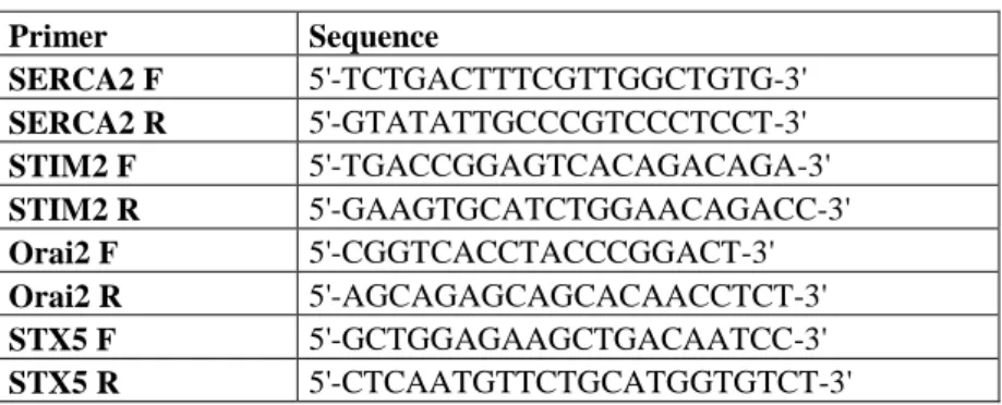
BP159925); the Primer3 homepage was used for primer designing. The primers (Table 1) were ordered from IDT Inc.
Table 1: Sequence of primers used in the experiments
Primer Sequence
SERCA2 F 5'-TCTGACTTTCGTTGGCTGTG-3' SERCA2 R 5'-GTATATTGCCCGTCCCTCCT-3'
STIM2 F 5'-TGACCGGAGTCACAGACAGA-3'
STIM2 R 5'-GAAGTGCATCTGGAACAGACC-3'
Orai2 F 5'-CGGTCACCTACCCGGACT-3'
Orai2 R 5'-AGCAGAGCAGCACAACCTCT-3'
STX5 F 5'-GCTGGAGAAGCTGACAATCC-3'
STX5 R 5'-CTCAATGTTCTGCATGGTGTCT-3'
Messenger RNA was isolated with the help of Dynabeads mRNA DIRECT Micro Kit (Invitrogen Co.). The Real-Time PCR reactions were carried out using a Bio- Rad thermal cycler.
The cDNS gained from the oocytes was inserted into E.coli bacteria with a plasmid vector using the Topo TA Cloning kit (Invitrogen). The bacteria were cultured at 37°C on LB agar plates supplemented with ampicillin overnight; the colonies carrying the plasmids were inoculated and then cultured again overnight in liquid LB medium at 37°C with constant shaking. The plasmids containing the given gene were purified by means of the Qiaprep Spin Miniprep Kit (Qiagen). The plasmid DNA was then digested with ECORI restriction enzyme and checked on an agarose gel whether or not the desired gene got inserted. The sequence of the insert was then checked. New primers were designed within the first PCR product in order to obtain shorter products that can be used for RT-PCR (Table 2). A standard curve for RT- PCR was generated using the DNA derived from plasmids.
Table 2: Oligonucleotide primers used for RT-PCR
Primer Sequence
ORAI2 F 5’-CACAACCTCAACTCGGTCAA-3’
ORAI2 R 5’-CTGCCAGGAAGAGCAGTGT-3’
SERCA2 F 5’-TCTGACTTTCGTTGGCTGTG-3’
SERCA2 R 5’-GATCATAATGACCCGGATGC-3’
STIM2 F 5’-ACCGGAGTCACAGACAGAAA-3’
STIM2 R 5’-CAATTATGAGGAGGGCGTGT-3’
STX5 F 5’-AGGATTTCGTGAGAGCCAAG-3’
STX5 R 5’-TTTGAAGTCATTGGACATGGAGG-3’
The statistical analysis was carried out by means of the SAS program using the Delta Delta Ct method. Transcript abundance was compared to that of the YWHAG gene.
3.3 Measuring Ca2+ influx in pig oocytes of different development stages
The pig oocytes used in these experiments were derived as described in the “The expression of genes influencing Ca2+ – oscillations in pig oocytes” chapter.
The oocytes were incubated in Ca2+-free medium in the presence of 10 µM CPA for two hours (to deplete the intracellular Ca2+- stores). They were then loaded with the Ca2+ indicator dye Fura 2-AM by incubation with the dye for half an hour. The Ca2+
measurements were carried out with an InCyt Im2 fluorescence imaging system attached to a Nikon Eclipse TE-2000U inverted microscope. After the first ~30- second baseline measurement a medium with elevated Ca2+ concentration (10 mM) was added to the oocytes and the changes in the intracellular Ca2+ concentration were monitored.
4 Results
4.1 Polymorphism of the prolactin receptor gene in mangalica
After the digestion with a restriction enzyme the separation of the different fragments on an agarose gel gave the following two different alleles: A allele - 127 bp, B allele - 92, 35 bp.
The results indicate that in mangalica the polymorphism of the prolactin gene exists and the A allele is related to the bigger litter size. Sows with AA genotype had 2.21 more piglets than sows with BB genotype. AA sows had 1.33 more piglets than the average of the population and had 1.77 piglets more than sows genotyped as AB.
The three genotypes differ significantly from each other (p<0,005): sows with AA genotype had bigger liter sizes than those with AB or BB genotype; however, there was no significant difference between the AB and BB genotypes (p> 0,005).
The frequency of the A allele was 32% and the rate of the AA genotype was lower (8%) which is in accordance with the results of other studies. It seems reasonable to suggest that an increase in the frequency of the A allele through selection would lead to an increase in the litter size as well.
AA genotype sows had the biggest litter sizes while BB sows had the smallest. In the experiments sows with AB genotype had also smaller litter sizes than those with AA genotype.
4.2 Expression of genes influencing Ca2+ oscillations in pig oocytes
The sizes of the PCR products obtained with the use of the first pairs of primers were the following: SERCA2 493 bp, Orai2 441 bp, STIM2 497 bp, STX5 499 bp.
The designed RT-PCR products were as follows: SERCA2 103 bp, STIM2 82 bp, Orai2 106 bp és STX5 114 bp.
Statistical analysis indicated a significant difference in the expression of two genes in oocytes of different development stages (p<0,05). There were no changes in the expression of the SERCA2 and STIM2 genes while the expression of Orai2 and STX5 genes reduced during the course of maturation.
Figure 1 shows the expression of the genes (SERCA2, Orai2, STIM2, STX5) in the oocytes of different developmental stages (GV, MI, MII).
Figure 1: The difference of gene expression of the observed genes in oocytes of various developmental stages (GV, MI and MII).
(a,bDifferent letters show significant differences; P<0,05)
The unchanged expression level of the SERCA2 gene may be the result of its role in oocyte development. The gene product is a component of the ER store which is equally important in all developmental stages during maturation; moreover, this organelle has additional functions in cellular communication as well. SERCA2 is the most sensitive to Ca2+ changes among all SERCA variants.
The role of the STIM2 and Orai2 proteins are still relatively unknown. Different studies showed that STIM2 has a role similar to STIM1 although it may be more involved in inhibition and Orai2 has a similar – although weaker – function as Orai1 does.
Upon store depletion STIM proteins form dimers (and oligomers) and attach to Orai proteins that leads to the opening of the Ca2+ channels of the plasma membrane. The over-expression of STIM1 only had no effect on store-operated Ca2+ influx while co- over-expression with Orai1 led to a significant increase in Ca2+ influx. On the other hand, over-expression of STIM2 with Orai1 inhibited the Ca2+ influx after the depletion of the stores although others reported that significantly higher level of STIM2 together with Orai1 can cause an elevated Ca2+ influx.
It seems that the ratio of Orai to STIM proteins is important regarding store-operated Ca2+ influx. Over-expression of the two proteins causes an elevated Ca2+ influx while their over-expression one-by-one inhibited SOCE. Our own results confirm these observations. Immature oocytes – in which the level of Orai2 is relatively high compared to STIM1 – show only a limited number of Ca2+ rises after fusion with sperm cells. During maturation the ratio of Orai proteins compared to STIM proteins decreases and this may be important for the oocytes to acquire the ability to display the repetitive Ca2+-oscillations in response to the fertilizing sperm.
The decrease in the expression of syntaxin 5 gene may be explained by its inhibitory role; its protein is linked to polycistin-2 and inhibits the Ca2+-current across the ER membrane. Therefore, its decrease is understandable as during oscillations the efflux of Ca2+ from the stores is mainly responsible for the generation of the Ca2+ signal.
4.3 Measuring store-operated Ca2+ influx in pig oocytes of different development stages
The following results were obtained during fluorescence measurements of the oocytes at different development stages: in the GV and MI phase a low Ca2+ influx can be seen while in MII phase oocytes a significant elevation in the intracellular Ca2+ levels can be seen following Ca2+ add-back (Fig. 2, 3 and 4).
Figure 2: Ca2+ peak induced by 10 µM CPA and 10 mM Ca2+ in a MII oocyte
This sudden elevation in the intracellular Ca2+ level may be able to activate the oocyte and could serve as a first step in parthenogenetic oocyte activation protocols.
This elevation is the result of an influx of extracellular Ca2+ into the cell and is induced by the depletion of the intracellular stores (store-operated Ca2+ entry, SOCE).
Figure 3: Ca2+ elevation induced by 10 µM CPA and 10 mM Ca2+ in GV oocyte
The results show that in mature oocytes a significant Ca2+-influx follows the depletion of the stores; this is important for refilling these stores and for the maintenance of the long-lasting series of Ca2+ oscillations caused by the sperm. In GV and MI phase oocytes there is no remarkable Ca2+ influx after depletion of the stores; this is in accordance with the observation that in the immature oocytes the sperm can produce only a short Ca2+ signal.
Figure 4: Ca2+ increase induced by 10 µM CPA and 10 mM Ca2+ in an MI oocyte
5 New scientific results
1) The polymorphism of the prolactin receptor gene exists in mangalica and the different alleles have different effects on the litter size. We found that sows carrying the A allele and the AA genotype had more piglets at the time of farrowing but they are present at a lower rate in the population. by selection and keeping the other allele this rate should be increased so the litter sizes would increase, too.
2) In pig oocytes the expression of the four genes that we studied changes during the course of maturation. The expression of syntaxin 5 and Orai 2 decreases while those of STIM 2 and SERCA 2 do not change significantly.
These genes were not studied before in pig oocytes.
3) In mature oocytes the depletion of the Ca2+ stores induces a Ca2+ influx across the plasma membrane after blocking it with CPA (cyclopiazonic acid) and adding a media containing an elevated level of Ca2+. The level of the influx increases as the oocyte matures and this is crucial during fertilization when repetitive Ca2+ signals generated by the sperm are required to induce proper embryonic development. We measured the Ca2+
influx in pig oocytes for the first time after a blockage with CPA.
6 List of scientific articles published in peer-reviewed journals:
Wang C - Lee K. - Gajdócsi E - Papp AB - Machaty Z. (2012): Orai1 mediates store-operated Ca2+ entry during fertilization in mammalian oocytes. Developmental Biology. 2012 365(2):414-23. (IF: 4,069)
Gajdócsi Erzsébet. – Kiho Lee – Chunmin Wang – John M. Challie – Bali Papp Ágnes – Macháty Zoltán (2011): A sertés petesejtek calcium oszcillációját
befolyásoló gének expressziója. Magyar Állatorvosok Lapja 2011/2 104-107 (IF:
0,201)
Pataki R. – Gajdócsi E. – Kiss R. - Tempfli K. - Varga E. - Konrád Sz. - Bali Papp Á. (2009): A prolaktin receptor gén alomszámra gyakorolt hatásának vizsgálata a mangalica sertésekben. Acta Agronomica Óváriensis, 51 73-82.
Gajdócsi E. – Bali Papp Á. (2009): Sertés géntérképezés, Magyar Állatorvosok Lapja 2009/3, 148 (IF: 0,2)
Gajdócsi E – Pataki R – Tempfli K – Bali Papp Á (2008): A prolaktin receptor gén hatása a mangalicák alomméretére. Animal welfare, etológia és
tartástechnológia, Gödöllő, Vol.4. Is. 2.
Varga E. –Petz Makkosné B. –Gajdócsi E. –Salamon I. –Bali Papp Á. (2008):
Vitrification of in vitro matured oocytes of Mangalica (Hungarian native breed pig) and Large White pig. Acta Veterinaria Hungarica 56 (3) (IF: 0,535)
IF:5,005
7 Conference presentations and posters
Nánássy L.- Gajdócsi E. – Dudás B. – Antal F. – Vereczkey A. (2013):
Evaluation of the efficiency of in vitro fertilization combined with Preimplantation Genetic Screening in a small setup. COGI konferencia, Vienna 24-27 Oct. 2013 (poster)
E. Gajdócsi – K. Lee – C. Wang – J.M. Challie – Á. Bali Papp – Z. Macháty (2011): Expression of genes influencing the calcium oscillation in pig oocytes.
ESDAR conference, Antalya, Turkey, 15-17 Sept. 2011 (poster)
Gajdócsi Erzsébet – Kiho Lee – Chunmin Wang – John M. Challie – Bali Papp Ágnes – Macháty Zoltán (2010): A sertés petesejtek calcium oszcillációját befolyásoló gének expressziója. 16. Szaporodásbiológiai Találkozó, Visegrád, 29-30 Oct. 2010 (presentation)
Gajdócsi Erzsébet. – Kiho Lee – Chunmin Wang – John M. Challie – Bali Papp Ágnes – Macháty Zoltán (2010): A kalcium-hullámok génexpressziója sertés petesejtekben. Óvári Tudományos Nap, Mosonmagyaróvár, 7 Oct. 2010 (poster)
E. Gajdócsi – R. Pataki – R. Kiss – K. Tempfli – Á. Bali Papp (2008):
Connection of prolactin receptor gene with Mangalica pigs’ litter-size. XXXII.
Óvári Tudományos Napok, Mosonmagyaróvár, 9 Oct. 2008 (poster)
E. Gajdócsi – R. Pataki – Á. Bali Papp (2008): The effect of the gene of prolactin receptor on Mangalica pigs’ litter-size. Second European Conference on Pig Genomics. Pig Genome II. , Ljubjana, 4-5 June (poster)
Gajdócsi E – Pataki R – Tempfli K – Bali Papp Á (2008): A prolaktin receptor gén hatása a mangalicák alomméretére. I. Gödöllői Állattenyésztési Tudományos Napok. Gödöllő, 11-12 Apr. 2008 (poster)
Gajdócsi E. – Pataki R. – Bali Papp Á. (2007): Az alomszámot befolyásoló gének vizsgálata sertés fajban. XXVIII. OTDK Agrártudományi szekció. Debrecen, 16-18 Apr. 2007 (presentation)
Pataki R. - Gajdócsi E – Varga E. - Bali Papp Á (2007): Az alomszámot befolyásoló gének vizsgálata sertésben. VII. Magyar Genetikai Kongresszus. XIV.
Sejt –és Fejlődésbiológiai Napok. Balatonfüred, 15-17 Apr. 2007 (poster)
Varga E – Gajdócsi E – Bali Papp Á (2006): Különböző fajtájú sertés petesejtek vitrifikációs hűtése, visszaolvasztás utáni fertilizációja és az embriók fejlődése.
Állatbiotechnológiai kutatások Magyarországon. Budapest, MTA Székház, 29 Sept.
2006 (presentation)
Varga E –Gajdócsi E – Bali Papp Á (2006): In vitro maturált sertés petesejtek aktiválása. XXXI. Óvári Tudományos Nap „Élelmiszeralapanyag előállítás - Quo vadis?”Mosonmagyaróvár, 6 Oct. 2006 Előadások és poszterek összefoglaló anyaga 64. (poster)