Physiological changes during nonivasive ventilation of COPD patients and cellular effects of cigarette
smoke
Ph.D. thesis
József Lukácsovits MD
Clinical Medicine Doctoral School Semmelweis University Budapest
Supervisor: Veronika Müller MD, Ph.D., associate professor
Official reviewers: Éva Vizi MD, Ph.D., division head Erna Sziksz Ph.D.
Head of the Final Examination Committee:
Prof. Endre Cserháti MD, Ph.D., D.Sc Members of the Final Examination Committee:
Prof. Attila Somfay MD, Ph.D.
János Varga MD, Ph.D., division head Zsuzsanna Orosz MD, Ph.D.
Budapest 2013
1. Introduction
Smoking plays a central role in the development of many illnesses. Every year more than 5 million people on the world die in some forme of disease caused by cigarette smoke. Smokers live about 10 years less than non smokers and have a very low quality of life in the last years of their life.
Smoking was the cause of about 100 million deaths worldwide in the twentieth century. About a third of adult population is currently an active smoker. Cigarette smoke has a verified etiological role in the development of several malignant and non-malignant illnesses. One of the most common pulmonary illness caused by cigarette smoke is chronic obstructive pulmonary disease (COPD). According to its definition COPD is a pathological condition characterized by not completely reversible airway obstruction, caused by chronic inhalation of toxic particles and gases. The lung tissue damaging mechanisms of cigarette smoke are not fully elucidated. Intensive current researches investigate the role of cigarette smoke induced apoptosis or necrosis in development of COPD and emphysema and the role of glucocorticoids and heat- shock proteins (HSP) in this pathological process. In advanced stage of COPD, the reduced pulmonary surface and ventilation/perfusion inhomogeneity can lead to hypercapnic respiratory failure. Long term noninvasive positive pressure ventilation (NPPV) used in this pathological condition improves arterial blood gases, respiratory function, health related quality of life and reduces the frequency of hospitalisation. On the other hand the effect of NPPV on long term survival of COPD patients is uncertain, for this reason many current researches investigate the physiological changes during different NPPV methods.
2. Aim
1. Breathing pattern analysis of short term high intensity (Hi)-NPPV in COPD patients.
2. Analyis of changes in respiratory muscle mechanics using different NPPV settings and comparison to spontaneous breathing in COPD patients.
3. Effect of Hi-NPPV on blood gas parameters and patients perception of dypnea.
4. Analysis of short term hemodynamic changes (cardiac output, oxigen transport capacity) following Hi-NPPV in COPD patients.
**
5. Production of stabile quality cigarette smoke extract (CSE).
6. Assessment of the effect of CSE treatment on alveolar epithelial cells.
7. Interaction of CSE and the glucocoticoid dexamethasone (DEX) on the proliferation and apoptosis of type II alveolar epithelial cells.
8. Role of HSP72 in CSE and/or DEX effects in alveolar epithelial cells.
3. Methods
3.1. Physiological changes during NPPV
We studied 15 patients who were admitted for the management of chronic hypercapnic respiratory failure due to COPD. At the time of the study, all patients were in a phase of clinical and haemodynamic stability. Enrolment criteria were the presence of chronic hypercapnic respiratory failure (i.e. pH
>7.35 and Pa,CO2 >50 mmHg), while exclusion criteria were the presence
of cancer, neuromuscular disease or serious heart failure. The study was approved by the local ethics committee and written informed consent was obtained from all patients.
The ventilator therapy was stopped at least 1 h before the beginning of each trial. The patients were studied in a semirecumbent position. The physiological data were recorded during spontaneous breathing (SB) at the beginning of the trial and during two different ventilatory settings over 30 minutes, in randomised sequence, separated by a return to SB for 10 min.
The patients were ventilated with a noninvasive ventilator and with a oro- nasal mask, tailored to each patient’s facial features.
We used two different ventilatory settings, the low intensity (Li)-NPPV and the high intensity (Hi)-NPPV. During Li-NPPV setting we used an IPAP of 17.7± 1.6 cmH2O, expiratory pressure (EPAP) of 4 cmH2O and back up rate of 12/min.
With Hi-NPPV, we aimed to reach the maximum tolerated inspiratory pressure (at least 50% greater than Li-NPPV) by increasing pressures stepwise by 0.5 cmH2O/min. This allowed us to reach mean inspiratory pressure values very close to those walues publiched by Windisch W. and co-workers. During Hi-NPPV we used an IPAP of 27.6 ± 2.1 cmH2O, EPAP was fixed at 4 cmH2O and respiratory rate was set to match the SB rate, usually 20–22 breaths/min.
Arterial blood gases were obtained from a radial artery during SB and at the end of each ventilatory trial. Respiratory flow was measured with a heated pneumotachograph and a differential pressure sensor placed between the mask and the Y-piece of the ventilator. Tidal volume (Vt) was obtained by integration of the respiratory flow. Breathing pattern was measured from the flow signal. Expired Vt was used for data analysis. We also measured
the difference between expired and inspired Vt to quantify the amount of air leakage.
Airway pressure was measured from a side port between the pneumotachograph and the face mask. Oesophageal pressure was measured with an oesophageal balloon positioned at the lower third of the oesophagus.
Gastric pressure was measured with a gastric balloon. Transdiafragmatic pressure (Pdi) was calculated as the difference between gastric and oesophageal pressure.
Diaphragmatic pressure–time product (PTPdi) - which is a good indicator of the respiratory muscle’s oxigen consumption - were calculated per breath (PTPdi/breath) and per minute minute (PTPdi/min) by the Pdi trace.
Dynamic PEEPi (PEEPi,dyn) was obtained from the Pdi signal, as the value of Pdi at the moment of zero flow. Ineffective efforts (IEs) were analysed from the Pdi traces and expressed as percentage of total breaths measured.
We also measured cardiovascular parameters using a noninvasive device (Finometer PRO; Finapres Medical Systems BV, Amsterdam, the Netherlands). The measured and calculated cardiovascular parameters were systolic arterial pressure (SAP), diastolic arterial pressure (DAP), mean arterial pressure (MAP), heart rate, stroke volume (SV) and cardiac output (CO). Dyspnoea was assessed using the Borg scale, with a range of 0– 10, during SB and the two ventilator trials.
Data were presented as mean ± SD. Comparisons for each sequence and each continuous variable were performed, using ANOVA for repeated measures with the Newman–Keuls post hoc test. The protection test for carry-over effects was also performed. Outcome measures were tested for normality with the Kolmogorov–Smirnov test, and when normality was not achieved, we used the Friedman test. Statistical analysis was performed with
Prism and Medcalc softwares. All tests were two-sided. A p-value < 0.05 was considered statistically significant.
3.2. Effect of cigarette smoke on lung epithelial cells
A549 human type II alveolar epithelial cell line was used. Cells were cultured in Dulbecco’s modified Eagle’s medium (DMEM), containing glucose, fetal bovine serum, antibiotics/antimicotics and L-glutamine. Cells were incubated in a humidified incubator with 5% CO2, at 37°C.
Cigarette smoke extract (CSE) was prepared by bubbling the smoke from two commercially available filter cigarettes through 16 ml of pre-warmed (37°C) serum-free cell culture medium. The cigarettes were machine smoked at a rate of 35 ml over a period of 2 s followed by a pause of 28 s.
This cycles of 2s/28s were repeated for the two cigarettes, matching the smoking habits of an average smoker. The resulting CSE was applied to epithelial cell cultures within 30 min after preparation. Stability of CSE solutions was checked with highperformance liquid chromatography-mass spectrometry.
For the treatment, A549 cells were cultured on 6-well pates in 2 ml medium. CSE and/or dexamethasone (DEX) at three different concentrations (0.1; 1.0; and 10 μM) was given to the medium. DEX free (0.0 μM) groups served as steroid naive controls (C). The cells were treated with CSE and/or DEX for 24 h at 37°C in a humidified atmosphere containing 5% CO2.
Cell number was measured using Cell-Dyn 3200. Apoptosis was assessed by flow cytometric (FACS) analysis after annexin-V/propidium-iodide staining. Hsp72 expression was analyzed using anti-Hsp72 antibody and
Cy5-conjugated secondary antibody staining. The cytometric analysis was carried out using a FACSAria cytometer.
Total ribonucleic acid (RNA) was isolated from the cells using RNeasy RNA isolations kit. RNA was reverse transcripted using SuperScript II RNase-H to generate first-strand complementary dezoxy-ribonucleic acid (cDNA). Using this cDNA, real-time PCR was performed (Roche Diagnostics, Mannheim, Germany) to determine Hsp72 and glyceraldehyde- 3-phosphate mRNA expression.
Hsp72 silencing (SiRNA) transfection was performed with transfection Neo FX™ transfection agent. Scrambled (Scr)-RNA was used similarly as negative control for the transfection. 24 h after transfection, cells were treated with CSE, DEX, or both. The samples were assayed by flow cytometry 24 h thereafter, measuring apoptosis, necrosis and Hsp72 protein expression.
All data are expressed as mean ± SD. After testing the normality with Shapiro–Wilk’s test, the non-parametric Mann–Whitney U test was used to determine the levels of difference among all treatment groups. P values
<0.05 were considered statistically significant.
4. Results
4.1. Physiological changes during noninvasive ventilation
All patients tolerated the experimental procedures well and completed the study. Eight out of 15 patients were randomised to receive Li-NPPV as the first intervention. Compared with baseline conditions, both Hi-NPPV and Li-NPPV significantly increased expired Vt and minute ventilation (VE)
(SB: 9.6 ±3.5 l ; Li-NPPV: 11.7±2.3 l ; Hi-NPPV: 15.2±2.0 l; SB vs. Li- NPPV p<0.01; SB vs. Hi-NPPV p<0.001; Li-NPPV vs. Hi-NPPV p<0.001), but these changes were statistically higher with Hi-NPPV.
The amount of air-leaks was significantly lower with Li-NPPV (Li-NPPV:
10.62±5.27 l/min; Hi-NPPV 16.11±7.88 l/min; p<0.05 ). The mean inspiratory flow was also higher with Hi-NPPV (Li-NPPV: 0.52±0.11 l/s ; Hi-NPPV 0.93±0.18 l/s p<0.05) and the duty cycle (Ti/Ttot) lower (Li- NPPV: 0.37±0.08 ; Hi-NPPV 0.27±0.05 p< 0.001) , whereas no statistical difference was observed in breathing frequency.
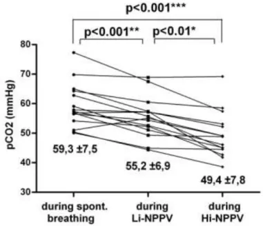
Tidal Pdi, PTPdi/breath and PTPdi/min were significantly reduced compared with SB, but the decrease in all these parameters was significantly greater using Hi-NPPV versus Li-NPPV (PTPdi/min: SB: 323±149 H2Ocm*s/min ; Li-NPPV: 132±139 ; Hi-NPPV 40±69 H2Ocm*s/min; SB vs. Li-NPPV p<0.001; SB vs. Hi-NPPV p<0.001; Li-NPPV vs. Hi-NPPV p<0.01) (Fig 1.) .
Fig 1. During NPPV the PTPdi were significantly reduced compared with SB, and this decrease was significantly greater using Hi-NPPV versus Li- NPPV.
Indeed, in nine out of 15 patients, the Pdi trace was flat and the pleural pressure (Ppl) even became positive during Hi-NPPV, suggesting a ‘‘true’’
controlled ventilation. For this reason, data on PEEPi,dyn are not presented.
Lung resistance was also significantly decreased during mechanical ventilation, but no difference was found between the two ventilator settings.
During both ventilatory settings, pH and PaCO2 improved significantly versus baseline conditions, the changes being significantly less pronounced using Li-NPPV than Hi-NPPV (PaCO2: SB: 59.3±7.5 mmHg; Li-NPPV:
55.2±6.9 mmHg ; Hi-NPPV 49.4±7.8 mmHg; SB vs. Li-NPPV p<0.001; SB vs. Hi-NPPV p<0.001; Li-NPPV vs. Hi-NPPV p<0.01) (Fig 2.).
Fig 2. During both ventilatory settings PaCO2 improved significantly versus baseline conditions, the changes being significantly less pronounced using Li-NPPV than Hi- NPPV.
No statistical differences were observed in dyspnoea score between the two modes of ventilation, but both resulted in a statistical reduction versus baseline (Borg score 6.93±1.1 in SB versus 3.13±1.64 in Li-NPPV p<0.001 and 4.67±1.04 in Hi-NPPV p<0.01).
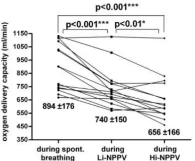
Mechanical ventilation induced a significant decrease in SAP, but without any difference between Hi-NPPV and Li-NPPV. NPPV also reduced SV, CO and cardiac index; the decrease was statistically more pronounced with Hi-NPPV than Li-NPPV. Similar changes were observed in the calculated oxygen delivery capacity (DO2) and the oxygen delivery corrected by body surface area (DO2/m2: SB: 577±110 ml/min/m2 ; Li- NPPV: 462±93 ml/min/m2 ; Hi-NPPV 409±104 ml/min/m2 ; SB vs. Li- NPPV p<0.001; SB vs. Hi-NPPV p<0.001; Li-NPPV vs. Hi-NPPV p<0.01 ) (Fig 3.).
Fig 3. NPPV induced a reduction in oxygen delivery capacity (DO2) and this decrease was statistically more pronounced with Hi-NPPV than Li- NPPV.
A statistically significant correlation was observed between the per cent changes from baseline CO and absolute changes from baseline Ppl in the Hi- NPPV trial (r = -0.68, p<0.01). We found similar correlations between changes from baseline airway pressure and per cent changes from baseline CO during Hi-NPPV (r= -0.59, p<0.05).
4.2. Cigarette smoke effect on lung epithelial cells
Under control conditions cells proliferated. Following DEX treatment no significant change in cell number was noted. In contrast, CSE treatment significantly reduced the cell number 24 h after incubation as compared to controls. CSE+DEX co-treatment dose-dependently and significantly increased the total cell count as compared to CSE treatment alone, reaching
similar number in both DEX (10) groups (C+DEX (10): 10.22±0.77×10⁵ cell/vial; CSE+DEX (10): 8.86±0.49×10⁵ cell/vial).
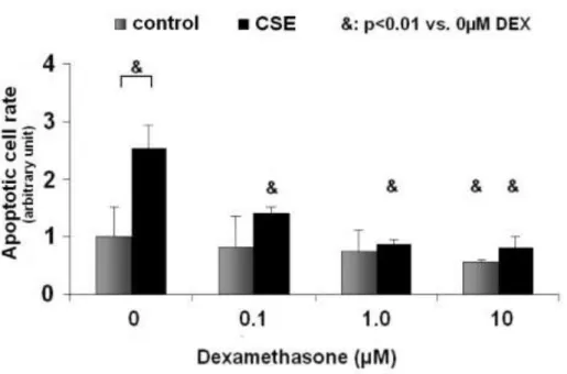
DEX slightly decreased the number of the apoptotic cells in controls, reaching statistical significance only in the C+DEX (10) group. In steroid- naive CSE-treated cells, apoptosis tripled as compared with the steroid naive controls. DEX treatment significantly reduced apoptosis in all CSE-treated groups (Fig 4).
Fig 4.: In steroid-naive CSE-treated cells, apoptosis tripled as compared with the steroid naive controls. DEX co-treatment significantly, dose dependently reduced apoptosis in all CSE-treated groups.
The ratio of necrotic cells did not differ in controls, whether DEX treatment was used or not. In contrast, CSE significantly increased the number of necrotic cells. Similarly to controls, DEX had no additional effect on necrosis in CSE-treated cells resulting in significantly higher necrotic cell
rate in all CSE groups (CSE: 6.24±1.02%; CSE+DEX (0.1): 6.12±2.11%;
CSE+DEX (1.0): 6.68±1.1%; CSE+DEX (10): 4.7±0.75%; p<0.05 vs.
respective control group).
Hsp72 mRNA expression did not change following DEX treatment in controls. Administration of CSE alone was associated with low level of Hsp72 mRNA, similar to the level observed in steroid naive controls. In contrast, DEX treatment resulted in significant increase in Hsp72 mRNA, already using 0.1 μM/μl concentration in the presence of CSE, and further significant increase was observed using 1 μM DEX. However, no further increase was detected in the CSE+DEX (10) cells.
Hsp72 protein expression of individual epithelial cells was low in steroid naive controls, similarly to the ratio of Hsp72 expressing cells. DEX treatment significantly decreased both the ratio of Hsp72 expressing cells and the cellular Hsp72 content of the cells in controls. In all CSE-treated groups, the ratio of Hsp72-positive cells was significantly higher as compared to respective controls (p<0.01).
DEX treatment significantly and dose-dependently increased the number of Hsp72 expressing cells, with the highest ratio measured in the CSE+DEX (10) group (Fig 5.).
Fig 5.: DEX treatment significantly and dose-dependently increased the number of Hsp72 expressing cells, with the highest ratio measured in the CSE+DEX (10) group.
CSE treatment significantly increased the cellular Hsp72 protein content in all (steroid naive and DEX treated) groups as compared to respective controls. The mean intensity of Hsp72 in Hsp72-positive cells increased with increasing doses of DEX following CSE treatment, compared to steroid naive controll group (p<0.01).
Transfection with siRNA was successful in steroid naive controls and CSE-treated cells. While scr-RNA did not change cellular Hsp72 protein expression, siRNA treatment resulted in significant decrease of cellular Hsp72 protein under control and CSE treated conditions. Similarly, Hsp72 protein expressing cell number significantly decreased in all groups following siRNA treatment. In C and CSE groups, siRNA reduced the number of Hsp72-positive cells by 80% and 60%, respectively, while it reached 75% in the CSE+DEX (10) group. The most marked decrease in
treatment, where significant reduction in the ratio of Hsp72- positive cells, and the reduction of the content of the expressing cells was observed.
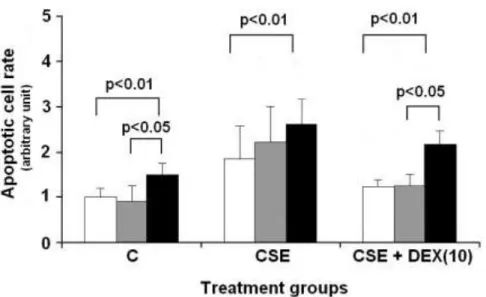
Parallel to the decreased expression of Hsp72 protein in siRNA treated cells, apoptosis significantly increased in all groups, indicating a direct link between cellular Hsp72 and apoptotic cell death (Fig 6.).
Fig 6.: In siRNA treated cells, apoptosis significantly increased in all groups (Control, CSE alone and CSE + DEX (10)).
5. Summary
Hi-NPPV in stable COPD patiens:
1. Significantly increases Vt and minute ventilation, while significantly decreases insiratory time and duty cycle.
2. Significantly increases Pdi amplitude and decreases PTPdi.
3. Acutely improves gas exchange and reduces the patient’s respiratory effort.
4. Significantly lowers cardiac output and other indices of cardiac performance, as well as oxigen transport.
**
5. With the developed „smoke machine” reproducible, stable quality of CSE was obtained
6. CSE significantly increased apoptosis of alveolar epithelial cells.
7. DEX significantly reduced CSE-induced cellular damage, by decreasing apoptosis.
8. Our transfection results confimed the central role of HSP72 in the increased survival following DEX-CSE treatment in alveolar epithelial cells.
6. Publications
Publications related to the dissertation:
Lukácsovits J, Carlucci A, Hill N, Ceriana P, Pisani L, Schreiber A, Pierucci P, Losonczy G, Nava S. (2012) Physiological changes during low
and high "intensity" noninvasive ventilation. Eur Respir J, 39: 869-875.
IF: 5,895 Gál K, Cseh A, Szalay B, Rusai K, Vannay A, Lukácsovits J, Heemann U, Szabó AJ, Losonczy G, Tamási L, Müller V. (2011) Effect of cigarette smoke and dexamethasone on Hsp72 system of alveolar epithelial cells. Cell
Stress Chaperones. 16: 369-378.
IF: 3,013
Other Publications:
Lukácsovits J, Nava S. (2013) Inspiratory pressure during noninvasive ventilation in stable COPD: help the lungs, but do not forget the heart.
Correspondence, From the autors. Eur Respir J, 41: 765-766.
IF: 5.895
Szondy K, Rusai K, Szabó AJ, Nagy A, Gal K, Fekete A, Kovats Z, Losonczy G, Lukácsovits J, Müller V. (2012) Tumor cell expression of heat shock protein (HSP) 72 is influenced by HSP72 [HSPA1B A(1267)G]
polymorphism and predicts survival in small Cell lung cancer (SCLC)
patients. Cancer Invest, 30: 317-322.
IF: 1,847
Máthé C, Bohács A, Duffek L, Lukácsovits J, Komlosi ZI, Szondy K, Horváth I, Müller V, Losonczy G. (2011) Cisplatin nephrotoxicity aggravated by cardiovascular disease and diabetes in lung cancer patients.
Eur Respir J, 37: 888-894.
IF: 5,895 Gál K, Cseh A, Szalay B, Rusai K, Vannay A, Lukácsovits J, Uwe H, Szabó AJ, Losonczy G, Tamási L, Kováts Z, Müller V. (2011) Dohányfüst és szteroid hatása tüdő-epithelsejtek hősokkfehérje (HSP) 72-rendszerére.
Med Thor, 64: 152-160.
Máthé C, Bohács A, Duffek L, Lukácsovits J, Komlosi ZI, Szondy K, Horváth I, Müller V, Losonczy G. (2011) Cardiovascularis betegségben és diabetes mellitusban szenvedő tüdőcarcinomás betegekben fokozódik a cisplatin nephrotoxikus hatása. Med Thor, 64: 33-41.
Lukácsovits J, Komáromi T, Tamási L, Magyar P, Losonczy G. (2009) Kétoldali intersticiális pneumonia miatt gépi lélegeztetett beteg esetismertetése. Med Thor, 52: 137-139.
Dr. Lukácsovits J fordítása. (2009) A Sepsis legutóbbi 100 éve. Med Thor, 62: 266-277.
7. Acknowledgements
I would like to acknowlwdge the generous help of Prof. Dr. György Losonczy, Prof. Dr. Pál Magyar, Dr. Veronika Müller and Prof. Dr. Stefano Nava in my scientific work and in the writing of my dissertation.