Dispersal by Air and Water—The Take-Off
C . T . INGOLD
Department of Botany, Birkbeck College, University of London, England
I. Introduction 137 II. Violent Spore Discharge 138
A. Ascomycetes 140 1. Discomycetes Type 141
2. Pyrenomycetes Type 143 3. Erysiphales Type 146
B. Basidiomycetes . . . . 147
C. Other Types of Discharge 155 III. Passive Spore Liberation 159 IV. Meteorological Conditions in Relation to Spore Liberation . . . . 162
A. Humidity and Rain 162 B. Temperature 1β3 C. Wind 163 D. Light 164 V. Periodicity of Spore Liberation 164
References . 166
I. INTRODUCTION
The dispersal story of a fungus can usually be divided into three major episodes: first, liberation of spores or their actual escape from immediate contact with the parent tissue; secondly, their dispersal in a viable condition to a greater or lesser distance; and thirdly, the coming to rest of the spores on solid substrata, on some of which germination and successful establishment may occur. This chapter is concerned essentially with the first episode although the second cannot completely be ignored, because the efficiency of take-off can be judged only in relation to subsequent dispersal. In particular it is important to consider the turbulence or nonturbulence of the air into which spores are liber
ated. Before discussing this matter, however, the various types of spore liberation will be considered.
This account will be largely concerned with organisms of importance in plant pathology although reference will often be made to sapro-
137
phytic types in which particular dispersal mechanisms have been more fully studied than in essentially similar pathogenic forms. However, certain examples, highly interesting in the general context of dispersal in fungi but of no phytopathological importance, will be ignored.
Thus the beautiful discharge mechanisms of Pilobolus, Ascobolus, and Coprinus spp. will receive no mention, nor will the wide range of dispersal types displayed in Gasteromycetes be considered (Ingold, 1953).
The conspicuous part of a fungus is essentially concerned with the production and liberation of spores, the feeding part being usually hidden away as a branched mycelium in the nutritive substratum. If we
hope to understand a reproductive structure in a fungus, the question must be asked: "How are the spores set free?" It is extraordinary how often, even for the commonest fungi, no really satisfactory answer can be given to this question.
It is convenient to recognize two contrasting types of spore libera
tion. In the first, the spores are actively and violently discharged. In the second, liberation is passive in the sense that the energy concerned comes from outside the fungus, the dislodgment of the spores being due to the kinetic energy of wind or rain.
II. V I O L E N T SPORE DISCHARGE
In connection with the violent discharge of projectiles the size of fungal spores (mostly less than 50μ in diameter) or small spore groups, certain basic mathematical expressions should be considered.
According to Stokes' Law, the rate of fall of a minute spherical particle in a fluid is given by
T 7 2p - δ 2
V =
9-ir
gawhere V = the terminal velocity of fall in the medium ρ = the density of the falling sphere
δ = the density of the fluid
g = the acceleration due to gravity (981 cm./sec.)
μ = the viscosity of the medium (1.8 X 10~4 in the case of air).
This can be simplified to
since for fungal spores the density is approximately 1.0, and since the density of the air can be neglected. Turning to the question of the horizontal discharge of a spore we find, according to Barlow (in Buller, 1909) that
Η qD
V (2)
where Η is the initial horizontal velocity of discharge and D is the distance of horizontal throw. Combining ( 1 ) and ( 2 ) , we get
(3) Thus for a given initial velocity of discharge, the distance of horizontal throw is proportional to the square of the radius of the spherical projectile.
The path of a spore projected horizontally is
y =
jL
l0geV~D)--D_
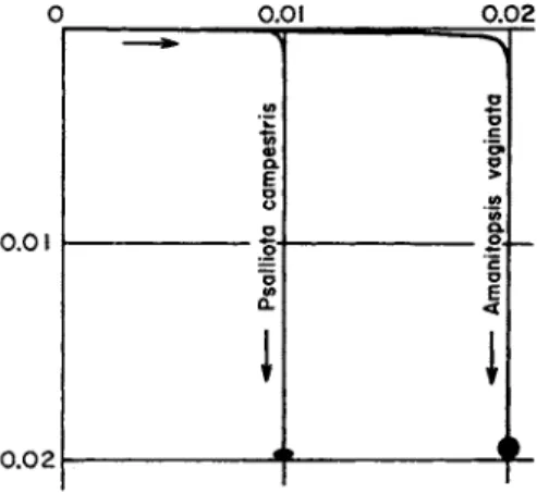
(4)y being the vertical distance of a point on the curve below the level of the point of departure, and χ the horizontal distance from the vertical axis through this point. This curve (Fig. 1) has been referred to by
o.oi 0 . 0 2
o.oi
0 . 0 2
stris ο
ο .c
:ampe; '& ο >
ΙΛ
σ ops
i
— Psallio 'Ξ
σ Ε
<
I
i r •
FIG. 1. The sporabolas of two spores shot horizontally from the hymenium.
The spores, drawn to scale, are shown below. The scale is in centimeters. (After Buller, 1909.)
Buller (1909) as a "sporabola." It is of interest to compare distances of horizontal discharge ( D ) and of vertical upward discharge (U):
C7 = D - | l o g , ( 5 ) *
k being given by 9μΙ2a2.
* My gratitude is expressed to Dr. R. Tiffen of Birkbeck College, London, for deriving this expression.
For particles the size of fungal spores U is almost as large as D. For example, in the common coprophilous pyrenomycete Sordaria fimicoUi, the spore projectiles are relatively large and an eight-spored mass may be shot horizontally to a distance of 10 cm. This projectile consists of eight spores, each 22 χ 13μ and surrounded by a sheath of mucilage 3μ wide. The total mass has a radius of 21.6/Λ. Substituting in ( 5 ) it is found that U = 9.82 cm. In fact it has not been found possible ex
perimentally to demonstrate a consistent difference between the distance of horizontal (10.0 cm.) and vertical discharge.* The difference is still less where smaller projectiles, those normally encountered in plant pathology, are concerned.
Violent discharge of the characteristic or "perfect" spores is the rule in two great groups of fungi, Ascomycetes and Basidiomycetes, although there are many individual examples in both groups in which discharge of ascospores or basidiospores is no longer active. Outside these two groups there are few examples of violent discharge, for in the Phyco- mycetes generally and in nearly all conidial fungi, including the conidial stages of Ascomycetes and the vast hordes of Fungi Imperfecti, libera
tion of spores is essentially passive. In this chapter violent discharge will be considered first.
A. Ascomycetes
In the majority of Ascomycetes the ascus is explosive although in many genera, widely scattered in any classification, this explosive character has been lost during the course of evolution or, perhaps, has never existed. Eurotium, Tuber, Chaetomium, and Ceratocystis (Ophio- stoma) are familiar examples.
The typical ascus is a cylindrical, turgid, elongated cell with a thin cell wall (two-layered in certain Ascomycetes), a thin lining layer of protoplasm, and a large central vacuole containing cell sap in which the ascospores (usually eight in number) are suspended toward the upper end of the ascus. The water relations of the ascus are presumably like those of most other living cells.
During the later stages of maturation the glycogen reserve (staining chestnut brown in iodine) disappears and is probably converted into sugar, which raises the osmotic pressure of the ascus sap. Unfortunately, careful plasmolytic determinations of the osmotic relations of maturing asci have not been made, nor is there any critical information about the osmotic pressure in ripe asci.
The ascus eventually bursts in a definite manner, most frequently
* Unpublished experiment by the author.
either by the flinging back of a small apical lid (operculate Ascomycetes) or by changes at the apex, producing a minute pore (inoperculate Ascomycetes). Depending on the size and form of the apical opening in relation to the size and form of the spores, the latter are discharged simultaneously (or apparently so) or in obvious succession.
Compared with the basidium, which can rarely throw a spore more than 0.2 mm. and never more than 1 mm., the range of the ascus is great
—being rarely less than 1 mm., usually of the order of 5 to 10 mm., and sometimes (e.g., in Ascobolus immersus and Pleurage fimiseda) as great as 500 mm. This very much affects Ascomycetes in relation to liberation of spores into turbulent air. Close to the ground or close to a host surface there is normally a layer of almost still, nonturbulent air commonly of the order of 1.0 mm. thick (Gregory, 1952). Most Ascomycetes are capable of shooting their spores through this laminar layer into the turbulent air beyond.
The structure of the fruit body in Ascomycetes in relation to violent spore discharge varies. Three major types can be recognized: ( 1 ) the Discomycetes type, in which the discharge occurs from extensive ex- posed hymenia; the Pyrenomycetes type, in which the asci are enclosed within a flask-shaped, true perithecium, or in a biologically similar pseudothecium, where the asci must elongate singly up the neck canal to the ostiole before discharge; and the Erysiphales type, in which the asci are completely enclosed within a cleistothecium, the wall of which must first be ruptured before an ascus can emerge to scatter its spores.
These types will now be considered separately.
1. Discomycetes Type
The organization of the apothecium in relation to spore discharge has been considered in some detail by Buller (1933). Although the examples he analyzed are not directly the concern of the plant pa- thologist, the principles of organization are fully applicable to genera of phytopathological importance such as Sclerotinia, Thialea, and Trichoscyphella.
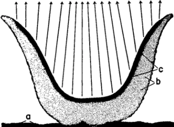
One example, Aleuria vesiculosa (Fig. 2 ) , a fairly common species on dunged soil, will be considered. The apothecium is cup-shaped and usually several centimeters in diameter, with a palisade-like lining layer of asci at various stages of development intermixed with paraph- yses. When an ascus bursts, an apical lid hinges back, the ascus wall contracts longitudinally and laterally, and the ascospores are shot to a distance of several centimeters. If contents of the asci were discharged at right angles to the hymenium, it is clear that many spores would
simply be shot onto an opposite hymenial surface. This would be par
ticularly true of those species with apothecia of a more vase-shaped form. However, this type of wastage does not occur, since the spores
FIG. 2. Aleuria vesiculosa. Above: L.S. of small apothecium. a, substratum;
by hypothecium; c, hymenium. The arrows show the direction of spore discharge.
Below: parts of the hymenium from the sides (A and B) and the bottom ( C ) of the apothecium. (After Buller, 1934.)
are shot freely into the air above the apothecium because of the photo- tropism of the individual ascus.
A special feature of most apothecia is the phenomenon of puffing, During a period of quiescence many asci ripen, but remain in a con-
dition of unstable equilibrium. In this state a touch or perhaps a sudden change in humidity, as occurs when an apothecium is breathed upon, is enough to cause all these ripe asci to discharge simultaneously, sending into the air a visible cloud of hundreds of thousands of asco- spores which drifts away like smoke. Buller has shown that this simultaneous bombardment sets the whole body of air just above the apothecium in motion, with the result that the spores are carried con- siderably farther than the distance to which a single ascus, discharging alone, could shoot its spores. Puffing may be of biological significance in the more efficient launching of the spores. Further, the cessation of discharge during periods of stillness tends to prevent the liberation of spores when the air is in a nonturbulent condition, that is, at such times when subsequent effective dispersal is less likely to occur.
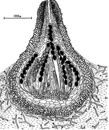
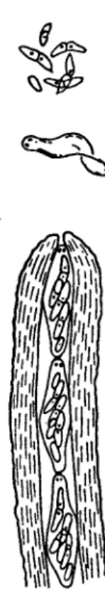
2. Pyrenornycetes Type
Many Pyrenornycetes, especially in the genera Nectria, Epichloe, Rosellinia, Mycosphaerella, Ophiobolus, Venturia, Endothia, etc., are parasites of higher plants, but again it is more convenient to consider, in the first instance, spore liberation in a saprophytic type, namely Sordaria fimicola (Ingold and Hadland, 1958). The structure of this common coprophilous species is illustrated in Fig. 3. The flask-like perithecium is filled with asci at various stages of development, and any free space between them is occupied by mucilage. In Pyrenornycetes generally no gas phase is present within an active perithecium. While remaining attached at its base, a ripe ascus elongates up the neck canal, which is lined by downward-projecting periphyses. When its tip pro- trudes slightly through the ostiole, the ascus bursts, shooting its spores to a distance of 1 to 10 om. Another ascus then elongates, and so on, the discharge of asci occurring one at a time in orderly succession. There is obviously no opportunity for "puffing" to take place. Empty asci retract into the perithecium, and soon liquify and disappear. In S. fimicola the distance of discharge is relatively great, a feature of many copro- philous fungi, mainly because of the large size of the spores and because of the fact that they tend to stick together. The whole contents of the ascus often forms a single projectile of eight spores. It has already been pointed out that the distance to which a minute spherical body is shot horizontally, with a given initial velocity (corresponding to the muzzle velocity of a gun), is directly proportional to the square of its radius.
In most plant pathogens the spores are relatively small, and the distance of discharge rarely exceeds 1 cm. and is more often half this distance.
However, such a distance of discharge is normally enough to launch the spores into turbulent air.
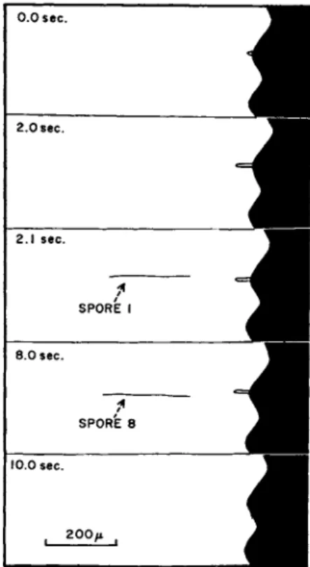
A slight variant of the type of discharge in Sordaria is found in Pyrenomycetes, with long thread-like spores such as those found in Clavicipitales (e.g. Claviceps, Epichloe, and Cordyceps). Here the spores from an ascus are shot away in succession. The process in Cordyceps is shown in Fig. 4. When the tip of the ascus protrudes from
FIG. 3. Sordaria fimicola. L.S. perithecium growing on nutrient agar.
the perithecium, the ascus dehisces by a pore, but at once a thread-like (300μ Χ 2μ) ascospore is forced into the pore, stoppering the ascus momentarily. It is then shot away like a dart from a blowpipe, and again the ascus is stoppered by the next spore. This process is repeated until, within the space of a few seconds, all eight spores are discharged.
Another departure from the more general Sordaria type is to be found in Endothia parasitica, Gnomonia rubi and probably in many other species with long-necked perithecia. In these species the asci are small and are produced in great numbers within the perithecium. At ma-
turity they become detached and are squeezed in single file up the rather long neck canal. When the tip of an ascus emerges through the ostiole, it bursts, discharging its spores into the air, and the next ascus below pushes out the empty envelope of the first one. This process can be seen in operation in Gnomonia rubi (Fig. 5 ) , where the neck of the peri-
0.0 sec.
2.0 sec.
2.1 sec.
8.0 sec.
SPORE 8 10.0 sec.
FIG. 4 . Cordyceps militaris. Profile of two projecting perithecia as seen when a stroma is laid on its side and viewed under low power of microscope. At 0 . 0 sec.
the tip of an ascus is beginning to project from its ostiole. At 2 . 0 sec it has reached its maximum extension. At 2 . 1 sec. the first spore flashes into view, then rapidly falls out of sight. At 8 . 0 sec. the last spore has been discharged. At 1 0 . 0 sec.
the empty ascus has retracted into the perithecium.
thecium is sufficiently transparent for microscopic observation of the column of ascending asci (Dowson, 1925). This type of perithecial behavior clearly allows very rapid spore liberation under suitable con- ditions, and less than a second may elapse between the discharge of successive asci. Such a speed of action would be impossible in Sordaria, where the interval between the bursting of successive asci is usually to be reckoned in minutes.
From this type of spore liberation it is but a short step to the con-
dition in Ceratocystis (Ophiostoma), where the asci not only become detached on ripening, but the ascus wall undergoes liquefaction. The naked asci, as discrete eight-spored droplets, still pass in single file up the perithecium neck, but as they emerge through the ostiole, no
FIG. 5. Gnomonia rubi. Neck of the perithecium in optical section showing passage of asci along neck canal. Above the ostiole the spores and empty membrane of a discharged ascus are shown. (After Dowson, 1925.)
discharge occurs, and they simply run together into a larger drop, poised at the end of the long, black neck. This step in evolution appears to be correlated with the transition from wind to insect dispersal.
3. Erysiphales Type
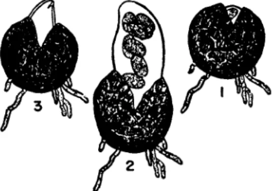
In the powdery mildews (Erysiphales) the cleistothecium is es
sentially a hibernating structure with a continuous hyphal wall around the ascus or asci within. In the spring, when conditions are favorable, the expanding asci must first rupture this wall before emerging to discharge their spores. This process is illustrated in Fig. 6 for Sphaero- theca mors-uvae (Salmon, 1914).
In some powdery mildews the ascus as well as the ascospores may be violently discharged. This has been described, for example, in Podosphaera leucotricha (Woodward, 1927). The expanding ascus ruptures the cleistothecium wall in an irregular manner. However, this
FIG. 6. Sphaerotheca mors-uvae. ( 1 ) swelling ascus is just bursting through cleistothecium wall; ( 2 ) fully swollen ascus is about to discharge its spores; ( 3 ) an instant later. (After Salmon, 1914.)
wall itself is elastic. As the enlarging ascus protrudes, it tends to slip out of the stretched cleistothecium shell. The jaws of this shell suddenly spring together, ejecting the whole ascus several centimeters into the air. The ascus itself then bursts, scattering the spores.
B. Basidiomycetes
In Basidiomycetes violent spore liberation is characteristic of Hy- menomycetes, Tremellales, Uredinales, and certain Ustilaginales. In Gasteromycetes, in which the whole hymenomycete equipment of spore liberation appears to have been lost, the basidium is no longer a spore gun, and consequently, novel methods of spore liberation have been developed along diverse lines. However, Gasteromycetes, being es- sentially saprophytic, are no concern of the plant pathologist.
The basidium of the Hymenomycetes consists of a single cell bearing apical sterigmata, usually four, with a basidiospore at the end of each.
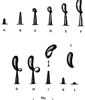
The basidiospore is poised asymmetrically on its sterigma, and when the basidium is ripe, its four spores are discharged in succession. On the spore, very near its junction with the sterigma, is a minute projection—
the hilum. It is this hilum that gives the highly characteristic appearance to discharged basidiospores, allowing them to be spotted readily on slides exposed by the aerobiologist. Just before a basidiospore is to be shot away, a drop of liquid makes its appearance at the hilum, and grows to a certain definite size; then the spore is discharged, usually to
a distance of 0.1 to 0.2 mm., carrying the drop with it (Fig. 7 ) . Im
mediately after the discharge of the four spores the basidium is ap
parently still turgid. Thereafter it slowly shrinks and finally seems to undergo autolysis.
Although studied fairly extensively, the mechanism of basidiospore discharge remains a mystery. It has been suggested (and for this there
FIG. 7. Calocera cornea. A-L, stages in the development of the basidiospore at the end of the sterigma and subsequent spore discharge. (After Buller, 1922.) is good observational evidence in the rust Gymnosporangium juniperi- virginianae) that at maturity a cross-wall separates spore and sterigma.
Discharge might thus be due to the sudden rounding-off at this region of the junction between a turgid spore and a turgid basidium (Prince,
1943).
If this is so, basidiospore liberation is essentially like conidium discharge in Conidiobolus or Sclerospora. However, because the sterigma is so narrow at its junction with the spore and because in the fully mature condition observation can only be made in air (since mounting
in water at once breaks the connection) it is impossible to be sure whether a cross-wall exists at the moment of discharge. Again, if the discharge is due to rounding-off at the interface between spore and sterigma, the beautiful asymmetry of its position on the sterigma would seem to have no significance, nor would the process of drop secretion at the hilum, which so regularly heralds discharge.
Another theory is that the basidiospore is discharged from its sterigma by a water-squirting mechanism. According to this view, the turgid basidium bursts at the end of the sterigma, discharging a liquid jet which carries the spore with it. Against this view is the fact that im- mediately after a spore has disappeared, the vacant sterigma appears to be closed at its apex, and no exudation of fluid normally takes place from it. Indeed if this type of discharge is to occur, the sterigma must become sealed immediately so that the turgidity of the basidium may be maintained for the discharge of the remaining spores. As a slight modification of this view, it has been suggested that the drop is exuded not from the spore itself but at the junction of the spore and the sterigma. It is supposed that, having grown to a certain size, the drop is shot away, carrying the spore with it. This view is supported to some extent by Muller's study of spore discharge in the mirror yeast Sporo- bolomyces, in which the aerial conidium is discharged in a manner that appears to be identical with basidiospore discharge in Hymenomycetes.
Miiller (1954) in a cinematographic study showed that, although nor- mally the spore and its exuded drop disappear simultaneously from the end of the sterigma, occasionally the drop suddenly disappears (pre- sumably having been discharged) leaving the spore behind on the sterigma. However, in this connection it must be remembered that the careful observations of Buller (1922, 1924, 1931, 1933) all go to show that the drop exudes from the spore itself and not from the junction of spore and sterigma.
It has also been suggested that the surface energy of the exuded drop might be mobilized in some way to effect spore discharge (Ingold, 1939). This view has the merit of bringing the exuded drop and the asymmetrical poising of the spore into the picture, but unfortunately there is no real evidence to support it, nor is it at all clear how this energy could be mobilized. It has been calculated, however, that there is enough surface energy available, if it could be utilized, to discharge the spore to the observed distance.
The essential features governing fruit-body construction in Hymeno- mycetes are the very short distance of basidiospore discharge and the fact that, since basidiospores are sticky, they cannot normally be dis- lodged by wind once they have settled on a surface. Upward-facing
hymenial surfaces are very rarely encountered in Hymenomycetes. Spores shot vertically to a distance of only 0.1 to 0.2 mm. would not usually reach the turbulent air above the laminar layer in contact with the hymenium, and would fall almost at once on the hymenial surface and become permanently stuck. In fact the hymenium in the sporophores of Hymenomycetes is mostly vertical, although it is sometimes horizontal and downward-facing, or it may occupy an intermediate, but still down
ward-facing, position. It should, perhaps, be remarked here that the position assumed by the hymenium in most toadstools and bracket
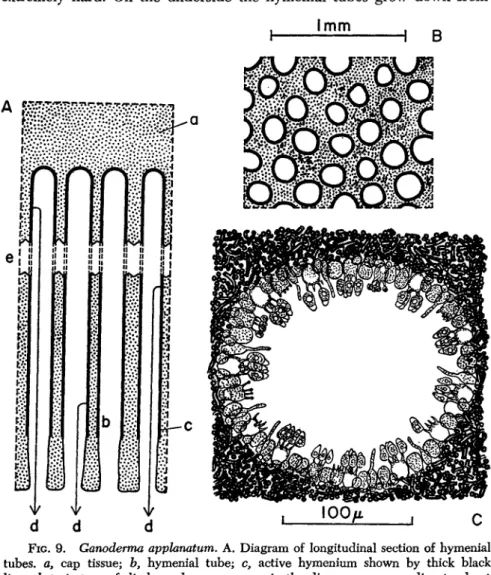
FIG. 8. Ganoderma apphnatum. Vertical section of a small sporophore in its second year growing on ash. a, wood; b, bark; c, upper "crust" of fungus; d, zoned fibrous cap tissue formed in first year; e, additional cap tissue produced in second year; /, hymenial tubes (the dotted line indicates the boundary between the tubes formed in the first year (above) and in the second (below).
fungi also tends to protect them from rain. This is of importance, since water on the surface disorganizes the hymenium, at least temporarily, in striking contrast to that of Discomycetes, which are not injured by the temporary presence of free water.
Since, for the plant pathologist (especially if he is a forester) the bracket polypores are probably of more significance than agarics, spore liberation in the larger Basidiomycetes will be considered in some detail in a polypore and more briefly in agarics, although the general principles in both are essentially similar.
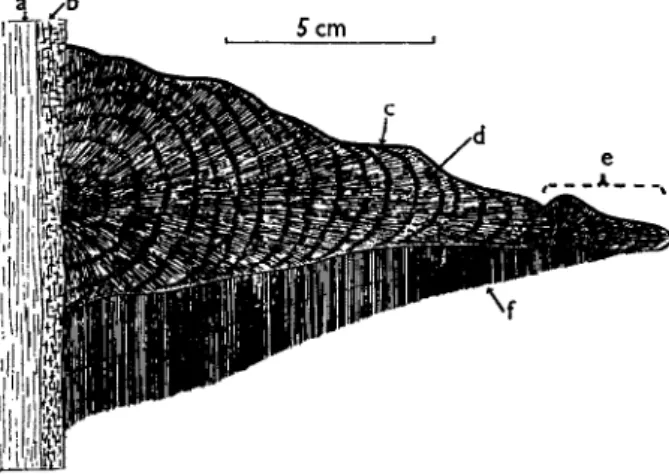
As a first example, Ganoderma applanatum will be taken. This fungus, with an almost world-wide distribution, is a wound pathogen of mature broad-leaved trees causing a heart rot. The mycelium occurs in the wood and the rigid sporophore forms a bracket or shelf broadly and firmly attached to the tree trunk. This woody fruit body is a perennial struc-
ture, the small specimen illustrated in Fig. 8 being 2 years old. Examples of 5 or 6 years of age are frequently to be found. The upper surface is extremely hard. On the underside the hymenial tubes grow down from
FIG. 9. Ganoderma appfonatum. A. Diagram of longitudinal section of hymenial tubes, a, cap tissue; b, hymenial tube; c, active hymenium shown by thick black line; d, trajectory of discharged spore; e, gap in the diagram corresponding to about 20 mm. in the actual specimen or 574 mm. at the scale of this figure. B. Transverse section in region of hymenial tubes. The thick black line around each pore is the hymenium. C. Drawing of a single hymenial tube seen in transverse section.
the sterile cap tissue, and these are long (up to 3 cm. in the specimen figured) and extremely narrow (0.1 to 0.2 mm. diameter) (Fig. 9 ) . During the growing season (May to September) they elongate, the meristematic growing region being at the orifice of each tube. Growth
ceases in autumn and winter, and is resumed in late spring. The arrest of growth has structural implications, and the result is that if a sporo
phore is broken vertically, successive annual layers of growth are clearly visible. Growth of the tubes is controlled by gravity so that each in
dividual tube develops in a perfectly vertical manner. This, combined with the rigidity of the fruit body and its firm attachment to a stout trunk, ensures that the tubes maintain their perfect vertically. This seems to be essential for effective spore liberation. Active hymenium lines each tube throughout most of its length, so far at least as the last two or three annual layers are concerned. In still older parts of the tubes the hymenium ceases to be functional, and the pores may become blocked with sterile hyphae.
The spores in this species are discharged horizontally to a distance of approximately 0.05 mm. (Ingold, 1957). They then fall under the influence of gravity down the narrow tube. If the tube were the merest fraction of a degree out of the vertical, clear fall would seem to be impossible and the spores would presumably become stranded on the hymenial surfaces lower down the tube. There is, however, just the possibility that the spores, which have been shown usually to carry a positive static electric charge (Gregory, 1957), are maintained in mid
stream during their fall down the hymenial tube by static electric forces.
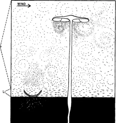
On emerging into the free air below the tubes, the spores are carried away by the wind. It is to be noted that by their very situation on the tree trunk, the sporophores are in a position to drop their spores directly into the turbulent air. As if to achieve this end, ground-inhabiting species such as bolets and most agarics are forced to raise their spore- producing surfaces above the substratum on stipes. As Gregory (1952) has pointed out, the Ascomycetes can shoot their spores through the surface laminar layer of air into the turbulent layer above, but the basidium is not a sufficiently powerful spore gun to achieve this, so that in Basidiomycetes the hymenial surfaces tend to be so placed that the discharged spores drop into turbulent air. This concept is illustrated diagrammatically in Fig. 10.
In Ganoderma applanatum spore liberation is on a gigantic scale, and Buller has estimated that a large sporophore may liberate millions of spores a minute, maintaining this rate more or less for the whole 6 months of its annual spore-fall period.
Ganoderma applanatum is an extreme example which serves to emphasize the principles of basidiospore escape from a sporophore.
Apart from species of Ganoderma and Fomes, bracket polypores have annual sporophores, and usually the pores are wider and shorter than those of Ganoderma. Thus in Polyporus squamosus, a common species
/ / / / /
/ / / / / / / / I
τ
\ \
\ \
\
\ I
\
\
\
\
\
\ \
<
1FIG. 10. A cup-fungus (Peziza sp.) and an agaric (Collybia radicata). The ground is shown black. Above this is a thin layer of laminar air ( L ) and on top of this turbulent air ( T ) . The Peziza has just discharged a puff of spores through the laminar air into the turbulent region. From the pileus of the Collybia spores are dropping into the turbulent air.
features. In the ordinary fleshy toadstool or mushroom, hymenium covers the gills (or lamellae), which hang down from the under surface of the cap (pileus), which is itself raised above the ground on a stipe. In the early stages the stipe may (e.g., in Pholiota spp.) or may not (e.g., in Psalliota spp.) be positively phototropic, but if this tropism does oper
ate in the young sporophore, it is finally lost and the stipe becomes negatively geotropic. This gives an approximate vertical orientation to causing a heart rot of broad-leaved trees (especially elm), the pores are 1 to 2 mm. wide and only about 1 cm. long. These features may be correlated with the comparative lack of rigidity in the fruit body
(Buller, 1909).
In the agaric, although the principles involved in spore liberation are essentially the same as in a polypore, there are certain special
the gills depending from the cap. But there is also a fine adjustment.
The individual gill is itself positively geotropic. If there is slight dis
placement from the vertical, each gill (near its insertion on the cap) undergoes growth movements which bring it again into the vertical plane. This is a special feature of agarics, but absent in polypores, in which readjustment of existing hymenial surfaces does not seem to be possible.
The type of spore, poised asymmetrically on a sterigma, which is violently discharged following drop secretion from a hilum has been termed a "ballistospore" (Derx, 1948). The basidiospores of Hymeno- mycetes are ballistospores; those of Gasteromycetes and of Ustttago are not. Further, certain ballistospores, especially those of the shadow yeasts
FIG. 11. Puccinia malvacearum. A sorus of germinating teliospores on the under side of a leaf of hollyhock (Althaea rosea). (After Buller, 1924.)
(Sporobolomycetaceae) cannot easily be regarded as basidiospores. We must now give some consideration to the ballistospores of rusts (Uredin- ales), which are clearly basidiospores, and to those of Tilletia caries, which can be classified as such only by a rather tortuous argument.
In rusts, the teliospore, usually after a winters rest, germinates to produce a curved transversely septate basidium (promycelium) bearing four basidiospores (sporidia) on its convex side. The organization of the telium in relation to the discharge of basidiospores in rusts has been illustrated by Buller (1931) in Puccinia malvacearum (Fig. 11). It is clear that the curvature of the basidium and the position in which basidiospores arise is of importance in relation to the free escape of the discharged spores.
In smuts the occurrence of ballistospores has been demonstrated, particularly in Tilletia caries, but it is clear that in Ustilago spp. violent
I m i l .. I
discharge does not take place. Buller (1933) has shown that when the brand spore of T. caries germinates on a moist surface a short pro- mycelium is formed. This becomes septate and soon grows away from the surface, probably because of negative hydrotropism. From the end cell a tuft of six to twelve needle-shaped spores (primary conidia) is formed. These have been regarded by most mycologists as the basidio- spores. While still attached, or after passive liberation, they conjugate, forming JT-shaped pairs. From one member of each pair a short sterigma is produced bearing an asymmetrically poised, sickle-shaped spore (secondary conidium) which is a typical ballistospore being violently discharged following drop excretion at the hilum (Fig. 12). If an
FIG. 1 2 . Tilletia caries. Germinating chlamydospore producing a promycelium bearing a crown of six filamentous primary conidia which, following pairwise con- jugation, are producing secondary conidia which are ballistospores. (After Buller,
1 9 3 3 . )
H-shaped pair of conjugated primary conidia is planted on a nutrient medium, such as malt agar, a fine, branched mycelium may be formed from which, in due course, a number of sickle-shaped ballistospores may arise on aerial sterigmata. In the field the ballistospore may germinate in contact with a host plant and cause infection, but on nutrient agar, and possibly under certain conditions in nature, a saprophytic mycelium is formed from which further ballistospores are produced. It is of inter- est to note that in Tilletia caries the record discharge distance for a ballistospore has been observed, namely, 1.0 mm.
C. Other Types of Discharge
We must now consider a number of examples in which the essential mechanism of discharge involves the sudden rounding-off of turgid
spores. Probably the most important example from the point of view of plant pathology is the aecium of rusts (Uredinales) from which the aeciospores are commonly shot to a distance of 5 to 15 mm. Neverthe
less in some species no violent liberation occurs. Although the force of propulsion is clearly provided by the sudden change in form of turgid spores, the details of the process are not absolutely clear. The mature aecium is usually a cup-like structure with a firm wall (pseudo- peridium) of thickened cells. At the base of the cup is a close-set pali
sade of basal cells, each producing an ever-growing file in which aecio
spores and intercalary cells alternate. Actually the basal cell cuts off a single terminal cell at a time; this divides to form a larger cell above, which becomes the spore, and a smaller below, which is the intercalary cell. Often this cell is delimited not by a wall parallel with the flat top of the basal cell, but by a curved wall at an angle to this plane. The result is that the intercalary cell arises in a corner-wise position—rather as a companion cell is carved off from a sieve tube. The intercalary cell remains thin-walled, while the wall of the associated aeciospore thickens.
Most workers have regarded this cell as an ephemeral structure, which soon breaks down, but it has been suggested (Savile, 1954) that it remains turgid until the maturation of its companion spore.
As the aeciospore develops, its wall differentiates into three layers, and at the same time the future germ pores are organized. In some rusts where a pore is to be formed, the wall is much thicker locally and prob
ably different chemically, producing a minute spherical plug, which becomes more or less free from the rest of the wall. In the spore deposit from a discharging aecium, these plugs can be seen either still adhering to the spores or lying free (Fig. 13). When the plug is eventually dis
placed, it leaves a very thin region in the spore membrane through which a germ tube may emerge if the spore eventually germinates.
It has been suggested that these spore plugs may play an essential part in aeciospore discharge—a part which can be illustrated by a simple model. "A tennis ball compressed over a marble on a table will be thrown farther upward when the confining pressure is suddenly removed than it will be if compressed against the table alone with the same force, and then released" (Dodge, 1924). The suggestion is that in the closely packed aecium the spore is indented by the pore plugs, but in the end the spore suddenly rounds off and is discharged. The pore plug acts as the marble in the model. The major difficulty of this theory is that in some rusts the aeciospores are actively discharged in spite of the absence of pore plugs. It may be, however, that the turgid, intercalary cells persisting among the mature spores act in the same way.
If the spore deposit from a discharging aecium is observed, it will
be seen to consist not only of single spores but also of groups of two, three, or more. Occasionally a clump of up to one hundred spores may be shot away as a single projectile.
It is perfectly clear that the mechanism of violent discharge in the rust cluster-cup can operate only under conditions of complete turgidity.
Even a slight reduction in turgor leads to a cessation of discharge. No
FIG. 13. Gymnosporangium myricatum A. and B. Spore prints formed after dissecting an aecium and laying it down on a glass slide in a damp chamber. Prepared from a photograph. C. Outline of spores showing plugs that still remained attached to spores after their flight and also free plugs. (After Dodge, 1924.)
spores are shot from aecia on wilting leaves. However, under damp conditions spores from the outermost layer of the aeciospores in the cluster-cup are violently discharged. These spores are followed by more, and so the cup tends to empty, but the spore supply is renewed from the base.
One of the best-known examples of spore discharge as a result of rounding-off of turgid structures is to be seen in Entomophthora coro- nata (— Conidiobolus villosus), which sometimes parasitizes aphids
(Martin, 1925). The single conidium is borne on an erect conidiophore, the tip of which bulges into the spore as a dome-shaped columella. At the interface between the two turgid structures, stresses are set up which
are relieved by the sudden eversion of the re-entrant part of the coni- dium. As a result, the spore springs into the air to a distance of a few centimeters.
The same type of discharge has been reported in Sclerospora philip
pinensis, causing downy mildew of maize (Weston, 1923). In this fungus, however, the tip of the conidiophore branch does not project into the conidium, but there is a flat surface of contact between the two. It seems that it is by rounding-off in this region that spore dis
charge occurs (Fig. 14). In S. philippinensis the spore is shot to only a very short distance—about 1 mm. This is probably due to the rela
tively small area of contact between the reacting structures.
FIG. 14. Sclerospora philippinensis. A. Conidiophore bearing eleven conidia.
B. Part of conidiophore more highly magnified; from the tip on the left the conidium has been discharged, that on the right being still attached. (Weston, 1923.)
It has been reported that the rounding-off mechanism of violent discharge operates in some powdery mildews (Erysiphales). Hammar- lund (1925) fitted capillary tubes over single conidiophores and watched spore liberation microscopically. He maintained that the mature conid
ium is shot to a distance of several spore lengths due to sudden rounding-off at the junction with the next spore in the chain. This observation, however, lacks confirmation, possibly because no later worker has used such an elegant technique as that employed by Hammarlund.
Although in Sclerospora, as we have seen, spores are discharged by
a rounding-off mechanism, generally in downy mildews (Peronospor- aceae) violent discharge is not very common. However, in a few other species it does occur, but by a mechanism quite unlike that found in maize mildew. Pinckard (1942) has described a case of this kind. In Peronospora tabacina (blue mold of tobacco) as the branched conidio- phore dries, violent twisting movements occur which may flick the finely attached spores into the air (Fig. 15). It might be supposed that
FIG. 1 5 . Peronospora tabacina. A. Conidiophore in damp air with attached conidia. Β and C. Changes on exposure to dry air. D. Recovery on return to damp conditions. (After Pinckard, 1 9 4 2 . )
even without these movements, the ripe spores would readily be de
tached by wind, and thus the biological significance of this hyposcopic discharge would be in doubt. However, Waggoner and Taylor (1958), as a result of spore-trapping carried out over tobacco seed-beds heavily infected with blue mold, found that spores were trapped mainly in the morning, when hyoscopic twirling of the drying conidiophores would be expected to occur. The absence of spores in the air during midday hours, when strong hyposcopic movements would be at a minimum, was taken to imply that forcible discharge is a necessity for spore liberation.
III. PASSIVE SPORE L I B E R A T I O N
In a large number of the fungi pathogenic to higher plants, the spores are not violently discharged, and spore liberation appears to depend on wind, rain splash, or the activity of insects.
In considering the classification of conidial fungi (Hyphomycetes or Moniliales) Mason (1937) laid emphasis on the biological importance of the difference between dry-spore types (Xerosporae) and slime-spore forms (Gloiosporae). In the former the conidia could be liberated by wind, but in the latter wind could not bring about liberation, and water or insects would seem to be indicated as agents in dispersal. However, from the point of view of dispersal the difference between Mason's two
types is not always clear-cut. Thus in some dry-spore fungi even the strongest winds fail to dislodge spores from their conidiophores, while some slime-spore species form their conidia in spore drops which eventu
ally evaporate, leaving dry spores which can be blown off the conidio
phore either singly or in groups. A common example is Trichoderma viride, a saprophyte in soil or on wood.
In a number of dry-spore species which rely on wind for spore liberation, the ripe dispersive units (propagules) are borne some dis
tance above the surface of the host on erect structures. This is so in such diverse fungi as downy mildews (Peronsporaceae), gray mold
(Botrytis cinerea), powdery mildews (Erysiphales), Omphalia flavida (gemmifers with gemmae), Cladosporium fulvum, and many others.
The advantage seems to be that the propagules are exposed directly in turbulent air above the nonturbulent layer, perhaps 50 to ΙΟΟμ thick, and which so often exists as a skin over the host surface. However, there are many dry-spore pathogenic fungi in which the spores are not raised to an appreciable extent above the surface of the diseased tissue. In this connection special mention must be made of most uredospores of rusts and brand spores of smuts. In these fungi the spores are, without doubt, freely liberated into the air in spite of the absence of violent discharge or of conidiophores raising the spores into a favorable posi
tion for liberation. It seems that the spores of such fungi, provided that they are sufficiently loose and powdery when mature, can be liberated freely when the diseased leaf, stem, or inflorescence on which they occur vibrates in the wind. This is the ancient principle of the sling shot.
Indeed, it would seem quite likely that long conidiophores only acquire an importance when fungi are very close to ground level or when the part of the host attacked is rigid. These remarks only serve to under
line our basic ignorance of this question. Further, we do not know in many cases whether spores become detached more readily under dry or under damp conditions.
In slime-spore fungi there is often doubt about the manner of their dispersal, although the agents concerned are likely to be insects, or water, or both. Among slime-spore fungi well-known to the phytopathol- ogist are Fusarium spp., Colletotrichum spp., the conidial stage of Nec- tria spp., most members of Sphaeropsidales, the pycnial stage of rusts, the Sphacelia stage of ergot, and Graphium spp. In the last three exam
ples insect dispersal of the minute sticky spores has been established, but in the others rain splash is probably the main factor involved in the take-off of the spores from the parent tissue. A well-known example of dispersal of rain splash is Colletotrichum lindemuthianum, the cause of anthracnose of Phaseolus spp. (dwarf and runner beans). The fungus
attacks the aerial parts of the host, producing dark, sunken lesions bear
ing pink acervuli. The slime spores from these are scattered by splash
ing rain onto healthy foliage and pods.
At this point brief mention should be made of the take-off from the host of bacteria pathogenic to higher plants. In bacteria, apart from certain Actinomycetes, there is no parallel with dry-spore conidial fungi.
However, in their dispersal there is a close parallel with slime-spore fungi. In a number of bacterial plant diseases, at some stage slimy masses of bacterial cells occur on the affected host, and the dispersal of these is due either to insects (as in Bacterium amylovorum, causing fireblight of pear and apple) or to rain splash. An example of the latter is Xanthomonas citri, the cause of citrus canker. The bacteria exude from scabby spots on leaves, young twigs, and fruits, and rain splash carries infection from diseased to healthy tissue.
The problem of the basic mechanics of splash dispersal has recently been investigated by Gregory and his co-workers (1959). The technique used was to allow water drops of definite size to fall onto a watery spore suspension exposed as a film of known thickness on a glass slide. The size and scatter of the reflected droplets was studied and they were examined to determine whether they carried spores. As a test organism Fusarium sohni, a slime-spore species, was generally used. It was found that a drop 5 mm. in diameter falling from a height of 7.4 m.
onto a spore-containing film 0.1 mm. thick produced over 5000 reflected droplets of which more than 2000 carried spores. The droplets ranged in size from 5μ to 2400μ. On the average the distance which they were scattered horizontally was 10-20 cm.
It is to be noted that the larger reflected droplets fall back within a small fraction of a second onto the substratum. The smaller ones, how
ever, may remain longer in the air and be rapidly reduced further in size by evaporation. Thus, as a result of rain splash, slime spores may become suspended in turbulent air and be dispersed in the same manner as dry spores. Although the lists of species recorded by aerobiologists invariably show a great preponderance of dry-spore fungi, there is always a slime-spore element and rain splash may be the principal factor involved in the contribution of this to the air spora.
A study, involving high-speed photography, was also made of splash liberation of conidia from a twig bearing abundant conidial stromata of Nectria cinnabarina. Large drops (5.0 mm. diameter) falling from a height into the twig each broke into thousands of droplets all of which carried spores.
Much further quantitative study, particularly in the field, is needed in connection with the problem of splash dispersal, but the work of
Gregory and his colleagues provides an inspiring model for future research.
I V . METEOROLOGICAL CONDITIONS I N R E L A T I O N TO SPORE L I B E R A T I O N
Liberation of fungal spores may be conditioned by external factors—
especially humidity, rain, temperature, wind, and light.
A. Humidity and Rain
These two factors, although usually correlated, are sometimes separ
able in their effects. As we have seen, rain may have a special effect in splash liberation of certain spores, but, further, some fungi require actual wetting if spore discharge is to take place. This is particularly true in Pyrenomycetes. Thus, for example, in Nectria galligena (Munson, 1939; Bulit, 1957), in Venturia inaequalis (Keitt and Jones, 1926; Hirst et al., 1955) and in Endothia parasitica (Heald and Studhalter, 1915) the discharge of ascospores is closely correlated with rainfall (Fig. 16).
1954
00 12 00 12 00 4 5
FIG. 1 6 . Relation of rainfall and concentration of Venturia inaequalis ascospores per cubic meter of air every 2 hours during May, 1 9 5 4 . (After Hirst et al., 1 9 5 5 . ) In some species (e.g., Hypoxylon pruninatum, Gruenhagen, 1945) it has been shown that a saturated atmosphere is not sufficient in itself to initiate spore discharge but that the perithecial stroma must first be wetted before discharge will begin. Although this relationship in Pyrenomycetes between spore discharge and rainfall is the normal one, there are exceptions. Thus Daldinia concentrica (Ingold, 1946) can discharge its spores under the driest of conditions, obtaining the water necessary for continued discharge from a water reserve in the stromatal
tissue, and Epichloe typhina (Ingold, 1948) can also discharge its spores even when the stroma is unwetted and the surrounding air is dry, relying on the water brought to it in the transpiration stream of the host.
High atmospheric humidity and rain are important for continued spore discharge not only in Ascomycetes but also for liberation of basidiospores and for the escape of aeciospores in rust. However, in a number of dry-spore fungi, low humidity may be important for the take-off. In unpublished work in my own department it has recently been established that the sporangioles of the mold Thamnidium elegans and the conidia of Trichoihecium roseum become detached by wind much more readily if it is dry than if it is damp, and the same may be true of other dry-spore fungi.
B. Temperature
The effect of temperature on spore liberation is probably largely associated with its influence on spore maturation. Thus in Ascomycetes, in the range normally encountered, an increase in temperature results in an increase in the rate of spore discharge (Ingold, 1939). The same is true of Basidiomycetes. In Lenzites betulina Buller (1909) has demon
strated that, although spore discharge can occur over the range 0° to 29° C , the rate of liberation is much lower at the extremes than in the middle region.
It has recently been shown (Ingold and Hadland, 1958) that tempera
ture also may affect the distance of spore discharge. Thus in Sordaria fimicofo the average distance of discharge of eight-spored projectiles is 6.30 cm. at 21 to 24° C. but only 5.23 cm. at a temperature of 7 to 10° C.
C. Wind
Wind is probably a major factor in connection with the take-off of many parasitic fungi although quantitative information is sadly lacking.
Mean-wind speed increases in a regular manner with height, and this may be expressed by the formula (Geiger, 1950)
where
t?i = wind velocity at unit (1 meter) distance above the ground v2 = wind velocity ζ units above the ground
and
a = an exponent which varies to some extent but near the ground is usually in the range of 0.2 to 0.3
This is, of course, a statistical "law," true on the average, but not neces
sarily giving a correct picture at any given moment. Theoretically at ground level the wind velocity is zero. Even under gale conditions a layer, perhaps only a few molecules thick, is still, or, if it is in motion, is nonturbulent. This "laminar layer" during the day is often a millimeter or less in thickness, but during the night with the disappearance of thermal turbulence, it may be reckoned in meters. However, although this laminar layer is normally present, it may locally and temporarily be invaded by violent eddies which may pick up dry spores and whirl them upward.
In connection with wind as a spore-liberating agent, the position of the spore-producing surfaces on diseased plants and the disposal of the spores on the conidiophores may be of great importance. It is to be hoped that wind-tunnel experiments will in the future add much to knowledge of the influence of wind on the escape of spores.
D. Light
Light is a minor factor in connection with the take-off of the spores of pathogens. Like temperature, it probably exerts its influence, if any, in conditioning spore maturation. It will be discussed later in connection with periodicity of discharge.
V . PERIODICITY O F SPORE L I B E R A T I O N
From the point of view of the spread of plant diseases, the time of day when the spores of a pathogen are set free may be of importance.
If the spores are liberated when atmospheric turbulence is high, their wide dispersal is favored, but this is likely to be an advantage only if they are of the type that can survive the relatively dry conditions often associated with turbulence. For delicate, short-lived spores it may be vital to the spread of the pathogen that liberation occur at a time of day when the air is damp and when potential infection drops, as the result of dew or rain, are likely to persist on the host leaves. Thus any daily cycle of spore liberation must be carefully studied by the plant pathologist.
Broadly speaking, this question of periodicity has been investigated in two different ways. First, the actual liberation of spores has been studied, often in the laboratory, and second, the daily cycle of spore- content in the air in the neighborhood of infected plants has been followed.
In considering any daily cycle of spore liberation in a particular case it must be borne in mind that each of the four major factors influ
encing the take-off of spores, namely, temperature, humidity, light, and
wind, tends to exhibit a daily cycle of behavior. Light, temperature, and wind velocity tend to be maximal in daytime, while humidity tends to be high at night associated with lower temperature and decreased atmos- pheric turbulence. Again, it is, perhaps, important to realize that periodic appearance of spores in a spore trap does not necessarily mean that they have been set free in a periodic manner. Let us consider, say, the apothe- cium of a Discomycete on the ground discharging spores at a uniform rate. During the night in fair weather there may be a thickish layer of still or laminar air close to the ground, and spores discharged into this would soon fall to the ground and would not be caught in a spore trap operating at the standard height of 1 m. During the day, with the onset of turbulent conditions and the reduction of the laminar layer to a bare millimeter or so, spores would be brought into the region sampled by the trap. Thus a periodicity might be recorded quite unconnected with spore liberation. Probably, however, there is normally a close correla- tion between liberation and trapping, provided the distance between the source and the trap is not too great.
Another general point which should be made is that periodic rhythm in spore liberation may be conditioned by periodic spore production or by the periodic operation of conditions which favor the escape of spores from a reservoir in the parent tissue.
In the powdery mildew of clover, caused by Erysiphe polygoni, it has been shown (Yarwood, 1936) that each conidiophore normally produces and abstricts one conidium each day and that this rhythm depends on the natural alternation of light and darkness. Spore liberation tends to occur during the daytime and in greatest abundance about noon. Childs (1940) has also reported a diurnal cycle of spore maturation in a num- ber of other powdery mildews, including species of Sphaerotheca and Podosphaera, and Yarwood (1957) has found a similar state of affairs in species of Micosphaera and Uncinula. However, in Erysiphe graminis on barley (Yarwood, 1936) no diurnal cycle was observable.
In Taphrina deformans a periodicity has been found (Yarwood, 1941) with spores discharged in greatest numbers in the evening. For the basidiomycete Corticium filamentosa, which forms its basidia on leaves of Hevea, nocturnal spore discharge has been demonstrated (Carpenter, 1949). Nocturnal discharge of ascospores occurs in Daldinia concentrica (Ingold, 1946; Ingold and Cox, 1955). This is an endogenous rhythm which continues for a number of days in continuous darkness at constant temperature. Under these conditions, however, the rhythm is finally lost, but is immediately restored on return to the normal periodic alternation of light and darkness.
In the saprophytic pyrenomycete, Sordaria fimicola, the periodicity of
spore discharge has also been rather fully studied, and it may be that the picture obtained will prove to be generally applicable to nonstromatal species (Ingold and Dring, 1957). In Sordaria there is no sign of an endogenous rhythm; the cycle is entirely dependent on the cycle of light changes in the day. Under conditions of illumination of 12 hours light (100 f .c.) and 12 hours dark in each day, spore discharge is at a low level during the period of darkness. With the onset of light, discharge rate rises to a maximum and thereafter falls, but not to the level of discharge in darkness. The change from light to dark always involves a rapid decline in rate. It has been shown (Ingold, 1958) that it is the short rays of light (400 to 500τημ) which are effective in stimulating discharge.
It should be remarked, however, that in the investigations using Daldinia and Sordaria, temperature has been kept constant and the water supply has been in no way limiting.
Mention has already been made of the periodicity in trapping of the spores of Peronospora tabacina. Waggoner and Taylor observed a maximum of spores in the morning, and associated this with the daily decrease of humidity in the early hours operating the hyposcopic mechanism of dispersal.
Work in Britain (Hirst, 19E>3) and in Nigeria (Cammack, 1955) shows that diurnal periodicity of spores in the air is the rule with such diverse types as the brand spores of Ustilago, the conidia of powdery and downy mildews, and the uredospores of rusts, although it remains to be seen if this is due to periodicity of spore liberation, to rhythm in the turbulent conditions of the air bringing spores to the spore trap orifice, or to a combination of both.
Much remains to be determined about the daily cycle of spore liberation, but from what is known the periodicities observed do not appear to have much selective value for the pathogen in the sense that the fungus achieves a greater infection as a result of restricting its spore output to certain times a day.
W e are left with a general picture of spore liberation in which one of the principal features is the lack of exact knowledge. However, plant pathologists generally are becoming increasingly interested in the quantative aspect of dispersal, including the take-off, in the pathogens they study, and in the next decade we may hope to see the development of a much more detailed picture.
R E F E R E N C E S
Bulit, J. 1957. Contribution έ letude biologique du Nectria galligena Bres. agent du chancre du pommier. Ann. inst. natl. recherche agr on. Ser. C7: 67-89.
Buller, A. H. R. 1909. "Researches on Fungi," Vol. I. Longmans, Green, London, pp. 1-287.
Buller, A, H. R. 1922. "Researches on Fungi," Vol. II. Longmans, Green, London, pp. 1-492.
Buller, A. H. R. 1924. "Researches on Fungi," Vol. III. Longmans, Green, London.
1-611.
Buller, A. H. R. 1931. "Researches on Fungi," Vol. IV. Longmans, Green, London, pp. 1-329.
Buller, A. H. R. 1933. "Researches on Fungi," Vol. V. Longmans, Green, London, pp. 1-416.
Buller, A. H. R. 1934. "Researches on Fungi," Vol. VI. Longmans, Green, London, pp. 1-513.
Cammack, R. H. 1955. Seasonal changes in three common constituents of the air spora of Southern Nigeria. Nature 1 7 6 : 1270-1272.
Carpenter, J. B. 1949. Production and discharge of basidiospores by Pellicularia filamentosa (Pat.) Rogers on Hevea rubber. Phytopathology 3 9 : 980-985.
Childs, J. F. L. 1940. Diurnal cycle of spore maturation in certain powdery mildews.
Phytopathology 3 0 : 65-73.
Derx, H. G. 1948. Itersonilia, nouveau genre de Sporobolomycetes a mycelium boucte. Bull. Botan. Gardens Buitenzorg Ser. Ill 1 8 : 465-^72.
Dodge, B. O. 1924. Aecidiospore discharge as related to the character of the spore wall. /. Agr. Research 2 7 : 749-756.
Dowson, W. J. 1925. A die-back of rambler roses due to Gnomonia rubi Rehm.
J. Roy. Hort. Soc. 5 0 : 55-72.
Geiger, R. 1950. "The Climate Near the Ground." Harvard Univ. Press, Cambridge, Massachusetts.
Gregory, P. H. 1952. Fungus spores. Trans. Brit. Mycol. Soc. 3 5 : 1-18.
Gregory, P. H. 1957. Electrostatic charges on spores of fungi in air. Nature 1 8 0 : 330.
Gregory, P. Η., E. J. Guthrie, and Μ. E. Bunce. 1959. Experiments on splash dis
persal of fungus spores. J. Gen. Microbiol. 2 9 : 328-354.
Gruenhagen, R. H. 1945. Hypoxylon pruinatum and its pathogenesis on poplar.
Phytopathology 3 5 : 72-89.
Hammarlund, C. 1925. Zur Genetik, Biologie und Physiologie einiger Erysiphaeeen.
Hereditas 6 : 1-126.
Heald, F. D., and R. A. Studhalter. 1915. Seasonal duration of ascospore expulsion of Endothia parasitica. Am. J. Botany 2 : 429-448.
Hirst, J. M. 1953. Changes in atmospheric spore content: diurnal periodicity and the effects of weather. Trans. Brit. Mycol. Soc. 3 6 : 375-393.
Hirst, J. Μ., I. F. Storey, W. C. Ward, and H. J. Wilcox. 1955. The origin of apple scab epidemics in the Wisbech area in 1953 and 1954. Phnt Pathol. 4 : 91-96.
Ingold, C. T. 1939. "Spore Discharge in Land Plants." Oxford Univ. Press, London and New York.
Ingold, C. T. 1946. Spore discharge in Daldinia concentrica. Trans. Brit. Mycol.
Soc. 2 9 : 43-51.
Ingold, C. T. 1948. The water-relations of spore discharge in Epichhe. Trans. Brit.
Mycol. Soc. 3 1 : 277-280.
Ingold, C. T. 1953. "Dispersal in Fungi." Oxford Univ. Press, London and New York. pp. 1-197.