Bronchiectasis bacterial etiology and the role of pleural c-reactive protein in prediction of parapneumonic
effusion
PhD thesis
Shimon Izhakian
Doctoral School of Clinical Medicine Semmelweis University
Supervisor: Dr. Sréter Lídia, MD
Official reviewers: Dr. Ildikó Horváth, MD Dr. Zsófia Lázár, MD
Head of the Final Examination Committee: Dr. Veronika Müller, MD
Members of the Final Examination Committee: Dr. Károly Nagy, MD Dr. Gyula Ostoros, MD
Budapest
2019
1 Table of content
1.
3 List abbreviation
4 . Introduction
1
3 1.1 The medical history of bronchiectasis
4 1.2 Epidemiology
4 1.3 Etiology
6 1.3.1 Abnormal structural lung conditions
6 1.3.2 Obstructive airways disease
7 1.3.3 Defects of muco-ciliary clearance
8 1.3.4 Channelopathies
8 1.3.5 Allergic bronchopulmonary aspergillosis (ABPA)
9 1.3.6 Immunodeficiency
10 1.3.7 Infections
10 1.3.8 Bronchiectasis in systemic diseases
11 1.3.9 Idiopathic bronchiectasis
12 1.4 Diagnostic evaluation of bronchiectasis
12 1.4.1 Clinical features
13 1.4.2 Laboratory tests
14 1.4.3 Radiology features of bronchiectasis
16 1.5 Microbiology of non-CF bronchiectasis
17 1.5.1 Haemophilus influenzae
18 1.5.2 Pseudomonas aeruginosa
19 1.5.3 Streptococcus pneumoniae
19
1.5.4 Staphylococcus aureus
20 1.5.5 Non tuberculous mycobacteria (NTM)
21
1.6 Treatment
21 1.6.1 Aims of treatment
21
1.6.2 Airway clearance
22
1.6.3 Long term antibiotics
28 1.6.4 Pulmonary rehabilitation
28 1.7 Parapneumonic effusion
28 1.7.1 As alarming sign
29 1.7.2 Definition of parapneumonic effusion
30 1.7.3 Clinical presentation
31 1.7.4 Epidemiology of parapneumonic effusion
32 1.7.5 Diagnosis of pleural effusion in adults
35
1.7.6 Diagnostic criteria
41 1.7.7 Challenges in diagnosis of parapneumonic effusion
41 1.7.8 The role of pleural CRP in parapneumonic effusion
43 Objectives
. 2
44 . Methods
3
44 3.1 Study subjects of bronchiectasis research
44 3.2 BAL procedure
45 3.3 Microbiologic samples
45 3.4 Study subjects of pleural effusion CRP research
46 3.5 Definitions of different types of pleural effusions
46 3.6 Laboratory tests
47 3.7 Statistical analysis
2
48 . Results
4
50 4.1 BAL bacterial isolation
50 4.2 BAL isolation according to age
50 4.3 Non- tuberculous mycobacterium (NTM)
51 4.4 Lobar bronchiectasis distribution
51 4.5 Lobar bacterial distribution in bronchiectasis
52 4.6 Resistance to antibiotics
52 4.7 Pleural effusions distribution
53 4.8 Pleural CRP level in pleural effusions
57 . Discussion
5
58 5.1 Lobar bacterial distribution in NCFB
60 5.2 The role of pleural CRP in parapneumonic effusion detection
62 . Conclusions
6
63 ry
a . Summ 7
64 Összefoglalás
8.
66 Bibliography
. 9
80 Bibliography of the candidate's publications
. 10
83 Acknowledgement
. 1 1
3 List abbreviation
Allergic bronchopulmonary aspergillosis ABPA
Acid-fast bacilli AFB
Area under the curve AUC
Alpha 1 antitrypsin deficiency ATT
Bronchoalveolar lavage
BAL
Bronchiectasis and long-term azithromycin treatment BAT
Bronchiectasis and Low-Dose Erythromycin Study BLESS
British thoracic society BTS
Cystic fibrosis CF
Cystic fibrosis transmembrane conductance regulator CFTR
Chronic granulomatous disease CGD
Congestive heart failure CHF
C-reactive protein CRP
Common variable immune deficiency CVID
Effectiveness of macrolides in patients with bronchiectasis using azithroymycin to control exacerbations
EMBRACE
Endotoxin lipo-oligosaccharides ELOS
Forced expiratory volume in 1 second FEV1
High resolution computed tomography scans HRCT
Health-Related Quality of Life HRQOL
Intravenous IV
Lactate dehydrogenase LDH
Left lower lobe LLL
Left upper lobe LUL
Non-cystic fibrosis bronchiectasis NCFBr
Negative predictive value NPV
Non-typeable H. influenzae NTHi
Non toberculous mycobacterium NTM
Positive predictive value PPV
Right lower lobe RLL
Rabin medical center RMC
Right middle lobe RML
Receiver operator characteristics ROC
Right upper lobe RUL
Soluble triggering receptor expressed on myeloid cells-1 STREM-1
X linked agammaglobulinemia XLA
Yellow nail syndrome YNS
4 1. Introduction
1.1 The medical history of bronchiectasis
Bronchiectasis was first described by Laennec (1) in 1819 as part of a wider work describing the use of his novel invention, the stethoscope. A century later, in 1919, A. Jex-Blake delivered a lecture at the Hospital for Consumption (London. UK) on the conditions which causes bronchiectasis (2). In this pre-antibiotic era a third of patients who were identified as having bronchiectasis were secondary to an episode of pneumonia or pleurisy, a third due to chronic bronchitis and a further third due to bronchial obstruction, the majority of which were malignant tumors.
1.2 Epidemiology
Recent data on bronchiectasis epidemiology has been collected from cohorts in Finland, New Zealand and the USA (3–5). The data from Finland suggested an incidence of 2.7 per 100,000 people, while in New Zealand an overall incidence in children of 3.7 per 100,000 was noted but showed wide variations with regards ethnicity. For example, children from a Pacific Island descent had an incidence of 17.8 per 100,000 compared with an incident of 1.5 per 100,000 for those of a Northern European descent.
Unsurprisingly, given the often-chronic nature of its development, the prevalence of bronchiectasis and hospital admission related to bronchiectasis increased with age.
Studies from the USA estimate a prevalence of 4.2 per 100,000 people in those aged 18–
34 years, increasing to 271.8 per 100,000 in people aged >75 years (5).
1.3 Etiology
Non-cystic fibrosis bronchiectasis (NCFBr) is characterized by irreversibly damaged and dilated bronchi with impaired muco-ciliary clearance leading to recurrent bacterial infection. Inflammation within the bronchial wall can be the result of an infection within the airway, inhalation of injurious agents or an endogenous condition such as an autoimmune disease (Table 1).
5
There are several causes of bronchiectasis:
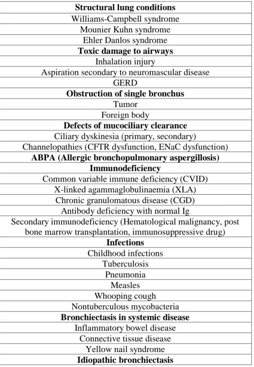
Table 1- Etiologies of bronchiectasis (9-40).
Structural lung conditions Williams-Campbell syndrome
Mounier Kuhn syndrome Ehler Danlos syndrome Toxic damage to airways
Inhalation injury
Aspiration secondary to neuromascular disease GERD
Obstruction of single bronchus Tumor
Foreign body
Defects of mucociliary clearance Ciliary dyskinesia (primary, secondary)
Channelopathies (CFTR dysfunction, ENaC dysfunction) ABPA (Allergic bronchopulmonary aspergillosis)
Immunodeficiency
Common variable immune deficiency (CVID) X-linked agammaglobulinaemia (XLA)
Chronic granulomatous disease (CGD) Antibody deficiency with normal Ig
Secondary immunodeficiency (Hematological malignancy, post bone marrow transplantation, immunosuppressive drug)
Infections Childhood infections
Tuberculosis Pneumonia
Measles Whooping cough Nontuberculous mycobacteria Bronchiectasis in systemic disease
Inflammatory bowel disease Connective tissue disease
Yellow nail syndrome Idiopathic bronchiectasis
6 1.3.1 Abnormal structural lung conditions
Congenital disorders affecting the structure of the bronchial tree can lead to bronchiectasis through a direct effect on the bronchial wall.
Williams–Campbell syndrome was first described in 1960 after the case reports of five children were studied by Williams and Campbell (6). Histological examination of the bronchial wall revealed a deficiency or absence of cartilage, mostly from the third division of the bronchi down.
Mounier–Kuhn syndrome (tracheobronchomegaly) is characterized by dilatation of the trachea and large bronchi, usually presented in young adults with central lobes bronchiectatic disease (Table 2). Its underlying pathology is not clearly understood but histological examination has shown atrophy of airway cartilage and smooth muscle.
1.3.2 Obstructive airways disease
Asthma and COPD conditions would lead to bronchiectasis as both have clearly been shown to cause airway inflammation and structural blockage of airways, either through bronchospasm or fixed airways obstruction, in the case of COPD.
1.3.2.1 Asthma
A number of studies have highlighted the presence of airway remodeling in chronic asthma patients using high-resolution CT (HRCT) scanning techniques. The airway remodelling can vary from mild airway wall thickening to blatant bronchiectasis.
Bronchial wall thickening has been found in up to 82% of asthmatic patients in a cohort (7) and in patients with mild asthma (8). As bronchial wall thickening is indicative of airway inflammation this suggests that a significant number of patients with asthma are at risk of developing bronchiectasis.
The prevalence of bronchiectasis in these studies is estimated at 17.5–40% (8). In the largest of these studies, which comprised of 463 patients with severe asthma, 40% of patients were shown to have evidence of bronchiectasis on HRCT scans (7-8). However, study participants were selected for HRCT on the basis of clinical indication, the most common being a suspicion of bronchiectasis.
These studies suggest that bronchiectasis is associated with a more severe obstruction and is more apparent in patients who present with a longer history of asthma symptoms, consequently a subgroup of severe asthma patients appear to be at risk of developing bronchiectasis (7-8).
7 1.3.2.2 COPD
COPD and bronchiectasis share common symptoms of cough with sputum production and susceptibility to recurrent exacerbations driven by new or persistent infection.COPD is diagnosed on the basis of poorly reversible airflow obstruction and is therefore a physiological diagnosis. While bronchiectasis is diagnosed in the presence of airway dilatation and airway wall thickening on imaging (usually computed tomography (CT)), and is therefore a structural diagnosis. Physiological criteria for the diagnosis of COPD and structural criteria for the diagnosis of bronchiectasis create the possibility for individual patients to fulfil both, resulting conceptually in either co-diagnosis or an overlap syndrome between the two conditions. A study of moderate-to-severe COPD patients demonstrated the prevalence of bronchiectasis to be up to 50% (9).
1.3.2.3 α1-antitrypsin deficiency (ATT)
α1-antitrypsin deficiency is classically associated with predominantly lower lobe emphysema. Bronchiectasis has also been associated with the enzyme deficiency, whether this is a direct consequence of the deficiency or secondary to the emphysema- associated airways obstruction is less clear. In a study of patients with severe AAT deficiency the vast majority of subjects had some evidence of bronchiectasis on a HRCT scan (70 out of 74 subjects), with 27% having clinically significant bronchiectasis with a correlation between forced expiratory volume in 1 second (FEV1) and bronchial wall thickness (10).
1.3.3 Defects of mucociliary clearance
Abnormalities of cilia structure and/or motility cause a decreased mucus clearance from the lung. These abnormalities can be due to:
A) Primary ciliary dyskinesia (PCD)- is a genetically heterogenous disorder with mutation in genes (DNAI1 and DNAH5) which code for proteins responsible for assembly of outer dynein arms.
As cilia are present throughout the body, patients with PCD will often present with multiple symptoms such as sinusitis, recurrent otitis media, infertility and defects of organ lateralisation with situs inversus or situs ambiguus. The triad of bronchiectasis, chronic sinusitis and situs inversus is also known as Kartagener’s syndrome.
B) Secondary ciliary dyskinesia- A number of noxious agents, both organic and inorganic, have been shown to affect the function of cilia in human airway epithelia. Certain
8
bacteria, such as Pseudomonas aeruginosa and Haemophilus influenzae, have been shown to disable mucociliary clearance by releasing products that inhibit ciliary beat frequency, allowing them to persist and propagate infection (11-12). Inhaled inorganic substances such as diesel particles (13) and cigarette smoke (14) have also been shown to have a direct effect on ciliary function, inhibiting ciliary beat frequency. It is important to note here that no causal role for tobacco smoking and the development of bronchiectasis has been made, indeed outside of COPD bronchiectasis appears to be a disease of the nonsmoker.
Aspiration of gastric contents is a well-recognized, but perhaps under diagnosed, cause of bronchiectasis. While aspiration of both acid and nonacid stomach contents leads to direct inflammation of the bronchial wall, ciliary function may also be affected by these agents.
1.3.4 Channelopathies
Defects in the ion channels of the epithelial layer can lead to dehydration of the airway surfaces, thereby affecting the depth of the periciliary layer and bringing the cilia into contact with the viscous mucus layer, further impeding its function. The most widely recognized of these defects is that found in CF. Here, the loss of a chloride channel known as the CF transmembrane regulator (CFTR) protein leads to the inability of the epithelial cells to excrete chloride.
1.3.5 Allergic bronchopulmonary aspergillosis (ABPA)
Allergic bronchopulmonary aspergillosis (ABPA) is a pulmonary condition caused by a hypersensitivity reaction to the ubiquitous environmental fungus Aspergillus fumigatus.
It is most commonly seen in patients with pre-existing asthma or CF and is clinically characterized by recurrent wheeze, pulmonary infiltrates and the development of bronchiectasis. The hypersensitivity reaction has mixed features of immediate hypersensitivity (type I), antigen–antibody complexes (type III) and inflammatory cell responses (type IV) (15). The inflammatory cell response seen in ABPA shows a predominance of T-helper cell type 2 (Th2) cells leading to a release of cytokines mediating allergic inflammation (as opposed to the Th1, cytotoxic pathway) (16). The type I hypersensitivity reaction causes local degranulation of mast cells and histamine release leading to bronchoconstriction. The combination of airway inflammation, which leads to viscous, eosinophil-laden mucus, plugging and airway obstruction and
9
bronchospasm leads to a reduction in mucociliary clearance and the development of bronchiectasis. As such bronchiectasis in ABPA is common. In three large case studies it was found that central bronchiectasis was present in 69–76% of patients with ABPA (17- 19).
1.3.6 Immunodeficiency
1.3.6.1 Primary Immunodeficiency
Defects in the immune system leave the lungs vulnerable to infection and in some cases the development of bronchiectasis can be the first indication of immunodeficiency. The most common forms of primary immune deficiencies observed in patients with bronchiectasis are common variable immune deficiency (CVID), X-linked agammaglobulinaemia (XLA) and chronic granulomatous disease (CGD).
Common variable immune deficiency (CVID)- is characterized by reduced levels of immunoglobulins (Igs) with associated recurrent bacterial infections. An increased risk of autoimmune conditions and malignancy has also been identified. The majority of patients present with recurrent pulmonary infections at a mean age 29 years (20). CVID is the most common primary immune deficiency to cause bronchiectasis. A case series undertaken in a UK population identified 68% of the patients with CVID as having evidence of bronchiectasis (21)
X-linked agammaglobulinaemia (XLA)- is caused by a mutation of a tyrosine kinase gene that is involved in the development of B- lymphocytes, leading to an absence of circulating B-lymphocytes and the absence of Igs. Given the severity of the immune deficiency it usually presents much earlier than CVID, usually being diagnosed in early childhood (22). Despite treatment with replacement Igs, chronic lung disease can still develop with the risk of developing bronchiectasis increasing with age (23)
Chronic granulomatous disease (CGD)- is a group of disorders characterised by a loss of phagocytic NADPH oxidase, without which phagocytes are unable to produce the reactive oxygen species required to kill ingested bacteria. Infections are mainly due to Staphylococcus aureus, Serratia marcescens, Salmonella sp., Klebsiella sp. and Burkholderia cepacia.
Antibody deficiency with normal Igs- In a study of patients with bronchiectasis and normal IgG levels, 11% were shown to have specific antibody production deficiencies with an inability to respond to pneumococcal and H. influenzae vaccines (24).
10 1.3.6.2 Secondary Immunodeficiency
The development of bronchiectasis in HIV-infected patients has been noted in a number of case series. While recurrent pulmonary infection is likely to be the major factor in the development of bronchiectasis in these patients, the development of lymphocytic interstitial pneumonia may also be implicated (25).
1.3.7 Infections
A number of childhood respiratory infections have been implicated in the pathogenesis of bronchiectasis. The most widely recognized infectious causes of bronchiectasis are measles and pertussis infection in the West (26), with tuberculosis being a major cause elsewhere. Globally, Mycobacterium tuberculosis infection remains a major cause of morbidity and mortality and a significant cause of bronchiectasis. In developed countries with screening programs and adequate access to treatment, the incidence of new infections remains low. However, the incidence of nontuberculous mycobacterial (NTM) pulmonary infections is increasing. These mycobacteria vary in pathogenecity with Mycobacterium avium complex (MAC) being the most pathogenic whilst other organisms, such as Mycobacterium gordonae and Mycobacterium abscessus, act as opportunistic pathogens and are only found in patients with underlying lung diseases.
The second of these clinical forms is also known as ‘‘Lady Windermere syndrome’’, and was first described in 1992 in a case series of 29 predominately elderly, female patients (27). The patients had MAC infection with bronchiectasis predominantly affecting the middle lobe and lingula. The authors postulated that persistent voluntary cough suppression could lead to chronic inflammatory processes in these poorly draining lung regions which are susceptible to MAC infection (27).
1.3.8 Bronchiectasis in systemic diseases
1.3.8.1 Inflammatory bowel disease
The development of bronchiectasis in patients with ulcerative colitis is well recognized phenomenon and the subject of a number of case series (28). Classically, bronchiectasis develops after resection of the large bowel, suggesting a common immune system response that becomes concentrated on the bronchial wall after the bowel is removed. The common embryonic origin and similar structures of bowel and bronchial wall (columnar epithelial and submucosal glands) add weight to this theory. The link between Crohn’s
11
disease and bronchiectasis is less clear with only a small number of case reports detailing their coexistence (29), perhaps too few to determine a definite association.
1.3.8.2 Connective tissue disease
A number of connective tissue diseases have been noted to be associated with bronchiectasis, largely based on case series reviews of small numbers of patients. The clearest association is that between rheumatoid arthritis and bronchiectasis (30).
Associations between bronchiectasis and Sjogren’s syndrome (31), systemic sclerosis (32), systemic lupus erythematosus (33), ankylosing spondylitis (34-35) and relapsing polychondritis (36) have all been made in small case series reviews.
1.3.8.3 Yellow nail syndrome (YNS)
YNS is a rare syndrome that was first described in 1964 by Samman and White (37) and is characterized by bronchiectasis, lymphoedema and a characteristic appearance of the nails. The underlying pathological defect is not clear, although a recent study revealing an association with chronic rhinosinusitis suggests a possible defect in an inflammatory pathway or mucociliary clearance rather than a structural defect within the lung itself (38).
1.3.9 Idiopathic bronchiectasis
In two large studies (39-40), which identified the cause of bronchiectasis in adults, a significant proportion of patients (26% and 53%, respectively) were found to have no identifiable cause and were labeled as having idiopathic bronchiectasis, the majority of whom were found to be female and nonsmokers. As all the patients studied had undergone rigorous clinical testing and their history had been reported, leading to the exclusion of all known causes, including genetic disorders, it is unlikely under recognition of known causes of bronchiectasis could have occurred.
Finally, another etiology of bronchiectasis is traction bronchiectasis. It is distortion of the airways secondary to mechanical traction on the bronchi from fibrosis of the surrounding lung parenchyma. Although the airways may become dilated in this situation, the other manifestations of bronchiectasis are lacking such inflammation-infection cycle. Traction bronchiectasis tends to have an upper lobe distribution in cases of radiation fibrosis and sarcoidosis, while the lower lobe is predominantly involved in cases of interstitial lung disease/ idiopathic pulmonary fibrosis (ILD/IPF).
12
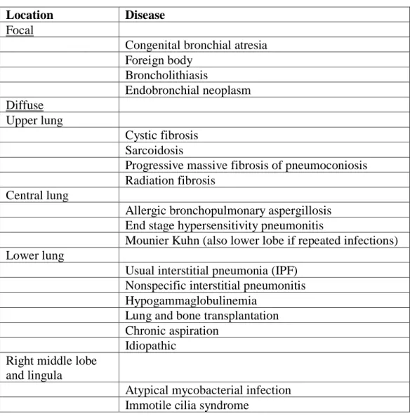
Table 2- Bronchiectasis lobar distribution and associated diseases (17,22,27).
Location Disease
Focal
Congenital bronchial atresia Foreign body
Broncholithiasis
Endobronchial neoplasm Diffuse
Upper lung
Cystic fibrosis Sarcoidosis
Progressive massive fibrosis of pneumoconiosis Radiation fibrosis
Central lung
Allergic bronchopulmonary aspergillosis End stage hypersensitivity pneumonitis
Mounier Kuhn (also lower lobe if repeated infections) Lower lung
Usual interstitial pneumonia (IPF) Nonspecific interstitial pneumonitis Hypogammaglobulinemia
Lung and bone transplantation Chronic aspiration
Idiopathic Right middle lobe
and lingula
Atypical mycobacterial infection Immotile cilia syndrome
1.4 Diagnostic evaluation
1.4.1 Clinical features
The classic clinical manifestations of bronchiectasis are cough and the daily production of mucopurulent and tenacious sputum lasting months to years. The older literature also described "dry bronchiectasis" with episodic hemoptysis and no sputum production, but this presentation is less common (41).
Other, less specific complaints include dyspnea, wheezing, and pleuritic chest pain.
Patients often report frequent bouts of "bronchitis" requiring therapy with repeated courses of antibiotics. There is generally a past history of repeated respiratory tract
13
infections over several years, although a single episode of severe bacterial pneumonia, pertussis, tuberculosis, and Mycoplasma infection can also result in bronchiectasis.
In a retrospective chart review of 103 patients with bronchiectasis who presented to a referral center, the following clinical findings were documented (41):
1) Symptoms – cough (98% of patients), daily sputum production (78%), dyspnea (62 %), rhinosinusitis (73 %), hemoptysis (27 %), and recurrent pleurisy (20 %).
2) Physical findings – crackles (75 %) and wheezing (22%) were common, with digital clubbing occurring in only 2 % of patients.
1.4.2 Laboratory tests
The following studies are typically part of the initial evaluation of a patient with bronchiectasis (42):
1.4.2.1 Routine tests performed
1) Serum immunoglobulins (IgG, IgA, IgM) and serum electrophoresis.
2) Specific antibody responses to tetanus and Streptococcus pneumoniae and Haemophilus influenzae type b capsular polysaccharides.
3) IgE and IgG to Aspergillus fumigatus +/- RAST testing.
4) Sputum for routine culture and for Acid fast bacilli
5) Autoantibodies (according to local policies but may include rheumatoid factor or anti- cyclic citrullinated protein antibody, Anti-nuclear antibody and testing for Sjogrens syndrome).
1.4.2.2 Tests performed in specific circumstances 1) Genotyping and sweat testing for cystic fibrosis:
a) Age <40 years or
b) Any age with persistent colonization by Staphylococcus aureus or Pseudomonas aeruginosa or
c) Evidence of mal-absorption, male infertility or d) Upper lobe disease.
2) Ciliary investigations (otitis media, chronic upper respiratory tract symptoms since childhood, dextrocardiaand infertility).
3) Bronchoscopy
14
a) Localized disease, bronchoscopy may be indicated to exclude proximal obstruction.
b) For patients in whom serial testing of sputum does not yield microbiological information and who are not responding well to treatment, bronchoscopic sampling of lower respiratory tract secretion may be indicated.
c) Bronchoscopy is indicated if high resolution CT (HRCT) suggests atypical mycobacterial infection and sputum culture is negative.
d) Cytological examination of bronchoscopic specimens can provide evidence supporting gastric aspiration.
4) Referral to clinical immunology specialist- clinical suspicion of immunodeficiency 5) α-1 antitrypsin level- basal emphysema.
1.4.3 Radiology
1.4.3.1 Plain radiograph
Chest x-rays are usually abnormal, but are inadequate in the diagnosis or quantification of bronchiectasis. Tram-track opacities are seen in cylindrical bronchiectasis, and air- fluid levels may be seen in cystic bronchiectasis (Figure 1). Overall there appears to be an increase in bronchovascular markings, and bronchi seen end on may appear as ring shadows (43). Pulmonary vasculature appears ill-defined, thought to represent peribronchovascular fibrosis (43-44).
15
Figure 1- Chest radiography showing cystic bronchiectasis with multiple cystic airspaces (45).
1.4.3.2 CT scan
The CT signs of bronchiectasis were first described by Naidichet et al. in 1982 (46).
Although initial studies using 8–10-mm slice thickness showed low sensitivity (47-49), the advent of HRCT led to markedly improved sensitivity, resulting in HRCT replacing bronchography as the diagnostic reference standard.
Optimal HRCT technique is important for maximizing diagnostic accuracy. Importantly, thin slices of 1–2 mm and a high-resolution lung reconstruction algorithm are used to optimize spatial resolution.
A number of features are helpful in diagnosing bronchiectasis (Figure 2) (43-44):
1) Bronchus visualized within 1cm of pleural surface A) Especially true of lung adjacent to costal pleura B) Most helpful sign for early cylindrical change 2) lack of tapering
3) Increasedbroncho-arterial ratio (BAR):
A) Diameter of a bronchus should measure approximately 0.65-1.0 times that of the adjacent pulmonary artery branch
B) Between 1 and 1.5 may be seen in normal individuals, especially those living at high altitude
C) Greater than 1.5 indicates bronchiectasis 4) A number of ancillary findings are also recognized:
A) Bronchial wall thickening: normally wall of bronchus should be less than half the width of the accompanying pulmonary artery branch
B) Mucoid impaction
C) Air-trapping and mosaic perfusion
16
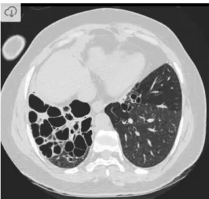
Figure 2 - High-resolution computed tomography image showing cystic bronchiectasis in the right lung (45).
1.5 Microbiology of non-CF bronchiectasis
Patients with bronchiectasis are commonly colonized with potentially pathogenic microorganisms in the airways (50). These microorganisms can cause lung infections and may produce a number of inflammatory mediators that can lead to progressive tissue damage and bronchial obstruction. The phenomenon of chronic infection, bronchial inflammation and progressive lung injury is called cole's ‘‘vicious cycle’’ hypothesis (Figure 3) and is also the reason why prompt evaluation of infection is important (51).
Figure 3 - Bronchiectasis pathophysiology (52).
Bronchial destruction
Release:
proteases, leukoxytes,
dead cells
Abnormal Mucus Clearance Microbial
colonization
Neutrophil inflammation
17
The most commonly cultured pathogens associated with the sputum of NCFBr are Haemophilus influenzae and Pseudomonas aeruginosa, with many isolated strains showing significant antibiotic resistance (53-55). Characterizations of the most common bacteria in bronchiectasis are:
1.5.1 Haemophilus influenzae
H. influenzae has been reported in 14–52% of patients with non-CF bronchiectasis. It is a Gram-negative coccobacillus with specific growth requirements, which can be difficult to isolate in the laboratory if mixed with other flora. Some H. influenzae possess a polysaccharide capsule and can be typed using type-specific anti-capsule antisera. Those with the type B capsule (Hib) can cause invasive infection with bacteraemia, and are most familiar as a cause of meningitis or epiglottitis. The use of Hib vaccine has greatly reduced the incidence of these life-threatening conditions. H. influenzae with capsule types other than type B are relatively rare and are far less pathogenic. The non- encapsulated strains, referred to as non-typeable H. influenzae (NTHi), are also less pathogenic than Hib and only rarely cause bacteraemia. They live as commensals in the human upper respiratory tract but can cause otitis media, sinusitis and conjunctivitis, often following a primary viral infection. NTHi are a common cause of lower respiratory infection in patients with underlying respiratory abnormalities including non-CF bronchiectasis (53). The Hib vaccine does not prevent infection with NTHi as it only contains the H. influenzae type B capsule antigen.
NTHi could be an oral contaminant in expectorated sputum; however, studies using a protected specimen brush (PSB) at bronchoscopy found NTHi in significant numbers in non-CF bronchiectasis, confirming its presence in the lower respiratory tract (54). In contrast Haemophilus parainfluenzae, a common commensal organism found in the upper respiratory tract, may be cultured from sputum but was not found in PSB samples.
In patients with COPD the presence of NTHi in sputum was associated with raised inflammatory cytokines, whereas patients with H. parainfluenzae in sputum had similar levels of cytokines to those who had no microorganisms cultured from their sputum, suggesting that even if present in the lower tract it does not have a direct pathogenic role (56-57).
There is little published data on the epidemiology of H. influenzae in non-CF bronchiectasis. It may be cultured repeatedly from the same patient over several years, but without typing data it is not known if this is the persistence of a single strain or
18
repeated episodes of infection. In COPD exacerbation NTHi were found in higher numbers and with the appearance of a new strain (58-59). However, other prospective study in COPD using molecular typing of H. influenzae and direct analysis of amplified DNA from sputum showed persistence of the same strain over prolong periods (60).
NTHi have various properties that can help explain their pathogenicity and ability to persist in the lung. They can adhere to mucus and to various cell types in the human respiratory tract using pili and other adhesion molecules. Virulence factors include the endotoxin lipo-oligosaccharides (LOS) and immunoglobulin (Ig) – A protease (61).
NTHi may be able to evade the immune response by varying its surface antigens.
Mechanisms: include phase variation of LOS (62), and changes to outer membrane proteins (63).
The prevalence of antibiotic resistant NTHi increases over time in patients with non-CF bronchiectasis (53). Many are resistant to amidopenicillins (e.g. amoxicillin, ampicillin) either due to production of b-lactamase or alteration of penicillin binding proteins.
Quinolone resistance is now recognized and resistance rates to trimethoprim and tetracycline are rising.
1.5.2 Pseudomonas aeruginosa
P. aeruginosa is a versatile non-fermentative Gram-negative bacillus that is found in a range of environments. P. aeruginosa is one of the most common isolates found in 12–
43% of non-CF bronchiectasis patients (64). Stable patients with P. aeruginosa have poorer lung function and more sputum production when compared with patients with other potentially pathogenic microorganisms (64). It has been associated with a poorer quality of life and more frequent hospital admissions (65). There is debate over whether infection with P. aeruginosa leads to a faster decline in lung function as is seen in CF, or whether it is a marker of more damaged lungs (66-67).
P. aeruginosa possesses a range of virulence factors, although their expression may differ between isolates that cause acute infection and those responsible for chronic infection.
Flagella, type IV pili lipopolysaccharide and exopolysaccharides contribute to the adherence to cells and surfaces. Type I and type II secretion systems export protein toxins, such as alkaline protease, elastase, exotoxin A and phospholipase C, while type III secretion systems inject exoenzymes directly into eukaryotic cells. Other extra-cellular
19
virulence factors include rhamnolipids, pyocyanin and hydrogen cyanide (68). Another pathogenicity factor is the ability to form alginate-enhanced biofilms (69) which contributes to the persistence of the organism rather than acute tissue damage and, together with other adaptations, promotes chronic infection.
P. aeruginosa can develop resistance by either:
1) Producing enzymes that destroy the antibiotic such as AmpC b-lactamase, carbapenemases or aminoglycoside modifying enzymes.
2) Modifying the antibiotic target, such as gyrA for quinolone resistance.
3) Reducing exposure either by a decrease in permeability or increased removal of the antibiotic from the bacterial cell (efflux). Efflux mechanisms often affect more than one class of antibiotics and therefore contribute to multi-drug resistance (70).
1.5.3 Streptococcus pneumoniae
S. pneumoniae is a Gram-positive coccus appearing in pairs and in short chains. It may be a harmless commensal in the oro-pharynx but can cause severe and invasive disease.
It can cause otitis media, sinusitis, lower airway infections in patients with damaged lungs such as non-CF bronchiectasis or COPD, but it is rare in CF. Although
S. pneumoniae can be found in up to 37% of patients with non-CF bronchiectasis, very little has been published on its role in this condition.
S. pneumoniae has a polysaccharide capsule that helps evade opsonisation, and isolates lacking the capsule are avirulent. There are over 90 capsule types and the capsule type may be one of several factors that determine the pathogenicity of an individual strain (71).
A polyvalent vaccine containing the most common serotypes is available and recommended for use in patients with chronic lung disease
S. pneumoniae can use a wide variety of molecules to adhere to host cells and produces an IgA protease and a toxin, pneumolysin that can promote invasion, inflammation and tissue damage (72). Pneumolysin is proinflammatory and has many actions including cytolysis, inhibition of cilial beating, and direct activation of the classical complement cascade.
The prevalence of antibiotic resistant S. pneumoniae has increased and in some countries very high rates of resistance to penicillin, macrolides and tetracyclines limit the treatment options. Penicillin resistance is due to modifications to penicillin binding proteins not by the production of a b-lactamase and, therefore, amoxicillin–clavulanate is ineffective.
1.5.4 Staphylococcus aureus.
20
S. aureus is a Gram-positive coccus found in clusters that may be part of the normal flora in the anterior nares, throat and on moist skin sites such as groin and axilla. Infection is by abscess formation, particularly in skin and soft tissues. It is a rare cause of characterized respiratory tract infection, but can cause severe pneumonia after influenza.
It is a common cause of early infection in CF but is less common in non-CF bronchiectasis where its presence may indicate undiagnosed CF (40). There is also an association of S.
aureus with ABPA in non-CF bronchiectasis (73).
1.5.5 Non-tuberculous mycobacteria (NTM)
Patients with bronchiectasis, as with other chronic lung diseases, are predisposed to infection with NTM. Few studies have undertaken a detailed analysis of NTM in the context of bronchiectasis. The prevalence of NTM in bronchiectasis may be higher than anticipated because of the non-specific symptoms and because routine screening is not usually undertaken. In one study, three cases of NTM were detected over 6 years in 91 patients with bronchiectasis (74). In another, NTM were found in 6% of bronchiectatic patients (55). No mycobacteria were isolated in a study of 150 patients over 3 years (40), but in this study sputum was sent only if no response to standard treatment occurred.
Patients with Mycobacterium avium complex infection may develop bronchiectasis over years. Middle-aged or elderly women seem particularly prone to this disease (27).
However, isolation of an opportunist mycobacterial species should not necessarily be interpreted as pathogenic. A ‘one-off’ isolate may have been inhaled shortly before the sample was provided. Persistent isolation (colonization) may occur without any change in clinical status. HRCT scan features can be helpful in confirming infection.
Once an opportunist organism has been isolated, prolonged follow-up may be required to decide whether this represents colonization or infection. Careful follow-up is mandatory because colonization can change to infection (42). This will include:
1) Clinical features - deterioration favoring infection.
2) Sputum examination- (repeated culture, smear positive, heavy growth.
3) Lung function- rapid decline.
4) HRCT- exudative ‘tree-in-bud’ bronchiolitis, mucus plugging, cavitating nodules, rapid progression of disease.
5) Treatment failure.
6) The species isolated will also influence the likelihood of infection: M. avium complex, M. kansasii, M. malmoense (75-76).
21
However, there is a lack of available data on the frequency of other important pathogens such as non- tuberculous mycobacterium (NTM), as well as the role of patient age and lobar distribution of the disease on bacterial profile.
1.6 Treatment
1.6.1 Aim of the treatment
The aims of bronchiectasis management are (42):
1) Reducing symptoms reduce exacerbation frequency.
2) Preserving lung function.
3) Improving health related quality of life.
Patient education is of great importance as many patients have a high treatment burden with physiotherapy regimes, inhaled and oral therapy regimes and frequent hospital visits.
Optimizing treatment compliance, rapid recognition of exacerbations and appropriate use of complex therapies requires active patient engagement. Patients should be advised to stop smoking as smoking will accelerate lung function decline and predispose to mortality (77). As with most other chronic illnesses, patients with bronchiectasis will be offered the annual influenza vaccination in accordance with national guidelines (42).
1.6.2 Airway clearance
Despite limited evidence, airway clearance techniques are widely considered a key component of management (42). There is little high-quality data comparing different techniques for effectiveness. The choice is therefore largely determined by patient choice and ease of use.
Chest physiotherapy- chest clearance techniques include physiotherapy regimes such as Active Cycle of Breathing Technique and Postural Drainage with or without adjuncts such as positive airway pressure devices, (e.g “Flutter” devices providing oscillatory positive pressure), or high frequency chest wall oscillation (78). Small, but significant improvements can be seen in exercise capacity, sputum volume and HRQOL (Health- Related Quality of Life) in patients who use chest physiotherapy compared with control (79-80). The availability of a physiotherapist or other health professional experience in teaching chest clearance techniques is invaluable for patient education.
Inhaled hyperosmolar agents- As an adjunct to standard chest physiotherapy, nebulized hypertonic saline can alter the mucus osmolality making it easier to clear (81). Hypertonic
22
saline can improve FEV1 when used in combination with chest physiotherapy (82).
Recent trials of inhaled mannitol, another hyperosmolar agent, suggest it can increase sputum volume compared to placebo, although the overall significance of this to patients is not entirely clear (83-84).
DNase- The experience of recombinant DNase acts as a cautionary tale in extrapolating results in cystic fibrosis to patients with non-CF bronchiectasis. Despite being effective in selected patients with CF, DNase was shown to be potentially harmful in a randomised controlled trial in NCFBE, reducing FEV1 (85). It is therefore not advised for use in this group of patients, and highlights the different pathophysiology in NCFB, compared with CF associated bronchiectasis.
Long Term Antibiotics 1.6.3
Long-term suppressive antibiotic therapy aims to reduce the bacterial load in the airways, interrupting the ‘vicious cycle’. According to the hypothesis, this should slow down disease progression and lead to improved symptoms and a reduction in exacerbation frequency (51).
Until recently there has been a lack of evidence to guide long term antibiotic therapy in NCFBr, but the recent publication of a number of randomized controlled trials have established clearly that long term antibiotic therapy can reduce exacerbations as well as providing other benefits.
1.6.3.1 Oral macrolide therapy
The BAT, BLESS and EMBRACE trails compare the effects of long term macrolide therapy (either 6 or 12 months) to placebo (86-88). All these trials have shown a significant reduction in exacerbation frequency compared to placebo during the treatment period. The BAT trail showed a median exacerbation frequency of 2 in the placebo group compared with 0 in the treatment group after 12 months (P<0.001)(87). Both 12-month trails showed a reduction in FEV1 decline for the treatment group, although these were small and of doubtful clinical significance (87-88). The main concern of macrolide therapy is a marked increase in macrolide resistance in oropharyngeal and other bacteria.
The BAT trial showed macrolide resistance of 88% in the treatment group compared to 26% on placebo (87). Azithromycin was associated with increased gastrointestinal side effects in the BAT trial, although erythromycin appeared to be better tolerated (88). There
23
have been other concerns regarding macrolides including an increased incidence of cardiovascular events although no cardiovascular complications were observed in these small RCT’s (89-90).
Macrolides have anti-inflammatory effects including inhibition of inflammatory cell migration, cytokine secretion and attenuation of the production of reactive oxygen species (91). Whether the benefit of macrolides is attributable to their antibiotic or anti- inflammatory effect is unclear.
BTS guidelines recommend consideration of long-term oral antibiotics for patients with
≥ 3 exacerbations a year or those chronically colonized with Pseudomonas aeruginosa (42). These guidelines were written before the publication of the three recent trials and given that the EMBRACE trial showed benefit in patients with one or more exacerbations per year these recommendations may change. In clinical practice, macrolides are most frequently used in patients with three or more exacerbations per year, patients with Pseudomonas aeruginosa and also in patients with less frequent exacerbations who continue to have significant impairment of quality of life despite standard treatment.
1.6.3.2 Inhaled antibiotic therapy- Nebulized or inhaled antibiotics deliver a high concentration of the drug to the airways, reducing systemic absorption and therefore theoretically are associated with fewer side effects compared with oral therapy. A 12 months randomized control trial comparing nebulized gentamicin to nebulized 0.9%
saline found a significant reduction in bacterial load, associated with decreases in exacerbation frequency and improvements in quality of life (92). This was associated with a reduction in airway and systemic inflammation (93). A 3-monthes follow-up review after treatment was stopped showed all outcome measures returned to baseline suggesting that this type of therapy needs to be continued long term for sustained benefit (92).
Pseudomonas aeruginosa colonization is associated with a worse prognosis in most studies (65). Inhaled antibiotics can suppress P.aeruginosa bacterial load and even achieved eradication of P.aeruginosa in 30% of patients treated in the nebulized gentamicin trial (92,94). Inhaled tobramycin has been successful in treating CF patients with chronic Pseudomonas aeruginosa infection (95). A study by Barker et al showed nebulized tobramycin also has benefits in non-CF bronchiectasis (95). During the study, tobramycin was given twice daily for 4 weeks. At 6 weeks the pathogen was eradicated in 35% of patients while all patients in the placebo group had persistent colonization.
There were some adverse effects with bronchospasm and cough. Larger studies of
24
tobramycin are needed in non-CF bronchiectasis. Several newer agents are now in late phase clinical trials including dry power inhaled ciprofloxacin, nebulized liposomal ciprofloxacin and inhaled colistin which was the subject of a recent phase III trial demonstrating improved quality of life and a reduction in exacerbations in compliant patients.
1.6.3.3 Anti-inflammatory therapy
Bronchiectasis is thought to be predominantly a neutrophil driven disorder, and neutrophils are largely resistant to the anti-inflammatory effects of corticosteroids. There is no role for oral corticosteroids in bronchiectasis out-with the treatment of ABPA (96).
Inhaled corticosteroids are currently indicated in patients with asthma, COPD or airway hyper-reactivity. They may have some benefits in bronchiectasis. Some studies have shown that regular high dose inhaled steroids reduce 24-hour sputum volume, reduce inflammatory markers in sputum and improve quality of life (97). However, they have not shown any significant improvement in lung function, or exacerbation frequency. A Cochrane review acknowledges they have short-term benefits but concludes there is insufficient evidence to recommend their routine use (97). Recent data in COPD shows an increase in pneumonia with the use of inhaled steroids (98). Whether this is true in bronchiectasis is uncertain but would be a concern in a population already at high risk of serious respiratory infections.
A large number of promising anti-inflammatory therapies specifically targeting neutrophils, such as neutrophil elastase inhibitors and CXCR2 antagonists are now entering clinical trials (99).
25
1.6.3.4 Pseudomonas aeruginosa eradication treatment
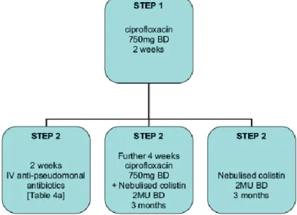
In keeping with recommendations in cystic fibrosis, most specialist centers will attempt eradication of P. aeruginosa upon first isolation. An attempt should be made to eradicate using 14 courses of oral ciprofloxacin. Failure to eradicate P. aeruginosa with oral treatment may lead to consideration of intravenous and/or nebulized eradication (Figure 4).
Figure 4 - The British Thoracic Society guidelines provides a useful algorithm for Pseudomonas aeruginosa eradication (42).
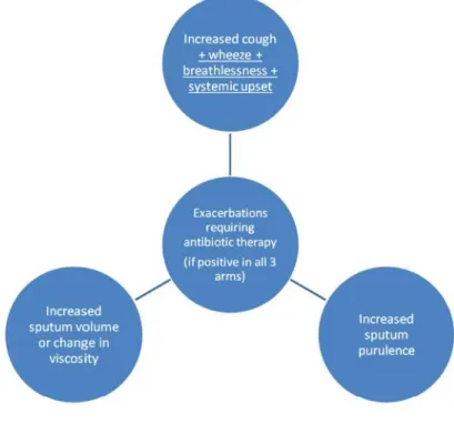
1.6.3.5 Treatment of Exacerbations An exacerbation is defined as (Figure 5):
1) Worsening local symptoms – cough, increases sputum volume, change in viscosity, increased sputum purulence.
2) Dyspnea, increase wheeze, hemoptysis.
3) Systemic upset.
26
Figure 5 - Definition of an exacerbation needing antibiotic therapy (42).
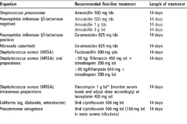
A sputum sample should be obtained at the initial presentation of an exacerbation and sent for culture. While pending sputum culture results, patients should receive treatment targeted towards organisms in previous sputum samples, based on previous sensitivity results. This emphasizes the importance of sending regular sputum samples while patients are clinically stable. Due to the higher bacterial loads and difficulty in achieving antibiotic penetration into biofilms, longer courses of therapy are given in bronchiectasis and most advocate 14 days for exacerbations. (42). If there is no previous bacteriology, first line treatment is amoxicillin 500mg three time a day or clarithromycin 500mg twice a day for 14 days (Table 3). By consideration of empiric therapy in patient with no previous bacteriology, the guideline does not take in consideration certain risk factors: patient age, bronchiectasis location, disease patter (bilateral vs unilateral).
27
Table 3- Common organisms associated with acute exacerbation of bronchiectasis and suggested antimicrobial agents (42).
BTS guidelines state that intravenous antibiotics may be considered in patients who fail to respond to oral therapy, or sputum culture reveals organisms to which no oral therapy is beneficial (e.g multidrug resistant pseudomonas aeruginosa) or where patients are systemically unwell (42). BTS guideline also suggests criteria for inpatient treatment which are:
1) Unable to cope at home.
2) Development of cyanosis or confusion.
3) Breathlessness with respiratory rate >25.
4) Circulatory failure.
5) Respiratory failure.
6) Temperature >38oC.
7) Intravenous therapy required in patients with clinical failure after oral antibiotics
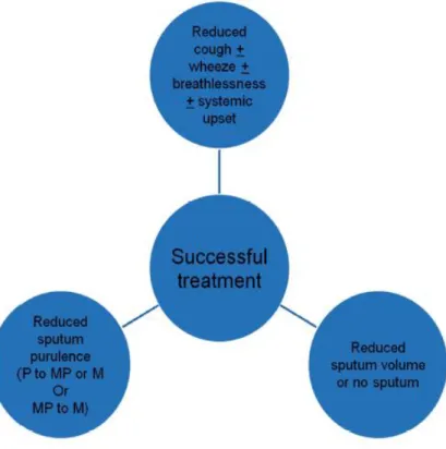
The goals of successful antibiotic treatment are defined in Figure 6.
28
Figure 6 - Definition of successful treatment of an exacerbation.
P, purulent; MP, mucopurulent; M, mucoid.
1.6.4 Pulmonary rehabilitation
Patients with bronchiectasis with significant breathlessness benefit from pulmonary rehabilitation and should be routinely referred for this service (100).
1.7 Parapneumonic effusion
1.7.1 Parapneumonic effusion as an alarming sign in pneumonia
Despite the advent of potent antibiotics, bacterial pneumonia still results in significant morbidity and mortality in the American population. The annual incidence of bacterial pneumonia is estimated to be 4 million, with approximately 25% of patients requiring hospitalization (101). Because as many as 40% of hospitalized patients with bacterial pneumonia have an accompanying pleural effusion (102), effusions associated with pneumonia, parapneumonic effusions, account for a large percentage of pleural effusions.
The morbidity and mortality rates in patients with pneumonia and pleural effusions are higher than those in patient with pneumonia alone. In one study of 1,424 patients
29
hospitalized with community acquired pneumonia, patients with pleural effusions were 2.7 times more likely to be treatment failure than were those without pleural effusions (103). In another study, the relative risk of mortality in patients with community acquired pneumonia was 7 times higher for patients with bilateral pleural effusions and 3.4 times higher for patients with unilateral pleural effusions of moderate or greater size as compared with other patients with community acquired pneumonia alone (104). In assessing risks of patients with community acquired pneumonia, the presence of a pleural effusion is given the same weight as a Po2 less than 60mmHg (105). Espana et al (106) recommended that any patient with pneumonia and a loculated effusion or an effusion greater than 2 cm in thickness on the decubitus be hospitalized. Some of the increased morbidity and mortality in the patients with parapneumonic effusion are due to mismanagement of the pleural effusion (107).
1.7.2 Definition of parapneumonic effusion
A parapneumonic effusion is a pleural effusion associated with lung infection (108).
Early in the course of parapneumonic effusion, the pleura become inflamed with leakage of cellular elements, protein and fluid into the pleural space, forming the effusion.
Subsequent bacterial invasion results in a frank empyema, the presence of which often requires thoracentesis. A delay in the diagnosis and initiation of proper therapy for infectious effusions leads to increases in the complication rate. These delays are more common in patients with coexisting heart failure or malignancy (109-110).
Parapneumonic pleural effusions are divided into three groups or stages based upon pathogenesis.
1. Uncomplicated parapneumonic effusion — An uncomplicated parapneumonic effusion forms when lung interstitial fluid increases during pneumonia and moves across the adjacent visceral pleural membrane. The pleural fluid is characterized by "exudative"
chemistries and an influx of neutrophils into the pleural space. An effusion forms when the resorptive capacity of the pleural space is exceeded; it resolves with resolution of the pneumonia.
2. Complicated parapneumonic effusion — A complicated parapneumonic effusion develops when there is bacterial invasion of the pleural space. Bacterial invasion typically leads to an increased number of neutrophils and the development of pleural fluid acidosis, which results from anaerobic utilization of glucose by the neutrophils and bacteria. In
30
addition, lysis of neutrophils increases the lactate dehydrogenase concentration in the pleural fluid to values often in excess of 1000 IU/L. Perhaps, because the bacterial count is low or because anaerobic cultures are not done, cultures of fluid from complicated parapneumonic effusions are sometimes falsely negative (111). Nevertheless, the typical morphotypes of anaerobes are usually seen on Gram stain and the characteristic putrid odor of the pleural fluid is considered diagnostic of anaerobic infection. Often, there is deposition of a dense layer of fibrin on both the visceral and parietal pleurae that can lead to pleural loculation (112). Whether spontaneous adhesions of the pleural space can result from pleural space inflammation without infection is unknown.
3. Thoracic empyema — Empyema develops when there is evident bacterial infection of the pleural liquid, resulting in either pus or the presence of bacterial organisms on Gram stain. A positive culture is not required for a diagnosis, since there are several reasons why bacteria may not be cultured from an empyema (see 'Bacteriology' below):
● Anaerobic organisms may be difficult to culture or anaerobic cultures are not done because they are not specifically requested
● Sampling is often performed after a patient has received antibiotics
● Sterile inflammatory fluid may be aspirated adjacent to an infected loculus of infection
● Current culture methods are insufficiently sensitive
1.7.3 Clinical presentation
The clinical presentation of the adult with parapneumonic effusion or empyema depends upon the patient's timing of presentation and immune competence, and also the specific organisms causing infection. Patients with pneumonia and uncomplicated parapneumonic effusion present earlier in the course of their pneumonia; those with empyema typically present later when bacteria from the untreated pneumonia have had time to colonize the pleural space. Infection with less virulent bacteria favors a later presentation; therefore, many empyemas complicate indolent anaerobic pneumonias, such as those following aspiration.
Common clinical features of bacterial pneumonia with parapneumonic effusion include cough, fever, pleuritic chest pain, dyspnea, and sputum production. However, patients may only have one or two of these symptoms. In general, the presenting symptoms, other than pleuritic pain and duration of fever, are not helpful in determining which patients
31
have pneumonia versus pneumonia with a parapneumonic effusion or empyema.
Compared to those with pneumonia alone, patients with empyema may report a longer course with several days of fever and malaise rather than hours. Among patients with pneumonia, the presence of a parapneumonic effusion was associated with an increased likelihood of being admitted, a longer hospital stays, and possibly increased mortality (113).
Physical examination may identify the presence of pleural fluid when the fine or coarse crackles, egophony (also known as e-to-a changes), and increased fremitus (palpable asymmetric increase in vibration with speech) typical of consolidation are replaced by decreased breath sounds and decreased fremitus. Occasionally, egophony will still be present at the upper edge of the effusion. Dullness to percussion is a potential feature of lung consolidation from either pneumonia or pleural effusion and is thus not a useful discriminating physical finding. Although clinical findings are helpful when present, they are often absent so radiographic imaging is crucial to the complete evaluation.
Radiographic and ultrasound imaging play a key role in the evaluation and management of parapneumonic effusions and empyema (114). In all patients with pneumonia, the chest radiograph should be evaluated for evidence of pleural fluid.
The presence of a complicated parapneumonic effusion or empyema is first suggested by a chest radiograph showing a pleural-based opacity.
1.7.4 Epidemiology of parapneumonic effusion
Pleural infection is rising in incidence across all age groups worldwide, confirmed by reports from the United States, Canda, Europe and Asia (115). The mortality rate of empyema has risen alarmingly. In Utah, death rates from empyema were sixfold higher in 2000-2004 compared to 1950-1975 (116). Overall in the United States, the incidence of empyema per 100,000 persons had roughly doubled between 1996 and 2008 with roughly equal increase occurring in all age groups (117). In this study, the increase was largely due to increase in nonpneumococcal empyema and Staph sp. empyema (55). The explanation for the increase in empyema incidence is not clear but has been attributed at least in part to the induction of the heptavalent pneumococcal conjugate vaccine (PCV7) IN 2000. After the introduction of this vaccine, there was a reduction in invasive pneumococcal disease, but the incidences of pneumococcal empyema in children and adults have both increased (115). The decrease in incidence of empyema from serotypes
32
covered by the vaccine was overcompensated by an emergence of disease caused by nonvaccine serotypes (particularly serotype 1) (115).
1.7.5 Diagnostic evaluation of a pleural effusion in adults
Determining the cause of a pleural effusion is greatly facilitated by analysis of the pleural fluid. Thoracentesis is a simple bedside procedure with imaging guidance that permits fluid to be rapidly sampled, visualized, examined microscopically and quantified for chemical and cellular content.
The indication for diagnostic thoracentesis is the new finding of a pleural effusion.
Observation, in lieu of diagnostic thoracentesis, may be warranted in uncomplicated heart failure and viral pleurisy. In the former setting, the clinical diagnosis is usually secure; in the latter, there is typically a small amount of fluid. However, if the clinical situation is atypical or does not progress as anticipated, thoracentesis should be performed.
Tests routinely performed on pleural fluid include cell count, pH, protein, lactate dehydrogenase (LDH), and glucose. Additional commonly performed tests in selected patients include amylase, cholesterol, triglycerides, N-terminal pro-BNP, adenosine deaminase, gram and AFB (acid-fast bacilli) stain, bacterial and AFB culture, and cytology.
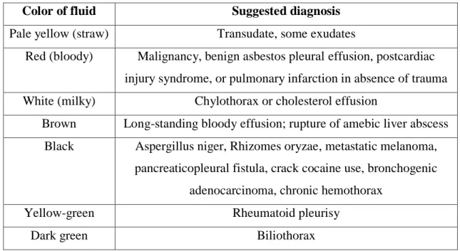
Initial diagnostic clues can be obtained by gross inspection of pleural fluid as it is being aspirated from the patient's chest (118). Observations that are helpful for diagnosis are listed in the table 4.
33
Table 4 – Colors of pleural fluid helpful in diagnosis (109).
Color of fluid Suggested diagnosis
Pale yellow (straw) Transudate, some exudates
Red (bloody) Malignancy, benign asbestos pleural effusion, postcardiac injury syndrome, or pulmonary infarction in absence of trauma White (milky) Chylothorax or cholesterol effusion
Brown Long-standing bloody effusion; rupture of amebic liver abscess Black Aspergillus niger, Rhizomes oryzae, metastatic melanoma,
pancreaticopleural fistula, crack cocaine use, bronchogenic adenocarcinoma, chronic hemothorax
Yellow-green Rheumatoid pleurisy
Dark green Biliothorax
The pleural fluid is next characterized as either a transudate or an exudate.
Transudates — Transudates result from imbalances in hydrostatic and oncotic pressures in the chest, as occur with CHF and nephrosis, or conditions external to the pleural space.
Examples of the latter include movement of fluid from the peritoneal, cerebrospinal, or retroperitoneal spaces, or from iatrogenic causes, such as crystalloid infusion through a central venous catheter that has migrated into the mediastinum or pleural space.
Nevertheless, transudates have a limited number of diagnostic possibilities that can usually be discerned from the patient's clinical presentation (table 5).
34
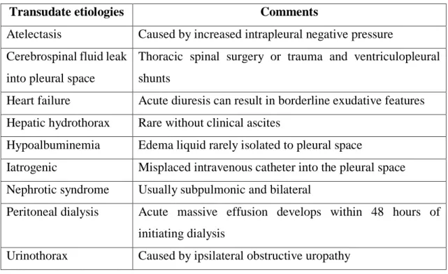
Table 5 - Causes of transudative pleural effusions (109).
Transudate etiologies Comments
Atelectasis Caused by increased intrapleural negative pressure Cerebrospinal fluid leak
into pleural space
Thoracic spinal surgery or trauma and ventriculopleural shunts
Heart failure Acute diuresis can result in borderline exudative features Hepatic hydrothorax Rare without clinical ascites
Hypoalbuminemia Edema liquid rarely isolated to pleural space
Iatrogenic Misplaced intravenous catheter into the pleural space Nephrotic syndrome Usually subpulmonic and bilateral
Peritoneal dialysis Acute massive effusion develops within 48 hours of initiating dialysis
Urinothorax Caused by ipsilateral obstructive uropathy
Exudates — In contrast, exudative effusions present more of a diagnostic challenge.
Disease in virtually any organ can cause exudative pleural effusions by a variety of mechanisms (Table 6), including infection, malignancy, immunologic responses, lymphatic abnormalities, noninfectious inflammation, iatrogenic causes, and movement of fluid from below the diaphragm.
Exudates result primarily from pleural and lung inflammation (resulting in increased capillary and pleural membrane permeability) or from impaired lymphatic drainage of the pleural space (resulting in decreased removal of protein and other large molecular weight constituents from the pleural space). Exudates can also result from movement of fluid from the peritoneal space, as seen with acute or chronic pancreatitis, chylous ascites, and peritoneal carcinomatosis.
35
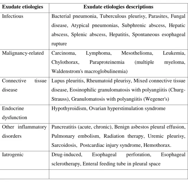
Table 6 – Causes of exudative pleural effusions (109).
Exudate etiologies Exudate etiologies descriptions
Infectious Bacterial pneumonia, Tuberculous pleurisy, Parasites, Fungal disease, Atypical pneumonias, Subphrenic abscess, Hepatic abscess, Splenic abscess, Hepatitis, Spontaneous esophageal rupture
Malignancy-related Carcinoma, Lymphoma, Mesothelioma, Leukemia, Chylothorax, Paraproteinemia (multiple myeloma, Waldenstrom's macroglobulinemia)
Connective tissue disease
Lupus pleuritis, Rheumatoid pleurisy, Mixed connective tissue disease, Eosinophilic granulomatosis with polyangiitis (Churg- Strauss), Granulomatosis with polyangiitis (Wegener's)
Endocrine dysfunction
Hypothyroidism, Ovarian hyperstimulation syndrome
Other inflammatory disorders
Pancreatitis (acute, chronic), Benign asbestos pleural effusion, Pulmonary embolism, Radiation therapy, Uremic pleurisy, Sarcoidosis, Postcardiac injury syndrome, Hemothorax.
Iatrogenic Drug-induced, Esophageal perforation, Esophageal sclerotherapy, Enteral feeding tube in pleural space
1.7.6 Diagnostic criteria
The Light's Criteria Rule is a traditional method of differentiating transudates and exudates that measures serum and pleural fluid protein and LDH (119). Abbreviated versions of Light's Criteria Rule have similar diagnostic accuracy and have been recommended for clinical use (120-121).
According to the traditional Light's Criteria Rule, if at least one of the following three criteria (ie, component tests of the rule) is fulfilled, the fluid is defined as an exudate (119):
1. Pleural fluid protein/serum protein ratio greater than 0.5, or 2. Pleural fluid LDH/serum LDH ratio greater than 0.6, or