R E S E A R C H Open Access
Serum leptin levels in relation to circulating
cytokines, chemokines, adhesion molecules and angiogenic factors in normal pregnancy and preeclampsia
Attila Molvarec1*, András Szarka1, Szilvia Walentin2, Gabriella Bekő3, István Karádi4, Zoltán Prohászka4,5 and János Rigó Jr1
Abstract
Objective:In this study, we determined circulating levels of C-reactive protein, several cytokines, chemokines, adhesion molecules and angiogenic factors along with those of leptin in healthy non-pregnant and pregnant women and preeclamptic patients, and investigated whether serum leptin levels were related to the clinical characteristics and measured laboratory parameters of the study participants.
Methods:Sixty preeclamptic patients, 60 healthy pregnant women and 59 healthy non-pregnant women were involved in this case-control study. Levels of leptin and transforming growth factor (TGF)-beta1 in maternal sera were assessed by ELISA. Serum levels of interleukin (IL)-1beta, IL-1 receptor antagonist (IL-1ra), IL-2, IL-4, IL-6, IL-8, IL- 10, IL-12p40, IL-12p70, IL-18, interferon (IFN)-gamma, tumor necrosis factor (TNF)-alpha, interferon-gamma-inducible protein (IP)-10, monocyte chemotactic protein (MCP)-1, intercellular adhesion molecule (ICAM)-1 and vascular cell adhesion molecule (VCAM)-1 were determined by multiplex suspension array. Serum C-reactive protein (CRP) concentrations were measured by an autoanalyzer. Serum total soluble fms-like tyrosine kinase-1 (sFlt-1) and biologically active placental growth factor (PlGF) levels were determined by electrochemiluminescence immunoassay. For statistical analyses, non-parametric methods were applied.
Results:There were significant differences in most of the measured laboratory parameters among the three study groups except for serum IL-1beta and TGF-beta1 levels. Serum leptin levels were significantly higher in
preeclamptic patients and healthy pregnant women than in healthy non-pregnant women. Additionally,
preeclamptic patients had significantly higher leptin levels as compared to healthy pregnant women. Serum leptin levels were independently associated with BMI in healthy non-pregnant women. In healthy pregnant women, both BMI and serum CRP concentrations showed significant positive linear association with leptin levels. There were significant positive correlations between serum leptin concentrations of healthy pregnant women and systolic blood pressure, as well as serum levels of IP-10, while their serum leptin levels correlated inversely with fetal birth weight. In preeclamptic patients, a significant positive correlation was observed between serum concentrations of leptin and IP-10. Furthermore, elevated serum leptin level and sFlt-1/PlGF ratio had an additive (joint) effect in the risk of preeclampsia, as shown by the substantially higher odds ratios of their combination than of either alone.
Conclusions:Simultaneous measurement of leptin with several inflammatory molecules and angiogenic factors in this study enabled us to investigate their relationship, which can help to understand the role of circulating leptin in normal pregnancy and preeclampsia.
* Correspondence: molvarec@freemail.hu
1First Department of Obstetrics and Gynecology, Semmelweis University, Budapest, Hungary
Full list of author information is available at the end of the article
© 2011 Molvarec et al; licensee BioMed Central Ltd. This is an Open Access article distributed under the terms of the Creative Commons Attribution License (http://creativecommons.org/licenses/by/2.0), which permits unrestricted use, distribution, and reproduction in any medium, provided the original work is properly cited.
Background
Preeclampsia, characterized by hypertension and pro- teinuria developing after the 20thweek of gestation in a previously normotensive woman, is a severe compli- cation of human pregnancy with a worldwide incidence of 2-10%. It is one of the leading causes of maternal, as well as perinatal morbidity and mortality, even in developed countries. Despite extensive research, the etiology and pathogenesis of preeclampsia are not completely understood. There is an increasing body of evidence that an exaggerated maternal systemic inflam- matory response to pregnancy with activation of both the innate and the adaptive arms of the immune sys- tem plays a central role in the pathogenesis of the dis- ease [1,2]. The excessive production of pro- inflammatory cytokines, chemokines and adhesion molecules may trigger a generalized endothelial dys- function characteristic of the maternal syndrome of preeclampsia [3]. In addition, an imbalance between angiogenic and anti-angiogenic factors has also been implicated in the development of this multifactorial disorder [4-6].
Leptin is a peptide hormone of 16 kDa molecular weight comprising 167 amino acids. The major source of leptin is the adipose tissue, but it can also be pro- duced by other organs, including the placenta [7]. This anti-obesity hormone decreases food intake and increases energy expenditure, thereby reducing body weight and adiposity [8]. It also modulates glucose metabolism by increasing insulin sensitivity [9] and acti- vates the sympathetic nervous system [10,11]. Further- more, leptin has been implicated in the control of the reproductive function, including embryonic development and implantation [12]. Leptin can also be considered as a pro-inflammatory cytokine that belongs to the type I cytokine superfamily and has structural similarity with interleukin-6 [13]. Increasing evidence suggests that lep- tin is involved in the regulation of innate and adaptive immune responses and inflammation [14]. Circulating leptin levels are significantly higher in pregnant than in non-pregnant women [7,15], and there is a further increase in complicated pregnancies, such as gestational diabetes mellitus, preeclampsia and intrauterine growth restriction [16-20].
In the present study, we determined serum leptin levels in a large number of healthy non-pregnant and pregnant women and preeclamptic patients. We also measured circulating levels of C-reactive protein and several cytokines, chemokines, adhesion molecules and angiogenic factors in a comprehensive manner, and investigated whether serum leptin levels were related to the clinical characteristics and measured laboratory parameters of the study participants.
Methods Study patients
Our study was designed as a case-control study. Sixty preeclamptic patients, 60 healthy pregnant women with uncomplicated pregnancies and 59 healthy non-pregnant women were involved in the study. The study partici- pants were enrolled in the First Department of Obste- trics and Gynecology and in the Department of Obstetrics and Gynecology of Kútvölgyi Clinical Center, at the Semmelweis University, Budapest, Hungary. All women were Caucasian and resided in the same geo- graphic area in Hungary. The preeclamptic patients and healthy pregnant women were matched on the basis of maternal age and gestational age at blood draw, and they were selected accordingly from the previously reported groups of 93 preeclamptic patients and 176 healthy pregnant women [21,22]. Exclusion criteria were multifetal gestation, chronic hypertension, diabetes mel- litus, autoimmune disease, angiopathy, renal disorder, maternal or fetal infection and fetal congenital anomaly.
None of the pregnant women were in active labor, and none had rupture of membranes. The healthy non-preg- nant women were consecutively selected in the early fol- licular phase of their menstrual cycle (between cycle days 3 and 5), and none of them received hormonal contraception.
Preeclampsia was defined by increased blood pressure (≥140 mmHg systolic or ≥90 mmHg diastolic on ≥2 occasions at least 6 hours apart) that occurred after the 20thweek of gestation in a woman with previously nor- mal blood pressure, accompanied by proteinuria (≥0.3 g/
24 h or≥1 + on dipstick in the absence of urinary tract infection). Blood pressure returned to normal by the 12th postpartum week in each preeclamptic study patient. Preeclampsia was regarded as severe if any of the following criteria was present: blood pressure≥160 mmHg systolic or ≥110 mmHg diastolic, or protei- nuria≥5 g/24 h (or≥3 + on dipstick). Pregnant women with eclampsia or HELLP syndrome (hemolysis, elevated liver enzymes, and low platelet count) were not enrolled in this study. Early onset of preeclampsia was defined as onset of the disease before the 34thweek of gestation (between the 20th and 33rd completed gestational weeks). Intrauterine growth restriction (IUGR) was diag- nosed if the fetal birth weight was below the 10th per- centile for gestational age and gender, based on Hungarian birth weight percentiles [23].
The study protocol was approved by the Regional and Institutional Committee of Science and Research Ethics of the Semmelweis University, and written informed consent was obtained from each patient. The study was conducted in accordance with the Declaration of Helsinki.
Biological samples
Fasting blood samples were taken from an antecubital vein into plain tubes, and then centrifuged at room tem- perature with a relative centrifugal force of 3000gfor 10 minutes. The aliquots of serum were stored at -80°C until the analyses.
Laboratory methods
Levels of leptin and transforming growth factor (TGF)- b1 in maternal sera were assessed by ELISA (DRG Inter- national, Mountainside, New Jersey, USA, Cat. No. EIA- 2395 and EIA-1864, respectively). Serum levels of inter- leukin (IL)-1b, IL-1 receptor antagonist (IL-1ra), IL-2, IL-4, IL-6, IL-8, IL-10, IL-12p40, IL-12p70, IL-18, inter- feron (IFN)-g, tumor necrosis factor (TNF)-a, inter- feron-g-inducible protein (IP)-10, monocyte chemotactic protein (MCP)-1, intercellular adhesion molecule (ICAM)-1 and vascular cell adhesion molecule (VCAM)- 1 were determined by multiplex suspension array on a Bio-Plex 200 analyzer (Bio-Rad Laboratories, Hercules, California, USA). We diluted the serum 1:4 in sample diluent. The samples were measured at low Photomulti- plier Tube (PMT) setting according to the instructions provided in the assay manual. The assays were per- formed using the Bio-Plex filter plates and manual vacuum manifold. We applied the Bio-Plex Manage- r™software. A 10-point extended broad range standard curve was used in order to maximize sensitivity for sam- ples that have very low levels of analytes. Any high end saturation points were removed from the standard curves for determining sample concentrations. Serum C- reactive protein (CRP) concentrations were measured by an autoanalyzer (Cobas Integra 800, Roche, Mannheim, Germany) using the manufacturer’s kit. Serum total soluble fms-like tyrosine kinase-1 (sFlt-1) and biologi- cally active placental growth factor (PlGF) levels were determined by electrochemiluminescence immunoassay (Elecsys, Roche, Mannheim, Germany, Cat. No.
05109523 and 05144671, respectively) on a Cobas e 411 analyzer (Roche, Mannheim, Germany).
Statistical analysis
The normality of continuous variables was assessed using the Shapiro-Wilk’sW-test. As the continuous vari- ables were not normally distributed, non-parametric sta- tistical methods were used. To compare continuous variables between two groups, the Mann-Whitney U- test was applied, whereas to compare them among mul- tiple groups, the Kruskal-Wallis analysis of variance by ranks test was performed. Multiple comparisons of mean ranks for all groups were carried out as post-hoc tests. The Fisher exact and Pearson c2 tests were used to compare categorical variables between groups. The Spearman rank order correlation was applied to
calculate correlation coefficients. Analysis of covariance (ANCOVA) and multiple linear regression analyses were undertaken, as a non-parametric method, with logarith- mically transformed values of serum leptin concentra- tions. Odds ratios (OR) with 95% confidence intervals (CI) were calculated by logistic regression. For all statis- tical analyses, p < 0.05 was considered statistically significant.
In the article, data are reported as median (interquar- tile range) for continuous variables and as number (per- centage) for categorical variables.
Results
Patient characteristics
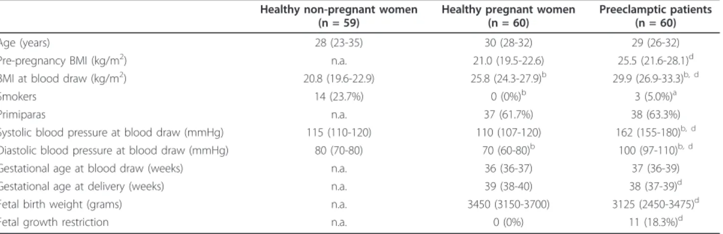
The clinical characteristics of the study participants are described in Table 1. There was no statistically significant difference in terms of age among the study groups.
Furthermore, no significant differences were observed in gestational age at blood collection and the percentage of primiparas between preeclamptic patients and healthy pregnant women. However, all of the other clinical fea- tures presented in Table 1, including smoking status, dif- fered significantly among our study groups. Fetal growth restriction was absent in healthy pregnant women, whereas the frequency of this condition was 18.3% in the preeclamptic group. Doppler abnormalities were observed in 7 fetuses of preeclamptic mothers (11.7%).
Twenty-one women (35.0%) had severe preeclampsia and 5 patients (8.3%) experienced early onset of the disease.
Laboratory parameters
The laboratory parameters of the study subjects are dis- played in Figure 1 and 2. As can be seen in the figures, there were significant differences in most of the mea- sured laboratory parameters among the three study groups except for serum IL-1band TGF-b1 levels. As shown in Figure 1, serum leptin levels were significantly higher in preeclamptic patients and healthy pregnant women than in healthy non-pregnant women. Further- more, preeclamptic patients had significantly higher lep- tin levels as compared to healthy pregnant women. The differences remained significant even after adjustment for BMI at blood draw in ANCOVA.
In the group of preeclamptic patients, no statistically significant differences were found in serum leptin con- centrations between patients with mild and severe pree- clampsia, between patients with late and early onset of the disease, or between preeclamptic patients with and without fetal growth restriction (data not shown).
Relationship of serum leptin levels of the study subjects to their clinical characteristics and laboratory parameters We also investigated whether serum leptin levels of the study participants were related to their clinical features
and laboratory parameters by calculating the Spearman rank order correlation coefficients (continuous variables) or by the Mann-Whitney U-test (categorical variables).
In healthy non-pregnant women, serum leptin concen- trations correlated significantly with body mass index (BMI) and serum CRP levels (Spearman R = 0.59, p <
0.001 and 0.39, p < 0.05, respectively). However, in mul- tiple linear regression analysis, only the association between leptin levels and BMI remained significant (standardized regression coefficient (b) = 0.53, p <
0.001). In the group of healthy pregnant women, we
found statistically significant positive correlations between serum leptin concentrations and BMI (R = 0.49, p < 0.001), systolic blood pressure (R = 0.38, p <
0.05), as well as serum levels of CRP (R = 0.46, p <
0.05), IL-6 (R = 0.35, p < 0.05) and IP-10 (R = 0.31, p <
0.05). In a multiple linear regression model involving BMI, serum CRP and IL-6 levels as independent vari- ables, both BMI and serum CRP concentrations showed significant positive linear association with leptin levels in healthy pregnant women (b = 0.43 and 0.45, respec- tively, p < 0.05 for both). There was a significant inverse correlation between serum leptin concentrations of healthy pregnant women and fetal birth weight (R = -0.36, p < 0.05), even after adjustment for gestational age at delivery in multiple linear regression analysis (b= -0.51, p < 0.001). In the preeclamptic group, a signifi- cant positive correlation was observed between serum levels of leptin and IP-10 (R = 0.36, p < 0.05). There was no other relationship between serum leptin concen- trations of the study subjects and their clinical features and measured laboratory parameters in either study group.
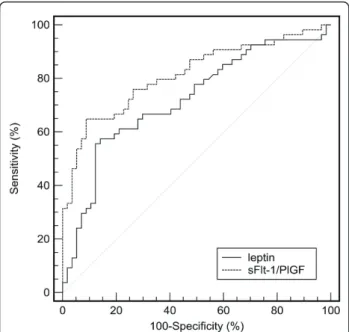
Using the Receiver Operating Characteristic (ROC) curve analysis (Figure 3), we determined cut-off values for serum leptin concentration (> 74.3 ng/ml, sensitivity:
57.6%, specificity: 84.7%; area under curve (AUC) with 95% CI: 0.72 (0.63-0.80)) and sFlt-1/PlGF ratio (> 31.2, sensitivity: 75.9%, specificity: 74.1%; AUC (95% CI): 0.81 (0.73-0.88)) to discriminate preeclamptic patients from healthy pregnant women. In a multiple logistic regres- sion model involving both variables, elevated serum lep- tin level and sFlt-1/PlGF ratio were found to be independent predictors of preeclampsia (odds ratios Table 1 Clinical characteristics of healthy non-pregnant and pregnant women and preeclamptic patients
Healthy non-pregnant women (n = 59)
Healthy pregnant women (n = 60)
Preeclamptic patients (n = 60)
Age (years) 28 (23-35) 30 (28-32) 29 (26-32)
Pre-pregnancy BMI (kg/m2) n.a. 21.0 (19.5-22.6) 25.5 (21.6-28.1)d
BMI at blood draw (kg/m2) 20.8 (19.6-22.9) 25.8 (24.3-27.9)b 29.9 (26.9-33.3)b, d
Smokers 14 (23.7%) 0 (0%)b 3 (5.0%)a
Primiparas n.a. 37 (61.7%) 38 (63.3%)
Systolic blood pressure at blood draw (mmHg) 115 (110-120) 110 (107-120) 162 (155-180)b, d
Diastolic blood pressure at blood draw (mmHg) 80 (70-80) 70 (60-80)b 100 (97-110)b, d
Gestational age at blood draw (weeks) n.a. 36 (36-37) 37 (36-39)
Gestational age at delivery (weeks) n.a. 39 (38-40) 38 (37-39)d
Fetal birth weight (grams) n.a. 3450 (3150-3700) 3125 (2450-3475)d
Fetal growth restriction n.a. 0 (0%) 11 (18.3%)d
Data are presented as median (interquartile range) for continuous variables and as number (percentage) for categorical variables n.a.: not applicable; BMI: body mass index
ap < 0.05 versus healthy non-pregnant women
bp < 0.001 versus healthy non-pregnant women
cp < 0.05 preeclamptic patients versus healthy pregnant women
dp < 0.001 preeclamptic patients versus healthy pregnant women
Figure 1Serum leptin levels of healthy non-pregnant and pregnant women and preeclamptic patients. Middle point:
median; Box: interquartile range (25-75 percentile); Whisker: range (excluding outliers).ap < 0.001 versus healthy non-pregnant women;bp < 0.001 preeclamptic patients versus healthy pregnant women.
Figure 2Serum levels of C-reactive protein, cytokines, chemokines, adhesion molecules and angiogenic factors in healthy non- pregnant and pregnant women and preeclamptic patients. Middle point: median; Box: interquartile range (25-75 percentile); Whisker: range (excluding outliers). CRP: C-reactive protein; IL: interleukin; IL-1ra: IL-1 receptor antagonist; IFN: interferon; TNF: tumor necrosis factor; TGF:
transforming growth factor; IP: interferon-g-inducible protein; MCP: monocyte chemotactic protein; ICAM: intercellular adhesion molecule; VCAM:
vascular cell adhesion molecule; sFlt: soluble fms-like tyrosine kinase; PlGF: placental growth factor.ap < 0.05 versus healthy non-pregnant women;bp < 0.001 versus healthy non-pregnant women;cp < 0.05 preeclamptic patients versus healthy pregnant women;dp < 0.001 preeclamptic patients versus healthy pregnant women.
with 95% confidence intervals: 9.16 (3.16-26.5) and 8.89 (3.34-23.7), respectively, p < 0.001 for both; after adjust- ment for BMI at blood draw: 5.03 (1.48-17.1), p < 0.05 and 11.3 (3.24-39.2), p < 0.001, respectively). Women with elevated serum leptin level and sFlt-1/PlGF ratio had substantially higher odds for having preeclampsia than those with elevated sFlt-1/PlGF ratio or serum lep- tin concentration alone (OR (95% CI): 80.5 (14.4-450), p
< 0.001 versus 9.0 (2.80-29.0), p < 0.001 and 9.33 (2.27- 38.4), p < 0.05, respectively), even after adjustment for BMI at blood collection in multiple logistic regression analyses (adjusted OR (95% CI): 65.9 (9.47-459), p <
0.001 versus 6.91 (1.55-30.7), p < 0.05 and 1.42 (0.15- 13.0), p > 0.05, respectively).
Discussion
In this study, we measured circulating levels of C-reac- tive protein, several cytokines, chemokines, adhesion molecules and angiogenic factors along with those of leptin in healthy non-pregnant and pregnant women and preeclamptic patients. According to our findings, serum leptin levels were independently associated with BMI in healthy non-pregnant women. In healthy preg- nant women, both BMI and serum CRP concentrations showed significant positive linear association with leptin levels. There were significant positive correlations between serum leptin concentrations of healthy preg- nant women and systolic blood pressure, as well as serum levels of IP-10, while their serum leptin levels
correlated inversely with fetal birth weight. In pree- clamptic patients, a significant positive correlation was observed between serum concentrations of leptin and IP-10. Furthermore, the combination of elevated serum leptin level and sFlt-1/PlGF ratio was found to be addi- tive for the risk of preeclampsia.
Leptin was first introduced as an adipocyte-derived signal of energy metabolism [24]. Indeed, the indepen- dent association of serum leptin levels with BMI in our healthy non-pregnant and pregnant women indicates that adipose tissue is a major source of circulating lep- tin. Later on, the placenta has been demonstrated to be an alternative source of leptin in the maternal circula- tion [7,25]. Maternal serum leptin levels have been reported to increase during the course of a normal preg- nancy with a peak at around 20-30 weeks of gestation and to decrease rapidly after birth [26-28]. In addition to BMI, serum levels of CRP were also directly related to those of leptin in our healthy pregnant group (unlike in non-pregnant women [29]), suggesting that systemic inflammation characteristic of the third trimester of nor- mal pregnancy might contribute to the increase in circu- lating leptin levels. In fact, inflammatory stimuli, including pro-inflammatory cytokines, can induce the expression of leptin and increase its serum level [30-32].
Therefore, the elevation of maternal circulating leptin levels in normal pregnancy could reflect - at least in part - an increase in adipocyte/trophoblast leptin synth- esis mediated by inflammatory stimuli. On the contrary, serum leptin levels were found to increase indepen- dently from BMI and CRP in our preeclamptic group, as shown by the lack of correlations with these variables.
Interestingly, placental expression of leptin has been observed to be raised in preeclampsia by several research groups, including ours [17,33-37]. An increas- ing body of evidence suggests that placental insufficiency and resultant placental hypoxia might be responsible for augmented placental production of leptin in this preg- nancy-specific disorder, leading to its elevated concen- tration in the maternal circulation [17,38-41].
Leptin is a pleiotropic molecule, which has also been implicated in the regulation of innate and adaptive immune responses by several ways [14,42]. This mole- cule alters the balance of T cell-derived cytokines in favour of a Th1-type response [43,44]. Leptin can also induce pro-inflammatory cytokine production of mono- cytes/macrophages [45,46]. It is noteworthy that the maternal syndrome of preeclampsia is characterized by a predominant Th1-type immune response with elevated amounts of pro-inflammatory molecules in the maternal circulation [3,47,48]. However, in this study, we did not find any association between serum levels of leptin and pro-inflammatory cytokines in preeclampsia, which might be explained - at least partly - by the fact that the
Figure 3Receiver Operating Characteristic (ROC) curves. Serum leptin levels (leptin, continuous line) and soluble fms-like tyrosine kinase-1 to placental growth factor ratio (sFlt-1/PlGF ratio, dashed line) to discriminate between preeclamptic patients and healthy pregnant women.
latter (especially TNF-a and IL-1b) have a very short half- life in the maternal circulation. Leptin may also be involved in blood pressure regulation through its central effect on sympathetic activity [10,11,49]. Accordingly, there was a significant positive correlation between serum leptin concentrations and systolic blood pressure in our healthy pregnant women. Nevertheless, the lack of relationship between leptin levels and blood pressure values in our preeclamptic group indicates that the pro- cess responsible for blood pressure elevation in this multifactorial disease is more complex and involves the interplay of numerous factors in addition to the sympa- thetic action of leptin.
An intriguing finding of our study is that increased serum levels of leptin were related to those of interferon- g-inducible protein (IP)-10 both in normal pregnancy and preeclampsia, as shown by the significant correla- tions between these variables. This is consistent with the in vitroobservation that leptin selectively induces the expression and secretion of IP-10 in human monocytic cells [50]. IP-10 (CXCL10) has pro-inflammatory and anti-angiogenic properties, and this chemokine has been proposed to be a potential link between inflammation and anti-angiogenesis in preeclampsia [51]. In our pre- vious study, IP-10 showed the strongest association with markers of endothelial dysfunction, as well as with renal and liver function parameters in healthy pregnant women and preeclamptic patients [3]. Therefore, it is tempting to speculate that IP-10 might play a connecting role between elevated circulating leptin levels and the generalized endothelial dysfunction characteristic of pre- eclampsia. On the other hand, leptin can promote angio- genesis at least partly through the up-regulation of vascular endothelial growth factor expression in endothe- lial cells [52,53]. Nevertheless, increased levels of anti- angiogenic factors such as sFlt-1 in the maternal circula- tion in normal pregnancy and preeclampsia might blunt this beneficial direct effect of leptin on angiogenesis.
Another remarkable observation of this study is that serum leptin levels correlated inversely with fetal birth weight in the group of healthy pregnant women. In pre- vious studies, elevated circulating leptin levels have been found in mothers with fetal growth restriction [18,19].
This relationship might reflect the state of inflammation and chronic stress in IUGR mothers. As reduced uteropla- cental blood flow plays an important role in the pathogen- esis of IUGR, resultant placental hypoxia might also account for the observed increase in maternal circulating leptin levels. Indeed, leptin expression was up-regulated in placental tissues of IUGR neonates [54]. Furthermore, maternal first trimester plasma leptin levels negatively cor- related with birth weight among normotensive and pree- clamptic women [55]. Interestingly, we have recently detected an inverse correlation between IP-10 levels and
fetal birth weight in healthy pregnant women, which raises the possibility that maternal leptin levels could influence fetal growth in normal pregnancy - at least in part - through the inhibitory effect of IP-10 on placental angio- genesis. Nevertheless, preeclampsia may disrupt the asso- ciation of maternal leptin levels with fetal growth, as shown by the lack of correlation with birth weight in our preeclamptic group. On the contrary, a recent meta-analy- sis demonstrated that cord blood leptin concentrations of small-for-gestational-age newborns are significantly lower than those of appropriate-for-gestational-age newborns [56]. Additionally, fetal circulating leptin levels positively correlate with birth weight in normal pregnancy [15,57].
In fact, leptin seems to play a crucial role in fetal growth [58], and it might be a potential link between poor fetal growth and the subsequent development of chronic dis- eases in later life [59].
Accumulating data indicate a central role of circulat- ing angiogenic factors and their antagonists in the pathogenesis of preeclampsia [4-6]. In this study, we also measured serum sFlt-1 and PlGF concentrations in normal pregnancy and preeclampsia by electrochemilu- minescence immunoassay. However, increased sFlt-1 and decreased PlGF levels were not related to serum leptin concentrations in women with preeclampsia, sug- gesting that alterations in angiogenic cytokine profile and circulating levels of leptin are independent mechan- isms in the pathogenesis of this multifactorial disorder.
Instead, elevated serum leptin level and sFlt-1/PlGF ratio had an additive (joint) effect in the risk of pree- clampsia, as shown by the substantially higher odds ratios of their combination than of either alone.
In our study, the similar circulating leptin concentra- tions of preeclamptic patients regardless of the severity, the time of onset of the disease or the presence of fetal growth restriction might be explained by the multifactor- ial etiology of preeclampsia. Several genetic, behavioural and environmental factors need to interact to produce the complete picture of this pregnancy-specific disorder.
Our research group reported various genetic (including leptin receptor gene polymorphisms) and soluble factors that were associated with the severity or complications of preeclampsia [60-63]. Nevertheless, it is also possible that the relatively small sample size of this study prevented to detect an effect in the subgroup analyses.
Simultaneous measurement of leptin with C-reactive protein, several cytokines, chemokines, adhesion mole- cules and angiogenic factors in this study enabled us to investigate their relationship, which can help to under- stand the role of circulating leptin in normal pregnancy and preeclampsia. Elevated leptin concentration in the first trimester of pregnancy appears to be associated with an increased risk of developing preeclampsia later in gestation [55,64,65]. Given the case-control design of
our study, we could not confirm this association.
Further longitudinal studies are required to clarify the predictive value of circulating leptin in preeclampsia.
Conclusions
Elevated serum leptin concentrations were associated with the mass of adipose tissue, systemic inflammation and sys- tolic blood pressure, as well as negatively with birth weight in the third trimester of normal pregnancy. In addition, increased serum levels of leptin were related to those of interferon-g-inducible protein (IP)-10 both in normal pregnancy and preeclampsia, suggesting that circulating leptin might contribute to the development of endothelial dysfunction and poor fetal growth through the anti-angio- genic effect of IP-10. Moreover, the combination of ele- vated serum leptin level and sFlt-1/PlGF ratio was found to be additive for the risk of preeclampsia.
Acknowledgements
This work was supported by a research grant from the Faculty of Medicine of the Semmelweis University, as well as by the János Bolyai Research Scholarship of the Hungarian Academy of Sciences.
Author details
1First Department of Obstetrics and Gynecology, Semmelweis University, Budapest, Hungary.2Central Laboratory, Kútvölgyi Clinical Center, Semmelweis University, Budapest, Hungary.3Department of Laboratory Medicine, Semmelweis University, Budapest, Hungary.4Third Department of Internal Medicine, Semmelweis University, Budapest, Hungary.5Research Group of Inflammation Biology and Immunogenomics, Hungarian Academy of Sciences, Budapest, Hungary.
Authors’contributions
AM conceived of the study, participated in its design and coordination, performed statistical analyses and drafted the manuscript. ASZ collected data and helped to draft the manuscript. SZW determined circulating angiogenic factors. GB carried out multiplex suspension array measurements.
IK participated in the coordination of the study. ZP measured serum leptin levels. JR participated in the design and coordination of the study. All authors read and approved the final manuscript.
Competing interests
The authors declare that they have no competing interests.
Received: 1 July 2011 Accepted: 9 September 2011 Published: 9 September 2011
References
1. Redman CW, Sacks GP, Sargent IL:Preeclampsia: an excessive maternal inflammatory response to pregnancy.Am J Obstet Gynecol1999, 180:499-506.
2. Saito S, Shiozaki A, Nakashima A, Sakai M, Sasaki Y:The role of the immune system in preeclampsia.Mol Aspects Med2007,28:192-209.
3. Szarka A, Rigo J Jr, Lazar L, Beko G, Molvarec A:Circulating cytokines, chemokines and adhesion molecules in normal pregnancy and preeclampsia determined by multiplex suspension array.BMC Immunol 2010,11:59.
4. Maynard SE, Min JY, Merchan J, Lim KH, Li J, Mondal S, Libermann TA, Morgan JP, Sellke FW, Stillman IE, Epstein FH, Sukhatme VP, Karumanchi SA:
Excess placental soluble fms-like tyrosine kinase 1 (sFlt1) may contribute to endothelial dysfunction, hypertension, and proteinuria in
preeclampsia.J Clin Invest2003,111:649-658.
5. Molvarec A, Szarka A, Walentin S, Szucs E, Nagy B, Rigo J Jr:Circulating angiogenic factors determined by electrochemiluminescence
immunoassay in relation to the clinical features and laboratory parameters in women with pre-eclampsia.Hypertens Res2010,33:892-898.
6. Molvarec A, Ito M, Shima T, Yoneda S, Toldi G, Stenczer B, Vasarhelyi B, Rigo J Jr, Saito S:Decreased proportion of peripheral blood vascular endothelial growth factor-expressing T and natural killer cells in preeclampsia.Am J Obstet Gynecol2010,203:567, e1-8.
7. Masuzaki H, Ogawa Y, Sagawa N, Hosoda K, Matsumoto T, Mise H, Nishimura H, Yoshimasa Y, Tanaka I, Mori T, Nakao K:Nonadipose tissue production of leptin: leptin as a novel placenta-derived hormone in humans.Nat Med1997,3:1029-1033.
8. Pelleymounter MA, Cullen MJ, Baker MB, Hecht R, Winters D, Boone T, Collins F:Effects of the obese gene product on body weight regulation in ob/ob mice.Science1995,269:540-543.
9. Ogawa Y, Masuzaki H, Hosoda K, Aizawa-Abe M, Suga J, Suda M, Ebihara K, Iwai H, Matsuoka N, Satoh N, Odaka H, Kasuga H, Fujisawa Y, Inoue G, Nishimura H, Yoshimasa Y, Nakao K:Increased glucose metabolism and insulin sensitivity in transgenic skinny mice overexpressing leptin.
Diabetes1999,48:1822-1829.
10. Shek EW, Brands MW, Hall JE:Chronic leptin infusion increases arterial pressure.Hypertension1998,31:409-414.
11. Aizawa-Abe M, Ogawa Y, Masuzaki H, Ebihara K, Satoh N, Iwai H, Matsuoka N, Hayashi T, Hosoda K, Inoue G, Yoshimasa Y, Nakao K:
Pathophysiological role of leptin in obesity-related hypertension.J Clin Invest2000,105:1243-1252.
12. Cervero A, Dominguez F, Horcajadas JA, Quinonero A, Pellicer A, Simon C:
The role of the leptin in reproduction.Curr Opin Obstet Gynecol2006, 18:297-303.
13. Otero M, Lago R, Lago F, Casanueva FF, Dieguez C, Gomez-Reino JJ, Gualillo O:Leptin, from fat to inflammation: old questions and new insights.FEBS Lett2005,579:295-301.
14. Fantuzzi G, Faggioni R:Leptin in the regulation of immunity, inflammation, and hematopoiesis.J Leukoc Biol2000,68:437-446.
15. Sarandakou A, Protonotariou E, Rizos D, Malamitsi-Puchner A, Giannaki G, Phocas I, Creatsas G:Serum leptin concentrations during the perinatal period.Am J Perinatol2000,17:325-328.
16. Vitoratos N, Salamalekis E, Kassanos D, Loghis C, Panayotopoulos N, Kouskouni E, Creatsas G:Maternal plasma leptin levels and their relationship to insulin and glucose in gestational-onset diabetes.Gynecol Obstet Invest2001,51:17-21.
17. Mise H, Sagawa N, Matsumoto T, Yura S, Nanno H, Itoh H, Mori T, Masuzaki H, Hosoda K, Ogawa Y, Nakao K:Augmented placental production of leptin in preeclampsia: possible involvement of placental hypoxia.J Clin Endocrinol Metab1998,83:3225-3229.
18. Mise H, Yura S, Itoh H, Nuamah MA, Takemura M, Sagawa N, Fujii S:The relationship between maternal plasma leptin levels and fetal growth restriction.Endocr J2007,54:945-951.
19. Kyriakakou M, Malamitsi-Puchner A, Militsi H, Boutsikou T, Margeli A, Hassiakos D, Kanaka-Gantenbein C, Papassotiriou I, Mastorakos G:Leptin and adiponectin concentrations in intrauterine growth restricted and appropriate for gestational age fetuses, neonates, and their mothers.Eur J Endocrinol2008,158:343-348.
20. Briana DD, Malamitsi-Puchner A:Reviews: adipocytokines in normal and complicated pregnancies.Reprod Sci2009,16:921-937.
21. Molvarec A, Prohaszka Z, Nagy B, Szalay J, Fust G, Karadi I, Rigo J Jr:
Association of elevated serum heat-shock protein 70 concentration with transient hypertension of pregnancy, preeclampsia and superimposed preeclampsia: a case-control study.J Hum Hypertens2006,20:780-786.
22. Molvarec A, Rigo J Jr, Nagy B, Walentin S, Szalay J, Fust G, Karadi I, Prohaszka Z:Serum heat shock protein 70 levels are decreased in normal human pregnancy.J Reprod Immunol2007,74:163-169.
23. Joubert K:Standards of the body mass and body length of birth in Hungary on the basis of the 1990-1996 nation-wide liveborn data.Magy Noorv L2000,63:155-163.
24. Zhang Y, Proenca R, Maffei M, Barone M, Leopold L, Friedman JM:
Positional cloning of the mouse obese gene and its human homologue.
Nature1994,372:425-432.
25. Green ED, Maffei M, Braden VV, Proenca R, DeSilva U, Zhang Y, Chua SC Jr, Leibel RL, Weissenbach J, Friedman JM:The human obese (OB) gene: RNA expression pattern and mapping on the physical, cytogenetic, and genetic maps of chromosome 7.Genome Res1995,5:5-12.
26. Sattar N, Greer IA, Pirwani I, Gibson J, Wallace AM:Leptin levels in pregnancy: marker for fat accumulation and mobilization?Acta Obstet Gynecol Scand1998,77:278-283.
27. Schubring C, Englaro P, Siebler T, Blum WF, Demirakca T, Kratzsch J, Kiess W:Longitudinal analysis of maternal serum leptin levels during pregnancy, at birth and up to six weeks after birth: relation to body mass index, skinfolds, sex steroids and umbilical cord blood leptin levels.Horm Res1998,50:276-283.
28. Henson MC, Castracane VD:Leptin in pregnancy.Biol Reprod2000, 63:1219-1228.
29. Wunder DM, Yared M, Bersinger NA, Widmer D, Kretschmer R, Birkhauser MH:Serum leptin and C-reactive protein levels in the physiological spontaneous menstrual cycle in reproductive age women.
Eur J Endocrinol2006,155:137-142.
30. Grunfeld C, Zhao C, Fuller J, Pollack A, Moser A, Friedman J, Feingold KR:
Endotoxin and cytokines induce expression of leptin, the ob gene product, in hamsters.J Clin Invest1996,97:2152-2157.
31. Sarraf P, Frederich RC, Turner EM, Ma G, Jaskowiak NT, Rivet DJ, Flier JS, Lowell BB, Fraker DL, Alexander HR:Multiple cytokines and acute inflammation raise mouse leptin levels: potential role in inflammatory anorexia.J Exp Med1997,185:171-175.
32. Faggioni R, Fantuzzi G, Fuller J, Dinarello CA, Feingold KR, Grunfeld C:IL-1 beta mediates leptin induction during inflammation.Am J Physiol1998, 274:R204-208.
33. Dotsch J, Nusken KD, Knerr I, Kirschbaum M, Repp R, Rascher W:Leptin and neuropeptide Y gene expression in human placenta: ontogeny and evidence for similarities to hypothalamic regulation.J Clin Endocrinol Metab1999,84:2755-2758.
34. Li RH, Poon SC, Yu MY, Wong YF:Expression of placental leptin and leptin receptors in preeclampsia.Int J Gynecol Pathol2004,23:378-385.
35. Enquobahrie DA, Meller M, Rice K, Psaty BM, Siscovick DS, Williams MA:
Differential placental gene expression in preeclampsia.Am J Obstet Gynecol2008,199:566, e1-11.
36. Sitras V, Paulssen RH, Gronaas H, Leirvik J, Hanssen TA, Vartun A, Acharya G:
Differential placental gene expression in severe preeclampsia.Placenta 2009,30:424-433.
37. Varkonyi T, Nagy B, Fule T, Tarca AL, Karaszi K, Schonleber J, Hupuczi P, Mihalik N, Kovalszky I, Rigo J Jr, Meiri H, Papp Z, Romero R, Than NG:
Microarray profiling reveals that placental transcriptomes of early-onset HELLP syndrome and preeclampsia are similar.Placenta2011,32(Suppl):
S21-29.
38. Grosfeld A, Turban S, Andre J, Cauzac M, Challier JC, Hauguel-de Mouzon S, Guerre-Millo M:Transcriptional effect of hypoxia on placental leptin.FEBS Lett2001,502:122-126.
39. Grosfeld A, Andre J, Hauguel-De Mouzon S, Berra E, Pouyssegur J, Guerre- Millo M:Hypoxia-inducible factor 1 transactivates the human leptin gene promoter.J Biol Chem2002,277:42953-42957.
40. Lepercq J, Guerre-Millo M, Andre J, Cauzac M, Hauguel-de Mouzon S:
Leptin: a potential marker of placental insufficiency.Gynecol Obstet Invest 2003,55:151-155.
41. Haugen F, Ranheim T, Harsem NK, Lips E, Staff AC, Drevon CA:Increased plasma levels of adipokines in preeclampsia: relationship to placenta and adipose tissue gene expression.Am J Physiol Endocrinol Metab2006, 290:E326-333.
42. Fernandez-Riejos P, Najib S, Santos-Alvarez J, Martin-Romero C, Perez- Perez A, Gonzalez-Yanes C, Sanchez-Margalet V:Role of leptin in the activation of immune cells.Mediators Inflamm2010,2010:568343.
43. Lord GM, Matarese G, Howard JK, Baker RJ, Bloom SR, Lechler RI:Leptin modulates the T-cell immune response and reverses starvation-induced immunosuppression.Nature1998,394:897-901.
44. Martin-Romero C, Santos-Alvarez J, Goberna R, Sanchez-Margalet V:Human leptin enhances activation and proliferation of human circulating T lymphocytes.Cell Immunol2000,199:15-24.
45. Loffreda S, Yang SQ, Lin HZ, Karp CL, Brengman ML, Wang DJ, Klein AS, Bulkley GB, Bao C, Noble PW, Lane MD, Diehl AM:Leptin regulates proinflammatory immune responses.Faseb J1998,12:57-65.
46. Santos-Alvarez J, Goberna R, Sanchez-Margalet V:Human leptin stimulates proliferation and activation of human circulating monocytes.Cell Immunol1999,194:6-11.
47. Saito S, Umekage H, Sakamoto Y, Sakai M, Tanebe K, Sasaki Y, Morikawa H:
Increased T-helper-1-type immunity and decreased T-helper-2-type
immunity in patients with preeclampsia.Am J Reprod Immunol1999, 41:297-306.
48. Saito S, Sakai M, Sasaki Y, Tanebe K, Tsuda H, Michimata T:Quantitative analysis of peripheral blood Th0, Th1, Th2 and the Th1:Th2 cell ratio during normal human pregnancy and preeclampsia.Clin Exp Immunol 1999,117:550-555.
49. Hall JE, Hildebrandt DA, Kuo J:Obesity hypertension: role of leptin and sympathetic nervous system.Am J Hypertens2001,14:103S-115S.
50. Meier CA, Chicheportiche R, Dreyer M, Dayer JM:IP-10, but not RANTES, is upregulated by leptin in monocytic cells.Cytokine2003,21:43-47.
51. Gotsch F, Romero R, Friel L, Kusanovic JP, Espinoza J, Erez O, Than NG, Mittal P, Edwin S, Yoon BH, Kim CJ, Mazaki-Tovi S, Chaiworapongsa T, Hassan SS:CXCL10/IP-10: a missing link between inflammation and anti- angiogenesis in preeclampsia?J Matern Fetal Neonatal Med2007, 20:777-792.
52. Sierra-Honigmann MR, Nath AK, Murakami C, Garcia-Cardena G, Papapetropoulos A, Sessa WC, Madge LA, Schechner JS, Schwabb MB, Polverini PJ, Flores-Riveros JR:Biological action of leptin as an angiogenic factor.Science1998,281:1683-1686.
53. Suganami E, Takagi H, Ohashi H, Suzuma K, Suzuma I, Oh H, Watanabe D, Ojima T, Suganami T, Fujio Y, Nakao K, Ogawa Y, Yoshimura N:Leptin stimulates ischemia-induced retinal neovascularization: possible role of vascular endothelial growth factor expressed in retinal endothelial cells.
Diabetes2004,53:2443-2448.
54. Struwe E, Berzl G, Schild R, Blessing H, Drexel L, Hauck B, Tzschoppe A, Weidinger M, Sachs M, Scheler C, Schleussner E, Dotsch J:Microarray analysis of placental tissue in intrauterine growth restriction.Clin Endocrinol (Oxf)2010,72:241-247.
55. Papastefanou I, Samolis S, Panagopoulos P, Tagia M, Bale C, Kouskoukis A, Galazios G:Correlation between maternal first trimester plasma leptin levels and birth weight among normotensive and preeclamptic women.
J Matern Fetal Neonatal Med2010,23:1435-1443.
56. Ren RX, Shen Y:A meta-analysis of relationship between birth weight and cord blood leptin levels in newborns.World J Pediatr2010,6:311-316.
57. Gursoy T, Aliefendioglu D, Aslan AT, Gunduz M, Haberal A, Senes M, Cakmak FN, Laleli YR:Preeclampsia disrupts the normal physiology of leptin.Am J Perinatol2002,19:303-310.
58. Briana DD, Malamitsi-Puchner A:The role of adipocytokines in fetal growth.Ann N Y Acad Sci2010,1205:82-87.
59. Briana DD, Malamitsi-Puchner A:Intrauterine growth restriction and adult disease: the role of adipocytokines.Eur J Endocrinol2009,160:337-347.
60. Rigo J, Szendei G, Rosta K, Fekete A, Bogi K, Molvarec A, Ronai Z, Ver A:
Leptin receptor gene polymorphisms in severely pre-eclamptic women.
Gynecol Endocrinol2006,22:521-525.
61. Molvarec A, Jermendy A, Nagy B, Kovacs M, Varkonyi T, Hupuczi P, Prohaszka Z, Rigo J Jr:Association between tumor necrosis factor (TNF)- alpha G-308A gene polymorphism and preeclampsia complicated by severe fetal growth restriction.Clin Chim Acta2008,392:52-57.
62. Molvarec A, Rigo J Jr, Lazar L, Balogh K, Mako V, Cervenak L, Mezes M, Prohaszka Z:Increased serum heat-shock protein 70 levels reflect systemic inflammation, oxidative stress and hepatocellular injury in preeclampsia.Cell Stress Chaperones2009,14:151-159.
63. Rosta K, Molvarec A, Enzsoly A, Nagy B, Ronai Z, Fekete A, Sasvari-Szekely M, Rigo J Jr, Ver A:Association of extracellular superoxide dismutase (SOD3) Ala40Thr gene polymorphism with pre-eclampsia complicated by severe fetal growth restriction.Eur J Obstet Gynecol Reprod Biol2009,142:134-138.
64. Ning Y, Williams MA, Muy-Rivera M, Leisenring WM, Luthy DA:Relationship of maternal plasma leptin and risk of pre-eclampsia: a prospective study.J Matern Fetal Neonatal Med2004,15:186-192.
65. Samolis S, Papastefanou I, Panagopoulos P, Galazios G, Kouskoukis A, Maroulis G:Relation between first trimester maternal serum leptin levels and body mass index in normotensive and pre-eclamptic pregnancies– role of leptin as a marker of pre-eclampsia: a prospective case-control study.Gynecol Endocrinol2010,26:338-343.
doi:10.1186/1477-7827-9-124
Cite this article as:Molvarecet al.:Serum leptin levels in relation to circulating cytokines, chemokines, adhesion molecules and angiogenic factors in normal pregnancy and preeclampsia.Reproductive Biology and Endocrinology20119:124.