TISSUE AND CIRCULATING PROGNOSTIC BIOMARKERS IN MALIGNANT PLEURAL
MESOTHELIOMA
PhD thesis
Thomas Klikovits, MD
Doctoral School of Clinical Medicine Semmelweis University
Consultant: Ferenc Renyi-Vamos, MD, Ph.D
Official reviewers: Erika Hitre MD, Ph.D Levente Fazekas MD, Ph.D
Head of the Final Examination Committee: Nándor Ács MD, Ph.D
Members of the Final Examination Committee: Marcell Szász MD, Ph.D Zoltán Heiler MD, Ph.D
Budapest
2017
- 2 -
Table of contents
1 Abbreviations ... 5
2 Introduction ... 9
2.1 Malignant pleural mesothelioma ... 9
2.1.1 Epidemiology ... 9
2.1.2 Etiology ... 11
2.1.2.1 Asbestos-related MPM development ... 11
2.1.2.2 Non-asbestos-related MPM development ... 16
2.1.3 Molecular background of MPM ... 17
2.1.4 Histology ... 21
2.1.5 Screening, clinical diagnosis, prognosis and staging of MPM ... 23
2.1.6 Therapeutic approaches ... 31
2.1.6.1 Systemic therapy ... 32
2.1.6.2 Surgery ... 35
2.1.6.2.1 Radical surgical approaches ... 35
2.1.6.2.2 Pleurectomy/Decortication ... 35
2.1.6.2.3 Extrapleural Pneumonectomy ... 36
2.1.6.2.4 Patient selection for surgery ... 38
2.1.6.2.5 Multimodality treatment ... 39
2.1.6.2.6 Palliative surgery ... 41
2.1.6.3 Radiotherapy ... 41
2.1.7 Prognostic tissue biomarkers in MPM ... 43
2.1.7.1 KI-67 ... 46
2.1.8 Prognostic circulating biomarkers in MPM ... 48
2.1.8.1 Complement component 4d (C4d) ... 50
2.1.8.2 Activin A ... 55
3 Objectives ... 59
3.1 Ki-67 index as a prognostic parameter in MPM ... 59
3.2 Circulating C4d as a prognostic biomarker in MPM ... 59
3.3 Circulating ActA as a prognostic parameter in MPM ... 60
- 3 -
4 Methods ... 61
4.1 General ethical considerations ... 61
4.2 Evaluation of Ki-67 index as a prognostic parameter in MPM ... 61
4.2.1 Study population ... 61
4.2.2 Tumor samples, staining, scoring and blood biomarkers ... 62
4.2.3 Statistical analyses ... 63
4.3 Evaluation of circulating C4d as a prognostic parameter in MPM ... 64
4.3.1 Study population ... 64
4.3.2 Tumor and blood samples, staining and blood biomarkers ... 64
4.3.3 Tumor volumetry ... 65
4.3.4 Statistical analyses ... 66
4.4 Evaluation of circulating ActA as a prognostic parameter in MPM ... 66
4.4.1 Study population ... 66
4.4.2 Blood samples ... 67
4.4.3 Tumor volumetry ... 67
4.4.4 Statistical analyses ... 67
5 Results ... 69
5.1 Ki-67 index as a prognostic parameter in MPM ... 69
5.1.1 OS is influenced by treatment modality and histological subtype ... 69
5.1.2 Ki67 index is not associated with histology and stage but significantly decreased in post-chemotherapy cases ... 70
5.1.3 Ki67 is an independent prognostic marker only in epithelioid MPM .. ... 72
5.1.4 Ki67 is an accurate marker predicting short-term survival in epithelioid MPM ... 74
5.2 C4d as a circulating prognostic biomarker in MPM ... 76
5.2.1 Lack of tumor cell-specific expression of C4d ... 76
5.2.2 C4d plasma levels are not elevated in MPM patients compared to HV or NMPD ... 77
5.2.3 Correlation of C4d with CT-based tumor volumetry ... 78
5.2.4 Correlation of C4d with circulating inflammatory-based markers .. 79
- 4 -
5.2.5 Plasma levels of C4d predict chemotherapeutic response after
induction treatment ... 79
5.2.6 Circulating C4d has a prognostic impact in MPM ... 80
5.2.7 Correlation of circulating C4d and C3a levels ... 81
5.2.8 Limited tumor cell-specific expression of C1q in MPM ... 82
5.3 Circulating ActA as a prognostic biomarker in MPM ... 84
5.3.1 Impact of ActA in a in distinguishing MPM from non-malignant pleural disease ... 84
5.3.2 ActA level has prognostic impact in epithelioid MPM only ... 85
5.3.3 Association of circulating ActA and fibrinogen ... 88
5.3.4 Correlation of ActA with tumor volume and chemotherapy treatment ... 88
6 Discussion ... 90
6.1 Diagnostic biomarkers in MPM ... 90
6.2 Prognostic factors and biomarkers in MPM ... 92
7 Conclusion ... 101
8 Summary ... 102
9 Összefoglalás ... 103
10 Bibliography ... 104
11 Bibliography of the candidate’s publications ... 129
11.1 Publications related to the thesis ... 129
11.2 Publications not related to the thesis ... 130
12 List of figures ... 133
13 List of tables ... 140
14 Acknowledgements ... 142
- 5 -
1 Abbreviations
ActA Activin A
AMIG Austrian Mesothelioma Interest Group AQP1 Aquaporin 1
AUVA Allgemeine Unfallversicherungs Anstalt BAL Bronchoalveolar lavage
BAP1 BRCA1(breast cancer 1)-associated protein1 Bcl-2 B-cell lymphoma-2
Bg8 Blood group 8 BSC Best supportive care C4d Complement component 4d CDH8 Cadherin 8
CDKN2A–ARF Cyclin-dependent kinase inhibitor 2A CFAP45 Cilia and Flagella Associated Protein 45 CHT Chemotherapy
CI Confidence interval
CNV Copy number variations COX-2 Cyclooxygenase-2 CRO Croatia
CRP C-reactive protein CT Computed tomography DDX3X DEAD-box helicase 3 DDX51 DEAD-Box Helicase 51 DFS Disease-free survival DHFR Dihydrofolate reductase DPP10 Dipeptidyl-peptidase 10 EBUS Endobronchial ultrasound EE Environmental exposure
EGFR Epidermal growth factor receptor ELISA Enzyme-linked immunosorbent assay EMT Epithelial-to-mesenchymal transition
- 6 -
EORTC European Organization for Research and Treatment of Cancer EPP Extrapleural pneumonectomy
ERCC1 Excision repair cross-complementation group 1 ERS European Respiratory Society
ESTS European Society of Thoracic Surgeons EUS Esophageal ultrasound
FDG-PET Fluorodeoxyglucose positron emission tomography FFPE Formalin fixed paraffin embedded
FNA Fine-needle aspiration FSH Follicle-stimulating hormone Gas-6 Growth arrest signal-6 GPS Glasgow Prognostic Score Gy Gray
HE Hematoxylin/eosin
HIOC Hyperthermic intraoperative chemotherapy HR Hazard ratio
HUN Hungary
HV Healthy volunteers
IARC International Agency for Research on Cancer
IASLC International Association for the Study of Lung Cancer IgM Immunoglobulin M
IHC Immunohistochemistry
IMIG International Mesothelioma Interest Group IMRT Intensity modulated radiotherapy
IRR Incidence rate ratio
MAC Membrane attack complex
MAP2K6 Mitogen-activated protein kinase kinase 6 MAPK Mitogen-activated protein kinase
MAPS Mesothelioma Avastin Cisplatin Pemetrexed Study MARS Mesothelioma and Radical Surgery
MASP MBL-associated serine protease MBL Mannan-binding lectin
- 7 - MCR Macroscopic complete resection MIF Migration inhibitory factor MMT Multimodal treatment
MOC-31 Mouse Monoclonal Primary Antibody 31 MPM Malignant pleural mesothelioma
mTOR Mechanistic target of rapamycin NA Not available
NCCN National Comprehensive Cancer Network NF2 Neurofibromin type 2
NFRKB Nuclear factor related to kappa B binding protein NKX6-2 NK6 homeobox 2
NLR Neutrophil-to-lymphocyte ratio
NLRP3 NOD-like receptor family, pyrin domain containing 3 NMPD Non-malignant pleural diseases
NSCLC Non-small cell lung cancer
nTiO2 Nano size titanium dioxide particles NTS Neurotensin
NTSR1 Neurotensin receptor 1 OPN Osteopontin
OR Odds ratio OS Overall survival
P/D Pleurectomy/decortication
PCBD2 Pterin-4-alpha-carbinolamine dehydratase 2 PDT Intracavitary photodynamic therapy
PFS Progression free survival PLR Platelet-to-lymphocyte ratio PS Performance status
RNS Reactive nitrogen species ROS Reactive oxygen species RT Radiotherapy
RTK Receptor-tyrosine kinase RYR2 Ryanodine receptor 2
- 8 - SCLC Small cell lung cancer
SD Standard deviation
SETD2 SET domain containing 2 SETDB1 SET Domain Bifurcated 1
SMART Surgery for Mesothelioma After Radiation Therapy
SMRP Serum-soluble mesothelin family proteins, soluble mesothelin-related peptide SV40 Simian virus 40
TBNA Transbronchial needle aspiration TKI Tyrosine-kinase-inhibitor
TNM Tumor node metastasis TS Thymidylate synthase
ULK2 Unc-51 Like Autophagy Activating Kinase 2 US United States
VATS Video-assisted thoracic surgery VATS-PP VATS partial pleurectomy VEGF Vascular endothelial growth factor WHO World Health Organization
WT-1 Wilms-Tumor Protein 1
- 9 -
2 Introduction
2.1 Malignant pleural mesothelioma
2.1.1 Epidemiology
In 2012, 14.1 million new cancer cases and 8.2 million cancer deaths occurred worldwide. Among these, lung and breast cancer are diagnosed most frequently and represent the leading causes of cancer death in men and women, overall and in less developed countries. However, in more developed countries, prostate cancer accounts for the most diagnosed malignancy and lung cancer represents the leading cause of cancer death in women [1]. In general, global cancer burden will shift to less developed countries within the next decades due to an increasing prevalence of risk factors and growing and aging populations [2]. By 2012, less developed countries accounted for only 57% of global cancer cases and 65% of cancers deaths, due to more frequent other causes of death, such as infection, and the younger age structure [1, 2].
Worldwide, liver, stomach and colorectal cancers are additionally frequently diagnosed among men, whereas stomach, cervical and colorectal cancer are frequent among women [1]. In more developed countries, prostate, colorectal, breast and lung cancer incidences tend to be higher, whereas liver, stomach and cervical cancer are more frequently diagnosed in less developed countries. These trends are predominantly attributable to infectious diseases, being more prevalent in less developed countries [3].
Additional risk factors for most frequently diagnosed cancers worldwide include smoking (lung, colorectal, stomach, liver cancer), overweight and physical inactivity (breast, colorectal). By applying effective risk factor prevention strategies (i.e. tobacco control, vaccination) and the broad use of early detection tests, a substantial proportion of cancer cases could be effectively prevented [1]. Besides highly prevalent cancer types, some less frequently diagnoses malignancies are gradually on rise and research must focus on prevention, early detection and new therapeutic targets in these diseases as well.
Malignant pleural mesothelioma (MPM) is a rare but devastating malignancy, arising from the pleural space. The tumor is known to be a rare disease; however, its incidence is increasing worldwide, probably as a result of widespread exposure to asbestos, known to be the main risk factor for MPM development. There is a significant variation
- 10 -
of MPM incidence among different areas worldwide. It ranges between 7 per million inhabitants in Japan and 40 per million in Australia [4]. The highest mesothelioma incidence rates are reported from some countries in Europe (UK, Italy, The Netherlands, Malta, Belgium) and in Oceania (Australia, New Zealand). In the UK for instance, the annual number of deaths from MPM increased continuously, being 153 in 1968 and 2.360 in 2010. In 2011, the numbers of deaths were 2.291. In the period of 2000-2011, incidence rates in the UK were 3.3-3.6 per 100,000 among men, and 0.5-0.7 among women [5]. In Italy, incidence in 2011 among men was 3.5 and 1.25 per 100,000 in men and women, respectively, and wide differences are noted among different geographic areas within the country [6]. Intermediate incidence rates are reported from a group of countries including large parts of Europe and the United States (US) [7, 8]. In Germany, 7.547 malignant mesotheliomas were reported to cancer registries diagnosed between 2009 and 2013, 90% of those being located in the chest. On average, 1.198 men and 312 women were affected each year. Regional clusters were predominantly located to the seaports of West Germany [9]. In the US, between 2003 and 2008 over 3000 cases were diagnosed each year, with a maximum of 3284 in 2005. In this period, the incidence was 1.93 per 100,000 among men and 0.41 among women [7]. Low incidence/mortality rates are reported from various countries of Central Europe, Ireland, Spain, and from several countries of Asia [10-12]. In Austria, there have been 276 cases of MPM approved by the “Allgemeine Unfallversicherungsanstalt – General accident insurance company” (AUVA) as being caused occupationally between 2010 and 2015. Of these, 53 were approved in 2014 only. In contrast to this, ten asbestos-related MPM cases were documented in 1995 and 41 in 2005. However, there is still uncertainty about the number of MPM cases not being reported to the AUVA and currently, there is no register on non-occupational MPM in Austria [12]. A comparison of the incidence of MPM worldwide and in Austria is depicted in Figure 1.
In Europe, the average incidence is 2 per 100.000 inhabitants. The frequency is highly dependent on the amount of asbestos removal, asbestos import and industrialization and the peak incidence is to be expected around 2020 due to the long latency period [4, 13].
In the US, MPM peaked in the 1980s to 1990s and is now plateauing. In men, incidence has been stable at 1.8 cases per 100.000 for the past 10 years, with peak values in the early 1990s (2.5 cases per 100,000 people), while in women, rates were 0.4 cases per
- 11 -
100.000 people, and did not change substantially over time [14]. Due to the long latency period and recent asbestos banning efforts, a possible reduction in the MPM health burden and a reduction in the number of newly diagnosed cases is expected in the near future, at least in developed countries [15].
Figure 1: Incidence of MPM in Austria compared to worldwide MPM incidence ([8]). Data are given as age-standardized rates per 100.000 in men.
Currently, however, MPM incidence is still increasing in most countries of the world, and a decrease can only be seen in countries where asbestos control measures were taken [5, 14]. Thus, the overall worldwide epidemic is still increasing and in countries that still produce and/or commercially use asbestos, such as China, India, Russia, a sharp rise in incidence might be expected in the future [16-18].
2.1.2 Etiology
2.1.2.1 Asbestos-related MPM development
Previous asbestos exposure is known to be the main risk factor for the development of MPM [19, 20]. Asbestos is classified into two main groups, the serpentines and the
3,40 3,20 2,85 2,50 2,08 2,00 1,84 1,76 1,50 1,40 1,40 1,40 1,23 1,11 1,07 1,00 0,90 0,82 0,60
0,0 0,5 1,0 1,5 2,0 2,5 3,0 3,5 4,0
UK Australia Netherlands New Zealand Malta Belgium Croatia Denmark Finland Cyprus Germany Norway Israel Sweden South Africa Austria Iceland Ireland Poland
- 12 -
amphiboles. The serpentines consist of one type, chrysotile (95% of asbestos in commercial use), with a characteristic short and curly fiber, also referred to as “white asbestos” due to its white color. The amphiboles, with straight, longer fibers, include crocidolite or “blue asbestos”, amosite, tremolite, actinolite and anthophyllite. These six fibers are collectively summarized as “asbestos” due to regulatory issues and their common known health risk [21, 22]. The risk of MPM development has shown to be dependent on the fiber type, as shorter fibers are assumed to be less carcinogenic [23].
Even though some studies claimed that chrysotile could generate mesothelioma only if it was contaminated with amphiboles [24], it has been clearly shown that chrysotile is an important carcinogen and risk factor for MPM development, and also for lung cancer [25]. Thus, in the current international perception, all types of asbestos are classified as class I carcinogens, according to the World Health Organization (WHO) and the International Agency for Research on Cancer (IARC). Furthermore, it is well recognized that exposure to asbestos is the major cause of both pleural and peritoneal mesothelioma, which resulted in the banning of asbestos production and import in several European countries at various time-points after 1970, and in the European Union as late as 2005 [4, 22].
Asbestos exposure is typically labor-dependent and is recognized as an occupational disease in many countries [8]. The mean latency period between exposure to asbestos and the onset of symptoms has been reported to be up to 40 years, and 99 % of cases show a latency of more than 15 years [26]. In most epidemiological studies, MPM is more common in men and some studies have claimed that its occurrence is correlated with sex. However, other studies have shown that MPM development is to be related to asbestos exposure and, typically, there is low asbestos exposure in women because the occupations associated with exposure are traditionally carried out by men [27].
Additionally, women with MPM have shown to exhibit a threefold better overall survival (OS) than men [14]. More recently, a shift has been observed from asbestos- removal workers to professionals involved in post-construction work, e. g., electricians, plumbers, or heat protection technicians [19].
There is a clear correlation between the amount of asbestos exposure and the incidence of MPM, however, no safe lower threshold has been identified. Furthermore, there is an increasing incidence of non-occupational asbestos diseases among housewives and
- 13 -
family members of asbestos workers, due to cleaning of contaminated work clothes, as well as a high environmental impact in the vicinity of mining and processing facilities [8, 28, 29]. In a certain region of Italy, an epidemic of MPM was registered among inhabitants who were never exposed to local asbestos factories, whereas a high proportion of cases had only one risk factor: living close to an asbestos cement factory.
The calculated risk was very high for those living <500 m from the factory and the fiber burden in the lungs of deceased cases was 10-fold that in those from other areas [30].
Another recent study reported a mesothelioma incidence rate ratio (IRR) of 12.92 for those living <500 m from an asbestos cement plant in Barcelona, Spain, while the ratios decreased to 0.70 and 0.23 for those living 500-2000 m and >2000-10000 m from the plant, respectively [31]. Accordingly, a dose dependent relation between asbestos exposure and risk for MPM development is well perceived.
Despite worldwide efforts to ban asbestos production and commercial use, there is still the additional risk for environmental exposure, whose impact on MPM development is not well studied. The quantification of environmental associated MPM occurrence is furthermore limited mainly by the lack of reliable assessment of type and amount of exposure [15, 32]. One major feature of environmental exposure associated MPM is a general higher disease burden in women than in men. The ratio (male:female) in several studies is often close or less than 1 (Figure 2).
- 14 -
Figure 2: Male to female ratios among malignant mesothelioma cases reporting overall exposure (occupational and environmental) and environmental exposure (EE) to asbestos [15]. EE, environmental exposure
Environmental occurrence of mesothelioma has been found in people growing up near natural asbestos resources (Turkey, Corsica, Cyprus) or in areas where asbestos was used for the whitening of house walls. In this regard, erionite, an asbestos-like mineral from the soil, was revealed as the main factor of mesothelioma in young people in some villages in Turkey, where more than 50% of inhabitants died from MPM [33]. In this area, the average annual mesothelioma incidence rates are 114.8 per 100,000 for men and 159.8 per 100,000 for women, or 88.3 times greater in men and 799 times greater in women, respectively, in comparison to world background incidence rates [34, 35].
Another study revealed that whitewashing the houses with soft tremolite in Metsovo, Greece, was the reason for a high number of mesothelioma cases in young women, as they used to do this work [36].
Inhaled asbestos fibers enter the pleural space through the alveoli or retrograde through the lymphatic vessels, causing cytotoxicity, DNA damage, frustrated phagocytosis and chronic inflammation (Figure 3) [37, 38]. Important key mechanisms of the mesothelial
- 15 -
cells, such as chromosomal aberrations and epigenetic changes, result in cellular dysfunction at gene, microRNA and protein expression levels [39, 40].
Asbestos fibers are usually detected and entrapped by alveolar macrophages into lysosomes. The NOD-like receptor family, pyrin domain containing 3 (NLRP3) inflammasome gets activated to cleave procaspase 1 to an active form. Fibrotic nodules are formed by the release of cleaved and activated prointerleukin-1-beta. Consequently, reactive oxygen species (ROS) and reactive nitrogen species (RNS) are produced within the macrophages which ultimately cause cellular and tissue damage. Apoptosis of alveolar macrophages lead to the production of various cytokines such as IL-1β tumor necrosis factor (TNF)-α, macrophage inflammatory protein (MIP)-1/2, monocyte- chemoattractant protein-1, and IL-8 to cause chronic inflammation and proliferation of collagenic fibers. Asbestos particles are then again released and newly recognized by nearby macrophages leading to the repetition of similar cellular response mechanisms.
Ultimately, partially cleaved asbestos fibers are transferred to regional lymph nodes, particularly at the opening of lymphatic vessels. Circulating and local immunocompetent cells will recognize these fibers repeatedly, recurrently and continuously, encountering for chronic local inflammation [41-43].
However, the question if the MPM progenitor cell arises from a highly differentiated mesothelial cell or a submesothelial multipotential cell is still unresolved yet. Normal mesothelial cells may regenerate from normal mesothelium and by the development of submesothelial multipotential stem cells [44]. Moreover, recent studies showed that also adipocytes, circulating multipotential fibrocytes and adult bone marrow-derived stem cells can differentiate into both epithelial and mesenchymal cells [45]. Despite this, it is still further unclear, if this MPM progenitor cell might arise from the parietal pleura more likely than from the visceral. It has been previously hypothesized that a parietal origin might be more likely since cases with only parietal affection (previously staged as T1a (TNM-7)) have a worse survival compared to cases in stage T1b (TNM-7), which involves the parietal and visceral pleura, indicating that the parietal lesion is an earlier event [46, 47].
- 16 - 2.1.2.2 Non-asbestos-related MPM development
In contrast to asbestos-related MPM development, the incidence of mesothelioma without asbestos contact is extremely low (<1: 1 million). Potential cofactors for MPM occurrence besides asbestos are synthetic materials (ceramics, nanoparticles), ionizing radiation, and SV-40 virus infections [48]. Exposure to nanoparticles such as carbon nanotubes has been shown to cause MPM similar to that caused by asbestos, and thus has become an environmental health issue [49]. In this regard, fiber-length is an important parameter in triggering chronic pleural inflammation. Recently, it has been demonstrated that nanofibers beyond 4 µm in length are pathogenic to the pleura and shorter nanofibers might exhibit a potentially lower risk of cellular damage [50].
Figure 3: Hypothesized sequence of events leading to pleural responses as a consequence of long fiber retention of asbestos and nanofibers at the parietal pleural stomatal openings leading to chronic inflammation and tumor induction [51].
Among nanofibers, nano size titanium dioxide particles (nTiO2) are one of the most commonly used metal nanoparticles in commercial products, such as cosmetics, sunscreens, food products, paints and drugs. nTiO2 have been shown to cause reactive oxygen species (ROS) leading to toxicity [52]. A recent study has shown that with respect to the toxicity of nTiO2 on human-derived mesothelial cells, the crystal form rather than the particle size has a greater effect on cellular absorption, and thus causes more cellular damage [53]. It is hypothesized that long fiber retention of nanofibers at the parietal pleural stomatal openings may lead to chronic inflammation and tumor induction eventually, similar to inflammation caused by asbestos exposure (Figure 3)
- 17 -
[51]. However, further studies shedding light on nanoparticles uptake and MPM induction are to be awaited.
Long-term effects of ionizing radiation have been made responsible for MPM development, however in a much smaller population than in asbestos exposed individuals [54]. Especially, individuals exposed to α-particle-emitting agents are at higher risk for MPM. Moreover, several studies showed a higher incidence of mesothelioma in patients treated with external beam radiotherapy for testicular cancer or lymphoma [55, 56].
Simian virus (SV) 40 was discovered in 1959 as a virus being endemic in rhesus monkeys, whose kidneys were used for primary cell cultures for the preparation of inactivated poliovirus vaccine (IPV) and live oral poliovirus vaccine. SV40 was shown to be oncogenic in rodents, causing mesothelioma, ependymoma, osteosarcoma and non-Hodgkin's lymphoma [57]. Formaldehyde was used at the 1950s to inactivate the poliovirus, but it did not completely turn down SV40 and thus there have been estimates that 30–100 million people in the USA and many more worldwide received potentially contaminated vaccines prepared during the years 1955–1963 [58]. In a meta-analysis including 528 MPM cases from 15 studies, the risk for the presence of SV40 DNA sequences in MPM tumor tissue was very high compared to controls (OR 17, 95% CI 10–28) [59]. Moreover, recent studies showed that rodents infected with SV40 were highly susceptible to asbestos-related carcinogenesis [60]. However, the link between SV40 infection and human cancer development is still controversial.
2.1.3 Molecular background of MPM
Because MPM is rare, genomic studies are limited and typically involve a small number of samples [61]. Nevertheless, due to the use of high-throughput analyses and a revolution in molecular characterization, the knowledge of cytogenetic and molecular changes in MPM has substantially increased in recent years [22]. Recent analyses have demonstrated frequent gained chromosomal regions in 5p, 7p, 7q, 8q, and 17q as well as frequent deletions of specific sites within chromosome arms 1p, 3p, 6q, 9p, 13q, 15q and 22q. Two of these regions are most frequently altered, the tumor suppressors cyclin- dependent kinase inhibitor 2A (CDKN2A–ARF) at 9p21 and neurofibromin type 2 (NF2) at 22q12 [62]. These have been known for a long time, but recurrent somatic
- 18 -
mutations in BRCA1 (breast cancer 1)-associated protein1 (BAP1) gene were more recently identified. BAP1 is a tumor suppressor gene located on 3p21, a chromosome region frequently lost in mesothelioma [61]. In contrast to many other solid tumor types, MPM has been reported to be rarely mutated in TP53 (Figure 4) [63].
Only limited data are available on whole genome sequencing of MPM cases. A recent study reported several chromosomal copy number variations (CNV) in a single MPM tumor sample [64]. In some chromosomes, copy number losses were detected (4, 14, 18, 19, and 10), others showed gains (5) or a combination of both (1, 8, 9, 11, 15, 16, 17, 21, and 22). Seventeen tumor-specific genes were identified, some of which are considered to be candidates for further investigation regarding future therapeutic options. Among genes of interest were mitogen-activated protein kinase kinase 6 gene (MAP2K6), dipeptidyl-peptidase 10 (DPP10), pterin-4-alpha-carbinolamine dehydratase 2 (PCBD2) and dihydrofolate reductase (DHFR). DHFR is of specific interest since it encodes an enzyme being important in the folate metabolism, which might be linked to reduced antifolate (ie. pemetrexed) treatment response in some MPM patients [65].
- 19 -
Figure 4: Frequency and types of genetic aberrations (mutation, amplification, loss, fusion/rearrangement or multiple alteration) among 23 pleural mesothelioma cases [63].
In this study, 3 further heterozygous point mutations were identified apart from CNVs.
These were noted in NK6 homeobox 2 gene (NKX6-2), cadherin 8 gene (CDH8), and nuclear factor related to kappa B binding protein gene (NFRKB).
Genetic alterations can be identified at DNA level (whole genome or exome sequencing) or at the mRNA level (i.e. transcriptome sequencing). Several reports are already available with these techniques in human MPM. To date, the largest and most comprehensive analysis has been reported by Bueno and colleagues [66]. In their study, transcriptomes (n=211), whole exomes (n=99) and targeted exomes (n=103) from 216 MPM patients were analyzed. By whole exome analysis, the following genes were found to be significantly mutated: BAP1, NF2, TP53, SET domain containing 2 (SETD2), DEAD-box helicase 3 (DDX3X), Unc-51 Like Autophagy Activating Kinase 2 (ULK2), Ryanodine receptor 2 (RYR2), Cilia and Flagella Associated Protein 45 (CFAP45), SET Domain Bifurcated 1 (SETDB1) and DEAD-Box Helicase 51
- 20 -
(DDX51). Furthermore, recurrent gene fusions and splice alterations were identified, being frequent inactivation mechanisms for NF2, BAP1 and SETD2. Through integrated analyses, alterations in Hippo, mTOR, histone methylation, RNA helicase and p53 signaling pathways in human MPM were identified. The authors concluded that incorporating genomic analysis for the detection of actionable alterations as part of new treatment strategies will help in developing rational individualized therapy in MPM patients.
Based on recent knowledge, it is likely that several mutations need to eventually accumulate for MPM development. The long latency period between asbestos exposure and MPM formation could support this hypothesis. However, most of the aforementioned mutations can be clustered in 4 main intracellular pathways:
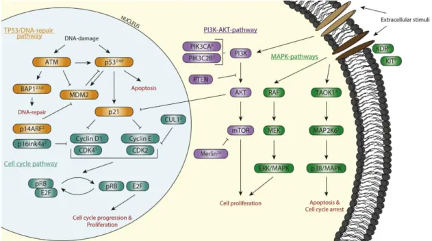
TP53/DNA repair, cell cycle regulation, mitogen-activated protein kinase (MAPK) and PI3K/AKT [67].
Figure 5: Affected pathways in MPM. Observed mutations cluster in four main pathways: the tumor protein p53 (TP53)/DNA-repair pathway (orange), the cell cycle pathway (blue), the mitogen-activated protein kinase (MAPK) pathway (green), and the phophatidylinositol-3 kinase (PI3K)-AKT pathway (purple) [67]
Each of these pathways is known to be important in cell growth, proliferation, and survival, processes that are all altered during tumor development [67].
- 21 -
Apart from somatic mutations in MPM, germline BAP1 mutation is the first gene reported to predispose for MPM development [68]. BAP1 is a nuclear protein, involved in transcriptional regulation, chromatin regulation, and forming part of multiprotein complexes that regulate cellular differentiation, gluconeogenesis, cell cycle checkpoints, transcription and apoptosis. The BAP1 gene is located on chromosome 3p21, a region that shows loss or deletion in 30–60% of mesotheliomas [68]. In families carrying BAP1 mutation, there is a dramatically increased incidence of malignant tumors, often diagnosed at earlier age compared to the general population [115]. In consequence, a
“BAP1 cancer syndrome” has been proposed, including mesothelioma, uveal and cutaneous melanoma and possibly other malignant tumors [69]. Moreover, germ-line BAP1 mutations have been described in families with extraordinarily high incidence of mesothelioma [69].
2.1.4 Histology
In 2015, the most recent World Health Organization (WHO) classification of tumors of the pleura has been published. While the histologic classification of MPM remains the same in the 2015 version as it was in the 2004 WHO classification, some new observations have been reported [70-72]. MPM derives from pleural stem cells, exhibiting both epithelioid and sarcomatoid growing patterns at the same time.
Depending on which component is predominant, three histological types of MPM can be distinguished: epithelioid (50–70 %), sarcomatoid (7–20 %) and a mixed or biphasic form (Figure 6) [73, 74]. Within the category of epithelioid MPM, a variety of morphologic subtypes are defined, including tubulopapillary, papillary, micro-papillary, trabecular, solid, and pleomorphic (Table 1) [71]. Especially the pleomorphic subtype of epithelioid MPM has been demonstrated to be associated with significantly poorer outcome, similar to that of patients with biphasic or sarcomatoid subtype [75].
Since there are morphological similarities of MPM to LADC, the histological diagnosis of epithelioid MPM can be challenging. However, besides hematoxylin and eosin (H&E) staining, additional immunohistochemistry can be a useful in distinguishing MPM from secondary malignances (including lung carcinomas) spreading to the pleura.
Specificity and sensitivity considerations support the use of calretinin, cytokeratins 5/6, WT-1, and podoplanin (D2-40) as positive mesothelial markers, and carcinoembryonic
- 22 -
antigen (CEA), B72.3 (recognizing tumor-associated glyocoprotein 72), Bg8 (blood group 8, detecting the Lewis Y antigen, type 2 chain), BerEP4 (differentiating glandular epithelium from mesothelium) and MOC-31 as positive carcinoma markers [71].
Figure 6: Examples of epithelioid a), biphasic (b) and sarcomatoid (c) MPM [73].
Table 1: Histological specification of malignant pleural mesothelioma [70, 72]
Epithelioid Sarcomatoid Biphasic mixed
– Tubulopapillary – Acinar
– Glandular – Adenomatoid
– Solid epithelioid patterns – Small cell
– Oat cell – Pleomorphic
Differential diagnosis:
metastatic carcinomas and other epithelioid tumors
Mimic malignant mesenchymal tumors:
leiomyosarcoma synovial sarcoma
Desmoplastic mesothelioma bland tumor cells
Differential diagnosis:
sarcomatoid carcinoma and other sarcomas
Combination of all epithelioid and sarcomatoid features Differential diagnosis:
Synovial sarcoma, other mixed or biphasic tumors
Sarcomatoid MPM consists of irregularly arranged elongated spindle cells, which show no uniformity in shape. The biphasic subtype shows a mixture of both, epithelioid and sarcomatoid elements.
The pathological diagnosis and differential diagnosis of MPM can be very challenging.
In a French study, the initial diagnosis of MPM was revised as false positive in 13 % of cases [76]. This can be explained in part by the fact that MPM can present in very heterogeneous forms on the one hand and must be distinguished from benign processes and other tumors. Such a differential diagnosis can be particularly difficult since mesothelioma-like features can also be found in some lymphomas, thymomas, and carcinomas, etc. However, Brcic and colleagues found a substantial interobserver reproducibility among two observers in the histological subtyping of MPM [74].
- 23 -
In small tissue biopsies, early invasive MPM is particularly difficult to be diagnosed, often disguised by cutting artefacts or the malorientation of sections, but may be suspected if there is nodular mesothelial cell proliferation. If definitive invasion cannot be identified, the diagnosis of “atypical mesothelial proliferation” is appropriate, and further sampling may be indicated. Distinguishing MPM from inflammatory pleural disease requires a full-thickness biopsy sample, with correct orientation of histological sections, perpendicular to the pleural surface [77, 78].
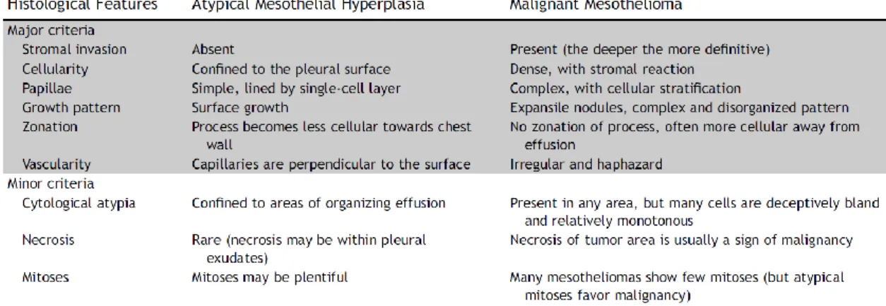
Moreover, some specific morphologic criteria are recognized in order to distinguish MPM from reactive mesothelial hyperplasia and organizing pleuritis and these are depicted in Table 2. However, the application of these criteria might be challenging due to size of the specimens, sampling problems, entrapment of mesothelial cells and superficial or tangential cuts [71, 79, 80].
Table 2: Tissue features of reactive atypical mesothelial hyperplasia versus epithelioid MPM. Adopted from [71]
2.1.5 Screening, clinical diagnosis, prognosis and staging of MPM
In current international guidelines (ie. European Society of Thoracic Surgeons/European Respiratory Society (ESTS/ERS) guidelines), no general screening methods in order to detect asymptomatic MPM are recommended. This is based on the low sensitivity of even advanced imaging techniques such as low-dose computed tomography (CT) in screening of asbestos workers [81]. Circulating biomarkers such as fibulin-3, osteopontin, mesothelin-related peptides and soluble mesothelin-related peptide (SMRP) have been extensively investigated in MPM and asbestos exposed individuals
- 24 -
[82-84]. However, none of them are yet to be considered as a reliable screening tool [81, 85, 86].
Specific clinical symptoms of MPM are comprised of dyspnea, cough and chest pain on initial examination. Dyspnea is often caused by pleural effusion and later by extensive restriction due to pleural and pulmonary tumor masses in the thoracic cavity. Chest pain might be diffuse, sometimes radiating into the shoulders, arms or abdomen. Tumorous invasion of the brachial plexus and the intercostal or paravertebral nerves can additionally cause neuropathic pain. Weight loss is a symptom of more advanced disease [73]. Since MPM development is associated with previous asbestos exposure (as described above), it is recommended to obtain a detailed occupational history [86].
Typically, MPM occurs as an initially unilateral disease. During disease progression, the tumor can, however, spread to the contralateral pleural cavity or into the peritoneum.
Compared with lung cancer, distant metastases in the extrathoracic lymph nodes or in other parenchymal organs are usually rare, although they do occur at more advanced stages [87].
In patients with suspected MPM and recurrent pleural effusions and/or pleural thickening, the recommended initial evaluation includes contrast-enhanced computed tomography (CT) scan of the chest, thoracentesis for cytological assessment of the effusion, pleural biopsy and general laboratory blood tests [73, 81, 85, 86]. Plain chest x-ray lacks sufficient sensitivity for routine diagnosis and staging as significant pleural effusions can mask pleural lesions.
Figure 7: Computer tomography of a patient with MPM showing circular involvement of the visceral and parietal pleura, pericardium and mediastinum. Pulmonary window (left) and mediastinal window (right).
Adopted from [73]
- 25 -
By radiological approaches, it can be difficult to distinguish between malignant and benign pleural disease and also to distinguish MPM from other malignant tumors spreading to the pleura such as metastatic carcinomas, sarcomas and thymomas [78].
A thoracoscopy is recommended to obtain adequate biopsies for histological verification, to optimally stage and to allow pleural fluid evacuation (with or without pleurodesis). This can usually be performed as a pleuroscopy or as video-assisted thoracic surgery (VATS). Fine-needle aspiration (FNA) is not routinely recommended for diagnosing MPM as its diagnostic yield is inferior compared to thoracoscopy [88].
MPM can be difficult to pathologically identify and it is therefore recommended to obtain biopsies from tissue of both abnormal and normal appearance. In case a VATS is not feasible or contra-indicated, ultrasound-guided true-cut biopsies are a good alternative [86]. When a biopsy is not possible, appropriate clinical and radiological features may assist in suggesting a diagnosis of MPM. In very rare cases, a surgical open biopsy might be necessary for definitive tissue diagnosis [8]. Micro-anatomical assessment of tissue biopsy samples permits to measure the level of host tissue invasion.
Immunohistochemistry (IHC) is pivotal in confirming the mesothelial origin of MPM cells, but cannot confirm their biological potential. A larger tissue biopsy and more targeted sampling approach (radiological or surgical (VATS or open procedure)) might result in a more reliable and definitive diagnosis. Thus, in the vast majority of cases, adequate tissue biopsies and the use of appropriate IHC for definitive, primary diagnosis of MPM are necessary. Consequently, definitive histopathological diagnosis of MPM by using frozen sections is not recommended [86]. Cytological features in effusions may permit a diagnosis of malignancy but reported sensitivities and specificities vary widely [77, 89]. In a high number of cases, MPM lacks significant cytological atypia and it is impossible to distinguish between benign, reactive mesothelial proliferations and MPM. Cytology sample cells may show variable atypia (usually low grade) and exhibit a mesothelial immune phenotype, but malignancy cannot be confirmed in most of the cases.
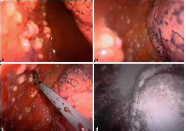
Because the precise diagnosis of MPM requires histopathological confirmation, thoracoscopy via VATS in operable patients remains the standard procedure for obtaining tissue and performing macroscopic staging at the same time (Figure 8) [73, 81].
- 26 -
Figure 8: VATS view of forceps biopsy taken from tumor nodules located on the parietal pleura of a patient with MPM. Note the black streaks of anthracotic pigment visible between lobules of the lung beneath the visceral pleura (a). Macroscopic view of the chest cavity after talc pleurodesis (d). Adopted from [73]
Thoracoscopy can be performed under local anesthesia or via surgical approach by VATS in general anesthesia. The VATS procedure allows the combination of a diagnostic procedure with an initial palliative/therapeutic step of talc pleurodesis.
Multiple and deep tissue biopsies obtained by a thoracoscopic procedure are strongly recommended by the Guidelines of the ERS and ESTS, except in the case of pre- operative contraindication or pleural symphysis [81]. Thoracoscopy should be preferred for diagnostic investigation as it allows complete visual examination of the pleura, taking multiple, deep and large biopsies (preferably including fat and/or muscle to assess tumor invasion) and providing a diagnosis in >90% of cases [81].
In cases being considered inoperable, the aforementioned diagnostic procedures should be performed obligatory before starting local or systemic treatment. In order to stage and assess patients diagnosed with MPM whether they are candidates for surgery, the National Comprehensive Cancer Network (NCCN) guidelines as well as the guidelines
- 27 -
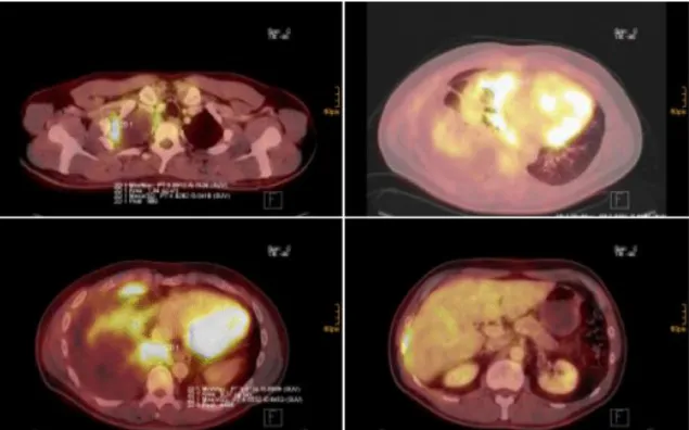
of the Austrian Mesothelioma Interest Group (AMIG) suggest to perform contrast enhanced CT of the chest and the abdomen and combined fluorodeoxyglucose positron emission tomography (FDG-PET) CT to rule out distant metastasis and involvement of the abdomen and the mediastinal lymph nodes (Figure 9) [85]. VATS or laparoscopy can be considered if contralateral pleural or peritoneal spread is suspected. If indicated, integrated FDG-PET/CT should ideally be performed before pleurodesis, since talc causes pleural inflammation, which can result in false positive findings by affected FDG avidity [90].
Figure 9: FDG PET-CT images: MPM of the right pleural cavity. Various slides of CT/PET fusion imaging showing pleural tumor apical right (top left), involving the visceral and parietal pleura of the costodiaphragmatic area (bottom left and right) and pericardium (top right). Adopted from [73]
In order to rule out involvement of mediastinal lymph nodes, histological confirmation has to be made either by endobronchial/endo-esophageal ultrasonography and transbronchial needle aspiration (EBUS/EUS-TBNA) or mediastinoscopy or VATS according to the lymph node station involvement and the involved side [91, 92].
A possible algorithm for diagnosis and staging as proposed by the NCCN is depicted in Figure 10.
- 28 -
Figure 10: Initial diagnostic and staging procedures for patients with MPM as proposed by the NCCN [85].
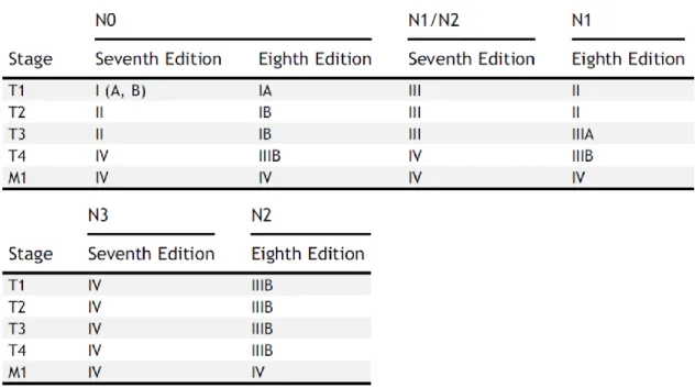
Staging procedures are standard in all malignant tumors including MPM. An appropriate staging system describes the anatomical extent of the tumor, correlates with prognosis and facilitates treatment decisions. In case of MPM, different staging systems have evolved during the past 30 years, most initially developed from small single-center experiences and predominantly retrospective surgical cases [93]. The first staging system by Butchart consisted of four stages and was based on observations from 29 patients only [94]. Another staging system as proposed by the International Mesothelioma Interest Group (IMIG) and the International Association for the Study of Lung Cancer (IASLC) was developed in 1995 [95]. Although the IMIG staging system could predict prognosis, it failed to be an independent prognostic factor when analyzed in the clinical setting using multivariate analysis [96]. The most recent staging system is based on a large international database analysis, set up by the IASLC and was recently published in 2016 [47, 97, 98].
- 29 -
Figure 11: TNM stage groupings of the revised TNM-8 versus the previous TNM-7 staging system in MPM. Adopted from [97]
The most important changes compared to the previous IMIG/IASLC staging system include: the incorporation of both clinical and pathological T1a and T1b into a T1 category and both clinical and pN1 and pN2 categories into a single N category (comprising ipsilateral, intrathoracic nodal metastases (N1)). Nodes that have been previously categorized as N3 are reclassified as N2 now (Table 3). Furthermore, measurement of pleural thickness on CT scans has been proposed for further studies, as tumor thickness and nodular or rind-like morphology were significantly associated with survival. These recently proposed revisions for the TNM-8 stage descriptors and groupings should provide a better estimation of outcomes. In the future, additional data collected from both surgically and non-surgically managed patients will also help to refine these stage groupings (Figure 11) [97].
- 30 -
Table 3: Definitions for T, N and M descriptors in the recently revised TNM-8 staging system for MPM.
Adopted from [97]
Several clinical prognostic factors for MPM have been reported. These are similar to those described in other solid tumor types. The prognosis for MPM highly depends on the tumor stage and histological subtype and, moreover, on the patient’s age and gender[99]. Patients with epithelioid MPM has a better OS than those with non- epithelioid tumors (median OS: 19 months vs. 13 months (biphasic) and 8 months (sarcomatoid)) [100]. In addition, performance status (PS) has shown to be an independent prognostic factor for OS [8]. Low hemoglobin levels, high platelet levels and high serum lactate dehydrogenase (LDH) are prognostically unfavorable characteristics. Numerous new laboratory markers with the purpose of facilitating decision making are have been evaluated, but no validated data on their prognostic value are available yet [101]. Additionally, our group has previously shown that high circulating c-reactive protein (CRP) and fibrinogen are generally associated with poor prognosis, independently from stage and histological subtype [102, 103].
- 31 -
In general, MPM is a devastating disease and despite many efforts regarding early detection and treatment, outcome remains poor. Even at early stages, minimal tumor burden and lack of distant metastases, median OS ranges from 18 to 23 months, with an expected 5-year survival rate of 15% only [97].
Figure 12: Overall survival according to the TNM-8 staging system for MPM. Adopted from [97]
In more advanced stages, palliative treatment approaches might be necessary and most of the patients die from tumor cachexia, body consumption and respiratory problems caused by secondary pneumonia and respiratory insufficiency with hypoxia and asphyxia.
2.1.6 Therapeutic approaches
Current treatment guidelines recommend that patients with MPM should be managed by a multidisciplinary team with experience in treating MPM. Treatment options in general include chemotherapy (CHT), radiotherapy (RT) and surgery. Selected cases with favorable prognostic parameters (i.e. clinical stage I, medically operable, good PS, epithelioid subtype) might be candidates for combined multimodality therapy [104, 105]. Definitive RT alone is not recommended for unresectable MPM, however, CHT alone is approved in this setting [85, 106]. Appropriate patients should undergo evaluation by medical oncologists, radiation oncologists, thoracic surgeons, diagnostic imaging specialists and pulmonologists in order to assess if they are amenable for multimodality treatment. Best supportive care is recommended for patients with PS 3 to
- 32 -
4. Observation may be considered for patients with PS 0 to 2 who are asymptomatic with minimal disease burden if CHT is planned when progression occurs [85].
2.1.6.1 Systemic therapy
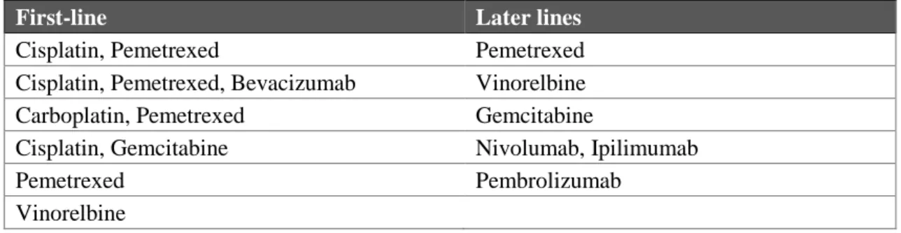
In general, CHT alone is recommended for patients with PS 0 to 2, clinical stage IV disease, sarcomatoid histology (due to the poor prognosis) or who are not candidates for surgery [85].
Prior to the early 2000s a nihilistic attitude persisted among clinical oncologists towards anti-MPM therapy because of the lack of response to standard therapies. Nevertheless, although the role of aggressive surgery remains controversial and no chemo- or targeted therapy has proved fully effective against MPM, robust evidence has emerged to support the use of chemotherapy and angiogenesis inhibition during the past decades [42].
In a meta-analysis including all phase II studies published between 1965 and 2001, cisplatin was found to be the most effective single agent and thus it became the mainstay for combinational therapeutic interventions in MPM [107]. Accordingly, after encouraging data of phase I and II pemetrexed (a multitargeted antifolate that inhibits thymidylate synthase, dihydrofolate reductase and glycinamide ribonucleotide formyl transferase) studies, a single-blinded phase III trial (Evaluation of Mesothelioma in a Phase III Trial of pemetrexed with Cisplatin, EMPHACIS) recruited 448 chemonaive patients with MPM randomly assigned to receive cisplatin at 75 mg/m2 plus either pemetrexed at 500 mg/m2 or placebo every 3 weeks [108]. The median OS in the combination arm was 12.1 months, compared to that of 9.3 months in cisplatin-alone patients. Based on this study, combination chemotherapy with pemetrexed and cisplatin (with folic acid and vitamin B12 supplementation) became the current standard of care for first-line systemic therapy in patients with unresectable MPM and good PS.
Although cisplatin monotherapy was never compared to placebo in a randomized trial, these results enforced the recommendation for combination chemotherapy. So far, all current major international guidelines endorse a combination of platinum-based CHT with modern antifolates (pemetrexed or raltitrexed) as the gold standard treatment in MPM [81, 85, 86, 109]. Still, it has to be mentioned that these recommendations are based on two randomized trials published in more than a decade ago [108, 110].
- 33 -
Accordingly, until 2015, no other combination therapy was able to significantly improve OS and progression free survival (PFS) compared to the cisplatin/pemetrexed doublet [86]. However, previous studies have demonstrated that vascular endothelial growth factor (VEGF) signaling plays a crucial part in mesothelioma cell physiopathology [111, 112]. The phase II/III randomized Mesothelioma Avastin Cisplatin Pemetrexed Study (MAPS) was initiated to assess the effect on survival of the VEGF inhibiting monoclonal antibody bevacizumab when added to the present standard of care, cisplatin/pemetrexed, as first-line treatment of advanced MPM[113]. Survival was improved in those who received cisplatin/pemetrexed/bevacizumab (18.8 months, versus 16.1 months in patients receiving CHT alone (hazard ratio 0.77 [95% CI 0.62–
0.95]). Although statistically significant, the overall improvement in OS (2.7 months) was still quite modest. Grade 3-4 adverse events were more common in the bevacizumab group (158 [71%] of 222 patients) (139 [62%] of 224 patients), and more patients stopped first-line treatment because of toxic effects in the bevacizumab group (53 [24.3%] of 218 patients) (13 [6.0%] of 217; difference 18.3% [11.7–24.9]).
However, these side effects were considered to be tolerable and thus those patients with MPM who are not candidates for clinical trials or who do not have access to such opportunities, but who do not have contraindications to bevacizumab, should be offered three-drug combination treatment as a new standard of care [85].
Some other recent phase II and III trials have assessed the safety and efficacy of different systemic combination treatments. A combination of carboplatin and pemetrexed was investigated in 3 large phase II trials with median OS ranging from 12.7 to 14 months [114-116]. A comparison of 1704 patients with inoperable MPM treated with ether cisplatin/pemetrexed or carboplatin/pemetrexed found that outcomes were similar and concluded that the carboplatin/pemetrexed regimen might be the better choice for patients with poor PS and/or comorbidities [117]. Cisplatin/gemcitabine was assessed in phase II studies and was found to be a valid treatment option for patients not eligible for pemetrexed treatment [118]. Additional acceptable fist-line single agent regimens include pemetrexed or vinorelbine monotherapy (Table 4) [119, 120].
Maintenance therapy after successful first-line systemic treatment might be effective for symptom control and maintaining quality of life [86]. However, the benefit of maintenance treatment in MPM is yet to be evaluated and several studies are ongoing.
- 34 -
For example, a recent phase III randomized trial has failed to show a benefit of adding maintenance thalidomide to pemetrexed-based CHT [121].
Second-line CHT options include pemetrexed (if not administered as first-line), vinorelbine and gemcitabine [122-124]. In patients with good response to first-line pemetrexed, rechallenging pemetrexed might be effective [125]. Limited data are available to guide second-line systemic therapy; however, several agents are investigated in clinical trials [126]. Preliminary data suggest that immune checkpoint inhibitors (nivolumab, pembrolizumab, tremelimumab, ipilimumab) might be effective in the second-line setting and thus currently phase II and III trials are ongoing [127, 128]. In a recent randomized phase IIb trial, the CTLA-4 inhibitor tremelimumab did not significantly prolong OS compared with placebo in patients with previously treated malignant mesothelioma [129]. However, preliminary phase II results from a prospective randomized trial evaluating the safety and efficacy of cisplatin/pemetrexed with the triple angiokinase-inhibitor (TKI) nintedanib revealed a trend for better OS and significantly better PFS for the experimental arm. The confirmatory phase III results are to be awaited [130].
Table 4: Principles of systemic therapy in MPM according to the most recent NCCN guidelines (2.2017).
Adopted from [85]
First-line Later lines
Cisplatin, Pemetrexed Pemetrexed
Cisplatin, Pemetrexed, Bevacizumab Vinorelbine
Carboplatin, Pemetrexed Gemcitabine
Cisplatin, Gemcitabine Nivolumab, Ipilimumab
Pemetrexed Pembrolizumab
Vinorelbine
Interleukins and interferons were tested in studies, as well as the application of targeted therapies with monoclonal antibodies. None of the following substances showed any survival benefit in several studies: gefitinib, erlotinib, or imatinib [131-134].
- 35 - 2.1.6.2 Surgery
2.1.6.2.1 Radical surgical approaches
Macroscopic complete resection (MCR) is goal of every surgical procedure with curative intent [135]. Currently, multimodality treatment including radical surgery as by extrapleural pneumonectomy (EPP) or pleurectomy/decortication (P/D) are the most commonly used surgical techniques for treating MPM with curative intention. However, only a number of mainly retrospective institutional reports using different multimodality regimens and different surgical techniques is available in the current literature, due to the difficulty of prospective randomized trials in this setting. Hence, the question whether P/D or EPP is the more appropriate technique to obtain improved OS in relation to the associated posttreatment quality of life is difficult to address. After the IMIG 2012 meeting and much discussion on the role of surgery and the value of MCR in MPM, the agreement is now that (I) surgical macroscopic complete resection and control of micrometastatic disease play a vital role in the multimodality therapy of MPM, as in case of other solid tumor types. (II) Surgical cytoreduction is indicated when MCR is deemed achievable and (III) the type of surgery (EPP or P/D) depends on clinical factors and on individual surgical judgment and expertise [135].
However, since MPM is a very heterogeneous disease with variability of clinical symptoms, stage, histology, tumor burden and biological behavior, a multidisciplinary discussion of each patient considering age, PS and individual prognosis is mandatory.
2.1.6.2.2 Pleurectomy/Decortication
As the technique of P/D includes a variety of surgical procedures with different clinical indications, the IASLC and IMIG have recently proposed a common nomenclature for these different techniques [136]. Partial pleurectomy was defined as a cytoreductive procedure with partial removal of the visceral and/or parietal tumor gross, without removing the lung or the intention for MCR. P/D was defined as complete resection of the parietal and visceral pleurae, and extended P/D as a technique with additional resection and reconstruction of the pericardium and diaphragm (Figure 13). In a recent systematic review investigating previous results of P/D, mortality and morbidity ranged from 0% to 11% and 13% to 43%, respectively. Median OS ranged from 7.1 to 31.7 months and disease-free survival (DFS) from 6 to 16 months [137]. A detailed analysis
- 36 -
suggested extended P/D to be associated with a higher number of perioperative morbidity whilst favoring the more aggressive approach for improved oncological outcome.
Figure 13: Surgical technique of extended pleurectomy/decortication for MPM. Adopted from [138]
2.1.6.2.3 Extrapleural Pneumonectomy
Extrapleural pneumonectomy (EPP) is a widely standardized surgical technique with en-bloc resection of the lung together with the parietal and visceral pleurae, the pericardium and diaphragm (Figure 15) [139]. The role of EPP has been recently under discussion after the publication of the Mesothelioma and Radical Surgery (MARS I) trial which aimed to assess the outcomes of patients after EPP or no EPP in the context of trimodal therapy within a prospective randomized trial [140]. Median OS was 14.4 months (5.3-18.7) for the EPP group and 19.5 months (13.4 to time not reached) for the no EPP group. EPP was associated with an unacceptably high perioperative mortality of 15.8% and morbidity of 68.8%. However, only 16 of the randomized patients have undergone EPP. The authors of the study concluded that radical surgery by EPP within a multimodality approach offers no benefit in OS and possibly harms patients.
However, the MARS I trial was designed to assess the feasibility of randomizing patients with MPM for surgery and not to evaluate morbidity or benefit in OS of EPP.
In order to sufficiently address this question an actual number of 670 patients to identify a significant survival benefit would have been needed [135]. A recent systematic review including 58 studies reported a median OS after EPP of 9.4 to 27.5 months, and 1-, 2-, and 5-year survival rates ranging from 36 to 83%, 5 to 59% and 0 to 24%, respectively.
Overall perioperative mortality ranged from 0 to 11.8%, and the perioperative morbidity
- 37 -
from 22 to 82% [141]. A recent retrospective evaluation of our own group together with colleagues from Toronto and Zurich including 251 patients completing EPP after platin- based CHT reported a 30-day mortality of 5% and perioperative complication rate of 30% [105]. However, according to the recent ERS/ESTS guidelines, EPP should only be performed at high volume centers within the context of a clinical study and a multimodality regimen [81]. Moreover, the current NCCN guidelines suggest to perform EPP in early stage patients only [85].
Figure 14: Surgical technique of extrapleural pneumonectomy for MPM. Adopted from [138]
Figure 15: Resection specimens after extrapleural pneumonectomy for MPM at the Division of Thoracic Surgery, Medical University of Vienna
The question whether to perform P/D or EPP in which clinical situation is difficult to address. Initial data from the IASLC Staging Committee Statistical Center including 1494 surgically-treated patients reported that stage I tumors resected by EPP were associated with a median OS of 40 months whereas those managed by P/D had a
- 38 -
median OS of 23 months. No differences in survival between EPP and P/D were identified in patients with higher-stage disease [100]. To date, the largest cohort of patients treated with P/D or EPP was published by Flores et al in 2008. Of 663 consecutive cases, the operative mortality was 7% for EPP (n = 27/385) and 4% for P/D (n = 13/278) [142]. OS was reported to be superior after P/D, however patients selected for EPP had more local advanced disease and P/D was applied in earlier stages. Another recently published meta-analysis compared the perioperative and long-term outcomes of EPP and extended P/D for selected surgical candidates [143]. A comparison between EPP and P/D revealed a significantly lower perioperative mortality and morbidity for patients who underwent extended P/D compared to EPP. Median OS ranged between 13-29 months for extended P/D and 12-22 months for EPP, with a trend favoring extended P/D. The authors concluded that selected patients who underwent extended P/D had lower perioperative morbidity and mortality with similar, if not superior, long- term survival compared to EPP, in the context of multi-modality therapy. However, it must be taken into account that in most of the included studies, P/D was usually chosen for earlier and EPP for more advanced stages.
2.1.6.2.4 Patient selection for surgery
Taken all this information together, patients undergoing surgery for MPM must be carefully selected to outweigh the potential risks with the respective procedures. Several studies have been published assessing clinical prognostic factors and biomarkers for estimating prognosis and proper patient selection. For example, non-epithelioid histology and nodal involvement have consistently been demonstrated to be associated with worse prognosis after EPP [144]. Furthermore, several blood biomarkers and combinations of several clinical factors as prognostic scores have been investigated to identify patients who are more likely to benefit from surgical treatment. Among these, pre-treatment CRP predicted benefit in OS from multimodality therapy including curative intent surgery [102]. Multivariate analysis confirmed that patients with low CRP levels have significantly improved OS compared to those with elevated levels independently from other relevant clinical factors. Moreover, circulating biomarkers such as fibrinogen and albumin were reported to influence prognosis and could be implemented into therapeutic algorithms [103, 145]. Our study group has recently
![Figure 2: Male to female ratios among malignant mesothelioma cases reporting overall exposure (occupational and environmental) and environmental exposure (EE) to asbestos [15]](https://thumb-eu.123doks.com/thumbv2/9dokorg/1383546.114285/14.892.130.766.123.577/malignant-mesothelioma-reporting-exposure-occupational-environmental-environmental-exposure.webp)
![Figure 10: Initial diagnostic and staging procedures for patients with MPM as proposed by the NCCN [85]](https://thumb-eu.123doks.com/thumbv2/9dokorg/1383546.114285/28.892.128.698.122.402/figure-initial-diagnostic-staging-procedures-patients-proposed-nccn.webp)
![Figure 12: Overall survival according to the TNM-8 staging system for MPM. Adopted from [97]](https://thumb-eu.123doks.com/thumbv2/9dokorg/1383546.114285/31.892.136.658.269.572/figure-overall-survival-according-tnm-staging-mpm-adopted.webp)
![Figure 13: Surgical technique of extended pleurectomy/decortication for MPM. Adopted from [138]](https://thumb-eu.123doks.com/thumbv2/9dokorg/1383546.114285/36.892.130.632.243.480/figure-surgical-technique-extended-pleurectomy-decortication-mpm-adopted.webp)
