Comparative analysis of prognostic histopathologic
parameters in subtypes of epithelioid pleural mesothelioma
Agnes Bilecz,
1Paul Stockhammer,
2,3Dirk Theegarten,
4Izidor Kern,
5Marko Jakopovic,
6Miroslav Samarzija,
6Thomas Klikovits,
3Mir A Hoda,
3Bal azs D € ome,
3,7,8Felicitas Oberndorfer,
9Leonhard Muellauer,
9J anos Fillinger,
10Ildik o Kov acs,
7Christine Pirker,
11Martin Schuler,
12Till Pl ones, €
2Clemens Aigner,
2Walter Klepetko,
3Walter Berger,
11Luka Brcic,
13,* Vikt oria Laszlo
3,* & Balazs Hegedus
1,2,*
12nd Institute of Pathology, Semmelweis University, Budapest, Hungary,2Department of Thoracic Surgery, Ruhrlandklinik,University Duisburg-Essen, Essen, Germany, 3Division of Thoracic Surgery, Department of Surgery, Comprehensive Cancer Center Vienna,Medical University Vienna, Vienna, Austria,4Institute of Pathology, University Hospital Essen,University Duisburg-Essen, Essen, Germany,5University Clinic of Respiratory and Allergic Diseases, Golnik, Slovenia, 6Department for Respiratory Diseases Jordanovac, University Hospital Center,University of Zagreb, Zagreb, Croatia, 7Department of Tumor Biology, National Koranyi Institute of Pulmonology,Semmelweis University, Budapest, Hungary, 8Department of Biomedical Imaging and Image-guided Therapy, Division of Molecular and Gender Imaging, Medical University of Vienna, Vienna, Austria,9Clinical Institute of Pathology, Medical University of Vienna, Vienna, Austria, 10Department of Pathology, National Koranyi Institute of Pulmonology,Semmelweis University, Budapest, Hungary, 11Institute of Cancer Research and Comprehensive Cancer Center, Department of Medicine, Medical University of Vienna, Vienna, Austria,12Department of Medical Oncology, West German Cancer Center, University Hospital Essen,University Duisburg-Essen, Essen, Germany, and13Medical University of Graz, Diagnostic and Research Institute of Pathology, Graz, Austria
Date of submission 28 December 2019 Accepted for publication 10 March 2020 Published onlineArticle Accepted14 March 2020
Bilecz A, Stockhammer P, Theegarten D, Kern I, Jakopovic M, Samarzija M, Klikovits T, Hoda M A, D€ome B, Oberndorfer F, Muellauer L, Fillinger J, Kovacs I, Pirker C, Schuler M, Pl€ones T, Aigner C, Klepetko W, Berger W, Brcic L, Laszlo V & Hegedus B.
(2020)Histopathology77, 55–66. https://doi.org/10.1111/his.14105
Comparative analysis of prognostic histopathologic parameters in subtypes of epithelioid pleural mesothelioma
Aims: Malignant pleural mesothelioma (MPM) is a rare malignancy with a dismal prognosis. While the epithelioid type is associated with a more favourable outcome, additional factors are needed to further stratify prognosis and to identify patients who can benefit from multimodal treatment. As epithelioid MPM shows remarkable morphological variability, the prognostic role of the five defined morphologies,
the impact of the nuclear grading system and the mitosis-necrosis score were investigated in this study.
Methods and results: Tumour specimens of 192 patients with epithelioid MPM from five European centres were histologically subtyped. Nuclear grading and mitosis–necrosis score were determined and cor- related with clinicopathological parameters and over- all survival (OS). Digital slides of 55 independent
Addresses for correspondence: Luka Brcic, Medical University of Graz, Diagnostic and Research Institute of Pathology, Neue Stiftingtalstraße 6, 8010 Graz, Austria. e-mail: luka.brcic@medunigraz.at
Viktoria Laszlo, Division of Thoracic Surgery, Department of Surgery, Comprehensive Cancer Center Vienna, Medical University Vienna, Spi- talgasse 23, 1090 Vienna, Austria. e-mail: viktoria.laszlo@meduniwien.ac.at
Balazs Heged}us, Department of Thoracic Surgery, Ruhrlandklinik, University Duisburg-Essen, T€uschener Weg 34, D-45239, Essen, Germany.
e-mail: balazs.hegedues@rlk.uk-essen.de
*These authors contributed equally to this study.
©2020 The Authors. Histopathology published by John Wiley & Sons Ltd.
This is an open access article under the terms of the Creative Commons Attribution License, which permits use,
cases from The Cancer Genome Atlas (TCGA) data- base were evaluated for external validation. Histologi- cal subtypes were collapsed into three groups based on their overlapping survival curves. The tubulopapil- lary/microcystic group had a significantly longer OS than the solid/trabecular group (732 days versus 397 days, P= 0.0013). Pleomorphic tumours had the shortest OS (173 days). The solid/trabecular vari- ants showed a significant association with high nuclear grade and mitosis–necrosis score. The mito- sis–necrosis score was a robust and independent
prognostic factor in our patient cohort. The prognos- tic significance of all three parameters was externally validated in the TCGA cohort. Patients with tubu- lopapillary or microcystic tumours showed a greater improvement in OS after receiving multimodal ther- apy than those with solid or trabecular tumours.
Conclusions: Histological subtypes of epithelioid MPM have a prognostic impact, and might help to select patients for intensive multimodal treatment approaches.
Keywords: epithelioid, grading, histological subtypes, mesothelioma, prognosis
Introduction
Malignant pleural mesothelioma (MPM) is the most common primary malignancy of the pleura. Due to its highly aggressive clinical behaviour it confers a dismal prognosis.1 MPM is divided into three major histological types; namely, epithelioid, sarcomatoid and biphasic.2Histological type is an important prog- nostic factor, also playing a substantial role in treat- ment decision-making.3–5 The epithelioid type is the most common type of MPM with the most favourable prognosis.6However, it is a heterogeneous entity and there is a lack of morphological prognostic factors for further stratification of epithelioid MPM (eMPM).
The presence of necrosis, the degree of nuclear aty- pia and mitotic count have been shown to have a prog- nostic role in eMPM.7–9Furthermore, the presence of prominent nucleoli and atypical mitotic figures was found to be of prognostic value, while intranuclear inclusions or a high nuclear/cytoplasmic ratio was not associated with worse patients’ outcomes, and the prognostic impact of chromatin structure alterations remains unclear.7,9 Immunohistochemical assessment of proliferation by Ki67 labelling was also shown to have a prognostic role in eMPM when using 10% or 15% as cut-off values.7,10 Furthermore, composite scores have been proposed and regarded as robust tools in the stratification of patient outcome.11The nuclear grade is based on a three-tier assessment of nuclear atypia and mitotic count.7,8Rosenet al. developed the mitosis–necrosis score (M/N score), which includes the presence of necrosis and a two-tier scoring of mitotic counts.8A more complex pathological grading system based on the presence of necrosis, the main histological subtype, Ki67 proliferation index and a four-tier evalu- ation of the mitotic count has also been developed for risk stratification of eMPM.12
There are limited data on the prognostic implications of histological subtypes in eMPM, but it is a promising emerging marker for predicting patient outcome, simi- larly to peritoneal mesothelioma13,14 and other malig- nancies, such as lung,15–17 gastric18 and bladder cancer.19The pleomorphic subtype has been shown to be a significant predictor of negative clinical outcome,20–24 while the microcystic/myxoid variant might have a posi- tive impact on overall survival (OS) for eMPM patients.22 A predominantly solid morphological subtype has been associated with worse outcome compared to non-solid subtypes; however, it was not found to be an independent prognostic factor.8The role of other individual architec- tural subtypes such as trabecular, tubulopapillary, micro- cystic and micropapillary are yet to be determined.2
Accordingly, we investigated the prognostic impact of five histological subtypes of eMPM and their associ- ation with the other proposed histopathological prog- nosticators, namely nuclear grade composed of scores for nuclear atypia and mitotic count7 and the M/N score based on the presence of necrosis and mitotic count.8Finally, we examined the association between OS and histological subtypes in subgroups of patients receiving multimodal therapy versus chemotherapy or best supportive care-only regimens.
Materials and methods
S T U D Y C O H O R T
Our multicentre cohort consisted of a total of 192 patients diagnosed with eMPM between 1994 and 2015 at the Medical University of Vienna, Vienna, Austria (n= 54), between 2000 and 2007 at the National Koranyi Institute of TB and Pulmonology, Budapest, Hungary (n= 30), between 2007 and 2012 at the University Clinic of Respiratory and Allergic
Diseases, Golnik, Slovenia (n= 67), between 2013 and 2014 at the University of Zagreb, School of Medicine, Jordanovac, Croatia (n= 9) and between 2016 and 2018 at the University Medicine Essen – Ruhrland- klinik, Essen, Germany (n= 32). The pathological diagnosis of eMPM was made by expert pulmonary pathologists following international histological and immunohistochemical criteria requiring a minimum of two positive mesothelial markers (calretinin, WT-1, D2-40, CK5/6) and at least two negative markers for carcinoma (such as Ber-EP4, TTF-1, CEA). Clinical data, including patients’ age, gender, date of diagnosis and date of death or last contact, were collected in accordance with each institute’s ethical guidelines and the latest Declaration of Helsinki. The retrospective analysis of MPM patients was approved in all partici- pating centres by the local ethics committees at the Medical University of Vienna (no. 904/2009), the University Hospital Center Zagreb (no. 02/21AG) and at the University Medicine Essen (17-7773-BO). The Institutional Review Boards of the University Clinic Golnik and the National Koranyi Institute of Tubercu- losis and Pulmonology granted a waiver for the retro- spective analyses. Samples were obtained by video- assisted thoracoscopy (n= 106), percutaneous pleural needle core biopsy (n= 28) or pleurectomy (n= 28).
In 30 cases the exact surgical sampling method was not specified. All tissue samples were formalin-fixed and paraffin-embedded (FFPE).
V A L I D A T I O N C O H O R T
We analysed 55 digital images of eMPMs openly avail- able at the Cancer Digital Slide Archive (CDSA), which correspond to diagnostic sections of specimens submitted by tissue source sites of The Cancer Genome Atlas (TCGA).25Of these 55 sections, six were frozen sections and 49 were FFPE specimens. Corresponding clinical variables and survival data collected by TCGA Research Network26were downloaded from the cBioPortal.27
E V A L U A T I O N O F H I S T O P A T H O L O G I C A L F E A T U R E S
One haematoxylin and eosin (H&E)-stained slide was provided for each case by expert pathologists from the participating centres and was classified by A.B.
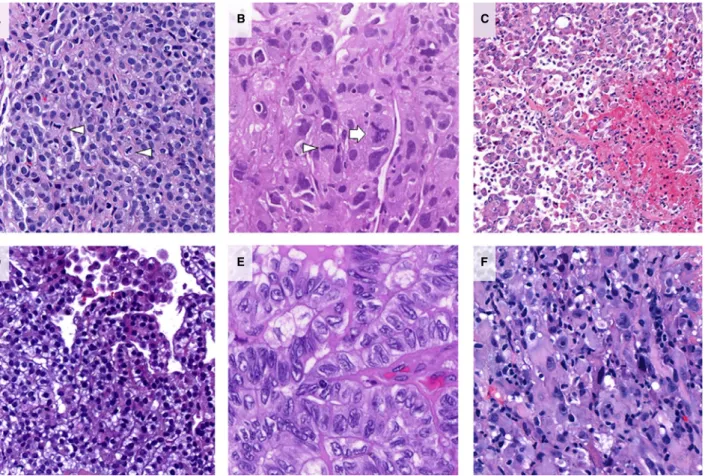
and L.B. Eventual discrepant cases were discussed together and consensus was reached. Classification was based on the predominant growth pattern and on the presence of pleomorphic cytological features (Figure 1).2,20,28 A sample was assigned pleomorphic if at least 10% of the tumour area consisted of anaplastic or giant tumour cells.20
Briefly, the following morphological features were used for the classification.28 Tubulopapillary pattern was defined by a combination of either tubular and/
or papillary structures consisting of cuboidal, slightly enlarged tumour cells arranged around connective tissue cores. Solid pattern was characterised by tumour cells of epithelioid appearance forming larger sheets or nests. Samples consisting of one or two lay- ers of relatively small, monomorphic cells forming thin cords often spreading within abundant desmo- plastic stroma were assigned trabecular pattern.
Microcystic pattern was characterised by tumour cells forming small cyst-like structures, while micropapil- lary pattern exhibited small papillae without the fibrovascular cores seen in tubulopapillary pattern.
Mitotic figures were counted in hot-spots at 9400 magnification and given as an average of mitotic fig- ures per 10 high-power fields (Figure 2A,B).8 The presence or absence of necrosis in the sample was also evaluated and used to calculate M/N score (0–2), as proposed earlier (Figure 2C).8A composite nuclear grade was calculated for each case based on nuclear atypia and mitotic count scores evaluated according to previous studies (Figure 2D–F).7,8
S T A T I S T I C A L A N A L Y S I S
OS was defined as the time from diagnosis to date of death or for censored patients as the time between diagnosis and date of last contact. The Kaplan–Meier method and log-rank (Mantel–Cox) tests were used to estimate OS and to calculate survival differences between groups, respectively. A multivariate Cox regression model including histology, M/N score and nuclear grade as variables was calculated to identify independent prognostic factors, hazard ratios (HR) and their corresponding 95% confidence intervals (CI). Fisher’s exact andv2tests were used to evaluate associations between histological subtypes, nuclear grade and M/N score, as well as between clinico- pathological variables and histological subtypes and treatment received. For all data comparisons, results were considered as statistically significant if P was
<0.05 based on two-sided tests. All calculations were performed using GraphPad Prism version 8.0 (Graph- Pad Inc., San Diego, CA, USA) and SPSS Statistics version 23.0 package (SPSS Inc., Chicago, IL, USA).
Results
We analysed tumour samples of 192 patients who had histopathologically confirmed diagnosis of eMPM
and for whom OS data were available. Median follow- up was 423 days. Median age of patients was 65.010.8 years; 143 (74.5%) of the patients were male. International Mesothelioma Interest Group (IMIG) stage at the time of diagnosis was available for 126 patients, 48.4% (61 of 126) of whom had early stage (IMIG I/II) disease, while 51.6% (65 of 126) had advanced stage (IMIG III/IV) disease. Among the 192 eMPM cases, the solid variant was the most common predominant pattern accounting for 52.1%
(100 if 192) of all samples, while 28.6% (55 of 192) were predominantly tubulopapillary, 10.4% (20 of 192) trabecular, 4.7% (nine of 192) microcystic, 3.1% (six of 192) pleomorphic and 1.0% (two of 192) micropapillary. According to the nuclear grad- ing system, 54.7% (105 of 192) were grouped in grade 1, 32.3% (62 of 192) in grade 2 and 13.0%
(25 of 192) in grade 3. Based on the presence of necrosis and mitotic counts, an M/N score of 0 was assigned to 45.8% (88 of 192) of the cases, score 1 to 40.1% (77 of 192) and score 2 to 14.1% (27 of 192) of the samples (Table 1).
Next, we analysed the OS of each histological sub- type of eMPM. Due to the very low number of micropapillary variants (n= 2) in our cohort, those were excluded from the survival analysis. We found that tubulopapillary and microcystic subtypes associ- ated with better prognosis (median OS 727 and 936 days, respectively), whereas patients with solid and trabecular patterns had a shorter median OS (397 and 394 days, respectively). The pleomorphic eMPM patients had the shortest median OS of 173 days, which was significantly worse than med- ian OS of tubulopapillary, microcystic and solid sub- types (P <0.0001, 0.0085 and 0.0277, respectively) and showed a trend for worse outcomes in compar- ison to predominant trabecular variant (P= 0.0906, Figure 3A).
Due to the rarity of microcystic and trabecular pat- terns the four subtypes, except for the pleomorphic variant, were collapsed into two groups. Based on their overlapping survival curves, specimens showing a predominantly microcystic pattern were merged with tubulopapillary variants, while trabecular
A B
D E
C
Figure 1.Histological subtypes of epithelioid malignant pleural mesothelioma (eMPM).A,Tubulopapillary pattern [haematoxylin and eosin (H&E)].B,Solid pattern (H&E).C,Trabecular pattern (H&E).D,Microcystic pattern (H&E).E,Pleomorphic features (H&E).
patterns were merged with solid pattern tumours for further analyses. The pleomorphic subtype was anal- ysed as a separate group. In univariate analyses, we found that patients with tubulopapillary/microcystic features had a significantly better OS than patients with solid/trabecular variants (medians 732 days ver- sus 397 days, P= 0.003, Table 2, Figure 3B).
Pleomorphic tumours were associated with signifi- cantly worse outcome when compared to solid/tra- becular variants (median OS 173 days versus 397 days, P= 0.039 Table 2, Figure 3B). As the pleomorphic variant showed a dramatically shorter OS than all other subtypes in our cohort, and several earlier studies suggested its exclusion from eMPM based on its very poor prognosis, we did not include it in our further survival analyses.20–23
Stage I/II disease was associated with a signifi- cantly better OS than stage III/IV (medians 650 days versus 421 days, P= 0.015, Table 2). The distribu- tion of tubulopapillary/microcystic and solid/trabecu- lar variants was similar among early (I/II) and
advanced stages (III/IV) of disease (P= 0.999, Sup- porting information, Figure S1A). Early-stage cases with tubulopapillary/microcystic features showed a tendency for longer OS (Mantel–Cox test, P =0.194;
Grehan–Breslow–Wilcoxon test P= 0.041; Support- ing information, Figure S1B), while among the advanced-stage patients the tubulopapillary/microcys- tic variants were associated with a significantly longer OS compared to the solid/trabecular variants (P =0.047, Supporting information, Figure S1C).
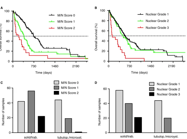
Patients with tumours of M/N scores 1, 2 and 3 had significantly different OS of 720 days, 386 days (P =0.0004) and 165 days (P= 0.0036), respec- tively (Figure 4A, Table 2). There was no significant difference in OS between nuclear grades 1 and 2.
However, patients with nuclear grade 3 had signifi- cantly worse OS when compared to patients with nuclear grade 2: median OS 123 versus 486 days (P =0.0002) (Figure 4B, Table 2).
Regarding the distribution of M/N scores as well as nuclear grades among the histological subtypes we
A B
D E F
C
Figure 2.Mitosis, necrosis and nuclear grading in epithelioid malignant pleural mesothelioma (eMPM).A,Bipolar mitoses [arrowheads, haematoxylin and eosin (H&E)].B,Bipolar (arrowhead) and atypical (arrow) mitoses (H&E).C,Coagulative necrosis (H&E).D,Mild nuclear atypia (H&E).E,Moderate nuclear atypia (H&E).F,Severe nuclear atypia (H&E).
found a significant association of solid/trabecular pat- terns with both higher M/N scores (P< 0.0001) and higher nuclear grades (P= 0.007) in comparison to tubulopapillary/microcystic variants (Figure 4C,D).
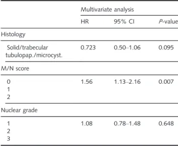
In multivariate analysis, including histological subtype, M/N score and nuclear grading as parame- ters, we found M/N score to be an independent prognostic factor in our MPM cohort (Table 3). His- tological subtype did not reach significance (P= 0.095).
We analysed the impact on OS of each individual factor – namely nuclear atypia, mitotic rate, presence of necrosis– used to calculate composite grades.
Patients with tumours exhibiting mild atypia (median Solid
Trabecular Tubulopapillary Microcystic Pleomorphic
Solid / trabecular
Tubulopapillary/microcystic Pleomorphic
100
80
Overall survival (%)
60
40
20 0
0 730 1460
Time (days)
2190
100 80
Overall survival (%)
60 40 20 0
0 730 1460
Time (days)
2190 A
B
Figure 3.Histological subtypes and patient outcomes.A,Overall survival (OS) of the five histological subtypes: tubulopapillary, microcystic, solid, trabecular and pleomorphic (P=0.0019, log- rank test).B,OS of collapsed groups: solid/trabecular [hazard ratio (HR)=Ref.], tubulopapillary/microcystic [HR=0.57, 95% confi- dence interval (CI)=0.41–0.80] and the pleomorphic subtype (HR=4.72, 95% CI=1.15–19.42). For all three curves:
P<0.0001, log-rank test. [Colour figure can be viewed at wileyon- linelibrary.com]
Table 1. Clinicopathological characteristics of the eMPM patient cohort
Total (n=192) Gender
Male 143
Female 49
Age (years)
MeanSD 65.010.8
Histology
Solid 100
Tubulopapillary 55
Trabecular 20
Microcystic 9
Pleomorphic 6
Micropapillary 2
Nuclear atypia
Mild 13
Moderate 132
Severe 47
Mitotic count
Low (≤1) 117
Intermediate (2–4) 41
High (≥5) 34
Necrosis
Yes 94
No 98
Nuclear grade
1 105
2 62
3 25
M/N score
0 88
1 77
2 27
IMIG stage (NA=66)
I/II 61
III/IV 65
NA, not available; SD, standard deviation; M/N, mitosis/necrosis;
eMPM, epithelioid malignant pleural mesothelioma; IMIG, Interna- tional Mesothelioma Interest Group.
OS 1197 days) had a significantly longer OS in com- parison to those with moderate or severe atypia (me- dian OS 501 days, P = 0.027 and 306 days, P< 0.001, respectively, Table 2, Supporting informa- tion, Figure S2A). High mitotic counts were associ- ated with shorter median OS in comparison to low mitotic rate (239 days, P < 0.001), while low and intermediate mitotic counts did not show a significant difference in median OS (545 and 501 days, respec- tively, P = 0.470, Table 2, Supporting information, Figure S2B). The presence of necrosis was also associ- ated with a significantly shorter OS in comparison to cases without necrosis (281 days versus 727 days, respectively, P< 0.0001, Table 2, Supporting infor- mation, Figure S2C).
We also performed a multivariate analysis of histo- logical variants and individual components of the composite scores. We found the presence of necrosis to be a strong independent prognostic factor (P <0.0001, Supporting information, Table S1).
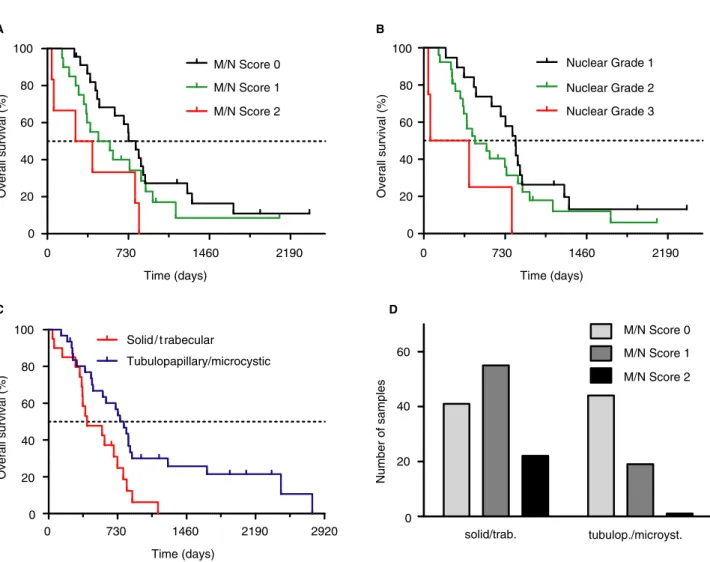
H I S T O P A T H O L O G I C A L A N A L Y S I S O F E M P M S A M P L E S F R O M T H E T C G A C O H O R T
For external validation, we analysed an additional set of eMPM samples derived from the TCGA for which scanned H&E-stained sections were available. The corresponding clinicopathological variables of these 55 patients are detailed in Supporting information, Table S2. We found 50.9% (28 of 55) of the samples to be of tubulopapillary pattern, 30.9% solid (17 of 55), 5.5% microcystic (three of 55), 5.5% trabecular (three of 55) and 7.2% micropapillary (four of 55).
No sample with pleomorphic features was identified.
In agreement with the results obtained in our multi- center MPM patient cohort, univariate analysis of the OS data (Supporting information, Table S3) showed a Table 2. Univariate survival analyses in the eMPM patient
cohort
Univariate analysis OS
(days) HR (95% CI) P-value Age
<70 years 495 0.92 (0.65–1.30) 0.619
≥70 years 463
Gender
Male 486 0.99 (0.69–1.44) 0.999
Female 469
Histology
Solid/trabecular 397 1 –
Tubulopap./microcyst. 732 0.58 (0.41–0.83) 0.003 Pleomorphic 173 2.65 (1.95–6.68) 0.039 Nuclear atypia
Mild 1197 1 –
Moderate 501 2.29 (1.32–3.97) 0.027
Severe 306 3.47 (1.88–6.42) <0.001 Mitotic count
Low (≤1) 545 1 –
Intermediate (2–4) 501 1.17 (0.75–1.87) 0.470 High (≥5) 239 2.48 (1.45–4.25) <0.001 Necrosis
Yes 281 2.38 (1.68–3.38) <0.0001
No 727
M/N score
0 720 1 –
1 383 2.01 (1.37–2.95) <0.0001
2 165 2.61 (1.39–4.97) <0.0001
Nuclear grade
1 555 1 –
2 486 1.10 (0.75–1.62) 0.531
3 123 3.75 (1.86–7.56) 0.0002
IMIG stage (NA=66)
I/II 650 0.60 (0.39–0.91) 0.015
III/IV 421
Table 2. (Continued)
Univariate analysis OS
(days) HR (95% CI) P-value Treatment (NA=76)
MMT 936 0.35 (0.23–0.55) <0.0001
CHT/BSC 340
NA, not available; SD, standard deviation; tubulopap., tubulopapil- lary; microcyst., microcystic; M/N, mitosis/necrosis; OS, overall sur- vival; eMPM, epithelioid malignant pleural mesothelioma; MMT, multimodal therapy; CHT, chemotherapy; BSC, best supportive care.
significantly inferior OS associated with solid/trabec- ular subtypes in comparison to tubulopapillary/mi- crocystic patterns (median 406 days versus 795 days, P= 0.01, Figure 5C). Histological grading was performed on 49 FFPE samples, while the six samples for which fresh frozen samples were only available were not included in grade analysis. The solid/trabecular subtypes showed a significant asso- ciation with higher M/N scores (P< 0.0001, Fig- ure 5D). Nuclear grade 3 and M/N score 2 cases were associated with significantly inferior OS than nuclear grade 1 (P= 0.01) and M/N score 0 (P= 0.023), respectively (Figure 5A,B). However, the low number of patients (n = 55) in the TCGA
validation cohort limited the feasibility of a multi- variate analysis.
T H E I M P A C T O F M O R P H O L O G I C A L S U B T Y P E S I N T H E M U L T I M O D A L T H E R A P E U T I C S E T T I N G
In order to identify whether tubulopapillary/microcys- tic and solid/trabecular epithelioid subtypes are asso- ciated with distinct outcomes after therapy, we evaluated differences in OS of patients with the treat- ment information available (n= 109, Supporting information, Table S4). Forty per cent of patients (44 of 109) received multimodal therapy (MMT) consist- ing of radical surgery plus chemo- and/or 100
M/N Score 0 M/N Score 1 M/N Score 2
Nuclear Grade 1 Nuclear Grade 2 Nuclear Grade 3
Nuclear Grade 1 Nuclear Grade 2 Nuclear Grade 3 M/N Score 0
M/N Score 1 M/N Score 2 80
Overall survival (%)
60
40
20
0
60
40
20
solid/trab. tubulop./microyst.
Number of samples
0
60
40
20
solid/trab. tubulop./microyst.
Number of samples
0
0 730 1460
Time (days)
2190
100
80
Overall survival (%)
60
40
20
0
0 730 1460
Time (days)
2190
A B
C D
Figure 4.Mitosis/necrosis (M/N) score and nuclear grading.A,M/N score is a significant prognostic factor in eMPM. In comparison to M/N score 0 [720 days, hazard ratio (HR)=Ref.], M/N score 1 [386 days, HR=2.01, 95% confidence interval (CI)=1.37–2.95,P<0.0001, log-rank test] and M/N score 2 (208 days, HR=5.03, 95% CI=2.43–10.46,P<0.0001, log-rank test) are associated with shorter overall survival (OS). B, Nuclear grading is a significant prognostic factor in epithelioid malignant pleural mesothelioma (eMPM). Nuclear grade 1 (555 days, HR=Ref.) is associated with longer OS in comparison to nuclear grade 3 (123 days, HR=5.64, 95% CI=2.69–11.83, P=0.0002, log-rank test), while nuclear grade 2 was not associated with significantly worse outcomes (486 days, HR=1.10, 95%
CI=0.75–1.62,P=0.531, log-rank test).C,M/N score significantly associates with histological subtypes of eMPM, solid/trabecular variants are associated with higher M/N scores (P<0.0001,v2test).D,Histological subtypes of eMPM show a significant association with nuclear grades, solid/trabecular variants show a higher frequency of higher nuclear grades (P=0.0008,v2test). [Colour figure can be viewed at wileyonlinelibrary.com]
radiotherapy, while 60% (65 of 109) received chemotherapy only (CHT) or best supportive care (BSC). Accordingly, we stratified the cohort into four subgroups based on solid/trabecular pattern eMPMs versus tubulopapillary/microcystic pattern eMPMs and MMT versus CHT/BSC treatment. We compared the distribution of several clinicopathological vari- ables between solid/trabecular and tubulopapillary/
microcystic subtypes treated with MMT or CHT/BSC (Supporting information, Table S5). Among the two subgroups with MMT there was no significant differ- ence in patients’ age, gender, stage or the tumours’
nuclear grade and M/N scores. Comparing the two subgroups with CHT/BSC we found that tubulopapil- lary/microcystic tumours were significantly associated with younger age, lower nuclear grades and M/N scores, but we did not identify any significant differ- ences in patients’ gender or stage.
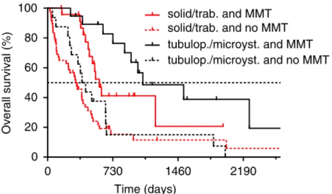
Interestingly, we found that among patients who received MMT those with tubulopapillary/microcystic pattern MPMs showed a trend for OS superior to patients with solid/trabecular pattern tumours (HR = 2.29, 95% CI = 0.95–5.12, P= 0.066, Fig- ure 6). Among those not receiving MMT there was no significant difference in OS between the two main groups of histological variants (HR =1.16, 95%
CI= 0.65–2.07, P= 0.617, Figure 6). Furthermore, both in the tubulopapillary/microcystic as well as in the solid/trabecular subcohorts, MMT provided a sig- nificant survival benefit (tubulopapillary/microcystic:
MMT versus CHT/BSC: HR= 2.67, 95% CI = 2.18– 3.08, P= 0.0006; solid/trabecular: MMT versus CHT/BSC: HR= 1.77, 95% CI= 1.24–2.31, P= 0.0018, Figure 6).
Discussion
In the current study, tumour samples from 192 patients with epithelioid MPM were re-analysed from the archives of five large central European thoracic centres. To the best of our knowledge, this is the sec- ond largest study so far to evaluate the prognostic role of different histological patterns of eMPM. More- over, this is the first study to directly compare the prognostic impact of morphological growth pattern, the nuclear grade and the M/N score. The three main histological types, epithelioid, biphasic and sarcoma- toid, are recognised as distinct categories of MPM by the most recent World Health Organisation classifica- tion2 and are a mandatory part of the final diagno- sis.4 According to the International Mesothelioma Interest Group’s 2017 update, the histological pattern of eMPMs is an optional part of a pathological report but– in the light of emerging data – is currently considered a potentially important prognostic fea- ture,29and is also recommended to be part of report- ing by the 2020 guideline of the European Network for Rare Adult Solid Cancers and the International Association for the Study of Lung Cancer.28 The fre- quency of the individual histological subtypes varies substantially in the literature; nevertheless, we observed their frequencies in our eMPM cohort to be within the range of previous studies.20,22,23
In the daily practice of MPM diagnostics, the amount of tissue available for histological work-up, subtyping and grading is often an issue. In our opin- ion, sampling heterogeneity might be an important factor regarding variable subtype frequencies among recent studies. Our cohort mainly consisted of surgi- cal biopsies. However, was not pre-selected based on sample size and included percutaneous core needle biopsies. This is partly a limitation of this study but also the reflection of a real-life situation from which our samples come.
Regarding outcome, we found that among eMPMs those of predominantly microcystic or tubulopapillary pattern were associated with the longest OS. This finding is similar to that of Brcicet al.23and Alchami et al.22 We found that the trabecular variant con- ferred a relatively poor prognosis, similar to the solid pattern. Regarding the trabecular variant, conflicting data are available in the current literature.20,23 Table 3. Multivariate Cox regression analysis in the MPM
patient cohort
Multivariate analysis
HR 95% CI P-value
Histology Solid/trabecular tubulopap./microcyst.
0.723 0.50–1.06 0.095
M/N score 0 1 2
1.56 1.13–2.16 0.007
Nuclear grade 1
2 3
1.08 0.78–1.48 0.648
CI, confidence interval; HR, hazard ratio; tubulopap., tubulopapil- lary; microcyst., microcystic; M/N, mitosis/necrosis; MPM, malig- nant pleural mesothelioma.
Furthermore, we were able to confirm the previously reported dismal prognosis of MPM exhibiting pleo- morphic features.20–23 Based on overlapping survival curves, we merged the tubulopapillary and microcys- tic variants, as well as solid and trabecular variants.
We found these two groups to have significantly dif- ferent OS. In an external validation cohort consisting of 55 digitised eMPM sections of the TCGA project we confirmed a similar significant difference in OS between these two groups.
The newly proposed M/N score aiming to further stratify patients with eMPM was a robust marker in our patient population. In multivariate analysis, we found M/N score to be the single independent
prognostic factor. On analysing the individual compo- nents of the composite grades, we identified the pres- ence of necrosis to be an independent factor defining prognosis. This result may be partially explained by the significant association we observed between solid/
trabecular pattern and higher M/N scores and higher nuclear grades. While nuclear grade 3 tumours showed a significantly shorter OS, we found no signif- icant OS difference between nuclear grade 1 and 2 tumours. This finding further supports the new EUR- ACAN/IASLC proposal on the use of preferentially two-tier grading of eMPM.28
Predominant histological subtypes30of invasive lung adenocarcinomas have been shown to have a stage- 100
80
Overall survival (%)
60 40 20 0
100 80
Overall survival (%)
60 40 20 0
0 730 1460
Time (days)
0 730 1460
Time (days)
2190
100 80
Overall survival (%)
60 40 20 0
0 730 1460
Time (days)
2190
2190 2920
A
C
B
60
40
20
solid/trab. tubulop./microyst.
Number of samples
0 D M/N Score 0
M/N Score 1 M/N Score 2
M/N Score 0 M/N Score 1 M/N Score 2 Nuclear Grade 1 Nuclear Grade 2 Nuclear Grade 3
Solid / t rabecular
Tubulopapillary/microcystic
Figure 5.Analysis of the The Cancer Genome Atlas (TCGA) epithelioid malignant pleural mesothelioma (eMPM) cohort.A,Mitosis/necrosis (M/
N) score is a significant prognosticator in eMPM. Compared to M/N score 0 [795 days, hazard ratio (HR)=Ref.], M/N score 1 (511 days, HR=1.47, 95% confidence interval (CI)=0.76–2.86,P=0.251, log-rank test] and M/N score 2 (330 days, HR=3.11, 95% CI=1.17– 8.23,P=0.023, log-rank test) was associated with shorter overall survival (OS).B,Higher nuclear grade showed a tendency towards shorter OS: nuclear grade 1 (823 days, HR=Ref.), nuclear grade 2 (459 days, HR=1.53, 95% CI=0.80–2.92,P=0.200, log-rank test) nuclear grade 3 (232 days, HR=4.91, 95% CI=1.45–16.59,P=0.010, log-rank test).C,Tubulopapillary/microcystic subtype (795 days, HR=Ref.) is associated with longer OS than solid/trabecular variants (406 days, HR=2.24, 95% CI=1.17–4.29,P=0.01, log-rank test).D,Solid/tra- becular subtypes were associated with higher M/N scores (P<0.0001,v2test). [Colour figure can be viewed at wileyonlinelibrary.com]
independent prognostic impact31and to be of predictive value for determining the patients’ subgroup that might benefit from adjuvant chemotherapy after complete sur- gical resection.32In our mesothelioma subcohort analy- sis, we investigated if histological patterns might be a useful marker for identifying patients who might benefit more from a more aggressive treatment approach. In this regard, we found a more pronounced OS difference between patients receiving multimodal therapy versus chemotherapy only or best supportive care in case of tubulopapillary/microcystic compared to solid/trabecu- lar MPM. These findings suggest that histological sub- types might be useful to risk-stratify eMPM patients prior to therapeutic decisions in multimodal treatment settings. Nevertheless, this observation needs further independent confirmation and prospective validation.
Acknowledgements
This work was supported by the EFOP (3.6.3-VEKOP- 16-2017-00009 Fund to A.B.), the Janos Bolyai Research Scholarship of the Hungarian Academy of Sciences (to V.L.), the UNKP-19-4 New National Excel- lence Program of the Ministry for Innovation and
Technology (to V.L.), the NRDI Office (KH130356 to B.D.) and the FWF Austrian Science Fund (I2872-B28 to B.H., I3522-B31 to V.L., I3977 and I4677 to B.D.).
Conflict of interest
None of the authors declare any conflict of interest.
References
1. Shavelle R, Vavra-Musser K, Lee J, Brooks J. Life expectancy in pleural and peritoneal mesothelioma. Lung Cancer. Int. 2017;
2017; 1–8.
2. Travis WD, Brambilla E, Burke AP, Marx A, Nicholson AJT eds. World Health Organisation classification of tumours of the lung, pleura, thymus and heart, 4th edn. Geneva, Switzerland:
WHO, 2015; 78–79.
3. Kindler HL, Ismaila N, Armato SG IIIet al. Treatment of malignant pleural mesothelioma: American Society of Clinical Oncology clinical practice guideline.J. Clin. Oncol.2018;36; 1343–1373.
4. Baas P, Fennell D, Kerr KM, Van Schil PE, Haas RL, Peters S.
Malignant pleural mesothelioma: ESMO clinical practice guide- lines for diagnosis, treatment and follow-up.Ann. Oncol.2015;
26(Suppl. 5); 31–39.
5. Calabro L, Rossi G, Maio M. New horizons from immunother- apy in malignant pleural mesothelioma. J. Thorac. Dis.2018;
10; S322–S332.
6. Meyerhoff RR, Yang C-FJ, Speicher PJet al. Impact of mesothe- lioma histologic subtype on outcomes in the surveillance, epi- demiology, and end results database.J. Surg. Res.2015;196;
23–32.
7. Kadota K, Suzuki K, Colovos Cet al. A nuclear grading system is a strong predictor of survival in epitheloid diffuse malignant pleural mesothelioma.Mod. Pathol.2012;25; 260–271.
8. Rosen LE, Karrison T, Ananthanarayanan V et al. Nuclear grade and necrosis predict prognosis in malignant epithelioid pleural mesothelioma: a multi-institutional study.Mod. Pathol.
2018;31; 598–606.
9. Habougit C, Trombert-Paviot B, Karpathiou Get al. Histopatho- logic features predict survival in diffuse pleural malignant mesothelioma on pleural biopsies. Virchows Arch. 2017;470;
639–646.
10. Ghanim B, Klikovits T, Hoda Met al. Ki67 index is an indepen- dent prognostic factor in epithelioid but not in non-epithelioid malignant pleural mesothelioma: a multicenter study. Br. J.
Cancer2015;112; 783.
11. Chapel DB, Churg A, Santoni-Rugiu E, Tsujimura T, Hiroshima K, Husain AN. Molecular pathways and diagnosis in malig- nant mesothelioma: a review of the 14th international confer- ence of the International Mesothelioma Interest Group. Lung Cancer2019;127; 69–75.
12. Pelosi G, Papotti M, Righi Let al. Pathologic grading of malig- nant pleural mesothelioma: an evidence-based proposal.J. Tho- rac. Oncol.2018;13; 1750–1761.
13. Cerruto CA, Brun EA, Chang D, Sugarbaker PH. Prognostic significance of histomorphologic parameters in diffuse malig- nant peritoneal mesothelioma. Arch. Pathol. Lab. Med.2006;
130; 1654–1661.
14. Krasinskas AM, Borczuk AC, Hartman DJet al. Prognostic sig- nificance of morphological growth patterns and mitotic index 100
80
Overall survival (%)
60 40 20 0
0 730 1460
Time (days)
2190 solid/trab. and MMT solid/trab. and no MMT tubulop./microyst. and MMT tubulop./microyst. and no MMT
Figure 6.Histological subtypes and multimodal therapy (MMT). His- tological subtypes showed a tendency for association with overall survival (OS) in patients treated with multimodal approaches. Within the patient subgroup who received MMT, tubulopapillary/microcystic subtypes [1068 days, hazard ratio (HR)=Ref.] were associated with longer OS than solid/trabecular subtypes [580 days, HR=2.29, 95% confidence interval (CI)=0.95–5.12,P=0.066, log-rank test].
Among patients who did not receive MMT, there was no significant difference in OS between tubulopapillary/microcystic (406 days, HR=Ref.) and solid/trabecular subtypes (327 days, HR=1.16, 95% CI=0.65–2.07,P=0.617). Within histological subtypes, MMT was associated with significantly longer OS; however, the bene- fit was more pronounced in the tubulopapillary/microcystic sub- group (MMT: 1068 days, HR=Ref. versus no MMT: 406 days, HR=2.67, 95% CI=2.18–3.08,P=0.0006, log-rank test) in com- parison to the solid/trabecular subgroup (MMT: 580 days, HR=Ref.
versus no MMT: 327 days, HR=1.77, 95% CI=1.24–2.31, P=0.0018, log-rank test). [Colour figure can be viewed at wileyonli- nelibrary.com]
of epithelioid malignant peritoneal mesothelioma.Histopathol- ogy2016;68; 729–737.
15. Postmus P, Kerr K, Oudkerk M et al. Early and locally advanced non-small-cell lung cancer (NSCLC): ESMO clinical practice guidelines for diagnosis, treatment and follow-up.Ann.
Oncol.2017;28; iv1–iv21.
16. Yu Y, Jian H, Shen L, Zhu L, Lu S. Lymph node involvement influenced by lung adenocarcinoma subtypes in tumor size≤3 cm disease: a study of 2268 cases.Eur. J. Surg. Oncol.2016;
42; 1714–1719.
17. Kris MG, Gaspar LE, Chaft JE, Kennedy EB, Azzoli CG, Ellis PM.
Adjuvant systemic therapy and adjuvant radiation therapy for stage I to IIIa completely resected non-small-cell lung cancers:
American Society of Clinical Oncology/Cancer Care Ontario Clinical Practice Guideline update. J. Clin. Oncol. 2017; 35;
2960–2974.
18. Gullo I, Carneiro F, Oliveira C, Almeida GMJP. Heterogeneity in gastric cancer: from pure morphology to molecular classifi- cations.Pathobiology2018;85; 50–63.
19. Fernandez MI, Williams SB, Willis DLet al. Clinical risk stratifi- cation in patients with surgically resectable micropapillary bladder cancer.BJU Int.2017;119; 684–691.
20. Kadota K, Suzuki K, Sima CS, Rusch VW, Adusumilli PS, Tra- vis WD. Pleomorphic epithelioid diffuse malignant pleural mesothelioma: a clinicopathological review and conceptual proposal to reclassify as biphasic or sarcomatoid mesothelioma.
J. Thorac. Oncol.2011;6; 896–904.
21. Galateau Salle F, Le Stang N, Nicholson AGet al. New insights on diagnostic reproducibility of biphasic mesotheliomas: a mul- ti-institutional evaluation by the international mesothelioma panel from the mesopath reference center. J. Thorac. Oncol.
2018;13; 1189–1203.
22. Alchami FS, Attanoos RL, Bamber AR. Myxoid variant epithe- lioid pleural mesothelioma defines a favourable prognosis group: an analysis of 191 patients with pleural malignant mesothelioma.J. Clin. Pathol.2017;70; 179–182.
23. Brcic L, Jakopovic M, Brcic Iet al. Reproducibility of histologi- cal subtyping of malignant pleural mesothelioma. Virchows Arch.2014;465; 679–685.
24. Ordonez NG. Pleomorphic mesothelioma: report of 10 cases.~ Mod. Pathol.2012;25; 1011.
25. Gutman DA, Khalilia M, Lee Set al. The digital slide archive: a software platform for management, integration, and analysis of histology for cancer research.Cancer Res. 2017;77; e75– e78.
26. National Cancer Instititute. The Cancer Genome Atlas. Avail- able at: https://www.cancer.gov/tcga. Date accessed: 22. 07.
2019.
27. Gao J, Aksoy BA, Dogrusoz Uet al. Integrative analysis of com- plex cancer genomics and clinical profiles using the cbioportal.
Sci. Signal.2013;6; pl1.
28. Nicholson AG, Sauter JL, Nowak AK et al. EURACAN/IASLC proposals for updating the histologic classification of pleural mesothelioma: towards a more multidisciplinary approach.J.
Thorac. Oncol.2020;15; 29–49.
29. Husain AN, Colby TV, Ordo~nez NGet al. Guidelines for patho- logic diagnosis of malignant mesothelioma 2017 update of the consensus statement from the international mesothelioma interest group.Arch. Pathol. Lab. Med.2018;142; 89–108.
30. Travis WD, Brambilla E, Nicholson AGet al. The 2015 World Health Organization classification of lung tumors: impact of genetic, clinical and radiologic advances since the 2004 classi- fication.J. Thorac. Oncol.2015;10; 1243–1260.
31. Warth A, Muley T, Meister Met al. The novel Histologic Inter- national Association for the Study of Lung Cancer/American Thoracic Society/European Respiratory Society classification system of lung adenocarcinoma is a stage-independent predic- tor of survival.J. Clin. Oncol.2012;30; 1438–1446.
32. Tsao M-S, Marguet S, Le Teuff Get al. Subtype classification of lung adenocarcinoma predicts benefit from adjuvant chemotherapy in patients undergoing complete resection. J.
Clin. Oncol.2015;33; 3439.
Supporting Information
Additional Supporting Information may be found in the online version of this article:
Figure S1. (A) Distribution of histologic variants among early and advanced disease stage patients.
(P= 0.999, Fisher’s exact test). (B) OS of tubulopapil- lary/microcystic and solid/trabecular histologic sub- types in the early stage subgroup of patients (median OS: 897 days, HR = Ref. versus median OS: 510 days, HR = 1.55, 95% CI [0.80–2.98],P = 0.194 by Man- tel-Cox test; P= 0.041 by Gehan-Breslow-Wilcoxon test) (C) OS of tubulopapillary/microcystic and solid/tra- becular histologic subtypes in the advanced stage sub- group of patients (median OS: 660 days, HR = Ref.
versus median OS: 401 days, HR = 1.75, 95% CI [1.00–3.06],P= 0.047, Mantel-Cox test).
Figure S2. (A) OS of tumors exhibiting mild (HR = Ref.), moderate (HR = 2.29, 95% CI [1.32– 3.97], P= 0.027) and severe nuclear atypia (HR = 3.47, 95% CI [1.88–6.42], P< 0.001). (B) OS and mitotic counts. Low (HR = Ref.) and intermediate (HR = 1.17, 95% CI [0.75–1.87],P= 0.470) numbers of mitotic figures are associated with a significantly better OS in comparison to high mitotic counts (HR = 2.48, 95% CI [1.45–4.25], P< 0.001) (C) The presence of necrosis is associated with significantly shorter OS (HR = 2.38, 95% CI [1.68–3.38],P< 0.0001).
Table S1. Multivariate Cox regression analysis of histologic variants and individual components of composite scores nuclear grade and mitosis-necrosis score in the eMPM patient cohort.
Table S2. Clinicopathological characteristics of the TCGA MPM patient cohort.
Table S3.Univariate survival analyses in the TCGA MPM patient cohort.
Table S4. Summary of therapeutic regimens patients in our exploratory subcohort received.
Table S5. Clinicopathologic characteristics of the exploratory subcohort in which we analysed differences in OS in the context of morphologic subtypes and therapy received.P-values were calculated by Fisher’s exact tests except for *where Chi-squared tests were used and
†where unpaired, two-tailedt-tests.
![Figure 1. Histological subtypes of epithelioid malignant pleural mesothelioma (eMPM). A, Tubulopapillary pattern [haematoxylin and eosin (H&E)]](https://thumb-eu.123doks.com/thumbv2/9dokorg/772833.34706/4.892.101.809.147.628/figure-histological-subtypes-epithelioid-malignant-mesothelioma-tubulopapillary-haematoxylin.webp)