MOLECULAR AND HISTOLPATHOLOGIC PROGNOSTIC FACTORS IN MALIGNANT
PLEURAL MESOTHELIOMA
PhD Thesis
Ágnes Bilecz, MD
Pathological Studies Doctoral School Semmelweis University
Supervisor: Balázs Hegedűs, Ph.D Official reviewers: Hajnalka Rajnai, MD, Ph.D
János Szőke, MD, Ph.D
Head of the Final Examination Committee: Attila Tordai, MD, D.Sc Members of the Final Examination Committee: Erika Tóth, MD, Ph.D
Attila Patócs, MD, Ph.D
TABLE OF CONTENTS
LIST OF ABBREVIATIONS ... 5
1. INTRODUCTION ... 11
1.1 Epidemiology of malignant pleural mesothelioma ... 11
1.1.1 Asbestos exposure and malignant pleural mesothelioma ... 13
1.1.2 Non-asbestos related MPM ... 15
1.1.3 Genetic predisposition to MPM ... 15
1.2 Clinical diagnosis and staging of MPM ... 16
1.3 Histopathologic features of MPM ... 20
1.3.1 Epithelioid type ... 22
1.3.2. Differential diagnostics of EMM regarding other carcinomas in the lung ... 24
1.3.3. Biphasic type ... 27
1.3.4 Sarcomatoid type ... 28
1.4 Treatment modalities in MPM ... 29
1.4.1 Systemic therapy ... 29
1.4.2 Radiotherapy ... 30
1.4.3 Surgical therapy ... 30
1.4.4 Multimodality treatment ... 32
1.4.5 Emerging therapeutic approaches ... 32
1.5 The molecular landscape of MPM... 35
1.5.1 Cell cycle regulation pathways ... 36
1.5.2 BAP1 and DNA damage repair ... 37
1.5.3 Hippo pathway ... 39
1.6 Telomere and telomerase ... 41
1.6.1 Structure and function ... 41
1.6.2 Telomere lengthening and telomerase in disease ... 42
1.7 TERT promoter mutations ... 44
1.7.1 Patomechanism of TERT promoter mutations ... 44
1.7.2 Germline mutations ... 45
1.7.3 Somatic mutations ... 45
1.7.4 Common single nucleotid polymorphism rs2853669 ... 46
1.7.5 TERT in MPM ... 47
2. OBJECTIVES ... 48
3. METHODS ... 49
3.1. Patient cohorts ... 49
3.2 Histological subtype analysis ... 51
3.3 Histological grading ... 52
3.4 BAP1 staining ... 53
3.5 Mesothelioma cell lines ... 54
3.6 DNA extraction and TERT promoter status analysis ... 56
3.7 TERT mRNA expression... 57
3.8 Cell viability assay... 58
3.9 Statistical analysis... 58
4. RESULTS ... 59
4.1 Histologic grading and subtype analysis ... 59
4.1.1 Clinicopathological characteristics of the patient collective ... 59
4.1.2 Histopathologic characteristics ... 59
4.1.3 Association between EMM subtypes and grade ... 61
4.1.4 Histopathologic parameters and disease outcome ... 61
4.1.5 Histopathologic parameters in the validation cohort ... 65
4.1.7 Differences in response to MMT among EMM subtypes ... 70
4.2 TERT promoter mutation ... 74
4.2.1 TERT promoter mutation and clinicopathological characteristics ... 74
4.2.2 TERT promoter mutation and histological subtypes of epitheioid MPM ... 76
4.2.3 TERT promoter mutation and BAP1 expression ... 77
4.2.4 TERT promoter status and patient outcomes ... 78
4.2.5 Patient outcomes and SNP rs2853669 ... 82
4.2.6 TERT mRNA expression and TERT promoter status ... 82
4.2.7 TERT promoter status and cell line formation ... 83
4.2.8 TERT promoter status and in vitro cisplatin sensitivity ... 84
5. DISCUSSION ... 86
5.1 Histologic subtypes of epithelioid mesothelioma ... 86
5.2 Pleomorphic subtype EMM confers dismal prognosis ... 88
5.5 TERT promoter mutation is an independent negative prognostic factor in
malignant pleural mesothelioma ... 89
5.6 TERT promoter mutation and implications in systemic treatment for MPM... 90
5.7 Mechanism of increased aggressivity of TERT promoter mutant MPM ... 90
5.8 Impact of the SNP rs2853669 in mesothelioma ... 91
5.9 Association of major molecular alterations and TERT promoter mutation ... 91
6. CONCLUSIONS ... 92
7. ÖSSZEFOGLALÁS ... 94
8. SUMMARY ... 95
9. REFERENCES ... 96
10. BIBLIOGRAPHY OF THE CANDIDATE’S PUBLICATIONS ... 121
10.1 Publications related to the thesis ... 121
10.2 Publications not related to the thesis ... 122
11. ACKNOWLEDGMENTS ... 123
LIST OF ABBREVIATIONS
3D CRT three-dimensional conformal radiation therapy
A adenine
AACR American Association of Cancer Research ACS active symptom control
AE1/AE3 pan cytokeratin antybody AE1/AE3 AJCC American Joint Committee on Cancer AKT Protein kinase B
ALT alternative lengthening of telomeres AP1 Activator Protein 1
ARID2 AT-Rich Interaction Domain 2 ASSI Argininosuccinate synthase 1 ATP Adenosine triphosphate
ATRX Alpha Thalassemia/Mental Retardation Syndrome X-Linked ATS American Thoracic Society
AUT Austia
BAP-1 BRCA1-associated protein-1
BAP1-TPDS BRCA1-associated protein-1 tumor predisposition syndrome BARD1 BRCA1-associated RING domain protein
Ber-EP4 anty-Epithelial cell adhesion molecule-antibody Ber-EP4
BG8 Blood group 8
BMM biphasic malignant mesothelioma
BRIP1 BRCA1 Interacting Protein C-Terminal Helicase 1 BSC best supportive care
BTS British Thoracic Society
C cytosine
c. codone
CANT1 Calcium Activated Nucleotidase 1 CAR Chimeric antigen receptor
CD80 cluster of differentiation 80
CDKN2A cyclin-dependent kinase Inhibitor 2A
cDNA complementary DNA
CEA Carcinoembryonic antigen
CHT chemotherapy
CI confidence intervall
CK cytokeratin
CK 5/6 cytokeratin 5/6
cKIT KIT proto-oncogene, receptor tyrosine kinase
CRO Croatia
CRP C Reactive Protein
CT computed tomography
CTLA4 cytotoxic T-lymphocyte-associated protein 4 D2-40 monoclonal anti-podoplanin-antibody D2-40 DAXX Death Domain Associated Protein
DNA deoxyribonucleic acid dsDNA double-strand DNA
E2F1 E2F Transcription Factor 1
ECOG PS Eastern Cooperative Oncology Group Scale of Performance Status EGFR epidermal growth factor receptor
EMM epithelioid malignant mesothelioma
EORTC European Organisation for Research and Treatment of Cancer ePD extended pleurectomy/decortication
EPP extrapleural pulmonectomy ERS European Respiratory Society
ETS E-twenty-six
EURACAN European Network for Rare Adult Solid Cancers EZH2 Enhancer of zeste homolog 2
FAK Focal adhesion kinase FAT4 FAT Atypical Cadherin 4 FDG fluorodeoxyglucose
FFPE Formalin fixed paraffin embedded
FISH fluorescent in situ hybridization
G guanine
GABPA/B GA Binding Protein Transcription Factor Subunit Alpha/Beta GATA3 GATA Binding Protein 3
GCDFP15 Gross cystic disease fluid protein 15
Gly glycine
Gy Grey
HE hematoxylin eosin
HIF1 Hypoxia Inducible Factor
HMGB-1 High mobility group box 1 protein HPF high power field
HR hazard ratio
HSV1716 Herpes simplex virus 1716
IASLC International Association for the Study of Lung Cancer IC50 half maximal inhibitory concentration
IFN-β interferon beta
IMIG International Mesothelioma Interest Group IMRT Intensity-modulated radiotherapy
IQR interquartile range KIBRA Kidney and brain protein
KPS Karnofsky performance status scale LADC lung adenocarcinoma
LATS2 Large Tumor Suppressor Kinase 2 Leu-M1 monoclonal anti-CD15-antibody Leu-M1 M/N score mitosis-necrosis score
MARS Mesothelioma and Radical Surgery MCR macroscopic complete resection MDM2 Mouse Double Minute 2,
MLLT3 MLLT3 Super Elongation Complex Subunit
MM malignant mesothelioma
MOC31 anty-Epithelial cell adhesion molecule-antibody MOC31 MPM malignant pleural mesothelioma
mRNA messenger RNA
MST1/2 Mammalian STE20-Like Protein Kinase 1/2 MTAP Methylthioadenosine Phosphorylase
mTOR mammalian target of rapamycin
mut mutant
MYC V-Myc Avian Myelocytomatosis Viral Oncogene Homolog
NA not available
NF2 Neurofibromatosis type 2 OD optical density
OR overall risk
OS overall survival
p. protein amino acid
p16 cyclin-dependent kinase inhibitor 2A p40 anty-deltaNp63-antibody
PARP Poly (ADP-ribose) polymerase PAX2 Paired box gene 2
PAX8 Paired box gene 8
PBRM1 Polybromo 1
PBS Phosphate-buffered saline PCR polymerase chain reaction PD pleurectomy/decortication PD(L)1 programmed death (ligand) 1
PET-CT Positron emission tomography–computed tomography PFS progressio-free survival
PI3K Phosphoinositide 3-kinase ( POT1 Protection Of Telomeres 1 pRb retinoblastoma protein PSEN1 presenilin-1
PTCH1 Patched 1
PTPRD Protein Tyrosine Phosphatase Receptor Type D
qRT-PCR quantitative reverse transcription polymerase chain reaction
RAD51 RAD51 Recombinase
RAP1 Repressor/Activator Protein 1 Homolog RNA ribonucleic acid
RPL41 Ribosomal Protein L41
RT raditherapy
SAV1 Salvador Family WW Domain Containing Protein 1 SE standard error
SETD2 SET Domain Containing 2, Histone Lysine Methyltransferase SETDB1 SET Domain Bifurcated Histone Lysine Methyltransferase 1
SLO Slovenia
SMM sarcomatoid malignant mesothelioma SMYD3 SET And MYND Domain Containing 3 SNP single nucleotid polymorphism
SP1 Specificity Protein 1 SQCC squamous cell carcinoma
SRB Sulforhodamine B
T thymine
TAZ Tafazzin
TCGA The Cancer Genome Atlas
TEAD transcription-enhancer activator domain transcripition factor TEK TEK Receptor Tyrosine Kinase
TERC Telomerase RNA Component TERT Telomerase Reverse Transcriptase
TERTp TERT promoter
THOR TERT hypermethylation oncological region TIN2 TRF interacting protein 2
TNM tumor, nodes and metastases
TP53 Tumor Protein P53
TPP1 TINT1/PTOP/PIP1
Trp tryptophan
TTF-1 thyroid transcription factor 1
UICC Union for International Cancer Control VATS video-assisted thoracoscopy
VGFR Vascular endothelial growth factor WBC white blood cell count
WHO World Health Organisation
wt wild-type
WT-1 Wilms' tumour 1
WWTR1 WW Domain Containing Transcription Regulator 1 YAP Yes Associated Protein
1. INTRODUCTION
1.1 Epidemiology of malignant pleural mesothelioma
Malignant mesothelioma (MM) is a rare malignancy arising from the mesothelial cells of serous membranes such as the pleura, peritoneum[1], pericardium[2], tunica vaginalis of the testis[3] and ovarial surface epithelium[4]. Malignant pleural mesothelioma (MPM) is the most common form of malignant mesothelioma accounting for 80-85% of the cases [5].
According to the World Health Organisation (WHO) mortality database 92,253 malignant mesothelioma deaths were reported in the period between 1994 and 2008 from 83 countries across the world. Worldwide, crude and age-adjusted mortality rates were 6.2 and 4.9 deaths per million population, respectively, the latter showing a yearly increase of 5.37%. During the studied time period malignant mesothelioma associated deaths occurred more frequently in the high-income countries of the Americas and Europe [6].
MPM’s incidence varies substantially across the world. MPM’s age standardized-rate incidence was 1.93 per 100,000 among men, and 0.41 among women in the United States. Standardized-rate incidences were 3.5 among men and 1.25 per 100,000 among men and women in Italy [7], while among males in Great Britain it was 3.4/100,000, 2.3/100,000 in France, and 3.2/100,000 in the Netherlands [8]. Lower MPM incidence rates are reported n Central and Eastern Europe, 1.84 in Croatia and 1 per 100,000 men in Austria [9].
MPM incidence and mortality not only shows spatial variations, but also changes over time. Number of MPM associated deaths has been rising during the 20th century due to the rising production and consumption of asbestos. In North America and Western Europe the rise in incidence is expected to level out in the near future and then decrease.
During the first decade of the 2000s Sweden already experienced a decrease in the number of MPM cases thanks to early adaptation of strict regulation of asbestos handling [10, 11]. In other countries like Italy, Netherlands and France show stagnant
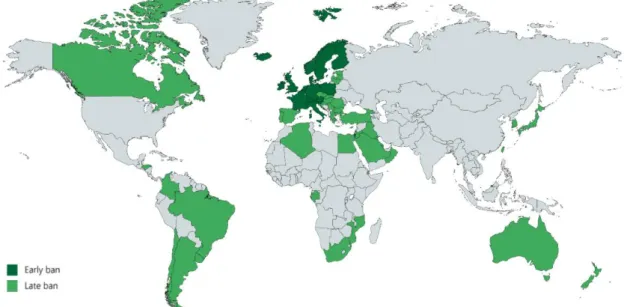
countries where there is no ban on asbestos (Figure 1) [15, 16] which causes a continued increase of MPM incidence worldwide [8].
Figure 1. The use and production of asbestos is currently banned in 67 countries. Early bans were introduced in Western Europe before 2000 (dark green). Several countries have implemented such measures only after 2000 (light green). The author’s drawing based on data from [17, 18].
Due to the long latency period after exposure MPM is most commonly diagnosed in patients older than 65 years [14]. 2% of all MPM patients are younger than 40 years, and they have a significantly better overall survival among all three main histological subtype, than those older than 40 (11 months vs. 8 months) [19].
MPM is approximately four times more common among males than females, which might be explained by males traditionally working in positions with higher risk of occupational asbestos exposure [19]. The French National Mesothelioma Surveillance Program calculated the MPM risk fraction attributable to occupational exposure for both genders, and estimated it to be 83.2% (95% CI 76.8-89.6) for men, while only 38.4%
(95% CI 26.8-50.0) for women [20]. Enviromental exposure, however is a higher burden for women, the male-to-female ratio being approximately 1, and MPM risk associated with environmental exposure in women being 38.7% and 20% in men [21].
Women with mesothelioma have been reported to have a significantly longer survival
compared to men, a phenomenon also in part attributable to differences in the doses of asbestos exposure [22].
1.1.1 Asbestos exposure and malignant pleural mesothelioma
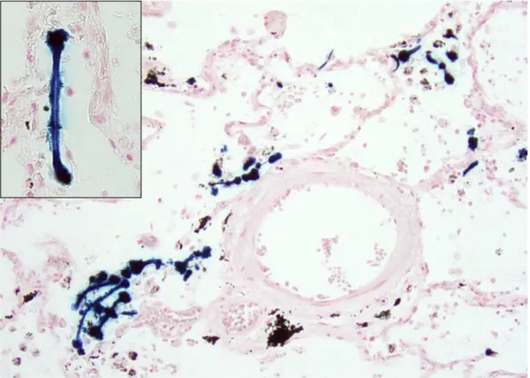
Of all MPM cases, approximately 80% are estimated to be linked to inhalation of asbestos fibers (Figure 2) [23], while only 10% of those substantially exposed to asbestos develop MPM [24]. Asbestos carcinogenesis is linked to DNA damage caused by direct mechanical interference of asbestos fibers whit chromosomes, as well as by reactive oxygen and nitrogen species secreted by mesothelial cells and macrophages [25]. The HMGB-1 mediated necrosis and chronic inflammation induced by the depositions of asbestos fibers also plays a role in the development of MPM [26].
Figure 2. Asbestos bodies, also known as ferruginous bodies are dumbbell-shaped, thin structures covered by a coat of proteins and iron-containing
Ecological correlations have been shown to be robust between a country’s historical asbestos consumption given in kg per person per year and its age-adjusted annual MM and MPM mortality rates [29].
Crocidolite, amosite and chrysotile are the three types of asbestos associated with the induction of MPM, the ratio of exposure specific risk of MPM from the three principal types of asbestos is estimated to be 500:100:1 [30]. Eternit workers and wives were typically exposed to a mixture of crocidolite and chrysotile, while railway stock workers were predominantly exposed to crocidolite, and amosite factory workers to amosite [31].
Regional clustering of MPM cases was observed within several Western European countries [32]. The hotspots were identified most commonly in the vicinity of harbors with oil refieneries or shipyards due to historical asbestos use in shipbuilding and repair (eg. South-East England [33], Genoa and Trieste, Italy [34]), asbestos mines and asbestos-cement industries (eg. Casale Monferrato, Italy ) or near railway carriage construction and repair sites (eg.Veneto, Italy) [35]. Men between the ages of 40 and 74 years in Scotland and England had an age standardized MPM incidence rate of 8.8 and 8.0 per 100,000, in the Trieste and Genova region of Italy 17.2 and 14.4 per 100,000 persons, respectively, while for the remaining European countries an incidence of 0.6- 4.2 per 100,000 was observed in the time period beween 1991 and 1995 [24].
A study carried out by the French National Mesothelioma Program identified industries associated with the highest risk for MPM. French men working in shipbuilding and repair had more than 9 times higher risk (OR=9.3, 95% CI: 5.20-16.06) for developing MPM compared to those never having worked in asbestos related occupations. Among others, the men working in the manufacturing of astbestos products, of metal constructions, plumbers, construction workers, electrical wiremen and those working in railroad equipment production were also at substantially higher risk for MPM [36].
Patients in household contact with workers exposed to asbestos also have an elevated risk for pleural disease [37].
A large pooled analysis of cohort studies including workers with occupational exposure and individuals with environmental asbestos exposure found the median age at the time of first exposure to be in the early- to mid-20s, and the median length of exposure to be 3.75 years (IQR 0.7-18.2). The median time between exposure and the diagnosis of
MPM was 38.4 years (IQR 31.3-45.3). The risk of developing MPM increased for 45 years after exposure, after that the increase in risk appeared to level out [31].
1.1.2 Non-asbestos related MPM
Approximately 20% of all MPM cases occur without asbestos exposure. The role of potential alternative risk factors remains unclear. Non-asbestos minerals that have a similar fibrous form and high biopersistence to that of commercial asbestos varieties also have carcinogenic potential, especially erionite [38]. An in vivo experiment showed that carbon nanotubules beyond the threshold length of 4 µm caused acute pleural inflammation, that is considered an early event in MPM carcinogenesis [39]. Exposure to ionizing radiation [40], and Simian virus 40-like virus infection [41] have been proposed as risk factors in a subset of MPM patients, however, their role needs further verification [42].
1.1.3 Genetic predisposition to MPM
The germline mutations in the gene encoding BRCA1-associated protein-1 (BAP-1) have recently been described as a predisposing genetic factor of MPM [43]. This high penetrance germline mutation causes a newly recognized cancer syndrome, namely the BAP-1 tumor predisposition syndrome (BAP1-TPDS), which is characterized by the development of distinct tumor types by the age of 55 years [44]. Carriers show an increased risk to develop peritoneal or pleural mesothelioma, but are also predisposed to other tumor types, such as atypical Spitz tumor [45, 46], cutaneous or uveal melanoma [47], renal cell carcinoma [48], breast cancer, basal cell carcinoma [49] and less frequently to further malignancies [50, 51]. The MM patients carrying these germline mutations are typically younger than those with sporadic MM, more than 60% of them are female, and they have a significantly longer overall survival compared to all MM patients [52, 53].
the germline CDKN2A mutation (c.301G > T, p.Gly101Trp) who developed malignant cutaneous melanoma and has a history of both melanoma and MPM in her family [54].
1.2 Clinical diagnosis and staging of MPM
The diagnosis of MPM is often challenging, as symptoms present at a late stage of the disease progression, and are non-specific [55]. The most common symptoms are dyspnoe and chest pain. Dyspnoe is caused by a typically unilateral pleural effusion.
Chest pain might be diffuse and dull, or less often of pleuritic nature [56]. Other patients present with weight loss, fatigue, or sweats. Local spread of the tumor into mediastinal structures can cause dysphagia, superior vena cava syndrome or recurrent laryngeal nerve palsy [57].
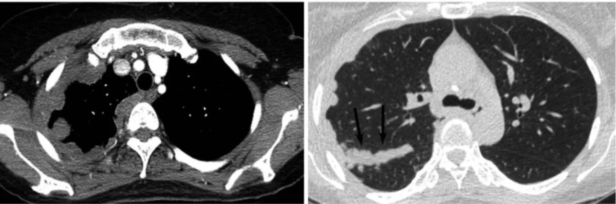
The diagnostic pathway for MPM proposed by the British Thoracic Society (BTS) includes chest radiography as first line imaging modality for patients with symptoms suspicious for MPM [58]. On radiographs, unilateral pleural effusions are present in 94% of the cases. Further findings typical for MPM include a diffuse thickening of the pleura, which might cause a loss in the lung volume, or show a spread along the interlobar fissures [59]. For patients with radiographic features of MPM the recommended second-line imaging method is venous-phase, contrast-enhanched CT of the thorax and the upper part of the abdomen (Figure 3).
Figure 3. Chest CT scan showing concentric and fissural (arrows) pleural thickening in the right thoracic cavity corresponding to MPM. Reprinted from: [60]
For a high rate of false positivity -especially after talc pleurodesis-, PET-CT is only recommended for assessment of patients’ eligibility for surgical resection, such as for evaluation of depth of chest wall invasion or for exclusion of distant metastases [58]. In each case, the diagnosis of MPM has to rely on pathologic evaluation, for there are no specific radiological or clinical features of the disease. There are several methods available for diagnostic sample acquisitions which differ widely in invasiveness and sensitivity. For patients presenting with unilateral pleural effusion, an ultrasound-guided pleurocentesis followed by the cytological evaluation of the pleural fluid is recommended [8, 61]. The sensitivity of cytology in the diagnosis of MPM varies substantially, ranging from 30% to 75%, mainly depending on the experience of the laboratories and the availability of ancillary testing [62-66].
The observed high false-negative rate might be explained by the fact that MPM cells lack specific features of malignancy, malignant epithelioid cells and reactive mesothelial cells share many cytological features, such as low nuclear to cytolplasmic ratios, cell clumps with scalloped borders. Another important factor is that the sarcomatoid component is usually not shed into the malignant effusion, that, as a consequence, results paucicellular [55]. In summary, the first diagnosis of MPM is often based on cytology, but in most cases a tissue biopsy is needed to assess invasion and to confirm the primary MPM diagnosis [61, 67]. However, in patients to frail for further invasive interventions, a diagnosis based on cytology alone is accepted [8, 68].
In patients who are candidates for chemotherapy or multimodal therapy, a tissue sample should be obtained. This might be carried out through video-assisted thoracoscopy (VATS) providing an opportunity to directly visualize any suspicious lesions throughout the pleural surface and to gain sufficiently large and deep tissue samples (Figure 4). Thoracoscopy allows a histologic diagnosis in more than 90% of the cases [69].
Figure 4. (A) and (B) Visualization of the pleural surface and MPM through VATS. (C) Forceps obtaining a tissue biopsy. (D) VATS view of talc pleurodesis [70].
If the extent of the disease does not allow a thoracoscopic approach, an open surgical biopsy might be carried out. In patients who are not fit for VATS or surgical biopsy and do not have a cytologic diagnosis, an imaging guided percutan core needle biopsy should be carried out [61]. Blind biopsies have a lower sensitivity due to sampling error [71] and higher complication rate including pneumothorax in 9.4% of the cases [72].
Initial staging of MPM is based on contrast enhanced chest and upper abdominal CT scan and usually an FDG PET-CT scan. If any of these suggest lesions suspicious for mediastinal lymph node metastases, these should be confirmed through an endobronchial ultrasound-guided fine needle aspiration biopsy or mediastinoscopy in cases where a radical surgical intervention is considered [61, 73-75]. Also, if suspicious lesions on the contralateral pleura or in the abdominal cavity are the only contraindication for radical surgery, a contralateral thoracoscopy or laparoscopy needs to be performed [61].
Individual patients’ functional status is commonly described using the Karnofsky performance status scale (KPS). It ranges from 0% (dead) to 100% (no sign of disease)
and measures the patient’s ability to carry out ordinary tasks [76]. The Eastern Cooperative Oncology Group (ECOG) Scale of Performance Status (PS) ranging between grade 0 and 5 is a similar measure of disease related changes in the amount of daytime spent in bed and the patient’s need for care [77].
The prognostic score system of the European Organisation for Research and Treatment of Cancer (EORTC) is a composit score developed to assess the prognosis of MPM patients. It includes patient’s gender, ECOG PS, the tumor’s histological subtype, certainty of the MPM diagnosis and white blood cell count (WBC) [78]. Male gender, non-epithelioid histology, an uncertain/possible diagnosis of MPM, WBC over 8.3x 10⁹/L and an ECOG PS other than 0 are associated with poor prognosis, and are summarized in the final score after multiplication with a constant specified for each [79]. The prognosis is considered poor if the EORTC score is below 1.27 [80].
The TNM staging system proposed by the IASLC is also used to predict patient outcomes and to help guide treatment decisions (Table 1) [81, 82].
Table 1. Definitions of T, N and M categories according to the IASLC proposal for the 8th edition of TNM, reprinted from [83].
Despite all efforts to achieve early detection, sometimes heroic surgical and oncological treatment, the prognosis of MPM remains dismal. Even in patients with disease limited to the pleura without lymph node or distant metastases (stage IA), the 5-year overall survival is only 16% (Figure 5) [82].
Figure 5. Overall survival of MPM patients based on the IASLC staging system from the 8th edition of TNM Classification of Malignant Tumors by UICC. Reprinted with the permission of Elsevier from [82]
The need for early detection of MPM in patients with known asbestos-exposure has emerged, and multiple screening methods, such as breath tests and circulating tumor markers were tested [84-88]. Nonetheless, screening remains not advised due to MPM’s low incidence even in a high-risk population, its subtle radiologic presentation and the lack of curative therapeutic options [8, 58, 61]. Although there are currently no biomarkers recommended for screening or as a single diagnostic test, biomarker testing is, however, used in the diagnosis of patients with suspicious cytology who are not fit enough for further invasive diagnostic procedures [89].
1.3 Histopathologic features of MPM
Mesothelioma remains a challenging histopathological diagnosis requiring expertise and extensive use of additional immunohistochemical markers. The French National Mesothelioma Surveillance Program reviewed the initial histological diagnosis in over
600 MPM cases with the involvement of at least three expert mesothelioma pathologists and supplemental immunohistochemical analysis. The study was able to confirm the diagnosis of MPM in only 67%. The study found false positive diagnoses in 13% of the initial MPM cases and an uncertain diagnosis was made in 17% of the reviewed cases [20]. The 2015 edition of the WHO Classification of Tumours of the Lung, Pleura, Thymus and Heart recognizes three main histological subtypes of diffuse malignant mesothelioma, namely the epithelioid (EMM), biphasic (BMM) and sarcomatoid (SMM) types [90] (Figure 6).
Figure 6. The major histological types of mesothelioma are (A) epithelioid, (B) biphasic and (C) sarcomatoid. (HE, 100x, author’s photomicrographs).
According to the recent IMIG recommendations, the distinction between these three subtypes is a mandatory part of the pathological reporting of all MPM cases, because the histological subtype is one of the most robust prognostic factor in MPM known to this date, and also influences crucial treatment decisions [63].
EMM is associated with the longes overall survival (12–27 months), BMM confers intermediate prognosis (8–21 months OS), while SMM is associated with dismal prognosis (7–18 months OS) [42]. Patients with EMM and BMM are more often treated by radical surgery, than those with SMM. EMM shows a survival benefit associated with multimodal therapy, however, the data in relation to BMM is unclear, and patients with SMM do not appear to benefit from macroscopic complete resection [91-93].
1.3.1 Epithelioid type
Epithelioid type MPMs are usually composed of bland, mostly cuboidal tumor cells with eosinophilic cytoplasm and inconspicuous nuclei, however marked atypia can also be present in a fraction of the cases. Mitoses are tipically infrequent [90].
EMM has a wide range of morphological subtypes, and often shows various growth patterns within the same tumor. The most common growth patterns of EMM include the solid pattern that consists of sheets of monomorphic, mostly cuboidal cells without specific architectural arrangement. Tubulopapillary EMM is composed of a mixture of tumor cells arranged around fibrovascular cores and tubular structures. The microcystic variant is composed of structures similar to adenomatoid tumors, forming a lace-like architecture of cysts of variable size. Microcystic morphological variants are sometimes associated with a myxoid stroma. The trabecular subtype is characterized by tumor cells arranged in thin rows embedded in desmoplastic stroma. Micropapillary subtype tumors are composed of small papillary structures lacking a fibrovascular core [90]. The pleomorphic variant is characterized by prominent giant cells and anaplastic tumor cells, often with multiple nuclei, nuclear enlargement and hyperchromasia [94]. The rare histological variant composed of plump, elongated epithelioid cells with marked cellular borders and a sheet-like growth has been termed transitional pattern [95]. EMM not only shows a wide variability in growth patterns, but also exhibits unconventional cytologic features in a minority of the cases. Variant cytologic features of EMM include deciduoid [96], lymphohistiocytoid [97] small cell [98], rhabdoid, signet ring and clear cell features [99].
The prognostic role of histomorphology, with an emphasis on growth patterns has been studied extensively in a variety of solid malignancies [100-102]. The 2011 IASLC/ATS/ERS proposal recommended the use of an architecture based classification for invasive lung adenocarcinoma (LADC), introducing the lepidic, acinar, papillary, solid and micropapillary predominant histological subtypes [103]. Growth patterns of lung adenocarcinomas have since been established as independent prognostic factors by several studies [104, 105]. A predominant lepidic growth pattern of LADC shows an indolent clinical behavior and excellent 5-year survival after surgical resection [106, 107]. Predominant solid and micropapillary patterns, however, were associated with
significantly shorter OS [108, 109]. The presence of non-predominant solid or micropapillary patterns in resection specimens was associated with intermediate patient outcomes: significantly worse than those without such areas, however, significantly better than those with predominant solid or micropapillary growth [110]. Solid and micropapillary patterns were associated with an elevated risk for lymph node metastases [106], and in case of solid predominant tumors with multiplex, early, extrathoracic recurrences [111]. Growth pattern based classification of LADC is not only of prognostic relevance, but might be associated with distinct driver gene alterations [112- 114], as well as a predictor of patients benefiting from adjuvant chemotherapy after surgical resection [115, 116]. Patients with early stage disease and a solid or micropapillary predominant component are found to benefit from adjuvant chemotherapy, while no significant benefit from adjuvant chemotherapy was found in the patients subgroup with acinar or papillary patterns [117]. Similarly, on eximanation of small biopsies of advanced stage patients receiving adjuvant platinum based therapy high-grade (micropapillary and solid predominant) patterns were significantly associated with an increased progression-free and overall survival in comparison to intermediate grade tumors [118].
In contrast to lung cancer, limited data is available in the literature on the potential prognostic role of the different predominant patterns in malignant pleural mesothelioma.
A study analyzing 114 EMM samples found 16 myxoid-microcystic variants and identified it as positive prognostic factors in EMM, being associated with significantly longer OS than solid, micropapillary and pleomorphic subtypes [119]. Another study found predominant solid pattern tumors associated with worse patient outcomes in comparison to non-solid variants among 708 EMM samples [120]. The tumors showing a transitional pattern are associated with exceptionally short OS [95]. Similarly, the pleomorphic subtype also shows an association with dismal clinical outcomes comparable to that of BMM [94, 95, 119, 121, 122]. Although the 2015 WHO classification of MPM included the pleomorphic and transitional patterns among the variants of EMM, the 2019 proposal of European Network for Rare Adult Solid Cancers/International Association for the Study of Lung Cancer (EURACAN/IASLC)
but also among the subtypes of SMM based on their dismal prognosis, a finding, however, that still needs confirmation [99].
Several studies investigated cellular features of mesothelioma tumor cells in correlation with patient outcomes. The presence of necrosis was found to be associated with worse prognosis in multiple studies, as well as the degree of nuclear atypia and elevated mitotic counts [120, 123, 124]. Additionally, more delicate nuclear and cytological features were also evaluated in EMM. While the presence of atypical mitoses and prominent nucleoli showed significant prognostic power, intranuclear inclusions and a low cytoplasmic/nuclear ratio did not exhibit such properties, and the impact of chromatin architecture and density is still ambigous [123, 124].
The resently established nuclear grading system predicts patient outcomes. It is based on a three-tier assessment of nuclear atypia, and a three-tier scoring of mitotic counts.
These scores are combined into nuclear grades I to III [120, 123].
A further grading system, the recently proposed mitosis-necrosis score is computed based on the presence of necrosis and a two-tier scoring of mitotic figures, the cut-off value being 5 per 10 high power fields [120]. In a recent validation study, both the three-tier nuclear grading and the mitosis-necrosis score was confirmed to be useful in predicting patient outcomes in a cohort where 87% of the tissue samples were small biopsies [125].
1.3.2. Differential diagnostics of EMM regarding other carcinomas in the lung
The distinction between EMM and lung carcinomas involving the pleura or pleural metastases is often challenging and requires the use of immunohistochemistry. Due to the variable specificity and sensitivity of the commonly used antibodies, the IMIG guideline for the diagnosis of MPM recommends the use of a minimum of two positive markers for confirmation of mesothelial origin, and two negative markers to exclude carcinomas [63].
Lung malignancies and metastases involving the pleura are far more common than MPM, thus, it is important to use a panel of organ-specific immunhistochemical markers selected based on the patient’s clinical history and the differential diagnosis.
The most commonly applied negative markers are listed in Table 2.
Table 2. List of immunohistochemical markers commonly negative in MPM and positive in carcinomas that frequently involve the pleura either through direct infiltration or through metastases [63, 90].
Markers negative in MPM and positive in carcinomas
Sensitivity Specificity vs. MPM Lung squamous cell carcinoma
P40 100% 97.5%
Claudin 4 95% 0%
MOC31 97-100% 85-98%
BG8 80% 93-97%
Adenocarcinoma markers
MOC31 95-100% 85-98%
Ber-EP4 95-100% 74-87%
BG8 90-100% 93-97%
CEA (monoclonal) 80-100% > 95%
Markers of lung origin
TTF-1 80% High
Napsin-A 80% High
Markers of breast origin
GCDFP15 30-40% High
Mammaglobin 50-85% High
Markers of renal origin
PAX8 70-100% Unknown
PAX2 80% Unknown
Claudin 4 90% 0%
CD15 (Leu-M1) 60% High
The most commonly used mesothelial markers include WT-1, calretinin, podoplanin (D2-40 and CK 5/6 [90]. The sensitivity and specificity of these markers are described
Table 3. The most common and sensitive mesothelial markers used in the immunohistochemical diagnosis of epithelioid MPM, and their reactivity in lung squamous cell carcinoma and adenocarcinoma [63].
Positive markers for epithelioid mesothelioma Marker Staining in
MPM
Positivity in MPM
Positivity in lung squamous cell carcinomas
Positivity in lung adenocarcinomas
WT-1 Nuclear, diffuse, strong
70-95% ˜0% ˜0%
Calretinin Nuclear and cytoplasmic, often diffuse, strong
˜100% 40% 5-10% (usually
focal) Podoplanin
(D2-40)
Membrane positivity, diffuse
90-100% 50% <15%
Cytokeratin 5/6
Citoplasmic,
diffuse 75-100% 100% 2-20% (focal)
Discrimination between reactive mesothelial proliferations and EMM is yet another diagnostic challenge. In addition to morphological characteristics, immunohistochemical detection of the loss of nuclear BAP1 staining is useful. The loss of nuclear BAP1 staining was detected by immunohistochemistry in 40-77% of epithelioid MPMs [126-128] and was found to be significantly associated with nonsynonimous genetic alterations of the BAP1 gene [129]. BAP1 negativity was exclusively observed in MPM but not in benign, reactive lesions of the pleura [130]. In a further study, the sensitivity, specificity, positive predictive value, and negative predictive value of the loss of nuclear BAP1 staining was estimated to be 61%, 100%, 100%, and 32%, respectively [127]. Another study proposes that combined use of MTAP – a highly sensitive surrogate marker of 9p21 deletions, a common event in
MPM – and BAP1 immunohistochemistry improves sensitivity of the distinction between MPM and benign proliferations [131].
1.3.3. Biphasic type
BMM contains an epithelioid component intermixed with a sarcomatoid or spindle cell component, both constituting at least 10% of the tumor area.
The diagnosis of biphasic MPM often represents a diagnostic challenge. In an international interobserver agreement study 42 patients’ MPM samples originally classified as biphasic MPM were reviewed by fourteen pathologists with special interest in mesothelioma. The 544 expert opinions on the diagnosis for 42 cases showed moderate interobserver correlation (weighted κ-value=0.45). The original diagnosis of BMM was agreed in 71% of the cases, in 17% the case was reclassified as EMM, and in 12% as pure SMM [95].
The identification of a sarcomatoid component is of outmost importance, since it is a negative prognostic factor and is associated with worse patient outcomes in a radical surgery setting [95]. Both the WHO and the EURACAN/IASLC recommend that the amount of spindle cell component be reported because of its possible prognostic role [90, 99]. Patients with BMM containing less than 20% sarcomatoid elements were found to have significantly longer median OS [95], while another study reported a similar association between the amount of sarcomatoid elements and OS using a cutoff or 50% [132].However, a frank sarcomatoid component of BMM is hard to be distinguished from reactive fibrosis accompanying an epithelioid MPM. The malignant spindle cell population almost invariably shows an at least focal positivity with pancytokeratin antibodies [133] and broad spectrum anti-keratin cocktails such as AE1/AE3 [90]. Reactive fibroblastic proliferations might also be positive with pancytokeratins, but are arranged in regular fascicles that respect mesothelial boundaries, in contrast to the haphazard appearance of a malignant proliferation [63].
Other ancillary techniques are helpful in this setting, such as the BAP-1
p16/CDKN2A status of the epithelioid and sarcomatous component was 100%. The non-neoplastic fibrous stroma showed intact p16/CDKN2A status in 100% of the cases.
The loss of the nuclear BAP-1 staining was reported in 38.5% of the BMM cases, but no such loss was observed in the atypical fibrous stroma of EMM cases [135].
1.3.4 Sarcomatoid type
SMM is composed fascicles of spindle cells arranged in a haphazard pattern. The sarcomatoid tumor cells show remarkable morphological variability, and might have plump or thin cytoplasm, nuclei with various degree of atypia and exhibit a wide range of mitotic counts [90]. Heterologous elements such as rhabdomyo-, osteo- or chondrosarcomatous components might be present [136]. Desmoplastic mesothelioma is a distinct subtype of SMM, and is characterized by dense, eosinophilic, hyalinized stroma, and bland, atypical spindle cells forming no remarkable structure (also known as patternless pattern) [90]. The pleomorphic and transitional patterns -currently regarded by the WHO classification as variants of EMM - might be reclassified as SMM subcategories in the future [94, 95].The main differential diagnoses for SMM are various metastatic or primary soft tissue sarcomas, which are mostly CK negative, while virtually all SMM show at least focal CK positivity [133]. The diagnostic role of broad spectrum keratins as positive markers is especially important, since mesothelial markers, such as WT1 and calretinin, only stain SMM cells in about 50% of the cases, and the D2-40 immunostaining, while highly sensitive, lacks specificity [137, 138].
GATA3 recently emerged as a positive marker for SMM that might play a role in distinguishing between SMM and the sarcomatoid carcinoma of the lung [139]. The homozygous deletion of p16/CDKN2A was detected through FISH in 100% of SMM samples by a recent study [140]. The diagnostic challenge of discriminating organizing pleuritis from low grade sarcomatous or desmoplastic MPM is similar to BMM.
1.4 Treatment modalities in MPM 1.4.1 Systemic therapy
Since the early 2000s the first line treatment of MPM patients not eligible for surgery has been a combination chemotherapy based on antifolate and platinum agents [141].
Cisplatin in combination with either raltitrexed or pemetrexed improves overall survival in comparison to cisplatin alone [8, 142, 143], and the combination of cisplatin and pemetrexed is the most commonly used frontline treatment to date [144]. Carboplatin is also an acceptable alternative to cisplatin in combination with antifolates, and might be better tolerated for patients of elder age or comorbidities [145]. In unresectable cases the median overall survival achievable through combination chemotherapeutic treatment was found to be approximately 12 months in a randomized trial [142], while on a population-based level median overall survival of patients treated with chemotherapy increased from 10.1 months observed before the introduction of combined chemotherapy to 13.1 months after that [146]. The combined use of bevacizumab and cisplatin plus pemetrexed provided significantly longer OS for patients newly diagnosed with MPM, and improved 20-month survival rates to 90%, and 40-month survival to 20% in contrast to 77% and 16% achived through cisplatin-pemetrexed only [147].
Based on these findings the combination containing bevacizumab is now included among the first line treatment regimens in the National Comprehensive Cancer Network’s Guidline [148].
The single randomized trial comparing the patient outcome between active symptom control (ASC) alone and ASC in combination vinorelbine as first-line chemotherapy treatment found a 2-month survival benefit for the latter group (7.6 months vs. 9.5 months) [149]. However, there is no established biomarker recommended for standard use for the prediction of patient’s response to first-line chemotherapy [141].
Among patients receiving first-line chemotherapy the median time to progression is 5 months, and 25% of the patients are refractory to first-line agents, thus, a large number of patients receive second-line treatment [13]. In spite of all efforts in developing
retrospective studies, but due to the study designs interpretation of these data remains difficult. Premetexed has been found to be effective as a single agent [153], however, its common inclusion in first-line regimens limits its use in second-line in a variety of cases, although rechallenge therapy remains an option still to be evaluated [154].
1.4.2 Radiotherapy
The application of RT alone is not recommended because of its poor efficacy and is only used either as part of palliative care in an attempt to control chest pain and other tumor mass related obstructive symptoms, or in multimodality treatment protocols [8, 155]. The results of RT in terms of local control are complicated by the complex growth of tumors along interlobar fissures and into diaphragmal recesses. The associated toxicity is high due to the vicinity of vital organs including the remaining lungs after pleural decortication [8, 156].
Recent retrospective studies analysed patient outcomes after receiving either intensity modulated radiation therapy or 3D conformal radiation as part of multimodality therapy.
One study found that of 2846 patients undergone surgical treatment, 213 (7%) received adjuvant RT. The study found a survival benefit after adjuvant RT only in stage I-II patients (p=0.024) in contrast to stage III (p=0.890) and IV patients (p=0.183) [157].
Another study analysed data of 24914 patients, 23.8% received surgical therapy only, and 3.1% surgery plus at least 40 Gy radiaton therapy. The two subgroups had 16.59 months and 21.4 months OS, respectively (p<0.001). In multivariable analysis, receiving chemotherapy, surgery plus radiotherapy and a higher socioeconomic status were found to be independent predictor of improved survival [158]. Analysis of retrospective data of The National Cancer Data Base in the United States identified IMRT as the most commonly used technique for adjuvant RT, and did not find a significant difference among patients receiving 3D CRT or IMRT [159].
1.4.3 Surgical therapy
Only few cases are eligible for radical intent surgery, mostly young patients with localized disease, good performance status and epithelioid histology [150, 160]. The
aim of radical procedures is to remove all visible tumor tissue, however, due to the highly complicated location of these tumors it is virtually impossible to achieve microscopically confirmed complete tumor-free resection margins [8]. The surgical procedures currently applied with the intent of achiving macroscopic complete resection include extrapleural pulmonectomy (EPP, also known as pleuropneumectomy), which involves the en bloc resection of both the parietal and visceral pleura, as well as the ipsilateral lung, or lung sparing options pleurectomy/decortication (PD) or extended pleurectomy/decortication (ePD), also meaning the removal of both pleural plates, but - if required- with the removal of the diaphragm and/or pericardium [161].
In patients who underwent extrapulmonal pneumonectomy a median overall survival of 12 months was observed, while those having received pleurectomy/decortication treatment had 16 months median overall survival, with operative mortality rates of 7%
and 4%, respectively [162]. Outcomes after EPP have been assessed in the MARS feasibility study, in which 50 patients all eligible for surgical resection were randomly assigned to either EPP plus hemithoracic irradiation of the affected side or to no EPP, both arms in combination with three cycles of platinum-based neoadjuvant chemotherapy and further adjuvant chemothrapy. In the no-EPP arm of the study an OS of 19.5 months (13.4-time not reached at the time of publication), while for patients receiving EPP as part of trimodality treatment OS was 14.4 months (5.3–18.7) [163].
Further systematic review of data on the efficacy and safety of EPP reported, that patients receiving EPP as part of trimodality treatment also involving adjuvant chemoradiotherapy had a median OS of 13-23.9 months, as well as perioperative mortality ranging between 0-11.8%, perioperative morbidity of 22-82% and major morbidity rates between 12.5 and 48% [164].
Due to a possibly more favorable patient outcomes, lower perioperative mortality rate and its feasibility for patients over 65 years [165, 166], as well as its superiority in QoL analyses [167] pleurectomy/decortication is becoming the preferred surgical intervention for MPM patients.
1.4.4 Multimodality treatment
Most guidelines on MPM management recommend the application of radical surgery only in a selected set of patients, in specialized centers, and favorably in combination with chemo- and/or radiotherapy [168].
In a study mainly including patients with epithelioid histology tumors (87.3%), a median OS of 35.6 months (15.4–42.6) and good locoregional disease control was observed among patients who were able to complete MMT. However, due to serious complications only 45% of the patients concluded induction chemotherapy, surgery and postoperative irradiation. Postoperative mortality was 11.1%, and 44.4% experienced major complications including rethoracotomy for haemothorax, acute respiratory distress syndrome, pulmonary embolism, cardiac or gastric herniation and bronchopleural fistula among others [169]. Further studies also suggest that surgery alone provides dissatisfactory results, and it be used in combination with other treatment modalities. However, questions regarding the preferred type of induction chemotherapy and radiation therapy are yet to be settled [170, 171].
1.4.5 Emerging therapeutic approaches
Given the dismal prognosis of MPM even in cases suitable for radical multimodal treatments, there are several novel therapeutic approaches currently tested in clinical trials, including antiangiogenic agents bevacizumab and nintedanib [147, 172], anti- mesothelin targeted therapy[173-175], anti-WT1 vaccination [176], arginin deprivation [177], dendritic cell vaccination [178], anti-CTLA4 antibodies [179], anti-PD(L)1 inhibitors [180, 181], FAK inhibitors [182], intrapleural viral therapy [183] (Figure 7).
So far, neither of these approaches provided the anticipated substantial improvement in survival.
Figure 7. Summary of novel therapeutic approaches for MPM management. Reprinted with the permission of Elsevier from [184].
Bevacizumab is used in combination with cistplatin and pemetrexed as first-line treatment, thus, the potential role of other angiogenesis inhibitors was also investigated.
In the phase 3 trial the addition of nintedanib to cisplatin and pemetrexed was compared to placebo plus cisplatin and pemetrexed in MPM patients not receiving surgical resection. The trial failed to confirm any of the positive effects of nintedanib on outcomes previously observed in a phase 2 trial [172].
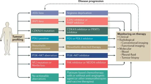
There are various genetic alterations in MPM that might be tested in biopsy samples and predict which molecularly targeted therapeutic approach is most likely to be beneficial for the patient (Figure 8).
Figure 8. There are several genetic alterations wich are proposed to have a predictive implication in targeted therapy of MPM. Reprinted with the permission of Springer Nature from [42].
Tumors harboring NF2 mutations might be targeted by FAK inhibitors, such as defactinib. However, a phase 2 clinical trial failed to prove any statistical difference in PFS, OS or quality of life between patients who after first line chemotherapy received defactinib maintenance treatment versus those who received placebo, and the result was found to be independent of NF2 mutation status [182].
Another potential target is the subgroup of ASS1 (argininosuccinate synthetase 1) deficient MPMs. The use of arginin-lowering agent ADI-PEGO20 in a phase 2 trial involving 68 patients, has provided a statistically significant improvement in median PFS (3.2 months versus 2.0 months) [177].
The anti-CTLA4 antibody tremalimumab failed to increase OS in patients pretreated with first and second line chemotherapy [179]. PD1 inhibition is studied in several clinical trials, of which the most promising so far has achieved 12-week disease control in 44% and 52% of the patients using either nivolumab or nivolumab plus ipilimumab, respectively [180]. The combination of durvalumamb and platinum plus pemetrexed chemotherapy has resulted of sufficient activity, a median PFS of 6.9 months and objective tumor response in approximately 50% of the cases [181].
To overcome the relatively immunosuppressing microenviroment typical of MPM, various immune-activating therapies have emerged and are currently tested in pilot studies involving a limited number of patients. Chimeric antigen receptor (CAR) T-cells extracted from the patients and then genetically engineered to be activated by MPM specific cell surface protein mesothelin and readministered the modified T-cells into the patients represent another novel direction that is currently being investigated in various solid malignancies [173]. Another approach is the presentation of allogenic tumor lysate to monocytes extracted from the patient and the re-injection of allogenic activated dendritic cells into the patient [178].
1.5 The molecular landscape of MPM
Molecular alterations in MPM include mutations and copy number alterations, as well as epigenetic changes. Strikingly, the most frequently involved genes are tumor suppressors and regulators of gene expression (Figure 9).
Figure 9. Frequency (%) of genetic alterations detected in MPM based on DNA- sequencing and copy number analysis of 87 samples from TCGA-MESO cohort [185].
Despite the growing number of high throughput genomic analyses and the increasing data on the molecular characteristics of MPM, no frequent oncogenic driver has been discovered to this date [188-190].
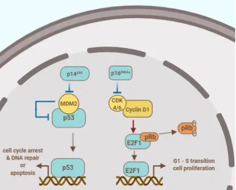
1.5.1 Cell cycle regulation pathways
The CDKN2A locus encodes the p16INK4a and p14ARF tumor suppressor proteins that are inhibitors of the cell cycle as depicted in Figure 10. The protein p16INK4a binds to cyclin dependent kinases (CDK4/6) and inhibits their kinase activity. Uninhibited CDK4/6 binds cyclin D1 and their complex phosphorylates the retinoblastoma protein (pRb) which releases the transcription factor E2F1. The latter protein promotes the transcripition of genes involved in the transition from G1 to S phase. The alternate reading frame product of CDKN2A, p14ARF inhibits MDM2, thus, activates p53 and prevents its MDM2-mediated degradation [191, 192]. The activation of transcription factor p53 results in the transcription of numerous genes involved in cell cycle arrest, senescence, apoptosis and differentiation [193].
Figure 10: The products of the CDKN2A gene p14ARF and p16 INK4a play a role in the regulation of cell cycle and apoptosis. The author’s drawing based on: [191, 193].
The loss of CDKN2A locus through homozygous deletion of 9p21 occurs in 67-83% of all MPM cases, while its frequency is up to 100% in SMM [140, 186, 191, 194, 195].
Less frequent causes for p16 inactivation are hypermethylation and point mutations of the gene CDKN2A [196, 197]. Several studies reported a strong association between the loss of CDKN2A and significantly shorter OS in MPM patients [197-199].
Recurrent mutations in the gene TP53 are relatively infrequent in MPM [186, 194], however, its reported frequency varies widely and was found to be 57% in one retrospective study [129], while only 16% by another recent study [200]. The germline mutation of the TP53 gene is associated with the Li-Fraumeni cancer syndrome. The patients carrying this type of TP53 mutations frequently develop breast cancer, carcinomas of the adrenal cortex or sarcomas, however, are only occasionally diagnosed with MPM [28].
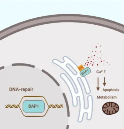
1.5.2 BAP1 and DNA damage repair
BRCA1-Associated Protein 1 (BAP1) is a deubiqitinating enzyme consisting of three main domains, namely the N-terminal ubiquitin carboxyl hydrolase domain, a middle portion containing binding sites for complex forming and a C-terminal domain also important in interactions with other proteins [44]. When located in the nucleus BAP1 acts as a tumor suppressor through regulation of the cell cycle and differentiation [201]
and plays an essential role in the repair of double strand DNA break repair through an interaction with a variety of recombination proteins, such as Breast cancer type 1 susceptibility protein (BRCA1) and BRCA1-associated RING domain protein (BARD1) [202] as shown in Figure 11. When located in the endoplasmic reticulum (ER) BAP1 modulates intracellular calcium levels and promotes apoptosis [203]. Cells with an impaired BAP1 function show reduced mitochondrial Ca2+ levels, and as a consequence are more likely to show a metabolic shift towards aerobic glycolysis [204], and are not able to initiate the apoptotic process through a Ca2+ release from the ER [28].
Figure 11. BAP1 has different functions in a nuclear localisation and in the endoplasmic reticulum. The author’s drawing based on [28].
The gene encoding BRCA1-Associated Protein 1 (BAP1) is located on chromosome 3’s short arm (3p21.1) and is one of the most frequently affected by genetic alterations in MPM. The frequency of alterations leading to the loss of BAP1 function is within a wide range between different studies, it is reported to occur in 23-63% of MPM cases [194, 200, 205-207]. The mechanisms of the inactivation of BAP1 include loss-of- function mutations, copy number loss of chromosome 3p21 and gene fusions [206].
Hotspot regions of the BAP1 gene with genetic alterations are exon 13 and 17, where a study identified variations in 38% and 25% of the patients, respectively [129]. The loss of nuclear localization of the BAP1 protein detected by immunohistochemistry correlates with the nonsynonimous variations of the BAP1 gene identified by next generation sequencing [129].
1.5.3 Hippo pathway
The Hippo pathway is a highly conserved signaling pathway that plays a regulatory role in organ growth, tissue regeneration and preventing tumorigenesis through restraining the cell cycle, controlling cellular differentiation and promoting apoptosis.
Figure 12. Main actors of the Hippo pathway. The author’s drawing based on [208]
The kinase cascade of the Hippo pathway (Figure 12) include the MST1/2-SAV1 complex that activates the LATS1/2-MOB1A/B complex through phosphorylation, which then phosphorylates the YAP/TAZ complex. When phosphorylated, the nuclear effector YAP/TAZ transcriptional coactivators are excluded from the nucleus and thus inactivated. When the activity of the Hippo pathway is low, YAP/TAZ is able to enter the nucleus, where it interacts with transcription-enhancer activator domain transcripition factor (TEAD) and activates the transcription of several target genes involved in cell proliferation and the evasion of apoptosis. Upstream regulators of the
Among these regulators, NF2/Merlin and KIBRA are cooperating proteins located at the apical membrane of cells that interact with LATS1/2 and through the activation of the Hippo pathway mediate contact inhibition in cell cultures [208].
In MPM the loss of function alterations of genes NF2 and LATS1/2 occur relatively frequently, while alterations of MST1 and SAV1 are also reported [206]. The inactivation of the negative regulators of YAP/TAZ complex leads to the constitutive activation of the complex [209]. Although oncogenic alterations of the YAP1 and WWTR1 gene (encoding TAZ) are relatively frequent in triple-negative breast cancers, non-small cell lung cancer, it is a rare occurrence in MPM [210, 211].
The frequency of genetic abnormalities affecting the NF2 gene is reported to be 14- 50%, mostly being missense, nonsense or splice site mutations, and less commonly losses of chromosome region 22q12 encoding NF2 [186, 194, 200, 205, 206, 212].
Alterations of the NF2 locus is reported to occure significantly more often in patients not exposed to asbestos [190].
Neurofibromatosis type 2 is associated with germ-line mutations in the NF2 gene, but this autosomal dominant disease is not associated with increased risk to develop MPM, even though there is an overlap between somatic mutations detected in MPM and those in hereditary neurofibromatosis [213].
The loss of function alterations of tumor suppressor LATS2 occur through the homozygous deletion of 13q12 encoding the LATS2 gene, which was reported to occur in 10 out of 45 MPM samples [214], or through somatic mutations of the LATS2 gene [206, 215]. The loss of LATS1 function occurs most frequently through a chromosomal translocation which leads to the fusion of the LATS1 and PSEN1 (presenilin-1) genes, and the fusion protein product lacks the kinase activity which is essential in the inhibition of YAP [215].
Other altered pathways include the mTOR, histone methylation and RNA helicases signaling pathways, which, however, occur in a small fraction of MPM cases [206].
1.6 Telomere and telomerase 1.6.1 Structure and function
The telomere region of eukaryotic chromosomes is located at the extremes of the chromosomes and is “capped” by a large nucleoprotein complex that prevents breakdowns and fusions between chromosome ends during mitosis [216]. Its DNA component contains several kb of the repetitive sentence d(TTAGGG) in humans [217].
Telomeric DNA is characterized by the protruding extreme of the G-rich strand, which is approximately 200 nucleotides long and is a consequence of the mechanism of terminal replication [218]. The overhang produced at the end of the lagging strand might form G-quadruplexes [219] or a T-loop which is a circle of curled-up single strand DNA forming a triple-strand structure at the very end, called displacement loop [220] (Figure 13A).
Figure 13. Telomeric region of human chromosomes. (A) G overhang forms a protective T-loop and through invading the dsDNA to form a D-loop. (B) Shelterin complex of telomere binding proteins protecting the chromosomes’ ends from triggering a DNA damage response. The telomerase complex consisting of the protein TERT and template RNA TERC components recognizes the 3’-end of the single strand G overhang
The telomeric region is bound by the shelterin complex in human cells that consists of six proteins, namely TRF1 and TRF2, RAP1, TIN2, TPP1 and POT1. TRF1 and TRF2 both need to build dimers to be able to bind to 5’-YTAGGGTTR-3’ sequences of double strand telomeric DNA. POT1 interacts with single strand G overhang at 5’- TAGGGTTAG-3’ sequences and interacts with the TRF1 and TRF2 homodimers through proteins RAP1, TIN2 and TPP1 (Figure 13B).
During cell division the length of the telomere decreases at each passage of the replication fork due to the inability of conventional DNA polymerases to fully duplicate the 3’ end of linear DNA molecules [222]. Telomerase plays an essential role in maintaining chromosomal integrity by preventing the loss of genetic material caused by incomplete terminal replication and compensating for the shortening of the telomere region through de novo addition of TTAGGG repeats. The telomerase enzyme complex is a reverse transcriptase containing the catalytic subunit TERT encoded by the gene hTERT in humans and the RNA template component TERC [221] (Figure 13B).
Telomerase is physiologically expressed in a strictly regulated manner in germ cells and stem cells, but its activity is restrained in somatic cells [216]. Telomere repression is a mechanism for the prevention of uncontrolled cellular proliferation. In cells lacking telomerase expression the erosion of the telomere leads to the activation of DNA damage response pathway and cells enter senescence [223]. TERT also plays telomere- independent roles both in cooperation with the TERC RNA template and independently of that. Such functions of TERT include the regulation of targets of the Wnt-pathway, the genesis of double strand precursors of silencing RNAs, and the maintenance of mitochondrial fitness [224].
1.6.2 Telomere lengthening and telomerase in disease
Telomere shortening can lead to impaired tissue regeneration and accelerated aging. On the other hand, constitutive expression of the telomerase permits uninhibited cell division and immortalization but is also associated with increased chromosomal instability [225].
The constitutive expression of the TERT gene and telomerase activity is detected in 85- 90% of all malignant tumors [223, 226], and is considered a hallmark of cancer
[227].There are several mechanisms underlying the telomerase reactivation and telomere lengthening in malignant cells.
Abnormal expression of positive regulators of the TERT gene such as the oncogene MYC induce TERT expression and leads to an increased telomerase activity [228, 229].
Epigenetic factors might also lead to an increase in TERT activity. The TERT promoter is generally not methylated in normal cells, however, hypermethylation of the promoter at the TERT hypermethylation oncological region (THOR) occurs in malignant cells and accounts for upregulation of TERT expression [230]. SMYD3 regulated histone H3-K4 trimethylation are factors leading to constitutive activation of the telomerase [231], as well as the recruitment of histone acetyltransferases or histone deacetylases that might cause telomere reactivation depending on the cellular context [232]. Viruses such as Epstein-Barr virus, cytomegalovirus, human papilloma virus, hepatitis B and C encode exogenous positive regulators of hTERT [233].
Telomerase-independent, recombination-based mechanisms of telomere maintenance, the so called alternative lengthening of telomeres (ALT) are reported in several human malignancies, and are associated with the loss of ATP-dependent helicase encoded by ATRX or the H3.3-specific histone chaperone DAXX both of which would otherwise repress ALT [234]. The loss of ATRX and DAXX function occurs most commonly in pancreatic neuroendocrine tumors [235] sarcomas [236, 237] and childhood glioblastomas [238].
According to recent data, rearrangements and focal amplifications of the TERT gene are relatively rare. Amplifications occur in approximately 4% of malignancies, but it is more common in lung adenocarcinomas and squamous cell carcinomas, as well as in ovarian cancer, adrenocortical and esophagus carcinomas [239]. Rearrangements have been identified in high-risk neuroblastomas, however do not seem to play a crucial role in telomerase derepression in other cancer types [240, 241].
![Table 1. Definitions of T, N and M categories according to the IASLC proposal for the 8 th edition of TNM, reprinted from [83]](https://thumb-eu.123doks.com/thumbv2/9dokorg/1373941.112780/19.892.127.752.652.1105/table-definitions-categories-according-iaslc-proposal-edition-reprinted.webp)

![Figure 7. Summary of novel therapeutic approaches for MPM management. Reprinted with the permission of Elsevier from [184]](https://thumb-eu.123doks.com/thumbv2/9dokorg/1373941.112780/33.892.130.764.126.616/figure-summary-therapeutic-approaches-management-reprinted-permission-elsevier.webp)
![Figure 12. Main actors of the Hippo pathway. The author’s drawing based on [208]](https://thumb-eu.123doks.com/thumbv2/9dokorg/1373941.112780/39.892.228.662.316.740/figure-main-actors-hippo-pathway-author-drawing-based.webp)
