ORIGINAL ARTICLE
Comparison of Nottingham Prognostic Index, PREDICT
and PrognosTILs in Triple Negative Breast Cancer – a Retrospective Cohort Study
Anita Sejben1 &Tibor Nyári2&Tamás Zombori1&Gábor Cserni1,3
Received: 28 May 2020 / Accepted: 10 June 2020
#The Author(s) 2020
Abstract
Triple-negative breast cancer (TNBC) represents a heterogenous subtype of breast cancer with generally poor prognosis. The prediction of its prognosis remains essential to clinicians in their therapeutical decision-making process. The aim of our study was to compare the validity of three multivariable analysis derived prognostic systems, the Nottingham Prognostic Index (NPI), PREDICT and PrognosTILs (a prognosticator including tumor infiltrating lymphocytes, TILs) in a series of TNBCs. Patients operated on with TNBC at the Department of Surgery, Bács-Kiskun County Teaching Hospital, Kecskemét between 2005 and 2016 were included. Clinical and pathological parameters and follow-up data were collected from medical charts. TILs were assessed retrospectively, following international recommendations. Estimated survivals of PrognosTILs, PREDICT and NPI were recorded and compared with real outcomes. Altogether 136 patients were included in this retrospective study. In univariate Cox analysis, type of surgery, pT, pN, stage, NPI and type of adjuvant therapy were the significant prognostic variables. The multivariate Cox-regression strengthened that NPI is an independent predictor of overall and disease-free survivals in TNBCs.
The NPI, PREDICT and PrognosTILs could be compared directly only in a ROC curve analysis: the sensitivities and specificities of these predicting systems are rather similar with area under the curve values falling between 0.7 and 0.8, and NPI having the highest values. Our findings reflect the diverse prognosis of TNBC and highlight the difficulties of predicting its outcome. None of the three multivariable prognosticators is inferior to the others, the NPI can reliably be used for TNBCs.
Keywords Triple negative breast cancer . Nottingham Prognostic Index . Predict . Tumor infiltrating lymphocytes . PrognosTILs . Prognosis
Introduction
Triple-negative breast cancer (TNBC) represents a heteroge- neous subtype of breast cancer (BC) defined by the lack of immunohistochemical expression of estrogen receptor (ER), progesterone receptor (PR) and human epidermal growth fac- tor receptor-2 (HER2), and by variable though distinct molec- ular profiles [1,2]. Epidemiological data on TNBC revealed
its higher prevalence among women of African ancestry, young BC patients and patients with Breast Cancer Gene-1 (BRCA-1) mutations [3]. The treatment of TNBC remains a challenge for clinicians due to its poor overall prognosis.
Distant hematogenous metastasis formation and local recur- rence are frequent and the treatment efficiency of TNBC is lower than in other types of BC [1,4,5]. By taking molecular profiles and BRCA deficiency into account, more personal- ized treatment methods are currently available [6]. Besides chemo- and radiotherapy, the role of immuno- and targeted therapy is increasing, both being currently under investigation with promising results [7–9].
Prediction of prognosis remains essential to clinicians in their decision-making process, helps stratifying patients by risk and better allows preparing individual treatment plans [10]. Various prognostic factors have already been presented in TNBC. Ovcaricek and coauthors described nodal status and age as independent prognostic factors for disease-free survival
* Anita Sejben
sejben.anita@gmail.com
1 Faculty of Medicine, Department of Pathology, University of Szeged, Állomás u. 1, Szeged 6725, Hungary
2 Department of Medical Physics and Informatics, University of Szeged, Szeged, Hungary
3 Bács-Kiskun Teaching County Hospital, Kecskemét, Hungary https://doi.org/10.1007/s12253-020-00846-8
/ Published online: 20 June 2020
(DFS), whereas for overall survival (OS), only nodal status proved to be an independent factor [11]. Urru et al. have dem- onstrated that tumor stage at diagnosis and positive lymph node ratio are relevant predictors of survival and tumor recur- rence, with the addition of Ki-67 status for recurrence predic- tion [12]. Asaga and coworkers have used a different ap- proach, and analyzed clinical response to preoperative system- ic chemotherapy [13].
The Nottingham Prognostic Index (NPI) was described by Haybittle and coauthors in 1982 and it was originally designed for primary operable BC. It takes tumor size, nodal stage and tumor histological grade into consideration [14]. On the basis of its equation and the values of the NPI, patients’could be divided into three prognostic categories according to the orig- inal article: Category I (good prognosis); Category II (moder- ate prognosis) and Category III (poor prognosis) [14,15].
Later the prognostic groups were subdivided to form the very good, the good, the moderate I, the moderate II, the poor and the very poor prognostic groups [16]. Different cut-off values and diverse definitions of NPI-based groups (ranging from three to ten classes) have been used by some research groups [10]. The NPI has been proven to be a valid prognostic tool in BC treatment [17].
A more complex prognostic model, PREDICT was pub- lished by Wishart and coauthors in 2010. The algorithm was developed from 5694 patients’data from the Eastern Cancer Registration and Information Centre. The selected patients were operated on for invasive breast cancer. Based on the factors that were found to hold independent prognostic value, an algorithm was established that includes the presence of ductal carcinoma in situ (DCIS) or lobular carcinoma in situ (LCIS), age at diagnosis, menopausal state, ER, PR, HER2 and Ki-67 status, invasive tumor size, tumor grade, method of tumor detection and number of positive lymph nodes [18].
PREDICT is also endorsed by the American Joint Committee of Cancer [19]. The on line calculator estimates OS for 5, 10 and 15 years. Although the tool generally re- ceived good ratings for validity, Maishman and coauthors’
results showed that PREDICT was a great tool only in long- term survival estimates, and overestimated short-time sur- vivals, especially in ER-positive tumors [17,20,21].
Tumor infiltrating lymphocytes (TILs) reflect prognosis in TNBC, since their higher proportion correlates with better outcome in this subset of breast tumors, and indicates the prominent role the immune system plays in TNBC. While TNBCs lack targeted therapy, the interest for immune modu- lators has increased [22,23]. Loi and coworkers conducted a pooled analysis of 2148 patients and identified the following factors that independently influence the prognosis of primary TNBCs: percentage of stromal TILs, age, tumor size, number of positive lymph nodes, histological grade and treatment.
Invasive disease-free survival (i-DFS), distant disease-free survival (d-DFS) and OS results were examined in 3 and 5 year
intervals [24]. Based on the results, an equation was devel- oped for survival estimates. For easier utilization, an online tool named PrognosTILs was developed for early stage TNBCs [25]. With this application, the 5-year and 10-year OS and DFS estimates can be calculated.
The aim of our study was to compare the validity of NPI, PREDICT and PrognosTILs in a series of TNBC cases.
Materials and Methods
Patients operated on for histologically verified triple negative, invasive breast carcinoma at the Department of Surgery, Bács- Kiskun County Teaching Hospital, Kecskemét between 2005 and 2016 were included in our consecutive and retrospective study. Follow up data (OS and DFS) were collected from medical charts. For these outcomes, patients were followed from the date of surgical treatment until the time of recurrence or tumor-related death; those alive without recurrence and those dying from other causes were censored at the time of the last follow-up and death, respectively.
The following clinical and pathological variables were ob- tained for analysis: age, gender, localization, type of surgical and adjuvant treatments, histological type and grade of cancer, vascular invasion, tumor size, pT and pN categories, and stage. The NPI was calculated with the following equation:
NPI = tumor size (cm) × 0.2 + nodal score (1 for pN0, 2 for pN1, 3 for pN2 or pN3) + number value from the histological grade [14]. The Nottingham Prognostic Groups were classi- fied as excellent (EPG): ≤2.4; good (GPG): 2.41–3.4;
moderate-1 (MPG1): 3.41–4.4; moderate-2 (MPG2): 4.41–
5.4; poor (PGP): 5.41–6.4 and very poor (VPPG):≥6.41 [16].
The predicted OS and DFS estimates of PrognosTILs were obtained from an online calculator [23,25]. The estimations were based on the following parameters: age, number of pos- itive lymph nodes, tumor size, histological grade, type of che- motherapy and proportion of stromal TILs. For the determi- nation of the latter, the International TILs Working Group (later acting as International Immunooncology Biomarker Working Group - IIOBWG) recommendations and rules were used [25,26]. To help in the estimation of stromal TILs, the online calibration system described by the IIOBWG and found at was also used [27,28]. After getting accustomed with the scoring system with a hundred cases evaluated in a study by the European Working Group for Breast Screening Pathology, the calibration (etalon) pictures for different rates of stromal TILs were screensaved and printed, and these printed pictures were compared with the microscopic images displayed on a monitor for at least three areas. The mean of these estimates was rounded to the closest 10% value also allowing for 5% and 1%, with the help of the calibration picture published in the first article of the IIOBWG for the latter value [29].
The anticipated OS evaluations of PREDICT were deter- mined with the online calculator, that required the following data: age, menopausal state, ER status, HER-2 status, Ki67 status, size of invasive tumor, grade of tumor, type of detec- tion, number of positive lymph nodes and presence of micrometastasis in the lymph nodes [18,19].
The Wilcoxon rank sum test was applied to analyze the correlation between recurrence or tumor-specific death and DFS or OS prediction rate of PrognosTILs and OS prediction rate of PREDICT. The OS and DFS data could not be correlat- ed directly with the survival predictions of PrognosTILs and PREDICT, therefore the patients were classified in the follow- ing four categories: patients alive, patients who died of disease (DOD), patients alive with and without recurrence. The calcu- lated OS and DFS survival predictions of PrognosTILs, the OS survival estimates of PREDICT and NPI scores were correlated with the 4 categories by receiver operating characteristic (ROC) curve analysis aiming to compare them and to find cut-off points. Patients DOD and patients alive categories were utilized in ROC curve analysis focusing on 5-year-OS prediction of PrognosTILs, PREDICT and NPI scores, while patients with recurrence and patients without recurrence categories were used in a ROC curve of 5-year-DFS estimates of PREDICT and NPI scores. The cut-off points identified by ROC curve analysis could show which OS and DFS rates of PrognosTILs, OS es- timates of PREDICT and NPI scores are related to more fre- quent recurrence and tumor-specific death, respectively.
NPI was analyzed with the Kaplan-Meier method and the subgroups were compared with the log rank test. Cox- regression was utilized as univariate analysis. The parameters found significant in the univariate models were entered in a multivariable Cox proportional hazard model to identify factors of independent prognostic significance. PrognosTILs and PREDICT survival estimates could not be included in the mul- tivariate analysis due to statistical reasons. Statistical models were fitted using SPSS Statistics V.23.0 software (IBM, SSPS 22.0, Armonk, NY USA). All statistical tests were two-sided and p< 0.05 values were considered statistically significant.
This retrospective study was approved by the institutional ethical committee of the Albert Szent-Györgyi Clinical Centre of the University of Szeged and the ethical committee of Bács- Kiskun County Teaching Hospital also gave a consent for the study.
Table 1 Clinical and pathological characteristics of patients evaluated and the results of univariate Cox-regression [pT, pN categories defined by AJCC [27: Amin-AJCC], CMF: cyclophosphamide, methotrexate and 5- fluorouracil; second generation systemic treatment refers to anthracycline based regimens without taxanes; third generation refers to taxane contain- ing regimens]
pOS pDFS
Age (years) n % p= 0.102 p= 0.207
30–39 12 9.5
40–49 15 11.9
50–59 37 29.3
60–69 35 27.8
70–79 21 16.7
80–91 6 4.8
Laterality p= 0.645 p= 0.958
Right 58 46.0
Left 68 54.0
Type of surgery p= 0.354 p = 0.017
Mastectomy 24 19.0
Breast conserving surgery 102 81.0
Histology diagnosis p= 0.626 p= 0.566
Carcinoma of no special type (NST) 112 88.8
Medullary carcinoma 7 5.6
Other 7 5.6
Grade p= 0.967 p= 0.88
2 5 4.0
3 121 96.0
pT p= 0.222 p= 0.009
pT1 67 53.1
pT2 55 43.7
pT3 1 0.8
pT4 3 2.4
pN p= 0.006 p < 0.001
pN0 75 59.6
pN1mi 8 6.3
pN1 31 24.6
pN2 9 7.1
pN3 2 1.6
pNx 1 0.8
Vascular invasion p= 0.573 p= 0.400
Absent 100 79.4
Present 26 20.6
Stage p= 0.05 p < 0.001
I 47 37.3
II 51 40.5
III 27 21.4
no data 1 0.8
Adjuvant therapy p= 0.151 p= 0.003
Chemotherapy 10 7.9
Radiotherapy 15 11.9
Both 85 67.5
Neither 16 12.7
Table 1 (continued)
pOS pDFS
Generation of chemotherapy p= 0.092 p= 0.303
Second generation 16 12.7
Third generation 73 57.9
Other (CMF) 6 4.8
No data 31 24.6
Results
Altogether, 136 patients who underwent surgical resection were included in our study. Ten patients (7.4%) were censored due to non-tumor related death. Tumor-specific death was found in 23 cases (16.9%), while 103 patients (75.7%) were alive at the last follow up, including 20 patients with recurrence (14.7%). The mean and median OS and DFS were 66.8 months and 57.5 months, 59.9 months and 41 months, respectively (range for OS: 7–170 months; range for DFS: 2–170 months).
Recurrence was observed in 43 cases, including 11 cases (25.6%) with local or regional recurrence, 23 cases (53.5%) with distant metastasis and two cases with both local and distant types of recurrence. The median time to recurrence was 41 months (range: 2–170 months) Novel malignancies were found in 3 cases (7.0%; ovary [n= 1] and lung cancer [n= 2]). The median follow up was 56 months (range: 7–170 months).
The basic clinical and pathological characteristics are displayed in Table1[30]. The mean and median age of the patients were 59.6 and 59 years, respectively (range: 32–91).
In univariate Cox-regression, the type of surgery, the pT and pN categories, the stage of the disease and the type of adjuvant therapy were found to be significant variables.
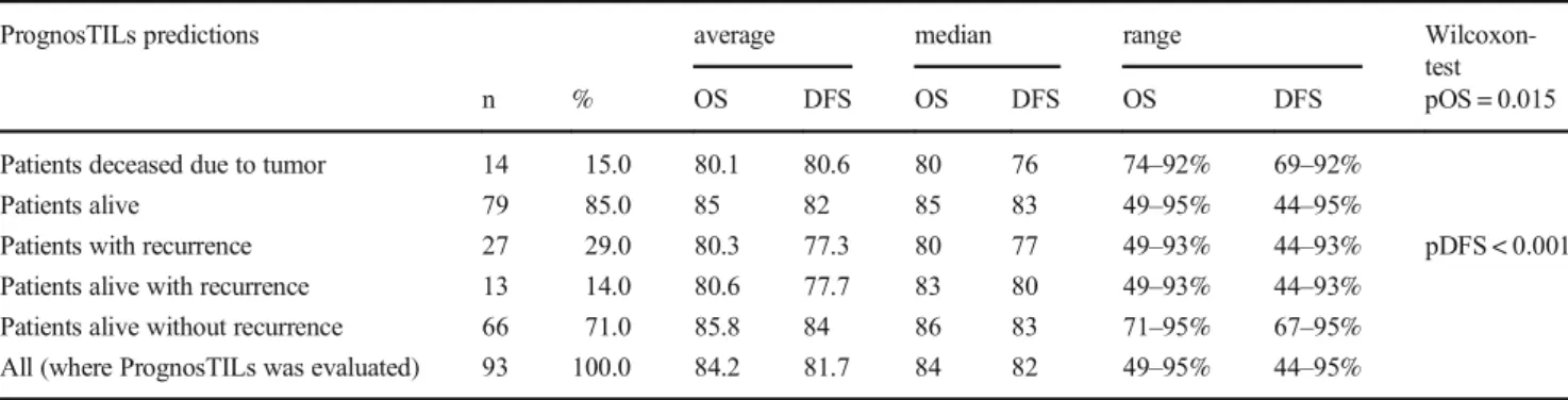
The predictions from PrognosTILs and PREDICT and the NPI scores were established in 93, 126 and 125 cases, respec- tively. Concerning the 5-year-OS and -DFS predictions of PrognosTILs, the mean, the median and the range of estimates are presented in Table2. The comparison of predicted survival estimates and outcomes revealed that the predicted OS estimates
of the patient DOD were significantly lower than those of pa- tients who were alive (p= 0.015); similarly, the predicted DFS estimates of patients with recurrence were significantly lower, than those of patients without recurrence (p< 0.001). Table3 highlights the mean, the median and the range of the 5-year- OS estimates of PREDICT. The statistical analysis strengthened, that the predicted OS estimates of patient DOD were significant- ly lower, than those of patients who were alive (p= 0.020).
The NPI-based GPG included only 3 cases, therefore this group was excluded from further evaluation. Figure1demon- strates the results of Kaplan-Meier analysis of the NPI subgroups.
Significant differences were detected between OS and DFS esti- mations of different prognostic groups, namely the OS estimates of MPG1 vs. PPG (p= 0.017), MPG1 vs. VPPG (p= 0.049), MPG2 vs. PPG (p= 0.026); and the DFS estimates of PPG vs.
MPG1 (p= 0.002), PPG vs. MPG2 (p= 0.035), PPG vs. VPPG (p= 0.013), VPPG vs. MPG1 (p< 0.001) and VPPG vs. MPG2.
(p= 0.001). In the univariate Cox-regression, NPI was found to be a significant prognostic variable (pOS = 0.022; HR:1.71, 95%CI:1.08–2.72; pDFS<0.001; HR:2.02, 95%CI:1.43–2.86).
Figure2displays the results of ROC curve analysis focus- ing on 5-year-OS estimates of PrognosTILs, PREDICT and NPI scores. The area under the curve (AUC) of PrognosTILs, PREDICT and NPI were 0.759, 0.762 and 0.792, respectively.
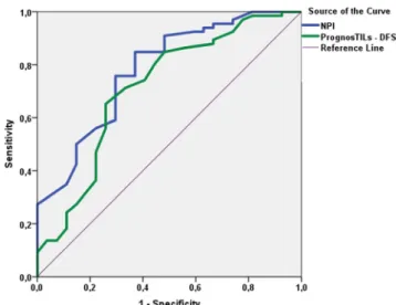
Figure3demonstrates the ROC curve analysis of 5-year-DFS estimates of PrognosTILs and NPI scores. The AUC values of PrognosTILs and NPI were 0.713 and 0.781, respectively.
The findings of ROC curve analyses drew attention to the similarities of these predictive systems concerning sensitivity Table 2 The 5-year overall survival (OS) and disease-free survival (DFS) predictions of PrognosTILs according to outcome. Significant differences were detected between OS predictions of patients who died of disease and patients alive, and DFS predictions of patients with and without recurrence
PrognosTILs predictions average median range Wilcoxon-
test
n % OS DFS OS DFS OS DFS pOS = 0.015
Patients deceased due to tumor 14 15.0 80.1 80.6 80 76 74–92% 69–92%
Patients alive 79 85.0 85 82 85 83 49–95% 44–95%
Patients with recurrence 27 29.0 80.3 77.3 80 77 49–93% 44–93% pDFS < 0.001
Patients alive with recurrence 13 14.0 80.6 77.7 83 80 49–93% 44–93%
Patients alive without recurrence 66 71.0 85.8 84 86 83 71–95% 67–95%
All (where PrognosTILs was evaluated) 93 100.0 84.2 81.7 84 82 49–95% 44–95%
Table 3 The basic characteristics of 5-year overall survival (OS) predictions of PREDICT according to outcome. The survival estimates of patients dying of tumor progression were lower than those of patients who were alive at last follow up
PREDICT estimates Wilcoxon-test
n % mean median range pOS = 0.020
Patients deceased due to tumor 23 18.3 62.9 65.5 9.2–85.1%
Patients alive 103 81.7 71.8 78.1 7.1–86.5%
All (where PREDICT was evaluated) 126 100 70.1 75.3 7.1–86.5%
and specificity and to the fact that they are not ideal for defin- ing cut-off values.
The multivariate Cox proportional hazard model revealed that among the variables found significant in univariate models (type of surgery, pT, pN, stage, adjuvant therapy and NPI), only NPI was an independent prognostic marker for triple negative breast cancer (pOS = 0.006; HR:1.66, 95%CI:1.16–2.37;
pDFS<0.001; HR:1.92, 95%CI:1.46–2.53).
Discussion
TNBCs are generally considered as the worst IHC based mo- lecular subtype of breast cancer, owing to their poor prognosis
and the limited therapeutic success associated with them.
Despite the overall bad prognosis of TNBC, there are some tumors that by definition fall into this category, but belong to a better prognostic group. These include rare tumors like tall cell carcinoma with reversed polarity, secretory carcinoma, non-high grade, i.e. classical adenoid cystic carcinoma [31–33]. Even without these low grade special type carcino- mas, the prognosis of TNBC is heterogeneous and depends on a number of prognostic factors.
The presence of distant metastasis, nodal status, tumor size and histological grade are established prognostic factors of breast carcinomas, and have their role in predicting the out- come of TNBCs as well. More recently the proportion of stromal TILs has also been recognized as an independent Fig. 1 Kaplan-Meier analysis of NPI. According to the log rank test
significant differences were observed between the overall survival (OS) estimates of MPG1 vs. PPG (p= 0.017), MPG1 vs. VPPG (p= 0.049) andMPG2 vs. PPG (p= 0.026); and the disease-free survival (DFS) esti- mates of PPG vs. MPG1 (p= 0.002), PPG vs. MPG2 (p= 0.035), PPG vs.
VPPG (p= 0.013), VPPG vs. MPG1 (p< 0.001) and VPPG vs. MPG2 (p= 0.001) [MPG1: Moderate Prognostic Group 1, MPG2: Moderate Prognostic Group 2, PPG: Poor Prognostic Group, Very Poor Prognostic Group]
Fig. 2 ROC curve analysis of 5-year overall survival predictions of TIL, PREDICT and NPI scores (area under the curve values for TIL, PREDICT and NPI were 0.759, 0.762 and 0.792, respectively)
Fig. 3 ROC curve analysis of 5-year disease-free survival predictions of PrognosTILs and NPI scores (area under the curve values for TIL and NPI were 0.713 and 0.781, respectively)
prognosticator of TNBCs [24], and the prognostic value of TILs was also found in a more recent meta-analysis [34].
When prognostic factors show divergent features, i.e. clini- cians are faced with a combination of factors toward good and bad prognosis, predictive models based on multivariable analysis of multiple prognostic factors are much more valu- able than isolated factors. The NPI is one such factor and was derived from the multivariable analysis of 387 patients with different molecular subtypes of breast cancer and was later validated in a series of 320 independent consecutive cases [35]. Several external studies have demonstrated its ability to give a prognostic classification of breast carcinomas [36–38].
Although the improvements in treatment have significantly altered the outcomes of breast cancer, and this improvement is also reflected in the NPI prognostic group-specific sur- vivals, the prognostic separation of breast cancers on the basis of the NPI was still found to be valid [39]. The PREDICT tool was derived from a much greater population and was also independently validated in a number of reports [17, 40].
PrognosTILs is a novel multivariable prognosticator model and calculator derived from the pooled analysis of 2148 indi- vidual patients’data from 9 studies on TNBCs proving the prognostic value of stromal TILs in the adjuvant setting [24].
This distinguishes it from NPI and PREDICT which were built on data from ER-positive and ER-negative tumors to- gether, and theoretically could mean that it is better fitted to predict the prognosis of TNBCs.
The significance of the NPI in TNBC was first examined by Albergaria and coauthors in 2001 with reassuring results.
NPI results correlated well with real survival data due to the facts that TNBCs are frequently high grade and large tumors [41]. PREDICT, to our knowledge has not yet been evaluated for TNBCs alone, whereas PrognosTILs is relatively recent for larger validation on comparison studies.
In univariate Cox analysis, type of surgery, pT, pN, stage, NPI and adjuvant therapy were found significant prognostic variables. We also found that lower 5-year OS and DFS pre- dictions of PognosTILs are related with more frequent tumor specific death and recurrence (pOS = 0.015, pDFS<0.001), while the lower 5-year OS predictions of PREDICT are as- sociated with higher rate of tumor specific death (p= 0.02).
Concerning the NPI, we demonstrated that there are signifi- cant differences among OS and DFS estimates of certain prognostic groups (Fig.1). PrognosTILs and PREDICT de- rived estimates of survival, as scale variables could not enter the Kaplan-Meier analysis. The direct comparison of the mul- tivariable prognosticators was performed with ROC curve analysis. Regarding the OS follow up data, PrognosTILs, PREDICT and NPI, while regarding the DFS follow up data, PrognosTILs and NPI were compared. All three predictors of outcome reflect fair performance with areas under the ROC curves falling between 0.7 and 0.8. The sensitivity and spec- ificity of these predicting systems are rather similar, although
there seems to be a tendency for NPI values to better predict outcome on the basis of the somewhat greater AUC values. In keeping with the results of Albergaria et al., the multivariate Cox-regression strengthened that NPI is an independent pre- dictor of OS and DFS in TNBCs (pOS = 0.006; HR:1.66, 95%CI:1.16–2.37; pDFS<0.001; HR:1.92, 95%CI:1.46– 2.53) [41]. Considering that the ROC curve analysis yielded similar results for the three multivariable prognosticators studied, it can be inferred that any of these is suitable to predict the outcome of TNBCs, and none of these is inferior to the others.
The results also show that TNBCs are prognostically het- erogeneous. No case was classified as of very good prognosis on the basis of the NPI, and only 3 cases fell into the good prognostic group. This is due to the fact that only 5 tumors were of histologic grade 2, whereas the remaining were high grade, and with this combination, their NPI value was imme- diately >4.
The lack of all prognostic markers for all cases and the fact that this was a single institution study of retrospective nature with limited number of cases are possible limitations of this work. A further limitation may be that values predicted by PrognosTILs and PREDICT, due to statistical reasons, could not be entered into the multivariate Cox-regression analysis, and could not be compared to NPI in this setting; but this drawback was compensated by the ROC curve analysis of the three prognosticators. Our study has strengths, as well.
To our knowledge, this study is the first to evaluate the value of PREDICT in TNBCs, and these multivariable prognostic tools have never been compared in a single study. Another advantage of the study design was the uniform evaluation of TILs with rigorous adherence to internationally agreed guidelines.
In conclusion, our findings reflect the diverse nature of TNBC and highlight the difficulties of predicting the outcome of this disease. Although the NPI seemed to give somewhat higher AUC values in the direct comparisons with PREDICT and PrognosTILs, none of the multivariable prognosticators is inferior to the others according to our data.
Authors’Contributions All authors contributed to the study conception and revision. Data collection was performed by Anita Sejben, Tamás Zombori and Gábor Cserni. Statistical analysis was performed by Tibor Nyári and Tamás Zombori. The first draft of the manuscript was written by Anita Sejben, Tamás Zombori and Gábor Cserni and all authors commented on previous versions of the manuscript. All authors read and approved the final manuscript.
Funding Information Open access funding provided by University of Szeged.
Data Availability The datasets generated during and/or analyzed during the current study are available from the corresponding author on reason- able request.
Compliance with Ethical Standards
Consent to Participate Not applicable.
Consent for Publication Not applicable.
This retrospective study was approved by the institutional ethical committee of the Albert Szent-Györgyi Clinical Centre of the University of Szeged.
Conflict of Interest The authors declare that they have no conflict of interest.
Code Availability There are no restrictions on the availability of mate- rials, data and code.
Open Access This article is licensed under a Creative Commons Attribution 4.0 International License, which permits use, sharing, adap- tation, distribution and reproduction in any medium or format, as long as you give appropriate credit to the original author(s) and the source, pro- vide a link to the Creative Commons licence, and indicate if changes were made. The images or other third party material in this article are included in the article's Creative Commons licence, unless indicated otherwise in a credit line to the material. If material is not included in the article's Creative Commons licence and your intended use is not permitted by statutory regulation or exceeds the permitted use, you will need to obtain permission directly from the copyright holder. To view a copy of this licence, visithttp://creativecommons.org/licenses/by/4.0/.
References
1. Aysola K, Desai A, Welch C, Xu J, Qin Y, Reddy V, Matthews R, Owens C, Okoli J, Beech DJ, Piyathilake CJ, Reddy SP, Rao VN (2013) Triple negative breast cancer–an overview. Hereditary Genet Suppl 2:001.https://doi.org/10.4172/2161-1041.S2-001 2. WHO Classification of Tumours Editorial Board (ed) (2019) WHO
classification of tumours, 5thedition–breast tumours. International Agency for Research on Cancer, Lyon
3. Siddharth S, Sharma D (2018) Racial disparity and triple-negative breast cancer in african-american women: a multifaceted affair be- tween obesity, biology, and socioeconomic determinants. Cancers (Basel) 10:514.https://doi.org/10.3390/cancers10120514 4. Anders C, Carey LA (2008) Understanding and treating triple-
negative breast cancer. Oncology 22:1233–1243
5. Al-Mahmood S, Sapiezynski J, Garbuzenko OB, Minko T (2018) Metastatic and triple-negative breast cancer: challenges and treat- ment options. Drug Deliv Transl Res 8:1483–1507.https://doi.org/
10.1007/s13346-018-0551-3
6. Park JH, Ahn JH, Kim SB (2018) How shall we treat early triple- negative breast cancer (TNBC): from the current standard to up- coming immuno-molecular strategies. ESMO Open 3(3):e000357.
https://doi.org/10.1136/esmoopen-2018-000357
7. Wahba HA, El-Hadaad HA (2015) Current approaches in treatment of triple-negative breast cancer. Cancer Biol Med 12:106–116.
https://doi.org/10.7497/j.issn.2095-3941.2015.0030
8. He MY, Rancoule C, Rehailia-Blanchard A, Espenel S, Trone JC, Bernichon E, Guillaume E, Vallard A, Magné N (2018) Radiotherapy in triple-negative breast cancer: current situation and upcoming strategies. Crit Rev Oncol Hematol 131:96–101.
https://doi.org/10.1016/j.critrevonc.2018.09.004
9. Lebert JM, Lester R, Powell E, Seal M, McCarthy J (2018) Advances in the systemic treatment of triple-negative breast cancer.
Curr Oncol 25:S142–S150.https://doi.org/10.3747/co.25.3954 10. Fong Y, Evans J, Brook D, Kenkre J, Jarvis P, Gower-Thomas K
(2015) The Nottingham prognostic index: five- and ten-year data for all-cause survival within a screened population. Ann R Coll Surg Engl 97:137–139.https://doi.org/10.1308/003588414X14055925060514 11. Ovcaricek T, Frkovic SG, Matos E, Mozina B, Bostnar S (2011)
Triple negative breast cancer– prognostic factors and survival.
Radiol Oncol 45:46–52.https://doi.org/10.2478/v10019-010-0054-4 12. Urru SAM, Gallus S, Bosetti C, Moi T, Medda R, Sollai E, Murgia
A, Sanges F, Pira G, Manca A, Palmas D, Floris M, Asunis AM, Atzori F, Carru C, D’Incalci M, Ghiani M, Marras V, Onnis D, Santona MC, Sarobba G, Valle E, Canu L, Cossu S, Bulfone A, Rocca PC, De Miglio MR, Orrù S (2018) Clinical and pathological factors influencing survival in a large cohort of triple-negative breast cancer patients. BMC Cancer 18:56.https://doi.org/10.
1186/s12885-017-3969-y
13. Asaga S, Kinoshita T, Hojo T, Suzuki J, Jimbo K, Tsuda H (2013) Prognostic factors for triple-negative breast cancer patients receiv- ing preoperative systemic chemotherapy. Clin Breast Cancer 13:
40–46.https://doi.org/10.1016/j.clbc.2012.09.013
14. Haybittle JL, Blamey RW, Elston CW, Johnson J, Doyle PJ, Campbell FC, Nicholson RI, Griffiths K (1982) A prognostic index in primary breast cancer. Br J Cancer 45:361–366.https://doi.org/
10.1038/bjc.1982.62
15. Galea MH, Blamey RW, Elston CE, Ellis IO (1992) The Nottingham prognostic index in primary breast cancer. Breast Cancer Res Treat 22:207–219. https://doi.org/10.1007/
BF01840834
16. Lee AH, Ellis IO (2008) The Nottingham prognostic index for invasive carcinoma of the breast. Pathol Oncol Res 14:113–115.
https://doi.org/10.1007/s12253-008-9067-3
17. Gray E, Marti J, Brewster DH, Wyatt JC, Hall PS, SATURNE Advisory Group (2018) Independent validation of the PREDICT breast cancer prognosis prediction tool in 45,789 patients using Scottish Cancer Registry data. Br J Cancer 119:808–814.https://
doi.org/10.1038/s41416-018-0256-x
18. Wishart GC, Azzato EM, Greenberg DC, Rashbass J, Kearins O, Lawrence G, Caldas C, Pharoah PD (2010) PREDICT: a new UK prognostic model that predicts survival following surgery for inva- sive breast cancer. Breast Cancer Res 12:R1.https://doi.org/10.
1186/bcr2464
19. https://breast.predict.nhs.uk/predict_v2.0.html. Accessed 28 May 2020
20. Candido Dos Reis FJ, Wishart GC, Dicks EM, Greenberg D, Rashbass J, Schmidt MK, van den Broek AJ, Ellis IO, Green A, Rakha E, Maishman T, Eccles DM, Pharoah PDP (2017) An up- dated PREDICT breast cancer prognostication and treatment bene- fit prediction model with independent validation. Breast Cancer Res 19:58.https://doi.org/10.1186/s13058-017-0852-3
21. Maishman T, Copson E, Stanton L, Gerty S, Dicks E, Durcan L, Wishart GC, Pharoah P, POSH Steering Group; Eccles D (2015) An evaluation of the prognostic model PREDICT using the POSH cohort of women aged⩽40 years at breast cancer diagnosis. Br J Cancer 112:983–991.https://doi.org/10.1038/bjc.2015.57 22. García-Teijido P, Cabal ML, Fernández IP, Pérez YF (2016)
Tumor-infiltrating lymphocytes in triple-negative breast cancer:
the future of immune targeting. Clin Med Insights Oncol 10:31– 39.https://doi.org/10.4137/CMO.S34540
23. Disis ML, Stanton SE (2015) Triple-negative breast cancer: im- mune modulation as the new treatment paradigm. Am Soc Clin Oncol Educ Book 2015:25–30.https://doi.org/10.14694/EdBook_
AM.2015.35.e25
24. Loi S, Drubay D, Adams S, Pruneri G, Francis PA, Lacroix-Triki M, Joensuu H, Dieci MV, Badve S, Demaria S, Gray R, Munzone
E, Lemonnier J, Sotiriou C, Piccart MJ, Kellokumpu-Lehtinen PL, Vingiani A, Gray K, Andre F, Denkert C, Salgado R, Michiels S (2019) Tumor-infiltrating lymphocytes and prognosis: a pooled in- dividual patient analysis of early-stage triple-negative breast can- cers. J Clin Oncol 37:559–569.https://doi.org/10.1200/JCO.18.
01010
25. https://cesp-proxy2.vjf.inserm.fr/shiny/prognosTILs/. Accessed 28 May 2020
26. Hendry S, Salgado R, Gevaert T, Russell PA, John T, Thapa B, Christie M, van de Vijver K, Estrada MV, Gonzalez-Ericsson PI, Sanders M, Solomon B, Solinas C, Van den Eynden GGGM, Allory Y, Preusser M, Hainfellner J, Pruneri G, Vingiani A, Demaria S, Symmans F, Nuciforo P, Comerma L, Thompson EA, Lakhani S, Kim SR, Schnitt S, Colpaert C, Sotiriou C, Scherer SJ, Ignatiadis M, Badve S, Pierce RH, Viale G, Sirtaine N, Penault- Llorca F, Sugie T, Fineberg S, Paik S, Srinivasan A, Richardson A, Wang Y, Chmielik E, Brock J, Johnson DB, Balko J, Wienert S, Bossuyt V, Michiels S, Ternes N, Burchardi N, Luen SJ, Savas P, Klauschen F, Watson PH, Nelson BH, Criscitiello C, O'Toole S, Larsimont D, de Wind R, Curigliano G, André F, Lacroix-Triki M, van de Vijver M, Rojo F, Floris G, Bedri S, Sparano J, Rimm D, Nielsen T, Kos Z, Hewitt S, Singh B, Farshid G, Loibl S, Allison KH, Tung N, Adams S, Willard-Gallo K, Horlings HM, Gandhi L, Moreira A, Hirsch F, Dieci MV, Urbanowicz M, Brcic I, Korski K, Gaire F, Koeppen H, Lo A, Giltnane J, Rebelatto MC, Steele KE, Zha J, Emancipator K, Juco JW, Denkert C, Reis-Filho J, Loi S, Fox SB (2017) Assessing tumor-infiltrating lymphocytes in solid tumors: a practical review for pathologists and proposal for a stan- dardized method from the International Immunooncology Biomarkers Working Group: part 1: assessing the host immune response, TILs in invasive breast carcinoma and ductal carcinoma in situ, metastatic tumor deposits and areas for further research. Adv Anat Pathol 24:235–251. https://doi.org/10.1097/PAP.
0000000000000162
27. http://virtuelle-mikroskopie.de/TIL-training/. Accessed 28 May 2020
28. Denkert C, Wienert S, Poterie A, Loibl S, Budczies J, Badve S, Bago-Horvath Z, Bane A, Bedri S, Brock J, Chmielik E, Christgen M, Colpaert C, Demaria S, Van den Eynden G, Floris G, Fox SB, Gao D, Ingold Heppner B, Kim SR, Kos Z, Kreipe HH, Lakhani SR, Penault-Llorca F, Pruneri G, Radosevic-Robin N, Rimm DL, Schnitt SJ, Sinn BV, Sinn P, Sirtaine N, O'Toole SA, Viale G, Van de Vijver K, de Wind R, von Minckwitz G, Klauschen F, Untch M, Fasching PA, Reimer T, Willard-Gallo K, Michiels S, Loi S, Salgado R (2016) Standardized evaluation of tumor-infiltrating lymphocytes in breast cancer: results of the ring studies of the in- ternational Immuno-oncology biomarker working group. Mod Pathol 29:1155–1164.https://doi.org/10.1038/modpathol.2016.109 29. Salgado R, Denkert C, Demaria S, Sirtaine N, Klauschen F, Pruneri G, Wienert S, Van den Eynden G, Baehner FL, Penault-Llorca F, Perez EA, Thompson EA, Symmans WF, Richardson AL, Brock J, Criscitiello C, Bailey H, Ignatiadis M, Floris G, Sparano J, Kos Z, Nielsen T, Rimm DL, Allison KH, Reis-Filho JS, Loibl S, Sotiriou C, Viale G, Badve S, Adams S, Willard-Gallo K, Loi S, International TILs Working Group 2014 (2015) The evaluation of tumor-infiltrating lymphocytes (TILs) in breast cancer:
Recommendations by an International TILs Working Group 2014. Ann Oncol 26:259–271. https://doi.org/10.1093/annonc/
mdu450
30. Amin MB, Edge SB, Greene FL, Schilsky RL, Gaspar LE, Washington MK, Sullivan DC, Brookland RK, Brierley JD,
Balch CM, Compton CC, Hess KR, Gershenwald JE, Jessup JM, Byrd DR, Winchester DP, Madera M, Asare EA, Madera M, Gress DM, Meyer LR (eds) (2017) AJCC Cancer staging manual, 8th edn. Berlin, Springer Nature
31. Foschini MP, Asioli S, Foreid S, Cserni G, Ellis IO, Eusebi V, Rosai J (2017) Solid papillary breast carcinomas resembling the tall cell variant of papillary thyroid neoplasms: a unique invasive tumor with indolent behavior. Am J Surg Pathol 41:887–895.https://doi.
org/10.1097/PAS.0000000000000853
32. Horowitz DP, Sharma CS, Connolly E, Gidea-Addeo D, Deutsch I (2012) Secretory carcinoma of the breast: results from the survival, epidemiology and end results database. Breast 21:350–353.https://
doi.org/10.1016/j.breast.2012.02.013
33. Kulkarni N, Pezzi CM, Greif JM, Suzanne Klimberg V, Bailey L, Korourian S, Zuraek M Rare breast cancer: 933 adenoid cystic carcinomas from the National Cancer Data Base. Ann Surg Oncol 20:2236–2241.https://doi.org/10.1245/s10434-013-2911-z 34. Gao GX, Wang ZH, Qu X, Zhang ZT (2020) Prognostic value of
tumor-infiltrating lymphocytes in patients with triple-negative breast cancer: a systematic review and meta-analysis. BMC Cancer 20:179.https://doi.org/10.1186/s12885-020-6668-z 35. Todd JH, Dowle C, Williams MR, Elston CW, Ellis IO, Hinton CP,
Blamey RW, Haybittle JL (1987) Confirmation of a prognostic index in primary breast cancer. Br J Cancer 56:489–492.https://
doi.org/10.1038/bjc.1987.230
36. Balslev I, Axelsson CK, Zedeler K, Rasmussen BB, Carstensen B, Mouridsen HT (1994) The Nottingham prognostic index applied to 9,149 patients from the studies of the Danish breast Cancer coop- erative group (DBCG). Breast Cancer Res Treat 32:281–290.
https://doi.org/10.1007/BF00666005
37. Sundquist M, Thorstenson S, Brudin L, Nordenskjöld B (1999) Applying the Nottingham prognostic index to a swedish breast cancer population. South east Swedish breast Cancer study group.
Breast Cancer Res Treat 53:1–8.https://doi.org/10.1023/a:
1006052115874
38. D'Eredita G, Giardina C, Martellotta M, Natale T, Ferrarese F (2001) Prognostic factors in breast cancer: the predictive value of the Nottingham prognostic index in patients with a long-term fol- low-up that were treated in a single institution. Eur J Cancer 37:
591–596.https://doi.org/10.1016/s0959-8049(00)00435-4 39. Blamey RW, Ellis IO, Pinder SE, Lee AH, Macmillan RD, Morgan
DA, Robertson JF, Mitchell MJ, Ball GR, Haybittle JL, Elston CW (2007) Survival of invasive breast cancer according to the Nottingham prognostic index in cases diagnosed in 1990-1999.
Eur J Cancer 43:1548–1555.https://doi.org/10.1016/j.ejca.2007.
01.016
40. Aguirre U, García-Gutiérrez S, Romero A, Domingo L, Castells X, Sala M, CAMISS Study Group (2019) External validation of the PREDICT tool in Spanish women with breast cancer participating in population-based screening programmes. J Eval Clin Pract 25:
873–880.https://doi.org/10.1111/jep.13084
41. Albergaria A, Ricardo S, Milanezi F, Carneiro V, Amendoeira I, Vieira D, Cameselle-Teijeiro J, Schmitt F (2011) Nottingham prog- nostic index in triple-negative breast cancer: a reliable prognostic tool? BMC Cancer 11:299.https://doi.org/10.1186/1471-2407-11- 299
Publisher’s NoteSpringer Nature remains neutral with regard to jurisdic- tional claims in published maps and institutional affiliations.
![Table 1 Clinical and pathological characteristics of patients evaluated and the results of univariate Cox-regression [pT, pN categories defined by AJCC [27: Amin-AJCC], CMF: cyclophosphamide, methotrexate and 5-fluorouracil; second generation systemic tre](https://thumb-eu.123doks.com/thumbv2/9dokorg/1090006.74379/3.892.457.819.107.241/pathological-characteristics-univariate-regression-categories-cyclophosphamide-methotrexate-fluorouracil.webp)