1
Bronchiectasis bacterial ethiology and the role of pleural CRP in prediction of parapneumonic effusion
PhD thesis
Shimon Izhakian
Doctoral School of Clinical Medicine Semmelweis University
Supervisor: Dr. Lídia Sréter, MD
Counselor: Dr. Mordechai Kramer, MD
Official reviewers: Dr. Ildikó Horváth, MD Dr. Zsófia Lázár, MD
Head of the Final Examination Committee: Dr. Veronika Müller, MD Members of the Final Examination Committee: Dr. Károly Nagy, MD
Dr. Gyula Ostoros, MD
Budapest, 2019
2 INTRODUCTION
Non-cystic fibrosis bronchiectasis (NCFBr) is characterized by irreversibly damaged and dilated bronchi with impaired mucociliary clearance leading to recurrent bacterial infection.
The most commonly cultured pathogens associated with the sputum of NCFBr are Haemophilus influenzae and Pseudomonas aeruginosa, with many isolated strains showing significant antibiotic resistance. P. aeruginosa persistence has been shown to be associated with poorer lung function, more extensive disease and poorer quality of life. P. aeruginosa has also been identified as a marker of bronchiectasis severity, although it remains unclear whether this is causal or associated with accelerated lung function decline. By contrast, H.
influenzae colonization is associated with fewer persistent infections and hospital admissions.
However, there is a lack of available data on the frequency of other important pathogens such as non-tuberculous mycobacterium (NTM), as well as the role of patient age and lobar distribution of the disease on bacterial profile.
A parapneumonic effusion is a pleural effusion associated with lung infection. A delay in the diagnosis and initiation of proper therapy for infectious effusions leads to increases in the complication rate. These delays are more common in patients with coexisting heart failure or malignancy. The existence of inadequate diagnostic criteria is a major reason for a delay in diagnosis. Pleural leukocyte counts, effusion cells, differential counts and Light's criteria do not reliably identify an infectious etiology. Although a pleural pH <7.20 and pleural glucose
<60 mg/dL are very good markers for identifying empyema they have limited usefulness for early detection of parapneumonic effusion. Moreover, other conditions, such as malignancy, tuberculosis, rheumatoid pleurisy, and lupus pleuritis, can cause pleural fluid acidosis or low pleural glucose, demonstrating that these indicators lack specificity for infection. Although a pleural white blood cell count >50,000 cells/µL may help accurately diagnose parapneumonic effusions, pleural white cell counts more often range between 10,000 to 50,000. As a result, they are not sensitive for diagnostic purposes. Although microbiologic studies provide definitive evidence of infection, positive cultures are seen in only 60% of parapneumonic effusions, and there is often a prolonged time to culture positivity.
Because the classic pleural biochemistry testing lacks both sensitivity and specificity, the development of a novel pleural biomarker for infection has been an area of active investigation.
3 OBJECTIVES
On the first part of our study we investigated the bacterial profile in patient with
bronchiectasis. There are several points which the British thoracic association guideline of bronchiectasis does not address:
1) By consideration of empiric therapy in patient with no previous bacteriology, the guideline does not take in consideration certain risk factors: patient age, bronchiectasis location, disease patter (bilateral versus unilateral bronchiectatic disease).
2) The guideline generally recommends on taking routinely sputum bacteriology but do not make special subgroup that prone to certain infections.
Thus, the aims of this study were to assess in bronchiectatic patients with infective exacerbations the following clinical parameters:
1) The direct influence of patient age on bacterial profile
2) Characterize the lobar bacterial distribution of the bronchiectasis 3) Report the resistance rates of P. aeruginosa and H. influenzae 4) Explore the isolated rate of non toberculous mycobacterium (NTM).
On the second part we studied the role of pleural CRP in detecting a parapneumonic effusion.
While we have very good criteria for defining empyema (pus appearance, pleural glucose<60, pleural PH<7.2) but with less usefulness for early detection of parapneumonic effusion before it becoming complicated.
Thus, the second aims of this study were:
1) Evaluating the sensitivity and specificity of pleural CRP levels in diagnosing parapneumonic effusions
2) Looking for the role of pleural CRP levels in distinguishing exudative from transudative effusions.
3) Evaluation the utility of pleural CRP as a biomarker of infection in the pleural space.
4 METHODS
Study subjects of bronchiectasis bacterial profile research
A retrospective cohort study of 339 individuals with a diagnosed infectious exacerbation of bronchiectasis occurring between January 2006 and December 2014 was conducted at the Rabin Medical Center (RMC), Petach Tikva, Israel.
The criteria for inclusion in the study were as follows:
1) Diagnosis of bronchiectasis established by high-resolution computed tomography (HRCT) within <1 year before entry into the study (168).
2) Acute infectious exacerbation of bronchiectasis.
3) Performance of a bronchoalveolar lavage (BAL) and bacterial cultures.
Medical charts were analyzed for demographic data, clinical and radiological reports, and microbiologic information. We reviewed the results of the BAL cultures and HRCT to characterize the bronchiectasis lobar distribution. The BAL culture results were organized into 9 groups: no bacteria (or "normal flora"), H. influenzae, Staphylococcus aureus, Streptococcus pneumoniae, other Streptococcus species (Groups A-F), P. aeruginosa, NTM, Enterobacteriaceae (Escherichia coli, Klebsiella,Proteus, Enterobacter, Serratia) and others (Moraxella catarrhalis, Stenotrophomona smaltophilia and Acinetobacter species). The patient population was divided into 2 groups to compare the distribution of the BAL culture results according to age, using the sample median age as the groups' separation line. The lobar distribution of bronchiectasis in the population sample and the bacterial distribution prevalence in each pulmonary lobe were also mapped. Each bacterial presentation in bilateral disease (both lung-diffuse distribution) was analyzed and compared to unilateral (one lung- local distribution) bronchiectatic disease. Finally, antibiotic susceptibility testing was performed for all isolated bacteria.
Study subjects of pleural CRP research
A retrospective, single-cohort study of clinically significant pleural effusions was performed at the Rabin Medical Center in Petach Tikva, Israel. The inclusion criteria in the study were as follows:
1) Ambulatory patients who were under outpatient observation at the Rabin Medical Center Pulmonary Institute and were diagnosed with a new pleural effusion
2) Hospitalized patients who were referred for pulmonary consultation from internal medical services and received a diagnostic thoracentesis.
The study population consisted of 244 individuals who were treated at our institution between January 2011 and December 2013. The diagnoses of pleural effusion were divided into five
5
categories parapneumonic, empyema malignant, post lung transplantation effusions and due to heart failure.
RESULTS
BAL bacterial isolation
A total of 339 patients were identified as having NCFBr, with a median age of 64 years (Table 1). Of the sample population, 133 (39%) were males and 206 (61%) were females, a 1:2 relation. 319/339 (94%) underwent a single BAL procedure while 20/339 (6%) underwent a repeated second procedure during the study period.
P value Age >64 years
(N = 162; 66 male)
% (n) Age ≤ 64 years
(N = 177; 67 male)
% (n)
NS 40.7% (66)
37.8% (67) No bacteria
0.0001
>
9.2% (15) 25.4% (45)
Haemophilus influenzae
NS 10.4% (17)
10.1% (18) Staphylococcus aureus
NS 3% (5)
5.6% (10) Streptococcus pneumonia
NS 6.1% (10)
6.7% (12) Other Streptococcus species
0.05 17.2% (28)
9.6% (17) Pseudomonas aeruginosa
NS 10.4% (17)
6.7% (12) Non-tuberculous mycobacterium
NS 9.2% (15)
4.5% (8) Enterobacteriaceae
61.7% (100) / NS 38.3 (62) 59.4% (105) /
40.6% (72) Unilateral / Bilateral
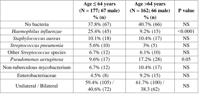
Table 1 - BAL microbiologic profile in patients according to age.
On initial assessment of 387 BAL culture results, 65.6% were positive for pathogenic microorganisms; the remainder yielded no bacteria (normal flora) (Table 2). The most common organisms were H. influenzae in 60 patients (15.5%), P. aeruginosa in 45 (11.6%) and S. aureus in 35 (9%). Less common organisms included NTM in 29 patients (7.5%), Enterobacteriaceae in 23 (5.9%), Streptococcus species in 22 (5.7%) and S. pneumoniae in 15 (3.9%). Of the total culture results, 25 (6.5%) were defined as "other" bacteria, including Moraxella catarrhalis, Stenotrophomonas maltophilia and Acinetobacterspecies.
6
Table 2- A total of 387 BAL cultures results in 339 patients with bronchiectasis 6.2 BAL isolation according to age
Univariate analysis revealed a significantly higher frequency of H. influenzae in the younger age group (25.4% in ≤64 years group vs. 9.2% in >64 years group, p<0.001). By contrast, P.
aeruginosa was found at a significantly higher frequency in those aged >64 years compared with the younger patient group (17.2% vs. 9.6%, respectively, p=0.05). There was also a higher but not statistically significant frequency of NTM (10.4% >64 years vs. 6.7% ≤64 years) and Enterobacteriaceae (9.2% >64 years vs.4.5% ≤64 years) detected in the older age group.
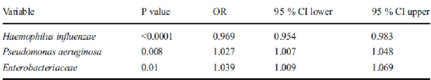
Univariate logistic regression showed a similar pattern, whereby the frequency of H.
influenzae was higher in younger patients (OR=0.969, p<0.0001, 95% CI 0.954-0.983), whereas the frequencies of P. aeruginosa (OR=1.027, p=0.008, 95% CI 1.007-1.048) and Enterobacteriaceae (OR=1.039, p=0.01, 95% CI 1.009-1.069) were much higher in older patients (Table 3).
Table 3 - Univariate logistic regression of H. influenzae, P. aeruginosa and Enterobacteriaceae odds ratio (OR) according to age.
Percentage of positive cultures (n) Type of Bacteria
34.4% (133) No bacteria
15.5% (60) Haemophilus influenzae
11.6% (45) Pseudomonas aeruginosa
9% (35) Staphylococcus aureus
7.4% (29) Non-tuberculous mycobacterium
6.5% (25) Others
5.9% (23) Enterobacteriaceae
5.7% (22) Other Streptococcus species
3.9% (15) Streptococcus pneumoniae
100% (387) Total
7 Non-tuberculous mycobacterium (NTM)
NTM were isolated in 6.3% (29/339) of the study sample, which increased in prevalence to 10.4% (17/162) in the >64 years of age group. These NTM included Mycobacterium avium- intracellulare (n=14), M. simiae (n=8), M. abscessus (n=3), M. chelonae (n=2) and M.
fortuitum (n=2).
The lobar distribution of NTM in all patients was as follows: 22.9% in the right upper lobe (RUL), 22.5% in the RML, 18.31% in the lingula, 14% in the right lower lobe (RLL), 12.6%
in the left upper lobe (LUL), and 8.4% in the left lower lobe (LLL).
Lobar bronchiectasis distribution
There was no significant difference between the lobar distributions of bronchiectasis in the younger versus the older group (Table 1).
The general lobar distributions of bronchiectasis in all 339 patients were as follows: 25.9% in the RML, 20.7% in the RLL, 20.4% in the LLL, 13.8% in the lingula, 13% in the RUL, and 6.2% in the LUL.
Lobar bacterial distribution in bronchiectasis
In the LLL, RLL, and RML of the lung, the most frequent pathogen was H. influenzae (23% - 28% of the total bacterial species in each lobe), and the second most common bacterial species was P. aeruginosa (19% - 21% in each lobe; Figure 1). In the lingula, H. influenzae comprised 14% and NTM comprised 13% of all bacterial species in this lobe. In the RUL, the most common bacterial species was P. aeruginosa, and the second most common was NTM, at prevalence rates of 18% and 17%, respectively. Finally, in the LUL, the most prevalent bacterial species were NTM, and the second most common was P. aeruginosa (9% and 6% of the total lobe's bacterial species, respectively).
8
Figure 1- Lobar bacterial distribution in bronchiectatic patients. The prevalence of each bacterium is represented in each lobe. RUL, right upper lobe; RML, right middle lobe; RLL, right lower lobe; LUL, left upper lobe; Lin, lingula; LLL, left lower lobe.
NTM was found to have a significantly higher prevalence in bilateral versus unilateral bronchiectasis (65.5% [19/29] vs.34.4% [10/29], respectively, p=0.004). The other bacterial groups showed no statistically significant differences in their prevalence when comparing unilateral and bilateral disease (Table 4).
Unilateral bronchiectasis Bilateral
bronciectasis Type of bacteria
NS 60.5% (81)
39.1% (52) No bateria
NS 61.6% (37)
38.3% (23) Haemophilus influenzae
NS 51.5% (17)
48.4% (16) Staphylococcus aureus
NS 53.3% (8)
46.6% (7) Streptococcus pneumoniae
NS 55.5% (25)
44.4% (20) Pseudomonas aeruginosa
0.004 34.4% (10)
65.5% (19) Non Tuberculus
Mycobacteria
NS 73.9% (17)
26% (6) Enterobacteriace
Table 4 - Bacterial distribution - unilateral versus bilateral bronchiectasis disease.
NS-non significant
9 Resistance to antibiotics
In this study, we reviewed the antibiotic sensitivity results of the two largest bacterial groups:
H. influenzae and P. aeruginosa. A total of 60 isolations of H. influenzae were observed; of these, 18.3% (11/60) showed resistance to sulfamethoxazole/ trimethoprim, 16.7% (10/60) to ampicillin and 1.7% (1/60) to ciprofloxacin. No resistance to cephalosporins, tetracycline or ertapenem was observed.
Of a total of 45 isolations of P. aeruginosa, 17.8% (8/45) showed resistance to ciprofloxacin, 6.7% (3/45) to gentamicin and amikacin, 4.4% (2/45) to piperacillin and 2.2% (1/45) to imipenem and ceftazidime; none showed resistance to tazocin and colistin.
Pleural effusion distribution
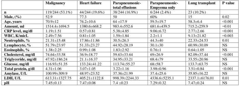
Of the 244 patients classified as having pleural effusions, 180 (73.7%) were diagnosed with exudative effusion, 44 (18%) were diagnosed with transudative effusion and 20 (8.1%) were excluded from the study due to lack of definitive diagnosis. The exudative effusion group was further divided into the following three subgroups according to the diagnosis: 119 (53.1%) malignant effusion, 38 (16.9%) parapneumonic effusion, and 23 (10.2%) lung transplant recipients (Table 5). Tuberculous pleural effusion was not diagnosed in any patient.
P value Lung transplant
Parapneumonic- Empyema only Parapneumonic-
total effusions Heart failure
Malignancy
23 (10.2%) 6/244 (2.4%)
38/244 (16.9%) 44/244 (19.6%)
119/244 (53.1%) n
0.02 15
50 60%
77.3 52.9
Male, (%)
0.001
>
58.3±6.4 59.5±19.7
64 ±17.9 76.2±10.6
70.9±12 Age, years
0.01
= 715.2±259.9
681.6±439.5 983.4±552.6
1380.6±668.2 1514.4±1694.5
Amount, ml
0.001
>
2.77±2.66 9.06±6.72
5.38±4.85 0.57±0.81
1.19±1.51 CRP level, mg/dl
0.003
= 9.13±21.82
2.2±1.1 1.59±1.84
0.81±1.05 2.49±7.56
WBC, K/micL
0.01 22.33±24.53
44.3±40 30.5±26.3
17.86±11.86 21.31±15.48
Neutrophils, %
NS 60.99±30.09
30.1±30 44.92±28.19
51.33±23.27 51.79±23.97
Lymphocyte, %
NS 0.64±1.05
0.76±1 1.83±2.92
0.99±1.08 1.38±2.25
Eosinophils, %
0.001
>
82.09±37.44 69±26.9
59.63±33.64 36.9±18.77
74.64±33.58 Cholesterol, mg/dl
NS 33.55±20.96
68.4±79 34.95±33.21
21.1±16.37 47.92±186.24
Triglyceride, mg/dl
NS 133.7±63.75
68±58.7 113.74±55.27
133.24±41.22 116.93±51.35
Glucose, mg/dl
0.001
>
3.51±0.84 3.98±0.96
3.22±1.16 3.07±1.05
4.31±1.06 Total protein, g/dl
NS 35.85±16.22
37.4±25.6 37.36±21.99
48.97±23.52 100.99±309.9
Amylase, U/L
0.01 2337.1±6176.01
4336.6±5235.1 998.39±2244.33
405.21±1122.8 613.31±1327.75
LDH, U/L
NS 7.47±0.24
7.29±0.32 7.4 ±0.23
7.47±0.08 7.45±0.13
pH
Table 5 - Clinical characteristics of the study population and pleural fluid parameters.
10 Pleural CRP level in pleural effusions
The pleural CRP levels differed significantly among all four groups (p<0.001, Figure 2). The mean values from highest to lowest were as follows: parapneumonic (5.38±4.85 mg/dl), lung transplant (2.77±2.66 mg/dl), malignancy (1.19±1.51 mg/dl) and heart failure (0.57±0.81 mg/dl). A backward logistic regression model selected CRP as the only predictor of parapneumonic effusion (OR=1.59, 95% C.I=1.37-1.89, p 0.0001> ).
Figure 2 -Pleural fluid CRP levels in effusions secondary to pneumoniae, malignancy, post- lung transplantation and heart failure. Each point represents one pleural fluid sample. The red points represent patients with empyema.
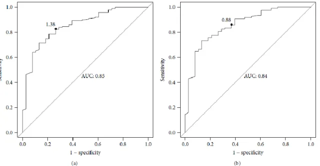
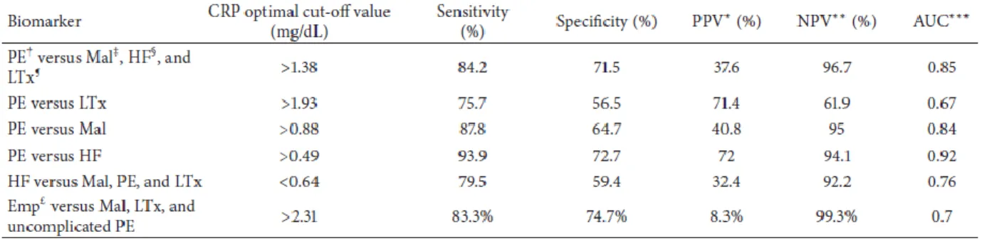
To determine the efficiency of pleural fluid CRP measurement in distinguishing parapneumonic effusion from the other 3 groups, we used ROC analysis (Figure 3). A CRP cut-off value of 1.38 mg/dl yielded 84.2% sensitivity, 71.5% specificity, 37.6% positive predicted value and 95.6% negative predicted value. Although the area under the curve (AUC) of the pleural CRP was as high as 0.85 (Figure 3A), it was lower for the following other pleural parameters: glucose (0.54), pH (0.61), neutrophils (0.59) and LDH (0.57). As a result, we could not calculate their optimal cut-off values.
11
Figure 3 - Receiver operator characteristic (ROC) analysis curves of pleural fluid CRP levels for differentiating between different effusion types.
3 (A) - ROC curve of CRP levels for differentiating parapneumonic pleural effusions from other types of pleural effusions such: malignant, heart failure and post-lung transplantation.
3 (B) - ROC curve of CRP levels for differentiating between parapneumonic and malignant effusions.
Figure 3 (C) - ROC curve of CRP for differentiating between parapneumonic and heart failure pleural effusions.
Figure 3 (D) ROC curve of CRP for differentiating empyema from other types of effusion such: malignant, heart failure and uncomplicated parapneumonic pleural effusions
12
CRP was a good marker for distinguishing parapneumonic effusion from: post-lung transplantation effusion (1.93 mg/dl cut-off value, 75% sensitivity, 56% specificity);
malignant effusion (0.88 mg/dl cut-off value, 87% sensitivity, 64% specificity- Figure 3B) and heart failure effusion (0.49 mg/dl cut-of value, 93% sensitivity, 72% specificity- Figure 3C).
CRP was a moderately good marker for differentiating between empyema and other types of effusions (2.31 mg/dl cut-off value, 83.3% sensitivity, 74.7% specificity, 8.3% positive predicted value and 99.3% negative predicted value- Figure 3D)
We also determined the efficiency of pleural fluid CRP measurements in distinguishing among heart failure effusion and the other 3 groups (Table 6). Using a cut-off value of 0.64 mg/dl, CRP exhibited 79.5% sensitivity, 59.4% specificity, 32.4% PPV and 92.4% NPV.
Table 6 - receiver operating characteristic curve analysis of the accuracy of biomarkers for identifying parapneumonic effusions.
Pleural white blood cells counts were significantly different among all four groups (p=0.003) with the following means: 9.13±21.82K/micL (lung transplant), 2.49±7.56K/micL (malignancy), 1.59±1.84K/micL (parapneumonic) and 0.81±1.05K/micL (heart failure).
Pleural neutrophil differentials were also significantly different among all four groups (p=0.01): 30.5±26.3% (parapneumonic), 22.33±24.53% (lung transplant), 21.31±15.48%
(malignancy) and 17.86±11.86% (heart failure).
13 CONCLUSIONS
Lobar bacterial distributionsin patients with bronchiectasis
1. We found that H. influenzae was more prevalent in younger patients who have bronchiectasis, whereas P. aeruginosa, Enterobacteriaceae and NTM predominated in older patients.
2. Different pathogens were associated with different lobar distributions.
3. In the lower lobes, H. influenzae was the dominant species isolated, whereas in the upper lobes and lingula it was non tuberculous mycobacterium (NTM). While Streptococci species were more prevalent in LLL and RUL, P. auriginosa was homogenously distributed in all the lobes. These findings can support decision making on empirical treatment.
4. Overall, the RML, RLL and LLL showed a greater tendency to develop bronchiectasis than other lobes.
5. On the base of these findings we recommend frequent screening of NTM in older patient (age>65) with bilateral disease and upper lobes bronchiectasis.
The role of pleural CRP in parapneumonic effusion detection
1. Pleural fluid CRP levels can be used to distinguish among parapneumonic effusions and other types of exudative effusions. CRP levels < 0.64 mg/dl are likely to indicate a pleural effusion from congestive heart failure, whereas levels ≥ 1.38 mg/dl are suggestive of an infectious etiology.
2. While we have clear criteria for empyema, we do not have such for parapneumonic effusion. In order to facilitate the initial clinical work up, we recommend using pleural CRP in addition to the conservative pleural parameters. This is especially important in patients with multiple comorbidities with unclear pleural effusion etiology.
14 Publications related to the dissertation:
1. Izhakian S, Wasser WG, Fuks L, Vainshelboim B, Fox BD, Fruchter O, Kramer MR.
Lobar distribution in non-cystic fibrosis bronchiectasis predicts bacteriologic pathogen treatment. Eur J ClinMicrobiol Infect Dis. 2016 May;35(5):791-6. I.F=2.857
2. Izhakian S, Wasser WG, Fox BD, Vainshelboim B, Kramer MR. The Diagnostic Value of the Pleural Fluid C-Reactive Protein in Parapneumonic Effusions. Dis Markers. 2016;2016:7539780. IF=2.137
Publications not related to the dissertation:
3. Vainshelboim B, Kramer MR, Fox BD, Izhakian S, Sagie A, Oliveira J.
Eur J Phys Rehabil Med. 2016 Dec 19. Supervised exercise training improves exercise cardiovascular function in idiopathic pulmonary fibrosis. IF=2.06
4. Unterman A, Fruchter O, Rosengarten D, Izhakian S, Abdel-Rahman N, Kramer MR.
Bronchoscopic Drainage of Lung Abscesses Using a Pigtail Catheter. Respiration.
2016 Dec 13. IF= 2.65
5. Vainshelboim B, Fox BD, Kramer MR, Izhakian S, Gershman E, Oliveira J. Short- term improvement in physical activity and body composition after supervised exercise training program in idiopathic pulmonary fibrosis. Arch Phys Med Rehabil.2016 Feb 8. IF=3.045
6. Izhakian S, Buchs AE. Characterization of Patients who were Mechanically Ventilated in General Medicine Wards. Isr Med Assoc J. 2015 Aug;17(8):496-9.
IF=0.879
7. Rapoport M, Harel N, Shasha Y, Barkan R, Kitaee E, Buchs A, Izhakian S, Aviel- Gadot E.Achievement of partial combined control of major diabetes targets in
primary care correlates with development of chronic complications in T2DM patients- -A real life data. Prim Care Diabetes. 2015 Dec;9(6):412-7. Epub 2015 Jun 15.
IF=1.57
8. Izhakian S., Wasser WG., Vainshelboim B., Fox BD., Kramer MR. Effect of jewish - arab ancestry and gender matching on clinical outcome of lung transplant patients in Israel. Isr Med Assoc J. 2016 Aug;18(18)470-3. IF=0.879
9. Izhakian S, Wasser WG, Fox BD, Vainshelboim B, Reznik JE, Kramer MR. The effectiveness of rabbit anti-thymocyte globulin in chronic lung
allograft dysfunction. Transplant Proc. 2016 Jul-Aug;48(6):2152-6. IF=0.867
15
10. Vainshelboim B, Kramer MR, Izhakian S, Lima RM, Oliveira J. Physical Activity and Exertional Desaturation Are Associated with Mortality in Idiopathic Pulmonary Fibrosis. J Clin Med. 2016 Aug 18;5(8). No Impact Factor.
11. Gorelik O, Izhakian S, Barchel D, Almoznino-Sarafian D, Tzur I, Swarka M, Beberashvili I, Feldman L, Cohen N, Shteinshnaider M. Prognostic significance of platelet count changes during hospitalization for community-acquired pneumonia.
Platelets. 2016 Sep 29:1-7. IF=3.2
12. Gorelik O, Izhakian S, Barchel D, Almoznino-Sarafian D, Tzur I, Swarka M, Beberashvili I, Feldman L, Cohen N, Shteinshnaider M. Changes in red cell
distribution width during hospitalization for community-acquired pneumonia: clinical characteristics and prognostic significance. Lung. 2016 Dec;194(6):985-995. IF=2 13. Abdel-Rahman N, Izhakian S, Wasser WG, Fruchter O, Kramer MR. Endobronchial
Enigma: A Clinically Rare Presentation of Nocardia beijingensis in an Immunocompetent Patient. Case Rep Pulmonol. 2015;2015:970548.