EFFECT OF ENDURANCE EXERCISE ALONE AND IN COMBINATION WITH IGF-1 ADMINISTRATION ON CELLULAR MARKERS INVOLVED IN SARCOPENIA
PhD thesis
Mohammad Mosaferi Ziaaldini
Doctoral School of Sport Sciences University of Physical Education
Supervisor: Dr. Zsolt Radák, professor, DSc
Official Reviewers: Dr. István Berkes, professor, DSc
Dr. Attila Bácsi, associate professor, PhD
Head of the Final Examination Committee: Dr. József Tihanyi, professor, DSc
Members of the Final Examination Committee: Dr. Gábor Pavlik, professor emeritus, DSc Dr. József Pucsok, professor emeritus, DSc
Budapest, 2015
DOI: 10.17624/TF.2015.02
1
Table of Contents
List of Figures ... 4
List of Tables ... 5
List of Abbreviations ... 5
1. Introduction ... 9
1.1. Aging process and its consequences ... 9
1.2. Sarcopenia phenomenon as a natural consequence of the aging process ... 10
1.2.1. Definition ... 10
1.2.2. Consequences ... 13
1.2.3. Etiology of sarcopenia ... 13
1.2.3.1. Sedentary life style ... 14
1.2.3.2. Chronic inflammation ... 15
1.2.3.3. Mitochondrial dysfunction ... 16
1.2.3.4. Hormonal changes ... 17
1.2.3.4.1. Growth hormone ... 18
1.2.3.4.2. Insulin and insulin-like growth factors ... 19
1.2.3.4.3. Testosterone and its precursors ... 20
1.2.3.4.4. Menopause ... 21
1.2.3.5. Neural degeneration ... 22
1.2.3.6. Malnutrition ... 23
1.2.3.7. Satellite cell dysfunction ... 24
1.2.4. Cellular mechanisms and signaling pathways involved in sarcopenia ... 26
1.2.4.1. IGF-1/Akt/mTOR... 26
1.2.4.2. MAPKs ... 28
1.2.4.3. FOXOs ... 29
1.2.4.4. TGFβ (Myostatin) ... 30
2
1.2.4.5. NF-κB ... 32
1.2.4.6. Apoptosis ... 33
1.3. Therapeutic strategies ... 36
1.3.1. Nutritional and pharmacological intervention ... 36
1.3.2. Caloric restriction (CR) ... 39
1.3.3. Exercise training ... 40
1.3.3.1. Resistance training... 40
1.3.3.2. Endurance training ... 42
1.3.4. Hormone therapy ... 46
1.3.4.1. IGF-1... 48
1.3.5. Combination of exercise and nutritional and pharmacological supplementation ... 50
1.4. Summary of introduction ... 52
2. Objectives... 53
3. Methods ... 54
3.1 Subjects ... 54
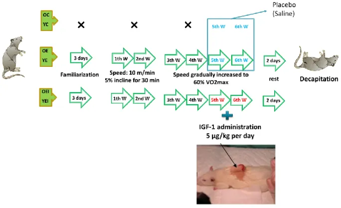
3.2 Exercise protocol ... 54
3.3 IGF-1 administration ... 56
3.3.1 Alzet pump ... 56
3.3.2. IGF-1 supplementation ... 57
3.4. Tissue preparation ... 57
3.5. Western blot analysis ... 58
3.6. Measurement of IGF-1 level ... 58
3.7. Measurement of ROS level... 60
3.8. Statistical analyses ... 60
4. Result ... 61
4.1 Cellular markers involved in protein synthesis ... 61
4.2 Cellular markers involved in protein degradation ... 66
3
4.3 Cellular markers involved in mitochondria biogenesis ... 69
4.4 Cellular markers involved in apoptosis ... 74
5. Discussion... 78
5.1 Aging negatively affected cellular markers involved in sarcopenia ... 78
5.2 Endurance training positively reversed some cellular markers involved in sarcopenia . 84 5.3 Endurance training combined with IGF-1 administration positively affected some cellular markers involved in sarcopenia... 90
6. Conclusions ... 94
7. Summary of thesis ... 95
7.1 Summary in English ... 95
7.2 Summary in Hungarian (Összefoglalás) ... 96
8. Bibliography ... 98
9. Bibliography of the author’s publications ... 122
9.1. Publications related to this study ... 122
10. Acknowledgements ... 123
4
List of Figures
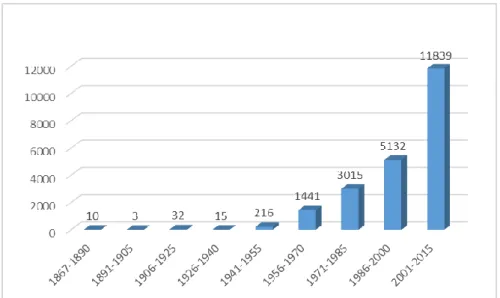
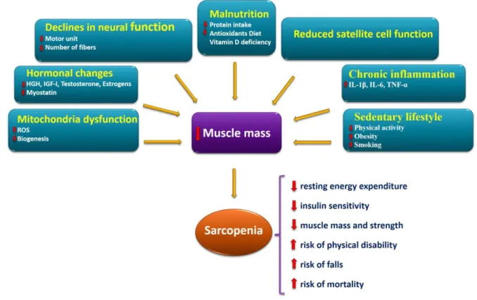
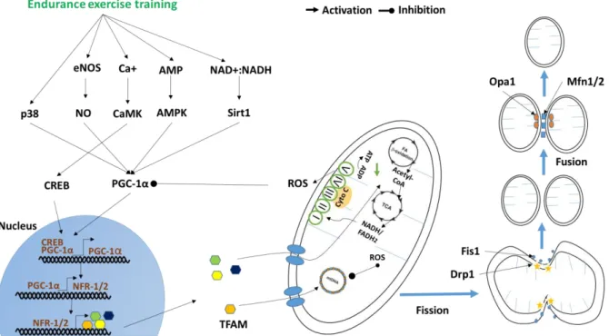
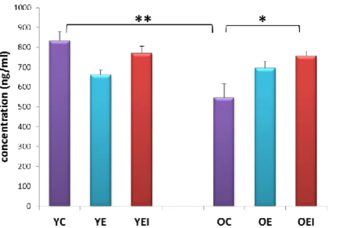
Figure 1. Percentage change in the world’s population by age: 2010-2050 [1] ... 9 Figure 2. Young children and older people as a percentage of global population: 1950-2050 [1] ... 10 Figure 3. Number of PubMed entries retrieved after entering the search term “muscle wasting OR sarcopenia” ... 11 Figure 4. Schematic model of main factors involved in the onset and progression of the sarcopenia and its consequences ... 14 Figure 5. Molecular mechanisms involved in sarcopenia which can be viewed as the result of a protein synthesis/degradation imbalance ... 26 Figure 6. Diagrammatic summary of endurance exercise training signaling pathways involved in mitochondria function in healthy mammalian skeletal muscle cell ... 46 Figure 7. The figures represent Speed, time and incline (ascent) of 6 weeks of endurance training on treadmill in young and old rats ... 55 Figure 8. Schematic design of the study protocol ... 56 Figure 9. Schematic representation of an Alzet osmotic pump ... 57 Figure 10. Effect of age, exercise and combination of exercise and IGF-1 administration on plasma level of IGF-1 ... 62 Figure 11. Effect of age, exercise and combination of exercise and IGF-1 administration on follistatin protein content ... 62 Figure 12. Effect of age, exercise and combination of exercise and IGF-1 administration on levels of pAkt (A), total Akt (B) and pAkt: Akt ratio (C) ... 63 Figure 13. Effect of age, exercise and combination of exercise and IGF-1 administration on levels of pmTOR (A), total mTOR (B) and pmTOR: mTOR ratio (C) ... 64 Figure 14. Effect of age, exercise and combination of exercise and IGF-1 administration on levels of pERK1/2 (A), total ERK1/2 (B) and pERK1/2:ERK1/2 ratio (C) ... 65 Figure 15. Effect of age, exercise and combination of exercise and IGF-1 administration on
Myostatin protein content ... 67 Figure 16. Effect of age, exercise and combination of exercise and IGF-1 administration on
ubiquitinated protein content ... 67 Figure 17. Effect of age, exercise and combination of exercise and IGF-1 administration on MuRF1 protein content ... 68 Figure 18. Effect of age, exercise and combination of exercise and IGF-1 administration on MuRF2 protein content ... 68 Figure 19. Effect of age, exercise and combination of exercise and IGF-1 administration on PSMA6 protein content ... 69 Figure 20. Effect of age, exercise and combination of exercise and IGF-1 administration on ROS level ... 70 Figure 21. Effect of age, exercise and combination of exercise and IGF-1 administration on PGC-1α protein content ... 71 Figure 22. Effect of age, exercise and combination of exercise and IGF-1 administration on SIRT1 protein content ... 71 Figure 23. Effect of age, exercise and combination of exercise and IGF-1 administration on SIRT3 protein content ... 72 Figure 24. Effect of age, exercise and combination of exercise and IGF-1 administration on Cyto C protein content ... 72 Figure 25. Effect of age, exercise and combination of exercise and IGF-1 administration on Cox 4 protein content ... 73
5
Figure 26. Effect of age, exercise and combination of exercise and IGF-1 administration on Nrf 2 protein content ... 73 Figure 27. Effect of age, exercise and combination of exercise and IGF-1 administration on TNF-α protein content ... 75 Figure 28. Effect of age, exercise and combination of exercise and IGF-1 administration on p53 protein content ... 75 Figure 29. Effect of age, exercise and combination of exercise and IGF-1 administration on Bcl-2 protein content ... 76 Figure 30. Effect of age, exercise and combination of exercise and IGF-1 administration on Bax protein content ... 76 Figure 31. Effect of age, exercise and combination of exercise and IGF-1 administration on apoptotic index (Bax: Bcl-2 ratio) ... 77
List of Tables
Table 1. Description of primary antibodies used in the study ... 59
List of Abbreviations
4E-BP1 4E-binding protein 1
AIF Apoptosis-inducing factor
ALS Acid labile subunit
AMPK AMP-activated protein kinase
ANT Adenine Nucleotide Translocase
Apaf-1 Apoptotic protease activating factor-1
ARC Apoptotic repressor with a caspase recruitment domain
Atg Autophagy-related protein
ATP Adenosine three phosphate
Bax Bcl-2-associated X protein
Bcl-2 B-cell lymphoma-2
CAD Caspase-activated DNase
Caspases Cysteine-aspartic proteases
COX Cytochrome c oxidase
CR Caloric restriction
CREB cAMP response element-binding protein
CS Citrate synthases
6
CSA Cross section area
CT Computed tomography
CyPD Cyclophilin D
Cyto C Cytochrome C
DHEA Dehydroepiandrosterone
DNA Deoxyribonucleic acid
Drp1 Dynamin-related protein 1
DXA Dual-energy X-ray absorptiometry
EAA Essential amino acid
EDL Extensor digitorum longus
eIF2B Eukaryotic initiation factor 2 subunit B
EIM Electrical impedance myography
EndoG Endonuclease G
ERK Extracellular signal-regulated kinase
ET Endurance exercise training
ETC Electron transport chain
ETS Electron transport system
EWGSOP European Working Group on Sarcopenia in Older People Fis1 Mitochondrial fission protein 1
FLRG Follistatin-related gene
FOXO Forkhead box O
FSH Follicle-stimulating hormone
GASP-1 GDF-associated serum protein-1
GH Growth hormone
GHR Growth hormone receptor
GHRH GH releasing hormone
H2O2 Hydrogen peroxide
HRT Hormone replacement therapy
IGF-1 Insulin-like growth factor 1
IGFBPs Insulin-like growth factor-binding proteins
IGFR1 Type 1 IGF receptor
IKK IκB kinase
7
IL-6 Interleukin 6
IMM Inner mitochondrial membrane
IR Insulin receptor
IWGS International Working Group on Sarcopenia
JNK c-Jun N-terminal kinases
LC3 Microtubule-associated protein 1A/1B-light chain 3
MAC Mitochondrial apoptosis-induced channel
MAFbx Muscle atrophy F-box
MAPKs Mitogen-activated protein kinases
Mfn1 Mitochondrial fusion 1
MGFs Mechano growth factors
MHC Myosin heavy chain
MKKs MAP kinase kinases
MKPs MAPK phosphatases
Mnk 1 MAPK-interacting kinase 1
MnSOD Manganese superoxide dismutase
MOMP Mitochondrial outer membrane permeabilization
MPS Muscle protein synthesis
MPT Mitochondrial permeability transition mPTP Mitochondrial permeability transition pore
MRFs Muscle regulatory factors
MRI Magnetic resonance imaging
mRNA Messenger RNA
mtDNA Mitochondrial deoxyribonucleic acid
mTOR Mammalian target of rapamycin
mTORC1 Mammalian target of rapamycin complex 1
MuRF1 Muscle RING-finger protein-1
Myf5 Myogenic factor 5
MyoD Myogenic differentiation 1 protein NAMPT Nicotinamide phosphoribosyltransferase
NF-кB Nuclear factor κb
OC Old control
8
OE Old exercise training
OEI Old exercise and IGF-1 treatment
OMM Outer mitochondria membrane
p70s6k 70-kilodalton ribosomal S6 protein kinase
PGC-1α Peroxisome proliferator-activated receptor-γ coactivator 1-α
PI-3K Phosphatidylinositol-3-kinases
PNPase Polynucleotide Phosphorylase
RDA Recommended dietary allowance
ROS Reactive oxygen species
RT Resistance exercise training
SDH Succinate dehydrogenase
SOCS-3 Suppressor of cytokine signaling-3
TCA Tricarboxylic acid
Tfam Transcription factor A, mitochondrial
TNF-R Tumor necrosis factor receptor
TNF-α Tumor necrosis factor alpha
TSC2 Tuberous Sclerosis Complex 2
UCP3 Mitochondrial uncoupling protein 3
UPS Ubiquitin-proteasomal system
VDAC Voltage dependent anion channel
VO2max Maximal oxygen consumption
YC Young control
YE Young exercise training
YEI Young exercise training and IGF-1 treatment
YY1 Yin Yang 1
9
1. Introduction
1.1. Aging process and its consequences
Aging is an undeniable and complicated biological process that is indicated by a general time-dependent decline in the physiological and biochemical functions of the main systems [1, 2]. Aging is also a remarkable risk factor for the advancement of cardiovascular diseases [3] and is associated with a significant decline in neuromuscular function and performance [4]. The percentage of elderly people in the world is increasing steadily (Figure 1) [5] due to the impressive rise in average life expectancy in the past century [6].
The rapid increase world’s aging population has led to concerns about the general health of elderly [7]. It is estimated that until year 2050, the elderly population will increase from 600 million as it was in the year 2000 to more than two billion (Figure 2) [8]. Several changes are observable along with aging, including a reduced capacity oxygen consumption, an impairment in cardiorespiratory adaptation and degradation of the nervous system, and decadence in muscle mass which characterized by a reduction in muscle mass and by a qualitative and quantitative alteration in muscle fibers [2]. After age 30, a change in body composition occurs. However, a reduction of about 0.23 kg muscle mass per year is expected from ages of 30 to 60 which accelerates to 2% annually from the age of 60 [9]. Indeed, data from the most recent longitudinal aging study suggest that muscle strength decreases at an intermittent rate of ~ 3% yearly between the ages of 70–79 years [10].
Figure 1. Percentage change in the world’s population by age: 2010-2050 [1]
10
Due to increasing longevity and the fact that the elderly have twice as many disabilities and four times as many physical limitations as people less than 60 years of age [11], it is imperative to understand the aging process and the mechanisms associated with healthy aging and conservation of functional independence at the end of life [12].
1.2. Sarcopenia phenomenon as a natural consequence of the aging process
1.2.1. Definition
One of the most well-known features of aging is a change in body composition and decline in lean body mass [13]. Age related skeletal muscle wasting, also called sarcopenia, plays a significant role in reducing performance [14]. The contribution of lean muscle mass in whole body weight is approximately ~50% in young adults, which declines to 25% after age of 75 years. The reduction of muscle mass is generally counteracted by gains in fat mass. The lower extremity muscle groups are more at risk for losing muscle mass, with to 40%
reduction in cross-sectional area (CSA) of the vastus lateralis between the age of 20 and 80 years [14]. Although sarcopenia has been widely recognized, however, its mechanisms remain poorly known and have not received proper attention until quite recently [13]. In the recent decades, research on sarcopenia and muscle wasting have grown considerably (Figure 3). The term sarcopenia has been derived from Greek sarx (flesh) and penia (loss),
Figure 2. Young children and older people as a percentage of global population: 1950-2050 [1]
11
letter by letter meaning deficiency of flesh [15, 16]. It has been suggested that up to 40% of muscle mass may be lost between the ages of 20 and 70 years. A reduction in skeletal muscle mass can start in the early 35 years of age and may speed up to 6% per decade after 30 year of age and 1.4% to 2.5% per year after age 60 [16].
Assessed on 13 January 2015 from www.pubmed.gov
In parallel, leg strength is declined by 10–15 % per decade until 70 years of age, and then by 25–40 % per decade [17]. Fast twitch fiber are more affected and their size may be reduced by 20-50%, despite the fact that slow twitch fibers are less affected, they may be still have a reduction by 1-25% in size. The decrement in total muscle mass is more than loss of muscle fiber size because of an extra loss of fibers [18]. For example, a 40–45% reduction in CSA of the type II muscle fibers was observed in older males compared to young subjects [19].
Using the difference definitions of sarcopenia makes it inconceivable to compare studies for understanding the pathophysiological processes and developing the targeted therapies [20].
There are no clear clinical outcome parameter for sarcopenia yet and the evaluation of muscle quality based on physical performance and strength is undesirable, because other parameters such as the neural controller, cardiovascular fitness and joint function are also involved in physical performance and strength. Despite the correlation between the amount of muscle mass and muscle strength, it has been demonstrated that muscle mass and muscle strength are two different entities and therefore the loss of muscle strength occurring with age can be described by the term ‘dynapenia’ [20].
Figure 3. Number of PubMed entries retrieved after entering the search term “muscle wasting OR sarcopenia”
12
In 2009, The International Working Group on Sarcopenia (IWGS) provided an operational definition for sarcopenia. This definition includes people with functional decline, movement- related problems, history of repeated falls, recent inadvertent body weight loss, post hospitalization, and chronic conditions (such as type 2 diabetes, chronic heart failure, chronic obstructive pulmonary disease, chronic kidney disease, rheumatoid arthritis, and cancer), and was more suitable in clinical settings [16]. On the other side, in 2010 the European Working Group on Sarcopenia (EWGSOP) developed a new definition for sarcopenia [21]. EWGSOP recommendations cover both low muscle mass and low muscle function for assessment of sarcopenia in clinical and research tests. EWGSOP offers three levels for sarcopenia including: The pre-sarcopenia level is specified by low muscle mass but no change in muscle strength or performance. The second level of sarcopenia, is specified by low muscle mass along with low muscle performance and the third level, known as severe sarcopenia, is characterized by decrease of all of three components, muscle mass, strength, and performance [8]. A formula has been used for assessment of sarcopenia as appendicular lean mass (sum of lean mass of both arms and legs) divided by height squared. Values higher than two standard deviations below the mean of a young reference population were categorized as sarcopenia [20]. The first step in the management of sarcopenia is to diagnose the condition.
Unfortunately, at the moment, there is no standard diagnostic criteria for detection low sarcopenia [22]. The most commonly used measurement techniques for muscle mass and body composition are Dual-energy X-ray absorptiometry (DXA), magnetic resonance imaging (MRI), computed tomography (CT) and electrical impedance myography (EIM) [15, 22, 23].
Due to practical problems in evaluating muscle mass, it is not easy to estimate the prevalence of sarcopenia. Many different methodologies have been used over the last 20 years, and new techniques are still being introduced. On average, it is estimated that 5–13 % of elderly people aged 60–70 years are impressed by sarcopenia, and the numbers raise to 11–50 % for those aged 80 or above [15]. In line with these data, other sources estimate the prevalence of sarcopenia in the range from 13% to 24% in adults over 60 years of age to more than 50% in persons aged 80 and older [24]. Current estimates suggest that ~200 million people worldwide will be affected by sarcopenia by the year 2050 [25]. Women show a higher decrease of muscle mass, especially after menopause. The reduction of muscle mass in aged people does not affect arms and legs the same way. Muscle wasting is higher in the lower limbs irrespective of the sex of the person. However, when Performance parameters, such as
13
muscle quality, are considered, it can be found a sex-dependent differences. Men undergo a greater loss in the upper limbs Compared to women whereas no differences in the decline of muscle mass in lower limbs have been observed [26].
1.2.2. Consequences
Sarcopenia is, therefore, a multifactorial consequence of aging and indicative a potent risk factor for the development of negative health-related conditions in the elderly [2].
Additionally, mobility disturbances resulting from muscle wasting is along with decreased quality of life and increased social and health care costs in elderly [27]. Sarcopenia is also related to acute and chronic disease states, increased insulin resistance, fatigue, falls, and mortality. Of the chronic disease states, sarcopenia has been especially associated with rheumatologic conditions, especially rheumatoid arthritis in women [28]. In addition, sarcopenia is a main risk factor of falls and disability among the elderly people. Dysfunction and physical inability in sarcopenic people are 2 to 3 times more likely [8]. Individuals with clinically sensible sarcopenia have 4 times higher risk of disability, three times more risk of balance impairment, and 3 times greater risk of falling. Therefore, sarcopenia is the single most prevalent etiology to falls and fall-related fractures in elderly people [13]. It has been estimated that direct healthcare costs related to sarcopenia in the United States of America in 2000 were $18.5 billion, which was around 1.5% of total healthcare expenses for that year [29]. It is estimated that a 10.5% reduction of the outbreak of sarcopenia could lead to a reduction of healthcare expenses by 1.1 billion US dollars per year in the United States [20].
1.2.3. Etiology of sarcopenia
Although the exact mechanisms involved in sarcopenia is still unclear, however, several factors have been proposed to be involved in the onset and progression of sarcopenia (Figure 4). Sarcopenia arises from several physiopathological factors including, but not limited to:
sedentary lifestyle [9, 14, 30, 31], chronic inflammation [13, 17, 30, 32], impaired satellite cell function [12, 27, 31, 32], malnutrition [9, 12, 13, 30, 32], declines in neural function [12, 13, 18, 32, 33], hormonal changes [12, 18, 30, 34], mitochondrial dysfunction [18, 34]. It is also probable that certain underlying mechanisms are of greater influence than others when considering any specific age group, gender, or association with comorbid states [4].
14 1.2.3.1. Sedentary life style
Physical activity has been defined as any action generated by the contractive activity of skeletal muscles that increments energy expenditure. Physical activity is including of daily routines like standing up from a chair and taking stairs, as well as Voluntary movements for health benefits such as walking or biking. Inactive persons are characterized by doing only basic physical activities such as standing, walking slowly and lifting light objects. Lifestyle styles related to nutrition, physical activity, exercise, alcohol consumption, and smoking have a Significant influence on the advancement of sarcopenia and the ability to prevent and treat the loss of muscle mass and function in old age [9].
Figure 4. Schematic model of main factors involved in the onset and progression of the sarcopenia and its consequences
One of the main factor involved in sarcopenia is reduced physical activity among older people [35]. It has been suggested that at least some of the increasing prevalence of sarcopenia after the age of 65 is due to decrease physical activity and smoking [36].
15
The connection between lean body mass and degree of physical activity and exercise are complex. Reduction in physical activity can alter body composition in some ways. One study studied relation between body composition and physical activity in older women over a 10 years period and found that grater levels of physical activity reduced the advancement of sarcopenia. The level of muscle loss is increased even greater when an older person has to spending a period of bed rest because of illness [37]. In addition, inactive and smoker persons had a greater risk for a reduction in health status compared with active and non-smoker people. The net impact of a healthier lifestyle on the process of healthy aging is likely going together with a compressed cumulative morbidity [38].
1.2.3.2. Chronic inflammation
An age-related disruption in the intracellular redox balance plays an important role in generating a chronic state of low-grade inflammation. Chronic cellular inflammation is intended for fundamental mechanism of aging and age-related diseases, and it may consider as a link between normal aging and age-related pathological processes [39]. There are several lines of evidence suggestting that the inflammation being associated with loss of muscle strength and mass with aging [40]. Number of studies have implicated increased levels of two pro-inflammatory cytokines, Interleukin 6 (IL-6) and tumor necrosis factor alpha (TNF-α), in the development of sarcopenia [41-44]. For example, it was observed a 2.8 fold increase in TNF-α expression in skeletal muscle of aged male compared to young male subjects [45].
An increased expression of TNF-α has also been reported in soleus and vastus lateralis of aged rats relative to young rats. Furthermore, it has been found that old animals have significantly higher plasma TNF-α levels than younger controls with corresponding elevations of IL-6 [45]. Animal studies have shown that the supplementation of IL-6 or TNF- α increases skeletal muscle breakdown, reduces the rate of protein synthesis, and decreases plasma concentrations of insulin-like growth factor 1 (IGF-1). The activation of the age related inflammatory process is thought to be the consequent of an up regulation of the transcription factor nuclear factor be (NF-кB), an important regulator of the innate immune response. This fundamental and increased activity of NF-кB that is associated with aging has been suggested to be one of the basic causes of sarcopenia [41]. Furthermore, reactive oxygen species (ROS) also seems to act as second messengers for TNF-α in skeletal muscle, activating NF-кB either directly or indirectly [39]. TNF-α is also linked to sarcopenia because
16
this pro-inflammatory cytokine is known to be associated with other factors that contribute to sarcopenia including protein degradation, ROS accumulation and apoptosis. In addition, TNF-α may be induced sarcopenia by promoting insulin resistance, impaired muscle repair, and amplifying the pro-inflammatory response by up-regulating IL-6 [45]. Notably, pro- inflammatory cytokines, in particular TNF-α, are potent stimulators of muscle proteolysis through activation of ubiquitin-proteasomal system (UPS). This pathway is thought to be responsible for the major part of muscle proteolysis and is stimulated by the repeated covalent binding of 76-amino acid ubiquitin monomers to proteins targeted for degradation [46]. Furthermore, there is evidence that due to hormone-cytokine receptor cross-talk, pro- inflammatory cytokines like TNF-α that cause muscle wasting may also operate by interfering directly with somatotrophic receptors, including insulin-like growth factor 1 (IGF- 1) [47].
1.2.3.3. Mitochondrial dysfunction
Mitochondria are subcellular self-autonomous organelles primarily responsible for the generation of energy and adenosine three phosphate (ATP) synthesis. Besides this, mitochondria has a significant role in amino acid and lipid metabolism and regulation of apoptosis [48]. There are a number of evidence supporting the hypothesis that mitochondrial function and biogenesis appear to be altered in skeletal muscles of older adults [28, 49-51], which in turn may contribute to altered skeletal muscle mass and function [28]. Alterations in mitochondria have been considered in aging, such as reduced total volume, elevated oxidative damage, and decreased oxidative capacity. These alternations may resulted in not only a loss of muscle mass and function with age, but other diseases associated with aging such as ectopic lipid infiltration, systemic inflammation, and insulin resistance [52]. It is well established that the amount of synthesis of mitochondrial, myosin heavy chain (a key contractile protein) and mixed muscle protein are decreased with age, along with oxidative capacity of skeletal muscle [53]. Muscle biopsy samples have determined that certain measures of mitochondrial content decrease with age as measured by electron microscopy, mitochondrial deoxyribonucleic acid (mtDNA) copy number, proteomics or the activities of key tricarboxylic acid (TCA) cycle enzymes such as citrate synthase [53]. Several hypotheses have been proposed to explain the decline in mitochondrial function with aging. These theories are included increased ROS production, chronic inflammation and/or mtDNA
17
damage [54]. According to the widely-accepted mitochondrial free radical theory of aging, mitochondrial dysfunction arising from oxidative damage to mtDNA is the central mechanism driving the aging process [55]. Mitochondria have their own DNA (mtDNA);
however, its role in mitochondrial protein encoding is only 1% of the approximately 1,000.
The bulk of the mitochondrial proteins are encoded by nuclear DNA and are transported to mitochondria from the cytoplasm [48]. The mtDNA is especially unprotected to oxidative damage because of its proximity to the electron transport chain (ETC) (the main cellular source of oxidants) and the lack of protective histones. Moreover, because of the density of mitochondrial genome (i.e., lack of intrones), each mutation is likely to affect gene integrity [27]. A defeat of replication of mtDNA may be the reason of a significant deletion in the mitochondrial genome; the shorter genome is replicated faster by inducing the formation of dysfunction or completely inactive mitochondria [2]. The relevance of mtDNA damage to sarcopenia is evidenced by reduced activity of complex I and IV of the ETC reported in aged skeletal muscles of various species. It should be noted that high levels of mtDNA deletions and ETC abnormalities in fibers often result in morphological distortions, including segmental atrophy, fiber splitting, and breakage [27]. Another important aspect to be considered is the motility of mitochondria which continuously undergo fusion (mitofusion) and fission (mitofission) events that actively alter their morphology [2]. The role of mitofusion and mitofission in aging human skeletal muscle still not fully understood, but are believed to be to be key components in regulating mitochondrial quality and function. By enhancing mitochondrial protein turnover, fusion and fission help the maintenance of mitochondrial and skeletal muscle health by avoiding the accumulation of protein damage that can evoke the stimulation of apoptotic and catabolic pathways. In order to remove damaged parts of mitochondria by exchanging and dilution, two mitochondrial membranes of separate mitochondria connect to each other by mitofusion process, whereas mitofission separates high damaged parts of mitochondria for removal by mitochondrial specific autophagy [56].
1.2.3.4. Hormonal changes
In humans, several hormonal systems appear a gradual reduction in activity during aging, as defined by their bioactive hormone concentrations [44]. Several studies have demonstrated age-related endocrine declines such as decreases in testosterone, estrogen, growth hormone
18
(GH) and IGF-1 [22, 40, 57, 58]. In addition to the well-documented decline in these anabolic hormones, other endocrine systems (including circulating levels of catecholamines) and paracrine/autocrine systems (including local IGF-1 production) may play an important role in sarcopenia [44]. These pathways also offer important potential opportunities for interventions [28]. We will discuss them in more detail in the following.
1.2.3.4.1. Growth hormone
Several hormones have been suggested for having effects on muscle mass, strength and function. Among them, GH has been one of the most studied [59]. GH is a single-chain peptide of 191 amino acids. The somatotrophs of the anterior pituitary gland are responsible for GH production and secretion. GH secretes in a pulsatory manner with a major boost at the onset of slow-wave sleep and less discernible secretory episodes a few hours after meals. GH production is controlled by the action of two hypothalamic factors, GH releasing hormone (GHRH), which stimulates and somatostatin, which inhibits GH secretion. During fasting, GH secretion is increased, whereas excess of glucose and lipids inhibits GH release [60]. The functions of GH are mediated through the growth hormone receptor protein (GHR), which subsequently activates the Janus kinase 2 (JAK2) signal transducer and activator of transcription pathway [44]. The main functions of GH are to increase the synthesis and releasing of systemic IGF-1 to stimulate local IGF-1 production in skeletal muscle [34]. IGF- 1 derived from circulation and/or tissue then stimulates amino acid uptake and the synthesis of nucleic acids and proteins. In addition, GH reduces lipogenesis and promotes lipolysis [61]. However, GH also has a number of IGF-1 independent actions. GH can affects other cellular processes in skeletal muscle, which may be important during aging. For example, GH acutely regulates muscle mitochondrial function by increasing the transcript levels of several key mitochondrial proteins and shifting fuel utilization toward increased fat oxidation [44].
The secretion of GH is maximal at puberty accompanied by very high circulating IGF1 levels, with a gradual decline during adulthood [60]. The circulating (blood-borne) levels of GH declines progressively after ~30 years of age at an average rate greater than 1% per annum [44]. Indeed, in aged men, daily GH secretion is 5- to 20-fold lower than that in young adults [60]. It is therefore not surprising that the age-related decline in GH was believed initially to be indirectly responsible for age-related changes in skeletal muscle via IGF-1 [44].
19 1.2.3.4.2. Insulin and insulin-like growth factors
Several studies have been suggested that IGF-1 is an imperative modulator of muscle mass, strength and function, not only during development, but also across the entire life span [34].
Much of the anabolic effects of GH is mediated via IGF-1 [19]. Skeletal muscle contains a population of heterotetrameric transmembrane receptors that bind insulin, IGF-1 and/or IGF- II to regulate various stages of myogenesis, including proliferation, differentiation and fusion of muscle precursor cell [44]. In addition to the mature IGF-1 produced by the liver, skeletal muscle is an important source of this hormone. Studies have demonstrated there are at least two different types of IGF-1 which produced by skeletal muscle. They are derived from the IGF-1 gene by alternative splicing. One of the splice variants is expressed in response to mechanical stress like physical activity and is called ‘mechano growth factor’ or MGF and the other is similar to the systemic or liver type (IGF-1Ea) important for providing the mature IGF-1 required to up-regulate protein synthesis [44]. There are six forms of insulin-like growth factor-binding proteins (IGFBPs) and IGFBP-3, -4, -5 and -6 are found in skeletal muscle. Overexpression of any of these IGFBP isoforms suppresses IGF-1 function by inhibiting its binding to IGF-1R [62]. The mammalian target of rapamycin (mTOR) signaling pathway is important for translation initiation and is therefore critical for muscle protein synthesis. One mechanism that activates mTOR signaling is the IGF-1/PI3k/Akt pathway [45]. In general, tissue responsiveness to IGF1 is altered with aging. Aging is associated with reductions in IGF1R content and phosphorylation in skeletal muscle [60].
Cross-sectional studies have been shown, circulating levels of IGF-1 decrease with age in both men and women, as well as in rodents [63]. In the older compared to the young males subjects, GHR and IGF-1 messenger RNA (mRNA) were reduced by 45% [19]. In addition, muscle production of MGF is decreased in old rats in response to mechanical overload.
Furthermore, both the density and affinity of the IGF type 1 receptor are reduced in the aged muscle [31]. Taken together, aging-related decline in IGF-1/Akt/mTOR signaling seems to significantly contribute to sarcopenia.
20 1.2.3.4.3. Testosterone and its precursors
During normal aging, the most notable and well characterized change in hormonal systems is the decrease in sex hormone production [64]. There is evidence that sex hormones such as testosterone, estrogens, and dehydroepiandrosterones (DHEAS), play an important role in the age-related onset of sarcopenia [58]. The available data suggest that low sex hormone concentrations are among the key mechanisms for sarcopenia and age-induced reduction in muscle strength and power. However, the underlying biological mechanisms by which age- induced sex hormone deprivation affects muscle mass and function are largely unknown [64].
It has been demonstrated that androgens, such as testosterone, regulate muscle mass in humans. Testosterone is secreted primarily by testicular Leydig cells in males and ovarian thecal cells in females, and directly binds to androgen receptors in skeletal muscle, resulting in transformation and dimerization of the receptor, followed by nuclear localization and subsequent DNA binding [44]. Sex steroids are found circulating in the bloodstream in three forms. The majority (~70 %) is strongly bound to sex hormone-binding globulin, with ~20 % bound to albumin and only 2–3 % circulating freely. The free and albumin-bound forms are considered biologically available components. Estradiol is produced from testosterone by the actions of aromatizing enzyme cytochrome P450 19A1. The effects of estradiol on gene expression are delivered through nuclear receptors (ESR1 and ESR2) [64]. Another hormone associated with muscle mass loss is DHEA, a pro-hormone that can transform into sex steroids, such as androgens and estrogens. DHEA plays important roles in the human body including increase in muscle mass, improvements in glucose and insulin levels, decline in fat mass and the reduction of breast cancer risk [65]. DHEAS may affect muscle function. In fact, skeletal muscle is capable to stimulate IGF-1 by converting DHEA into active androgens and estrogens and to stimulate IGF-1 which is important in muscle growth and recovery [58].
With aging, free testosterone levels are decreased in men and this decline parallels the decrease in muscle mass and strength [65]. Aging is associated with low testosterone which may lead to decreased muscle mass and bone strength, and thereby to more fractures and complications. Testosterone has proven effects to increase muscle mass and muscle function, but along with these benefits, there are also problematic side effects [57]. In males, levels of testosterone decrease by 1% per year, and those of bioavailable testosterone by 2% per year from age 30. In women, testosterone levels drop rapidly from 20 to 45 years of age [40]. A substantial number of older men are hypo gonadal. Hypogonadism has been defined as a total testosterone concentration of <9.26 nmol/L (2 SD below the mean for healthy young men).
21
As a result, approximately 20% of men >60 years and 50% men >80 years are categorized as hypogonadal [22]. In men, age-related decrease in serum testosterone levels has been linked with loss of skeletal muscle mass, strength, and physical performance [66]. However, it seems that the influence of sex hormones to maintain muscle mass and strength is greater in men than in older women [67]. Circulating levels of DHEA decline with age, especially at menopause in women. This decrease in DHEA has been shown to be associated with a decline in muscle mass and physical function [65]. It is suggested that the age-related decline in estrogen and testosterone are related to increases in levels of the pro-inflammatory cytokines IL-6 and TNF-α, which may promote the loss of muscle mass during sarcopenia [45].
1.2.3.4.4. Menopause
Menopause is defined as the permanent cessation of menstruation due to loss of ovarian follicular activity and is a sign of the end of a normal female fertility. Menopause is begun by a period of menstrual cycle irregularity, known as the menopause transition or peri- menopause, which usually begins in the mid-40s. The menopause transition is characterized by many hormonal changes that lead to a dramatic reduction in the ovarian follicle numbers.
A remarkable reduction in inhibin B seems to be the first endocrine marker of the menopause transition with follicle-stimulating hormone (FSH) levels being marginally raised. Significant decline in estrogen and inhibin with marked increases in FSH occur only at the late stage of menopause transition. At the time of menopause, FSH levels elevate to 50% of final post- menopausal concentrations while estrogens levels have reduced to about 50% of the premenopausal concentrations [65]. The decreases in estradiol and estrone concentrations occur much faster, within a six month period around the menopause. The changes are more dramatic in estradiol. Both estradiol and estrone levels continue to decrease further during the first three postmenopausal years. The postmenopausal status is characterized by the presence of a very low constant systemic estradiol level. This is in contrast to the cyclic estradiol production during premenopause. Circulating estrone, synthesized from the adrenal steroids in peripheral tissue, becomes the most abundant estrogen in the circulation [64]. There is a correlation between menopause due to decreased estrogen levels and sarcopenia in women. A reduction of estrogen can be resulted in body composition changes, including a loss of muscle mass, but an increase in adipose tissue as well as a redistribution of body fat to the
22
visceral region. It was suggested that estrogen may reduce fat accumulation within skeletal muscle and may have a direct connection with lipoprotein lipase (which catalyzes triglyceride utilization). Therefore age-associated decline in estrogen levels can be related at least in part to increased intramuscular fat in postmenopausal and decreased muscle strength [45]. One mechanism by which the loss of estrogen may be contributing towards age-related sarcopenia may be due to the increase of pro-inflammatory cytokines, such as TNF-α or IL-6 [24].
Furthermore, estrogen is able to effect directly on muscle mass through estrogen beta- receptors on the skeletal muscle cell membrane. Therefore, it should be a close potential mechanistic relation between decrease in estrogen levels and an impaired in protein synthesis [40]. Both of menopause and sarcopenia are associated with decline in muscle mass, however, menopause lead to a rapid while sarcopenia refers to the loss of muscle mass with age. Because the muscle mass is affected by many factors, and these factors related to aging and menopause, Therefore It is difficult to determine the relative contribution of menopause on the initiation and progression of sarcopenia [65].
1.2.3.5. Neural degeneration
Muscle contraction is initiated and sustained through the successive recruitment of motor units; a motor unit defined as an alpha-motor neuron and all the muscle fibers it innervates [44]. All skeletal muscles are composed of motor units and each motor unit contains a motor neuron and muscle fibers. Motor units can be differentiated to two main types based on the fiber type present in the motor unit. Slow motor units are generally involved of type I fibers while fast motor units mainly composed of type II fibers [65]. One of the main endogenous factors of sarcopenia is likely related to the decrease in motor neuron function. It has been suggested that decline of muscle innervation play a crucial role in the sarcopenic process since innervation is important to the maintenance of muscle mass, as well as strength [2].
Fortunately, many fibers are re-innervated by other motor neurons thereby minimizing the loss of functional muscle fibers. However, the process is insufficient to fully compensate for denervation resulting in atrophy and progressive loss of muscle fibers. Fast motor neurons seem to be preferentially affected and over time the denervation/re-innervation process may result in loss and atrophy of type II fibers and fiber type grouping of particularly type I fibers [5]. Individuals over 65 years of age have a large volumetric decline in areas of the brain that are involved in producing voluntary muscle contraction [10]. Indeed, in man, a 25% decrease in the number of a-motor neurons occurs with aging [68]. The number of motor units is
23
almost steady until age of 60 years, but after that rapidly declines is occurred at a rate of 3%
per year, as it would be expected 60% loss of motor units at age of 80 years [33]. However, the extent of these motor unit losses appears to vary considerably, and could be influenced by the neurotrophic effects of circulating growth factors (e.g. IGF-1) that can promote motor neuron survival [44].
1.2.3.6. Malnutrition
As a natural consequence of aging, it has been reported that food intake reduced by ~25%
between 40 and 70 years of age, leading to an increased risk of having inadequate nutrient intakes among older people. The growing number of the existing data shows that nutrition can have an important moderating effect on sarcopenia particularly in relation to protein, vitamin D and antioxidant nutrients [69]. Malnutrition leads to loss of muscle mass. It has been shown that aging is associated with a gradual reduction in food intake, which prepares to energy-protein malnutrition. Furthermore the elderly may be due to the use of diet, weight loss and cholesterol control, inadvertently reduce protein intake [70]. Inadequate caloric intake, also known as anorexia of aging, can lead to the development of a reduction in the availability of amino acids and consequently reduction of protein synthesis [46].
Furthermore, the anorexia of aging may cause the malnutrition, which is related to modulation of different hormones including testosterone, leptin, growth hormone, and IGF-1 that contribute to muscle wasting [71]. The multiple complex mechanisms and interactions leading to reduced food intake with aging include early satiety secondary to decreased relaxation of the fundus, increased release of cholecystokinin in response to fat intake, increased leptin levels, which may in part be due to increase in fat mass with aging, and the effects of neurotransmitters such as opioids and neuropeptides [4]. Recent data indicate that lean mass in older adults is significantly and positively associated with dietary protein intake.
Inadequate protein intake seems to be an important factor for sarcopenia progression in older adults [46]. The current recommended dietary allowance (RDA) of protein is 0.8 g/kg/day [9]. It has been shown that 15% of those over 60 years eat less than 75% of the RDA [4].
Furthermore, based on nitrogen balance studies, it has been recommended that aging population needs greater protein (1.14 g/kg/day) relative to the young (0.8 g/kg/day) [9]. One of the reasons that older people need more protein than the recommended amount, can be due to the phenomenon “anabolic resistance,” a blunted response of muscle protein synthesis
24
(MPS) following taking of dietary protein in the elderly compared to the young. Interestingly, this anabolic resistance is associated with decrease in IGF-1 levels in old age. IGF-1 activates mTOR which in turn regulates MPS by initiating translation. Thus, disruption in mTOR signaling leads to decreased capacity and efficiency of protein synthesis [9]. Nutrients, especially free amino acids, are sensed by the mTOR kinase, which then inhibits autophagy [72]. Vitamin D has recently received considerable attention as a potential factor involved in sarcopenia [69]. It is evident that vitamin D plays an important role in bone and muscle metabolism. Several mechanisms have been suggested for the role of vitamin D in muscle function [22]. Human skeletal muscle has a receptor for 1, 25-dihydroxyvitamin D. The change of muscle fibers as well as muscle differentiation-related genes (Myogenic factor 5 (Myf5), myogenin, E2A, and so on) occurs independently of calcium metabolism changes in vitamin D receptor-deleted mice [73]. Several studies have shown the beneficial effects of vitamin D on skeletal muscle and its ability to prevention of muscle damage [9].
1.2.3.7. Satellite cell dysfunction
While the underlying causes of sarcopenia have yet to be elucidated completely, one potential mechanism involves the age-related decline in muscle regenerative capacity, possibly as a consequence of a decreased number and/or function of quiescent skeletal muscle precursor cells (satellite cells) [43]. Myofibers are post-mitotic cells, and their nuclei do not proliferate.
New myonuclei are provided by a population called satellite cells [74]. Satellite cells are a heterogeneous collection of adult muscle stem cells that are normally quiescent. They were first identified more than 50 years ago as a unique population of nuclei that were
“sandwiched” between the sarcolemma and the basement membrane of the muscle fiber.
While satellite cells might become activated to the changing cellular niche, they do not be exposed until a considerable injury or stress (e.g., exercise loading) occurs [32]. In response to injury, satellite cells are activated to form myoblasts create new fibers by fusion together.
In response to an Injury the IGF synthesis increases and stimulates both satellite cell proliferation and differentiation into myoblasts [62]. After proliferation, satellite cells will fuse together with existing myofibers [75]. Satellite cells represent the endogenous source of muscle precursor cells which undergo activation, proliferation and differentiation to form
‘new’ muscle fibers, a process regulated by the muscle regulatory factors (MRFs) [44] such as myogenic differentiation 1 protein (MyoD), MRF4, Myf5, and myogenin. Specifically, MyoD and Myf5 are involved in stimulating myoblasts to enter differentiation and join the
25
muscle lineage, whereas MRF4 and myogenin are suggested to mediate terminal differentiation of myoblasts [76]. Pax7 regulates Myf5 and MyoD expression levels in satellite cells. The satellite cell pool is reproduced by the part of activated satellite cells that maintain a high level of Pax7 [32]. It is thus possible that alterations in MRF expression or activity may play a role in muscle wasting during aging [5]. The proliferation and fusion of the satellite cell is regulated by specific growth factors protein, (mainly IGF-1) but is also influenced by hormones such as GH, testosterone, and estrogen [2] and several signaling pathways, such as Wnt-ß-Catenin, DeltaL/Jagged1-Notch or Smad2/3- transforming growth factor ß (TGF-ß)/Activin/Myostatin [13]. With aging, Notch activation declines due to a fall in MAPK activity, thereby reducing satellite cell activation. This process is compounded by accumulation of cyclin-dependent kinase due to an elevation in the levels of TGF-ß. It can then deactivates satellite cells and suppresses their regenerative function to injury [33].
Reductions in Notch signaling is associated with reduced satellite cell proliferation and an inability to generate myoblasts in following muscle injury [32]. Wnt signaling is another mechanism witch has also been shown to be involved in satellite cell proliferation and differentiation in skeletal muscle regeneration [71]. By several studies, it has been demonstrated an age-related decrease in satellite cell number in rodents and humans. It has been reported that the mean number of satellite cells decreased in type II, but not type I fibers of the vastus lateralis muscle of healthy elderly men, which may help to explain the various responses of fast type II fibers compared with slow type I fibers with aging [44]. Although the mechanisms involved in satellite cell function changes associated with aging is unclear, however, the reduced efficiency of anabolic hormones such as IGF1 and testosterone is seems to be a key factor. In this regard, recent studies have been demonstrated that a spliced variant of IGF1, known as mechano-growth factor (MGF), may also play a critical role in satellite cell proliferation [46].
26
1.2.4. Cellular mechanisms and signaling pathways involved in sarcopenia
Approximately 50 percent of total body weight is composed of lean muscle mass in young adults, but declines with aging to 25% at 75–80 years of age. The loss of muscle mass is more obvious in in the lower limb muscle groups. For example, it has been reported about 40%
reduction in the CSA of the vastus lateralis between the ages of 20 and 80 years [77].
Maintenance of skeletal muscle mass is mainly dependent on the balance between protein synthesis and breakdown. An increase in protein breakdown leads to the muscle atrophy, whereas an increase in protein synthesis leads to muscle hypertrophy [78]. Numbers of cellular mechanisms and signaling pathways are involved in age-related skeletal muscle wasting in mammals (Figure 5). These are discussed in more detail in the following section.
1.2.4.1. IGF-1/Akt/mTOR
One of the central pathways to muscle size control is the PI3K/Akt pathway, a pathway modulated by IGF-1 and insulin. Stimulation of protein synthesis and hypertrophy involves these hormones interacting with their respective tyrosine kinase receptors to phosphorylate
Figure 5. Molecular mechanisms involved in sarcopenia which can be viewed as the result of a protein synthesis/degradation imbalance
27
IRS-1 and activate PI3K/Akt signaling that activates mTOR, and in turn, phosphorylates the targets 70-kilodalton ribosomal S6 protein kinase (p70S6K) and 4E-binding protein 1 (4E- BP1) [79]. Compared with wild-type controls, transgenic mice that overexpress IGF-1 under the control of muscle-specific promoters has increased muscle mass, CSA, and maximum isometric force [80]. The effects of IGF-1 are mediated generally by the type 1 IGF receptor (IGFR1), which has tyrosine kinase activity and signals through the PI3K/AKT pathway.
IGF-1 also binds to the Insulin receptor (IR) but with much lower (about 100-fold lower) affinity than to the IGF1R. There are six IGFBPs. Most serum IGF-1 is found in a tripartite complex with IGFBP3 and the acid labile subunit (ALS). IGF-IGFBP complexes are able to leave the circulation and effect on tissue unless they are bound to the ALS. In serum, they increase the circulating half-life and delivery of IGF-1 to tissues. In tissues, IGF function can be modulated due to a higher affinity for IGFs than the receptors. Releasing IGFs from IGFBPs Can occur by proteolysis of IGFBPs or binding of the IGFBPs to the extracellular matrix [74]. Akt/protein kinase B is a ser/thre kinase that has been shown to be a critical signaling component for the regulation of cellular metabolism, growth, and survival [81]. The Akt family is composed of three members: Akt1, Akt2 and Akt3. These three isoforms share over 80 % homology and are expressed in a tissue specific manner, thus the Akt1 and Akt2 isoforms are predominantly expressed in skeletal muscle, the brain, heart and lungs and Akt3 is more expressed in the brain and testicles [82]. Akt plays a number of roles that may be important in sarcopenia. These roles are included the inhibition of apoptosis and protein degradation in skeletal muscle by increasing phosphorylation and inactivation of the pro- apoptotic protein Bad and forkhead box O (FOXO) transcription factors, respectively [39].
The activation of Akt at same time reduced atrophy by phosphorylating FOXO transcription factors, preventing translocation to the nucleus where they would otherwise promote the transcription of atrophy-related genes muscle RING-finger protein-1 (MuRF1) and muscle atrophy F-box (MAFbx), both of which are ubiquitin ligases that degrade proteins [79]. In response to activation of Phosphatidylinositol-3-kinases (PI-3K), phospholipid generation increase inside the plasma membrane, which in turn recruit and activate AKT kinase, resulting in activation of mTOR and p70S6K [62]. The mTOR acts as a main integrator of a broad range of signals that regulate protein synthesis and cell growth. Moreover, mTOR indirectly inhibits the translation initiation factor elF4E through directly phosphorylation of the protein 4E-BPI. Other mTOR role in increased protein synthesis and muscle mass due to its impact on decreasing phosphorylation of S6K kinase, leading to the increase of skeletal muscle cross-sectional area [82]. Furthermore, Yin Yang 1 (YY1) physically interacts with
28
mammalian target of rapamycin complex 1 (mTORC1) and mediates mTOR-dependent regulation of mitochondrial gene expression via an YY1– Peroxisome proliferator-activated receptor-γ coactivator 1-α (PGC-1a) complex [72]. An important negative regulatory element in this pathway is the protein Tuberous Sclerosis Complex 2 (TSC2). The TSC2 can negatively regulates p70S6K activation in response to IGF-1 by downregulating mTOR activation. Phosphorylation of TSC2 by AKT on critical residues, leads to reduce its negative regulation on mTOR and consequently increase protein translation. This ability of TSC2 to regulate translation is linked directly to cellular energy status [62]. Recent studies demonstrate an age-related decline in both systemic and locally derived IGF-1, which may be responsible, at least in part, for the age-related decrease in skeletal muscle mass and function because of decrease activity of the Akt signaling pathway. Numerous of studies have indicated cross-talk between ROS, the proinflammatory cytokine TNF-α, and IGF-1 [39].
Skeletal muscle biopsies from older male subjects show a reduction in the cross section area (CSA) of type II muscle fibers by 40–45%, in parallel with a 45% decreased GHR protein and IGF1 mRNA levels, as well as increased TNF-α and suppressor of cytokine signaling-3 (SOCS-3) mRNA levels, when compared with younger donors. Furthermore, total Akt, but not pAkt, proteins levels increased by 2.5-fold, resulting in a 30% decline in the efficiency of Akt phosphorylation in older subjects [60].
1.2.4.2. MAPKs
Mitogen-activated protein kinases (MAPKs) are protein Ser/Thr kinases that induct extracellular signals into a broad range of cellular processes. In the eukaryotic cells coordination of multiple MAPK pathways regulate several cellular processes such as gene expression, cell division, metabolism, motility, survival, apoptosis, and differentiation [83].
The MAPK family of proteins is composed of four distinct signaling modules in skeletal muscle: 1) extracellular signal-regulated kinase (ERK) 1/2; 2) p38 MAPK; 3) c-Jun N- terminal kinases (JNK); and 4) ERK5 or big MAPK. These MAPK subunits are activated by cytokines, growth factors, and cellular stress [84]. MAPKs are stimulated by phosphorylation on regulatory tyrosine and threonine residues by upstream MAP kinase kinases (MKKs), and are deactivated by dephosphorylation on by MAPK phosphatases (MKPs). Despite the important role of MAPKs in myogenesis, relatively little information have been known about the role of the MAPKs in fiber type establishment. One function for ERK1/2 has been
29
indicated in type I fiber expression by increases myosin heavy chain (MHC) type I expression via activation of Ras. The ERK1/2 signaling pathway can stimulates several substrates, such as p90RSK, leading to the initiation of transcription factors and the ribosomal subunit S6 [54]. ERK1/2 can also activate kinases associated with protein translation such as Mnk1 and its downstream substrate, eIF4E. One study recently found that the higher baseline levels of ERK1/2, p90RSK and Mnk1 in aged compared to young muscle, is possibly a compensatory mechanism by the skeletal muscle with increasing age, trying to increasing protein synthesis [39]. It has also been shown that p38 MAPK actives the MHC type IIx (intermediate) gene expression in myoblasts [54]. In this regard, It has been indicated exposing myotubes to either TNF-α or Hydrogen peroxide (H2O2) led to activation all the p38, ERK1/2, and JNK [39]. Although role of MAPK in the aging process in vertebrates is still not clearly understood, however, some studies in drosophila as well as c. elegans have reported a contribution for MAPKs for increase of longevity. Whereas the role of p38 MAPK in enhancement of longevity is controversial, JNK activity promote longevity by antagonizing insulin signaling. Additionally, decreased ERK1/2 activity throughout aging is known to promote senescence [54].
1.2.4.3. FOXOs
FOXOs transcription factors consist a large family of proteins Identified by a protected DNA- binding domain termed the FOXO [39]. The FOXO family members which play a considerable role in skeletal muscle include three isoforms: FOXO1, FOXO3 and FOXO4 [72]. The FOXO isoforms are predominantly located in the nucleus where they are activated.
However, when they are phosphorylated, mainly by Akt protein, these FOXO proteins are displaced to cytosol, and they are not able to induce the transcription of genes involved in muscle atrophy [82]. Thus, when Akt is active, protein breakdown is suppressed, and when FOXO is induced, protein synthesis is blocked. The FOXOs activity is regulated by several post-translational modifications, such as phosphorylation, acetylation and mono- and polyubiquitination. Adding an additional level of complexity, the regulatory consequences of these changes appear to be specific for individual FOXO members [72]. Thus the FOXOs proteins may very well play a role in the loss of muscle mass or muscle nuclei with aging [85]. Recent studies have been provided evidences that FOXO1 suppresses the efficiency of anabolic pathways in skeletal muscle via increased expression and reduced phosphorylation
30
of the translational repressor protein 4E-BP1 and impaired signaling via reductions in mTOR and Raptor levels. Based on these observations, the possibility can be considered that, in mammalian skeletal muscle, FOXO1 may not only stimulates the catabolic processes through activation of ubiquitin ligases, but may also inhibits anabolic pathways. FOXO1 may be an important therapeutic target for human diseases in which anabolism is impaired [39]. FOXOs are among the age related transcription factors which are involved in the redox regulation.
Increased FOXO1 mRNA has been reported in aged muscle using standard microarray analysis, while another study demonstrated increased atrogin-1 mRNA in aged rats [39].
Furthermore, It has been found FOXO1 expression in nuclei of aged muscle was higher than those of young muscle [85]. It has also been identified that the FOXO-regulated ubiquitin E3- ligases Atrogin-1/MAFbx and MuRF1 is common in muscle atrophy caused by a range of etiologies [86]. In this regard, it has recently been shown that under energy stress situation AMPK increased activation of FOXO3 in myofibers, inducing expression of atrogin-1 and MuRF1 [72]. In skeletal muscle, FOXO4 is believed to be the most common expressed member of the FOXOs [86]. FOXOs activity is also regulated by direct or indirect functions of co-factors and by interaction with other transcription factors. An interaction has been demonstrated between FOXOs with PGC-1a. Under catabolic conditions similar to the effect observed for expression of constitutively active FOXO3 during aging and sarcopenia, maintaining high levels of PGC-1a protects muscle mass.
1.2.4.4. TGFβ (Myostatin)
Other factors that have been shown to modulate muscle regeneration belong to the family of TGFßs, which are known to suppress myogenic differentiation [43]. Myostatin, a member of TGFß superfamily, is one of the main signaling pathway that regulates skeletal muscle growth. Myostatin is produced by skeletal muscle and negatively regulates muscle growth [72]. It is expressed in both embryonic and adult skeletal muscle, suggesting that myostatin acts as a regulator of both prenatal and postnatal myogenesis [87]. Myostatin, similar with other family members, after synthesis to a precursor protein then is cleaved by furin proteases to produce the active C-terminal dimer [40]. Studies indicate that myostatin regulates cell cycle progression and myogenic regulatory factor levels, thereby controlling myoblast proliferation and differentiation during developmental myogenesis. In addition, myostatin
![Figure 1. Percentage change in the world’s population by age: 2010-2050 [1]](https://thumb-eu.123doks.com/thumbv2/9dokorg/1306588.105110/10.892.233.635.401.701/figure-percentage-change-world-s-population-age.webp)
![Figure 2. Young children and older people as a percentage of global population: 1950-2050 [1]](https://thumb-eu.123doks.com/thumbv2/9dokorg/1306588.105110/11.892.224.695.107.396/figure-young-children-older-people-percentage-global-population.webp)