EFFECT OF ENDURANCE EXERCISE ALONE AND IN COMBINATION WITH IGF-1 ADMINISTRATION ON CELLULAR MARKERS INVOLVED IN SARCOPENIA
PhD thesis
Mohammad Mosaferi Ziaaldini
Doctoral School of Sport Sciences University of Physical Education
Supervisor: Dr. Zsolt Radák, professor, DSc
Official Reviewers: Dr. István Berkes, professor, DSc
Dr. Attila Bácsi, associate professor, PhD
Head of the Final Examination Committee: Dr. József Tihanyi, professor, DSc
Members of the Final Examination Committee: Dr. Gábor Pavlik, professor emeritus, DSc Dr. József Pucsok, professor emeritus, DSc
Budapest, 2015
1 Introduction
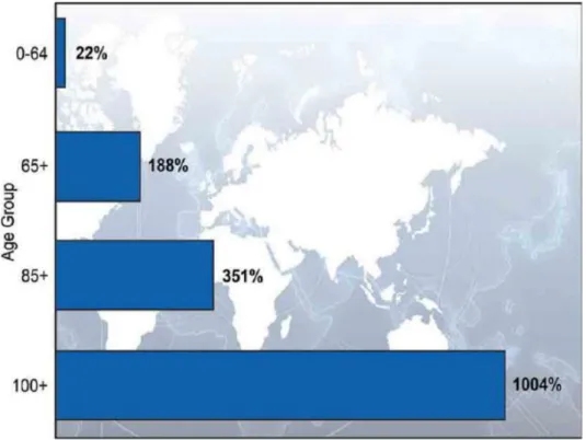
Aging is an undeniable and complicated biological process that is indicated by a general time- dependent decline in the physiological and biochemical functions of the main systems. The percentage of elderly people in the world is increasing steadily (Figure 1) due to the impressive rise in average life expectancy in the past century.
Figure 1. Percentage change in the world’s population by age: 2010-2050.
After age 30, a change in body composition occurs. However, a reduction of about 0.23 kg muscle mass per year is expected from ages of 30 to 60 which accelerates to 2% annually from the age of 60. One of the most well-known features of aging is a change in body composition and decline in lean body mass. Age related skeletal muscle wasting, also called sarcopenia, plays a significant role in reducing performance. On average, it is estimated that 5–13 % of elderly people aged 60–70 years are impressed by sarcopenia, and the numbers raise to 11–50
% for those aged 80 or above. Sarcopenia is, therefore, a multifactorial consequence of aging and indicative a potent risk factor for the development of negative health-related conditions in the elderly. It has been estimated that direct healthcare costs related to sarcopenia in the United States of America in 2000 were $18.5 billion, which was around 1.5% of total healthcare expenses for that year.
2
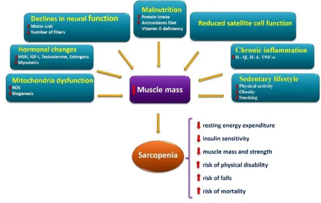
Although the exact mechanisms involved in sarcopenia is still unclear, however, several factors have been proposed to be involved in the onset and progression of sarcopenia (Figure 2).
Sarcopenia arises from several physiopathological factors including, but not limited to:
sedentary lifestyle, chronic inflammation, impaired satellite cell function, malnutrition, declines in neural function, hormonal changes, and mitochondrial dysfunction. It is also probable that certain underlying mechanisms are of greater influence than others when considering any specific age group, gender, or association with comorbid states.
Several studies have been suggested that IGF-1 is an imperative modulator of muscle mass, strength and function, not only during development, but also across the entire life span. In general, tissue responsiveness to IGF1 is altered with aging. Aging is associated with reductions in IGF1R content and phosphorylation in skeletal muscle. Cross-sectional studies have been shown, circulating levels of IGF-1 decrease with age in both men and women, as well as in rodents. In the older compared to the young males subjects, GHR and IGF-1 mRNA were reduced by 45%.
Figure 2. Schematic model of main factors involved in the onset and progression of the sarcopenia and its consequences
There are a number of evidence supporting the hypothesis that mitochondrial function and biogenesis appear to be altered in skeletal muscles of older adults, which in turn may contribute
3
to altered skeletal muscle mass and function. Alterations in mitochondria have been considered in aging, such as reduced total volume, elevated oxidative damage, and decreased oxidative capacity. These alternations may resulted in not only a loss of muscle mass and function with age, but other diseases associated with aging such as ectopic lipid infiltration, systemic inflammation, and insulin resistance.
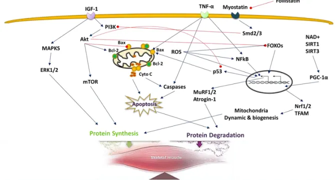
Maintenance of skeletal muscle mass is mainly dependent on the balance between protein synthesis and breakdown. An increase in protein breakdown leads to the muscle atrophy, whereas an increase in protein synthesis leads to muscle hypertrophy. Numbers of cellular mechanisms and signaling pathways are involved in age-related skeletal muscle wasting in mammals (Figure 3). These are discussed in more detail in the following section.
Figure 3. Molecular mechanisms involved in sarcopenia which can be viewed as the result of a protein synthesis/degradation imbalance.
Along with increased longevity, the prevalence and cost of sarcopenia are likely to rise. It is associated with elevated risk of cardiac, pulmonary, and metabolic disease processes, which further contributes to the socioeconomic burden. This global aging phenomenon has led to an increased morbidity and greater need for hospitalization and/or institutionalization. Healthy style of life is essential for older people to remain independent and to continue to actively take part in family and community life. Developing new therapies and strategies to prevent and treat sarcopenia not only will help to improve the quality of life for patients, but also will help to reduce the economic and productivity burdens for society in general. In this regard, in order
4
to provide further insight into the development of more effective therapy, it is important to explore the mechanism and etiology of sarcopenia. Among factors, free testosterone, physical activity, cardiovascular disease, IGF-1 in men and total fat mass and physical activity in women are significantly associated with muscle mass. In spite of the documented benefits of aerobic exercise on cardiovascular and metabolic health for older adults, the influence of this type of exercise on skeletal muscle mass and function is less understood. Furthermore, comparison of master athletes with sedentary controls demonstrated that training may not be sufficient to prevent skeletal muscle loss in older adults. Therefore, it seems that factors, such as nutrition in conjunction with appropriate physical activity can help attenuate the age related physical and muscle mass decline and maintain quality of life.
Objectives
As mentioned in the previous above, reduced systemic anabolic hormone levels, especially IGF-1 and reduced mitochondrial efficiency with aging are two potential factors involved in sarcopenia. Despite strong evidence for the effectiveness of RT, the effect of ET on the prevention and reduction of age-related muscle loss is still not well understood. Furthermore, available data from previous studies on hormone therapy is ambiguous. Although, it has been shown that the combination of exercise and hormone supplementation is more effective than either alone in attenuating muscle atrophy.
In our study, we aimed to elucidate the effects of endurance training with or without IGF-1 administration on four of the main mechanisms, involved in the onset and progression of sarcopenia including, decreased rate of protein synthesis, increased protein degradation, alteration in mitochondrial biogenesis and increased apoptotic signaling.
It was hypothesized that endurance exercise training alone and in combination with IGF-1 treatment has beneficial effects on age-related muscle atrophy, and in general can attenuate the process of sarcopenia. The following assumptions were made:
H1. Aging influences the cellular markers involved in sarcopenia.
H2. Endurance exercise training influences the cellular markers involved in sarcopenia.
H3. IGF-1 administration enhances positive effects of endurance exercise on the cellular markers involved in sarcopenia.
5 Methods
Subjects
Fifteen young (3 months old) and 15 old (26 months old) male Wistar rats were used in the study. We chose 26-month-old rats because previous studies have reported that sarcopenia is evident at 22 months age in this species. Subjects then were assigned to one of the following groups: young control (YC), young exercised (YE), young exercised and IGF-1-treated (YEI), old control (OC), old exercised (OE), and old exercised and IGF-1-treated (OEI). The investigation was carried out according to the requirements of The Guiding Principles for Care and Use of Animals of the European Union and approved by the local ethics committee.
Exercise protocol
Exercised rats were introduced to treadmill running for 3 days; then for the next 2 weeks the running speed was set at 10 m/min, with a 5% incline for 30 min/day, 5 days per weeks. The running speed and duration of the exercise were gradually increased to 60% of VO2 max of the animals. Therefore, on the last week of the 6-week training program, young animals ran at 22 m/min, on a 10% incline, for 60 min, whereas old animals ran at 13 m/min, on a 10% incline for 60 min. At the end of the study the animals were anesthetized with intraperitoneal injections of ketamine (50 mg/kg) and were sacrificed. This occurred two days after the last exercise session, to avoid any metabolic effects of the final run (Figure 8). Quadriceps muscle was carefully excised and stored at -80 ◦C.
IGF-1 administration
In the last 2 weeks of the study, treated animals received 5 μg/kg per day, 0.5 μL/hr IGF-1, whereas non-treated animals received saline via the pumps. With the help of the Alzet pumps, the 2-week supplementation of IGF-1 or saline could be maintained at constant flow, thus avoiding daily injections and their possible disturbance of behavioral and cognitive functions of the animals.
6 Tissue preparation
Frozen vastus lateralis samples were weighed (~100 mg) and homogenized (1:10 w/v) in ice- cold buffer (20 mM Tris–HCl pH 8, 137 mM NaCl, 2% NP-40, 10% glycerol) supplemented with phosphatase and protease inhibitors. The homogenates were incubated at 4 °C for 30 min, and then centrifuged at 12,000 g for 20 min at 4 °C. Then, the supernatant was collected and Bradford assays were used to determine supernatant protein concentrations. Proteins were diluted in 2× SDS sample buffer (1:1) and then heated to 95°C for 5 minutes.
Western blot analysis
Ten to 20 μg of protein were electrophoresed on 6–15% vol/vol polyacrylamide sodium dodecyl sulfate polyacrylamide gel electrophoresis (SDS-PAGE) gels. Proteins were electrotransferred onto polyvinylidene fluoride (PVDF) membranes. The membrane was blocked with 5% milk–Tris-buffered saline with 0.1% tween-20 solution and then incubated with a primary antibody overnight at 4°C. Primary antibodies are described in and were diluted 1:500 to 1:1000 with 5% milk–Tris-buffered saline with 0.1% tween-20. Blots were incubated with horseradish peroxidase–conjugated secondary antibody diluted 1:3000 with 5% milk–
Tris-buffered saline with 0.1% tween-20. After incubation with the secondary antibody, membranes were washed repeatedly and then were incubated with chemiluminescent substrate and protein bands were visualized on X-ray films. The bands were quantified by Image J software and normalized to B-actin, which served as an internal control.
Measurement of IGF-1 level
After sacrificing the animals, blood was collected, supercharged ethylenediaminetetraacetic acid (EDTA) was added, and the samples were centrifuged at 3000 × g, for 10 min at 4°C.
Plasma was separated and kept at −80°C. A Quantikine Mouse/Rat IGF-1 Assay Kit (R&D Systems, cat. no. MG100) was used to detect IGF-1 levels according to the description of the supplier. Briefly this assay employs the quantitative sandwich enzyme immunoassay technique. A monoclonal antibody specific for rat IGF-1 has been pre-coated onto a microplate.
Standards, control, and samples were pipetted into the wells and any rat IGF-1 present was bound by the immobilized antibody. After washing away any unbound substances, an enzyme- linked polyclonal antibody specific for mouse/rat IGF-1 was added to the wells. Following a
7
wash to remove any unbound antibody-enzyme reagent, a substrate solution was added to the wells. The enzyme reaction yielded a blue product that turned yellow when the Stop Solution was added. The intensity of the color measured was in proportion to the amount of rat IGF-1 bound in the initial step. The sample values were then read off the standard curve. The optical density of each well were determined within 30 minutes. microplate reader and wavelength was set to 450 nm and 570 nm respectively.
Statistical analyses
Statistical significance was assessed by the IBM SPSS program version 21. Data were tested with Shapiro-Wilk’s W normality test. Parametric data were analyzed by one-way ANOVA, followed by Tukey’s post hoc test. Kruskal-Wallis ANOVA followed by Mann-Whitney U test was applied to evaluate the differences in non-parametric results in case of those variables where post-hoc analysis was required. The significance level was set at p < 0.05.
Result
H1: Aging negatively affected cellular markers involved in sarcopenia
Maintaining muscle mass is achieved by balance between protein synthesis and protein degradation. An increase in muscle mass can come about due to either an increase in protein synthesis or a decrease in degradation, while a decrease in muscle mass can occur as a result of decreasing protein synthesis or increasing protein degradation. Two major signaling pathways control skeletal muscle growth: the IGF1–Akt/PKB–mTOR pathway acts as a positive regulator of muscle growth, and the myostatin–Smad3 pathway acts as a negative regulator.
Our result demonstrated that aging was associated with a reduced level of IGF-1 and expression level of follistatin -two important protein synthesis stimulators in mammalian skeletal muscle- in old rat.
8
H2: Endurance training positively reversed some cellular markers involved in sarcopenia Physical inactivity is a significant contributing factor to age-related sarcopenia. It is well established that sedentary elderly have less skeletal muscle mass and high prevalence of disability. Exercise training is considered as a simple, feasible, and inexpensive strategies available to prevent the onset of sarcopenia and reduce the rate of functional decline. Previous studies have clearly shown the effectiveness of resistance-type exercise interventions on skeletal muscle mass and functional capacity in the elderly. In addition, endurance type exercise training has been shown to improve muscle mass and strength, and increase performance capacity in both the young and elderly.
The data from our study showed that 6 weeks endurance exercise training did not lead to significant changes in the level of IGF-1 and protein content and phosphorylated of Akt, pAkt, mTOR, pmTOR, pERK1/2, but significantly increased the amount of follistatin and decreased pERK1/2 in the old exercise group compared to old control.
H3: Endurance training combined with IGF-1 administration positively affected some cellular markers involved in sarcopenia
Our third study question was about the effect of combination of endurance training and IGF-1 treatment on some cellular markers involved in sarcopenia. To the best of our knowledge, our study is the first, which has studied the effects of combined endurance training and IGF-1 treatment on skeletal muscle cellular markers of sarcopenia in old rats. However, previous studies investigated the effects of combined endurance training and administration of testosterone, growth hormone, fresh red orange juice, melatonin, resveratrol, 17 beta-estradiol and perindopril and caloric restriction on aged skeletal muscle in humans and rodents.
We found that two weeks of IGF-1 administration significantly increased serum IGF-1 levels in OEI compared to OC. The most remarkable result to emerge from our data is that protein expression of Bcl-2 was significantly higher in OEI than in OC and OE. Furthermore, Bax to Bcl-2 ratio as an apoptotic index was significantly lower in OEI vs. OC. Our result also demonstrated that protein contents of SIRT3 and Nrf2, but not SIRT1, PGC-1a, Cyto C and Cox 4, were significantly increased in OEI compared with OC.
9 Conclusions
In conclusion, our findings demonstrate that aging is associated with activation of signaling pathways involved in sarcopenia. Based on our study, it seems that among cellular factors that are responsible for protein synthesis and degradation, increased activity of proteolysis signaling, such as FOXO and apoptosis are contributed more to the onset and progression of sarcopenia than decreased anabolic pathways.
In regard to our finding, short term ET may attenuate sarcopenic conditions in old rats. This protective effect of ET seems to be due to, at least in part, improvement of mitochondria biogenesis and function as well as reduced levels of factors involved in protein degradation, such as MuRF1, in aged skeletal muscle.
According to the results of this study, combination of ET and IGF-1 administration can enhance the effects of ET alone on sarcopenic rats. This reinforcing effect of IGF-1 is likely to be through the modulation of activator and inhibitor factors of apoptosis such as TNF-α and Bcl- 2, respectively.
However, in order to achieve the best combination therapy it is essential for future researches to examine the impact of the intensity and duration of ET, as well as that of different doses of the hormone at various level of sarcopenia in both old female and male subjects.
10 Publications related to this study
Mosaferi Ziaaldini M, Koltai E, Csende Z, Goto S, Boldogh I, Taylor AW, Radak Z. (2015) Exercise training increases anabolic and attenuate catabolic and apoptotic processes in aged skeletal muscle of male rats. Experimental Gerontology. In press. (Impact Factor: 3.529)
Torma, F., Koltai, E., Nagy, E., Ziaaldini, M. M., Posa, A., Koch, L. G. … Radak, Z. (2014).
Exercise Increases Markers of Spermatogenesis in Rats Selectively Bred for Low Running Capacity. PLoS ONE, 9(12), e114075. doi:10.1371/journal.pone.0114075. (Impact Factor:
3.53)