expression in canine mammary tumours:
Prognostic and diagnostic role
OZLEM OZMEN
pDepartment of Pathology, Faculty of Veterinary Medicine, Burdur Mehmet Akif Ersoy University, Istiklal Yerleskesi, 15030, Burdur, Turkey
Received: April 22, 2020 • Accepted: May 8, 2020 Published online: November 21, 2020
ABSTRACT
Mammary tumours are among the most common tumours in dogs and are of interest due to their similarities to human breast tumours. Insulin-like growth factors (IGFs) are considered important in cell growth and development. The aim of this study was to investigate the immunohistochemical expression of IGF-I and IGF-II in benign and malignant canine mammary tumours. In this study, 10 benign and 10 malignant mammary tumours from the archives of the Department of Pathology were used, and five normal breast tissues were used as controls. It was observed that the expression of IGF-I and IGF-II was low to absent in benign tumours and increased in malignant tumours. The expression of IGF-II was higher than that of IGF-I. This study showed that IGF-I and IGF-II can be used as criteria for malignancy in canine mammary tumours. The results also indicate that IGF-I and IGF-II may be used as early diagnostic markers, and their inhibition may be used for the treatment of canine and human mammary tumours in the future.
KEYWORDS
dog, mammary tumour, IGF-I, IGF-II, immunohistochemistry
INTRODUCTION
Mammary tumours are the second most common skin tumours among dogs, and their incidence rate is 27.1% (Cohen et al., 1974; Johnston et al., 2001). It is known that steroid hormones such as progesterone and oestrogen play a role in the pathogenesis of mammary tumours (Queiroga et al., 2005; Illera et al., 2006). It has been reported that growth hormone (GH) and thus insulin-like growth factor (IGF) also contribute to tumour development when exogenous progestin is administered for contraceptive purposes (Concannon et al., 1980;
Selman et al., 1994; Mol et al., 1995, 1996, 1997; Johnston et al., 2001).
IGFs play roles in many physiological and pathological processes throughout life. For example, growth, metabolism, longevity, and diseases such as cancer, obesity, eating disor- ders, and neurodegenerative diseases are affected by the IGF pathway. Therefore, IGF signals are critical and must be regulated carefully for effective targeting of the system in under- standing the underlying mechanisms, not only in increments but also in the timing and cell-/
tissue-specific regulation of the IGF system components. However, some aspects are still not well understood. The IGF system is very complex with multiple elements at play. The IGF system is conventionally derived from three ligands (IGF-I, IGF-II, and insulin); their receptors–IGF-I receptor (IGF-IR), mannose 6-phosphate/IGF-II receptor (M6P/IGF-IIR), insulin receptor (IR), and hybrid IR (IGF-IR)–comprise at least six IGF-binding proteins (IGFBP1–6), acid labile subunit (ALS), and binding protein proteases. The IGF system clearly plays a role in regulating tumour growth and developing resistance to drug treatment (Mancarella and Scotlandi, 2018).
Acta Veterinaria Hungarica
68 (2020) 3, 269–274 DOI:
10.1556/004.2020.00044
© 2020 Akademiai Kiado, Budapest
RESEARCH ARTICLE
*Corresponding author.
E-mail:ozlemozmen@mehmetakif.edu.
tr, Tel.:þ90 (248) 2132170
The IGF system, known as growth and differentiation factor, released by GH stimulation, exists in two forms: IGF- I and IGF-II (Engstr€om et al., 1998; Samani et al., 2007;
Bergman et al., 2013). IGF-I is a basic peptide structure synthesised in the liver after birth, while IGF-II is a neutral peptide structure synthesised from various somatic tissues in the early embryonic and fetal periods (Engstr€om et al., 1998). Dogs express GH mRNA and receptor in normal and cancerous breast tissue. Due to the mitogenic effect of IGF-I, it plays a role in both growth and mammary tumour for- mation (Concannon et al., 1980; Van Garderen et al., 1999;
Queiroga et al., 2005, 2008, 2010). In transgenic mice with increased IGF-I synthesis, IGF-I causes ductal hypertrophy in the mammary glands and prevents breast involution after lactation (Kleinberg, 1998). Although there are studies on plasma GH and IGF-I concentrations and expression in breast tissue in canine mammary tumours, the role of IGF-II has not been fully elucidated yet.
The aim of this study was to determine whether IGF-I and IGF-II were expressed in different benign and malignant mammary tumours by immunohistochemistry, and to investigate whether there was a correlation between the expression rate and malignancy criteria. Numerous reports suggest a significant relationship between IGF and tumour malignancy in humans and experimental animals. There are also some publications about the release of IGF-II in canine malignant tumours. The results of this study show that IGF- I and IGF-II may be used to evaluate benign and malignant tumours, and due to the close relationship between increased expression and malignancy, their inhibition may be used for treatment approaches in the future.
MATERIALS AND METHODS
In this study, a total of 20 canine mammary tumours (10 malignant and 10 benign) and 5 normal breast tissues collected from the paraffin-embedded block archive of the Department of Pathology were used. All samples were selected from formalin-fixed and routinely processed cases that had presented for routine diagnosis. For histopathology and immunohistochemistry, paraffin blocks were cut to a thickness of approximately 5
m
m with a Leica RM2155 rotary microtome (Leica Microsystems, Wetzlar, Germany).All slides were stained routinely with haematoxylin and eosin (HE). After this evaluation, sections were stained immunohistochemically in order to demonstrate IGF-I and IGF-II expression. Commercial kits were used for immu- nohistochemical examination of IGF-I [IGF-I (W18):
sc-74116, Santa Cruz Biotechnology, Inc. Heidelberg, Ger- many, 1/100 dilution] and IGF-II [Anti-IGF2 antibody (ab170304), Abcam, Cambridge, UK, 1/100 dilution], using a routine streptavidin–biotin peroxidase technique, accord- ing to the manufacturer’s instructions. The Mouse and Rabbit Specific HRP/DAB Detection Kit–Micropolymer (ab236466) was used as the secondary antibody. The pri- mary antibody was omitted in negative controls. All the slides were analysed for immunopositivity.
To evaluate the severity of the immunohistochemical reaction of tumour cells with markers, semiquantitative analysis was performed using a grading score ranging from () to (þþþ) as follows: ()5negative, (þ)5focal weak staining, (þþ) 5 diffuse weak staining, (þþþ) 5 diffuse strong staining. For evaluation, 10 different areas were examined under 403 objective magnification in each sec- tion. Morphometric analyses and microphotography were performed using the Database Manual Cell Sens Life Science Imaging Software System (Olympus Co., Tokyo, Japan).
Statistical analysis
Statistical analysis was carried out in SPSS (Statistical Package for Social Sciences) 15.0 software (SPSS Inc., Chi- cago, Ill, USA). Results are expressed as mean±SD. In the statistical evaluations, a one-way analysis of variance test was used to evaluate any differences between the immuno- histochemical scores of malignant and benign tumours and normal control mammary gland. Duncan’s multiple com- parison method was used.P values <0.05 were accepted as statistically significant.
RESULTS
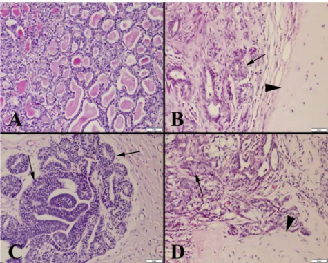
Severe hyperaemia in vessels was the most common histo- pathological finding in all mammary tumours. Haemor- rhagic foci were noted in some malignant tumours. The typical glandular structure with irregular shapes was pre- served in benign tumours, but excessive proliferation, necrosis, and loss of the glandular appearance were frequently observed in malignant tumours.
In mammary adenomas, a tissue architecture similar to normal mammary gland structure was noted. There was prominent fibrous tissue between the mammary glands.
Mitosis and pleomorphism were not observed (Fig. 1).
Ductal adenoma characterised by slight papillary growth limited to the ducts was diagnosed in a dog. Cartilage and osseous metaplasia was common in benign mixed tumours, but no necrosis, anaplasia, or mitosis was observed.
In malignant mammary tumours, epithelial cell struc- tures varied from cubic to flat, and cystic structures, papillary extensions, and atypical cells were seen in the mass. The number of mitotic figures varied from case to case and was usually high. In addition, slight connective tissue proliferation was detected in many malignant tu- mours. Excessive necrotic areas were generally observed.
Inflammatory cell infiltrates, mostly in neutrophil leuko- cytes and, in some cases, mononuclear cells were found around the necrotic areas. In malignant mixed tumours, cartilage and osseous metaplasia with pleomorphic cells was commonly observed.
The criteria for malignancy were evaluated according to mitotic activity, anaplastic, pleomorphic cells, and changes in general histological structures. The presence of meta- static masses was the most important criterion for malig- nancy. The results of histopathological diagnosis are shown inTable 1.
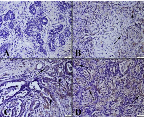
The relationships between IGF-I and IGF-II expression and tumour malignancy, which were the basis of our study, were evaluated by immunohistochemical methods. These markers were widely expressed in malignant tumours, but their expression was low to absent in benign tumours.
Different reactions in terms of IGF-I and IGF-II expression were noted in different areas of the same tumour mass. There was an increase in IGF-I and IGF-II expression, especially in areas where malignancy was more prominent. It was observed that both epithelial cells and mesenchymal cells had positive reactions for IGF-I and IGF-II in mammary tumours, and these reactions coincided with malignancy. The activity was higher in metastatic tumours. It was also noted that the cells with the most intense mitotic activity in the tumour mass were also those that expressed the most IGF-I and IGF-II.
IGF-I was found to have increased expression in benign and malignant tumours. Expression was found in both mammary gland cells and myoepithelial cells (Fig. 2). Negative or very slight IGF-II expression was observed in benign tumours, and severe expression was noted in malignant tumours (Fig. 3).
IGF-II was expressed markedly in tumour cells compared with IGF-I. The results of statistical analysis of the immu- nohistochemical scores are shown inTable 2.
DISCUSSION
Breast cancers have been considered among the most important causes of death in both humans and animals in Fig. 1.Histopathological appearance of the tumours. (A) Mammary adenoma, typical irregularly shaped glandular appearance; (B) Benign
mixed tumour, excessive cartilage metaplasia (arrow head) and mammary glands with slight proliferation (arrow); (C) Mammary adenocarcinoma with excessive proliferation (arrows) and loss of the glandular lumen; (D) Malignant mixed tumour, excessive proliferation in mammary glands, loss of tissue architecture (arrow), and cartilage metaplasia (arrowhead). Haematoxylin and eosin (HE). Bars550mm
Table 1.IGF-I and IGF-II expression scores of the mammary tissue and benign and malignant mammary tumours with pathological
classification
No. Tissue or diagnosis of the tumours IGF-I
IGF- II
1 Normal mammary tissue
2 Normal mammary tissue
3 Normal mammary tissue
4 Normal mammary tissue
5 Normal mammary tissue
6 Benign mixed tumour þ þ
7 Benign mix tumour þ þ
8 Benign mixed tumour þ þ
9 Benign mixed tumour þ þ
10 Benign mixed tumour þ þ
11 Mammary adenoma þ þ
12 Mammary adenoma þ þ
13 Mammary adenoma þ þ
14 Mammary adenoma þ þ
15 Mammary adenoma þ þ
16 Mammary adenocarcinoma þþ þþ
17 Mammary adenocarcinoma þþþ þþþ
18 Mammary adenocarcinoma þþ þþþ
19 Mammary adenocarcinoma þþþ þþþ
20 Mammary adenocarcinoma þþ þþ
21 Malignant mixed tumour þþ þþþ
22 Malignant mixed tumour þþþ þþ
23 Malignant mixed tumour þþþ þþ
24 Malignant mixed tumour þþ þþþ
25 Malignant mixed tumour þþ þþþ
recent years. For this reason, studies on the detection or treatment choice of biomarkers that can be used in the diagnosis and treatment of cancer are at the forefront of the prognosis. For the detection of these biomarkers, new techniques to measure both tissue and plasma levels are
being developed (Baker, 2000; Pepe et al., 2001; Dia- mandis and Yousef, 2002). The aim of this study was to determine the effect of IGF-I and IGF-II expression on the benign or malignant behaviour of canine mammary tu- mours.
Fig. 3.IGF-II expression in the tumours. (A) Slight expression (arrow) in a mammary adenoma; (B) Moderate expression (arrows) in a benign mixed tumour; (C) Marked expression (arrows) in a mammary adenocarcinoma; (D) Increased expression (arrows) in a malignant
mixed tumour. Streptavidin–biotin peroxidase method. Bars550mm
Fig. 2.IGF-I expression in the tumours. (A) Very slight expression in a mammary adenoma; (B) Slight expression (arrows) in a benign mixed tumour; (C) Marked expression (arrows) in a mammary adenocarcinoma; (D) Increased expression (arrows) in a malignant mixed
tumour. Streptavidin–biotin peroxidase method. Bars550mm
Although the expression of GH and IGF-I proteins in the mammary tissue of humans is known, the number of studies investigating IGF-I and IGF-II expression in canine mammary tumours is limited (Klopfleisch et al., 2010; Queiroga et al., 2010). IGF-I mediates the metabolic effects of GH. The IGF-I level increases after adolescence and decreases after this period, while the highest IGF-II level occurs in the fetal period.
In adulthood, IGF-II has been shown to be expressed in the liver and brain tissue and has an important role in cell pro- liferation, growth, and differentiation (Engstr€om et al., 1998;
Bergman et al., 2013). IGF-I is released from the stromal part of the mammary gland, while IGF-II is synthesised from both stromal and epithelial cells (Mol et al., 1999). In the present study, intense IGF-II expression was also observed in the mammary gland epithelium and myoepithelial tissues in dogs.
Generally, studies on human breast tumours are per- formed in either mouse models or cell cultures. However, due to the close relationship between human and canine mammary tumours, researchers have started to use canine mammary tumours as an alternative in the study of human breast cancers in recent years.Hawai et al. (2013)as well as Sultan and Ganaie (2018)showed that a number of factors can be adapted to human and canine tumours. These include homologous genome sequencing, genetic differ- ences, spontaneous tumour development, coexistence, and many other factors. Similarly, Queiroga et al. (2011), Mol (2013) and Carvalho et al. (2016) have shown many simi- larities, such as the same markers used in numerous immunohistochemical analyses and the presence of inflam- matory reactions with the same role in the tumour micro- environment. These comparisons between human and canine mammary tumours have been increasing in recent years (Nerurkar et al., 1989; Sorenmo et al., 2009; Cassali, 2013). Therefore, studies on canine breast tumours are also applicable to human tumours. The results of this study also showed that IGF-I and IGF-II expression may be important in the malignancy of human mammary tumours.
This study was carried out on 10 malignant and 10 benign canine mammary tumours and 5 normal mammary tissues in our department archives. For this purpose, tumour masses were first re-evaluated, and possible misdiagnoses were eliminated. Then, sections immunohistochemically stained with IGF-I and IGF-II were investigated to
determine whether there was a relationship between malig- nancy and IGF-I and IGF-II release. Even though the number of tumours used in our research was low, it is still one of the important studies on this subject.
The incidence of malignant mammary tumours in dogs varies between 41% and 53% (Misdorp et al., 1999; Lana et al., 2007). In a study of 4,755 Beagle dogs, the incidence was lower than 34% (Benjamin et al., 1999). Researchers reported that advanced circuit tumour operations were higher in non-mated and elderly females. In our archive of study material, the incidence of malignant tumours was found to be high and consistent with the classicalfindings (Misdorp et al., 1999; Misdorp, 2002).
The results obtained in our study show that IGF-I and IGF-II expression in canine mammary tumours is signifi- cantly different between benign and malignant tumours and that IGF-I and IGF-II expression increases significantly with malignancy. Since the study was conducted retrospectively, it was not possible to determine whether there was a rela- tionship between IGF-I and IGF-II expression and survival time. However, it is suggested that increased expression of IGF-I and IGF-II in malignant tumours may be related to decreased survival in dogs. More detailed studies on this topic are needed to shed light on the subject. In addition, the plasma levels of IGF-I and IGF-II should be determined and correlated with tissue levels in future studies.
The data obtained in this study suggest that IGF-I and IGF-II may be useful in the evaluation of mammary tumour malignancy and that the inhibition of IGF-I and IGF-II may be used in the treatment of canine or human mammary cancers in the future.
ACKNOWLEDGEMENT
This study was supported by the Scientific Projects Com- mission of the Burdur Mehmet Akif Ersoy University (Project number: 0387-MP-16).
REFERENCES
Baker, S. G. (2000): Identifying combinations of cancer markers for further study as triggers of early intervention. Biometrics 56, 1082–1087.
Benjamin, S. A., Lee, A. C. and Saunders, W. J. (1999): Classification and behavior of canine mammary epithelial neoplasms based on life-span observations in beagles. Vet. Pathol.36,423–436.
Bergman, D., Halje, M., Nordin, M. and Engstr€om, W. (2013):
Insulin-like growth factor 2 in development and disease: a mini-review. Gerontology59,240–249.
Carvalho, M. I., Silva-Carvalho, R., Pires, I., Prada, J., Bianchini, R., Jensen-Jarolim, E. and Queiroga, F. L. (2016): A comparative approach of tumor-associated inflammation in mammary cancer between humans and dogs. Biomed Res. Int. 4917387.
Cassali, G. D. (2013): Comparative mammary oncology: Canine model. BMC Proc.7,Suppl. 2, K6.
Table 2.Statistical analysis results of IGF-I and IGF-II expressions in benign and malignant mammary tumours
Control Benign Malignant
P value IGF-I 0.00±0.00a 1.00±0.00b 2.10±0.31c <0.001 IGF-II 0.00±0.00a 1.00±0.00b 2.60±0.51c <0.001
*Statistical analysis of the scores was performed by analysis of variance (ANOVA). Differences between the groups were analysed by Duncan’s test; the values represent the mean±standard deviation (SD).
**The differences between the means of groups marked with different superscript letters in the same column are significantly different (P< 0.001).
Cohen, D., Reif, J. S., Brodey, R. S. and Keiser, H. (1974): Epide- miological analysis of the most prevalent sites and types of canine neoplasia observed in a veterinary hospital. Cancer Res.
34,2859–2868.
Concannon, P., Altszuler, N., Hampshire, J., Butler, W. R. and Hansel, W. (1980): Growth hormone, prolactin, and cortisol in dogs developing mammary nodules and an acromegaly-like appearance during treatment with medroxyprogesterone ace- tate. Endocrinol.106,1173–1177.
Diamandis, E. P. and Yousef, G. M. (2002): Human tissue kalli- kreins: a family of new cancer biomarkers. Clin. Chem. 48, 1198–1205.
Engstr€om, W., Shokrai, A., Otte, K., Granerus, M., Gessbo, A., Bierke, P., Madej, A., Sjolund, M. and Ward, A. (1998): Tran- scriptional regulation and biological significance of the insulin like growth factor II gene. Cell Prolif.31,173–189.
Hawai, S. M., Al Zayer, M., Ali, M. M., Niu, Y., Al Awad, A., Al Jofan, M., Al Jarbou, A. and Al Tuwijri, S. (2013): Dogs: active role model for cancer studies–A review. J. Can. Ther.4,989–
995.
Illera, J. C., Perez-Alenza, M. D., Nieto, A., Jimenez, M. A., Silvana, G., Dunner, S. and Pe~na, S. (2006): Steroids and receptors in canine mammary cancer. Steroids71,541–548.
Johnston, S. D., Root Kustritz, M. V. and Olson, P. N. S. (2001):
Disorders of the mammary glands of the bitch. In: Johnston, S.
D., Root Kustritz, M. V. and Olson, P. N. S. (eds) Canine and Feline Theriogenology. 1st edition. Saunders, Philadelphia. pp.
243–256.
Kleinberg, D. L. (1998): Role of IGF-I in normal mammary development. Breast Cancer Res. Treat.47, 201–208.
Klopfleisch, R., Hvid, H., Klose, P., da Costa, A. and Gruber, A.D.
(2010): Insulin receptor is expressed in normal canine mam- mary gland and benign adenomas but decreased in metastatic canine mammary carcinomas similar to human breast cancer.
Vet. Comp. Oncol.8,293–301.
Lana, S. E., Ruttemans, G. R. and Withrow, S. J. (2007): Tumors of the mammary gland. In: Withrow, S. J. and Vail, D. M. (eds) Small Animal Clinical Oncology. Saunders Elsevier, St. Louis, Missouri. pp. 619–636.
Mancarella, C. and Scotlandi, K. (2018): IGF system in sarcomas: a crucial pathway with many unknowns to exploit for therapy. J.
Mol. Endocrinol.61,T45–T60.
Misdorp, W. (2002): Tumors of the mammary gland. In: Meuten, D. J. (ed.) Tumors in Domestic Animals. State University of California, California. pp. 575–606.
Misdorp, W., Else, R. W. and Hellemn, E. (1999): WHO Histo- logical Classification of Mammary Tumors of the Dog and Cat.
Definitions and Explanatory Notes. Armed Forces Institute of Pathology, Washington. pp. 18–27.
Mol, J. A. (2013): Comparative breast cancer research– Lessons from companion animals. BMC Proc.7,Suppl. 2, K9.
Mol, J. A., Lantinga-van Leeuwen, I. S., van Garderen, E., Selman, P. J., Oosterlaken-Dijksterhuis, M. A., Schalken, J. A. and Rijnberk, A. (1999): Mammary growth hormone and tumourigenesis–Lessons from the dog. Vet. Q.21,111–115.
Mol, J. A., Selman, P. J., Sprang, E. P., van Neck, J. W. and Oos- terlaken-Dijksterhuis, M. A. (1997): The role of progestins, insulin-like growth factor (IGF) and IGF-binding proteins in the normal and neoplastic mammary gland of the bitch: a re- view. J. Reprod. Fertil. Suppl.51,339–344.
Mol, J. A., van Garderen, E., Rutteman, G. R. and Rijnberk, A.
(1996): New insights in the molecular mechanism of progestin- induced proliferation of mammary epithelium: induction of the local biosynthesis of growth hormone (GH) in the mammary glands of dogs, cats and humans. J. Steroid Biochem. Mol. Biol.
57,67–71.
Mol, J. A., van Garderen, E., Selman, P. J., Wolfswinkel, J., Rijnberk, A.
and Rutteman, G. R. (1995): Growth hormone mRNA in mam- mary gland tumors of dogs and cats. J. Clin. Invest.95,2028–2034.
Nerurkar, V. R., Chitale, A. R., Jalnapurkar, B. V., Naik, S. N. and Lalitha, V. S. (1989): Comparative pathology of canine mam- mary tumors. J. Comp. Pathol.101,389–397.
Pepe, M. S., Etzioni, R., Feng, Z., Potter, J. D., Thompson, M. L., Thornquist, M., Winget, M. and Yasui, Y. (2001): Phases of biomarker development for early detection of cancer. J. Natl Cancer Inst.93,1054–1061.
Queiroga, F. L., Perez-Alenza, M. D., Silvan, G., Pe~na, L., Lopes, C.
and Illera, J. C. (2005): Role of steroid hormones and prolactin in canine mammary cancer. J. Steroid Biochem. Mol. Biol.94, 181–187.
Queiroga, F. L., Perez-Alenza, M. D., Silvan, G., Pe~na, L., Lopes, C.
and Illera, J.C. (2008): Crosstalk between GH/IGF-I axis and steroid hormones (progesterone, 17b-estradiol) in canine mammary tumours. J. Steroid Biochem. Mol. Biol.110,76–82.
Queiroga, F. L., Perez-Alenza, M. D., Silvan, G., Pe~na, L., Lopes, C.
and Illera, J. C. (2010): Serum and intratumoural GH and IGF-I concentrations: Prognostic factors in the outcome of canine mammary cancer. Res. Vet. Sci.89,396–403.
Queiroga, F. L., Raposo, T. and Carvalho, M. I. (2011): Canine mammary tumours as a model to study human breast cancer:
Most recentfindings. In Vivo25,455–466.
Samani, A. A., Yakar, S., LeRoith, D. and Brodt, P. (2007): The role of the IGF system in cancer growth and metastasis: overview and recent insights. Endocrine Rev.28,20–47.
Selman, P. J., Mol, J. A., Rutteman, G. R. and Rijnberk, A. (1994):
Progestin treatment in the dog. I. Effects on growth hormone, insulin-like growth factor I and glucose homeostasis. Eur. J.
Endocrinol.131,413–421.
Sorenmo, K. U., Kristiansen, V. M., Cofone, M. A., Shofer, F. S., Been, A. M., Langeland, M., Mongel, C. M, Grondahl, A. M., Teige, J.
and Goldschmidt, M. H. (2009): Canine mammary gland tumors:
A histological continuum from benign to malignant; clinical and histological evidence. Vet. Comp. Oncol.7,162–172.
Sultan, F. and Ganaie, B. (2018): Comparative oncology: Integrating human and veterinary medicine. Open Vet. J.8,25–34.
Van Garderen, E., Van der Poel, H. J. A., Swennenhuis, J. F., Wissink, E. H. J., Rutteman, G. R., Hellmen, E., Mol. J. A. and Schalken, J. A. (1999): Expression and molecular characteriza- tion of the growth hormone receptor in canine mammary tissue and mammary tumors. Endocrinology140,5907–5914.