SIGMA-1 RECEPTOR AGONISM:
THE NOVEL TREATMENT OPTION IN RENAL DISEASE
Ph.D. Thesis
Ádám Hosszú
Doctoral School of Clinical Medicine Semmelweis University
Supervisor: Andrea Fekete, M.D., Ph.D.
Official reviewers:
Attila Szijártó, M.D., D.Sc.
Szilveszter Dolgos, M.D., Ph.D.
Head of the Final Examination Commitee:
Prof. Zoltán Benyó, M.D., Ph.D.
Members of the Final Examination Commitee:
Gábor Kökény, M.D., Ph.D.
Kristóf Dede, M.D., Ph.D.
Introduction
Today chronic kidney disease (CKD) is a global health crisis creating a huge social and financial burden. More than 10% of the worldwide population is affected by CKD and the number of cases is constantly increasing. End-stage renal disease (ESRD) is the final stage of CKD characterized by complete loss of kidney function.
Growing incidence of ESRD can be attributed to the rapidly escalating incidence of causative disorders, mainly diabetes mellitus and hypertension. More than 400 million people suffer from diabetes worldwide and diabetic nephropathy (DNP) develops in around one- third of these cases. Increasing global trends suggest that DNP will continue to drive the prevalence of CKD and ESRD in the future.
Acute kidney injury (AKI) is also a relevant risk factor for the development of CKD that accounts for 2 to 3% of ESRD cases annually. However AKI represents a much larger portion of the renal disease burden on the long run due to the significantly increased long-term risk of CKD and ESRD following AKI, even if renal function recovers initially. This relation seems to be bi-directional as CKD patients are more vulnerable to AKI as well.
For ESRD patients renal replacement therapy (dialysis or kidney transplantation) is the sole treatment option. Kidney transplantation (KTx) is the preferred treatment as it is associated with improved survival and quality of life. Although short-term outcomes of KTx have improved substantially due to advances in surgical technique and immunosuppression, long-term outcomes have remained largely unchanged over the past decades. The factors affecting long-term outcome may be either alloantigen-dependent (e.g. HLA matching, HLA immunization etc.) or alloantigen- independent (e.g. donor type and age of both the donor and recipient,
KTx. IRI is unavoidable and the duration of storage and cold ischemia time correlate with delayed graft function.
Notable morphologic features of ischemic AKI include the loss of proximal tubule brush border, depolarization and patchy loss of tubular cells and tubular cast formation as well as peritubular capillary congestion, endothelial damage and leukocyte accumulation. The endothelium of microcirculation plays a pivotal role in the pathophysiology of IRI. Even under normal conditions the kidney regions housing nephron segments with very high energy requirements sustain relative hypoxia due to lower blood flow and exchange of oxygen. Relative hypoxia worsens after ischemia, which leads to prolonged cellular injury and cell death. Renal blood flow is reduced by up to 50%, which persists in the outer medulla even during reperfusion. Release of vasoconstrictors such as endothelin is enhanced and abundance of vasodilators such as endothelium- derived nitric oxide (NO) is decreased causing vasoconstriction in small arterioles and peritubular capillaries.
Chronic vasoconstriction is a relevant pathognomic feature of DNP as well, where hyperglycemia activates the renin-angiontensin- aldosterone system (RAAS) leading to ischemia. Renal blood flow is decreased due to vasoconstriction, which further activates RAAS and increases the production of reactive oxygen species (ROS). These processes lead to functional and structural damage of the kidneys.
Inflammation is also an important contributor both of acute and chronic ischemic injury. Cells of the immune system accumulate and attach to the endothelium where they release ROS, proteases and inflammatory cytokines, all of which aggravate kidney injury in acute renal injury models as well as DNP.
Very recently a new molecule, the Sigma-1 receptor (S1R) has become the center of attention in brain ischemia and stroke as a possible mediator of key protective mechanisms. S1R is vastly expressed in the brain where it is localized in the endoplasmatic reticulum (ER), but upon ligand stimulation it translocates to the cytosol.
In models of brain ischemia S1R agonist treatment reduced infarct areas, presumably by promoting cell survival and reducing the inflammatory response. S1R stimulation was also effective in minimizing infarct size in cardiac hypertrophy. This protection seemed to be mediated by the upregulation of protein kinase B (Akt)- endothelial nitric oxide synthase (eNOS) signaling.
These preliminary results in brain and cardiac ischemia suggest that S1R activates vasodilatative and protective pathways.
Therefore it is rather presumable that S1R agonists could trigger similar mechanisms in renal IRI. Since renal IRI has high morbidity and mortality and treatment options are still very limited better understanding of the underlying molecular mechanisms are essential for developing novel renoprotective therapies.
In our preclinical experiments - during my PhD work - we aimed to investigate the promising option of S1R agonist treatment in acute and chronic kidney injury.
Objectives
The purpose of our experiments was to investigate the pathomechanisms of IRI-induced AKI in order to identify new therapeutic targets that can be used in KTx as well as in DNP. Based on the current literature demonstrating that S1R is protective in brain and heart ischemia the main aim was to investigate the effect of S1R and its modulation in renal diseases.
Our aims were the following:
1. To investigate the intrarenal and subcellular localization of S1R in the normal and ischemic kidney
2. To analyze the molecular mechanism of S1R - mediated effects 3. To determine the possible renoprotective effect of S1R agonism in acute (renal IRI) and chronic (DNP) models of kidney disease
4. To evaluate the protective effect of S1R agonist pretreatment in KTx
Methods
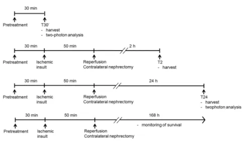
Renal ischemia/reperfusion injury model and treatment groups
Adult, male Wistar rats weighing 200±15g were used in all experiments. Renal ischemia was accomplished by cross-clamping the left renal pedicles for 50 min. Before the end of the ischemic period the contralateral kidney was taken out, the clips were removed. Sham animals underwent laparotomy of the same duration without clamping. At pre-determined times of reperfusion, blood samples were collected, the remnant kidneys were harvested.
To test the effect of dehydroepiandrosterone (DHEA) rats were pretreated first 25 hours, then 1 hour before the surgical procedure with:
(i) isotonic saline as vehicle (ii) DHEA (4 mg/bwkg)
To investigate the effect of fluvoxamine (FLU) animals were treated 30 min prior to the ischemic insult as follows:
(i) isotonic saline as vehicle (ii) FLU (20 mg/bwkg)
(iii) FLU (20 mg/bwkg) and NE100 (1 mg/bwkg)
To test the NO-mediated effect the following pretreatment was applied 30 min prior to the ischemic insult:
(i) FLU (20 mg/bwkg) and L-NAME (10 mg/bwkg, non- selective NOS inhibitor)
(ii) FLU (20 mg/bwkg) and L-NIO (20 mg/bwkg, selective eNOS inhibitor)
(iii) FLU (20 mg/bwkg) and 7-NI (25 mg/bwkg, selective nNOS inhibitor)
Figure 1. Experimental design of renal IRI
Rat model of kidney isograft autotransplantation
Kidneys were perfused with cold Custodiol perfusion solution, then removed from the animal: kidneys were placed into a container for 2 hours filled with:
(i) cold Custodiol perfusion solution
(ii) cold Custodiol perfusion solution containing 0.003 mg/mL FLU
After 2 hours kidneys were placed back into the rats and end-to-end anastomoses of the renal artery, vein and ureter were performed.
Contralateral kidneys were removed. Total warm ischemia time was 35 min.
Figure 2. Experimental design of renal autotransplantation
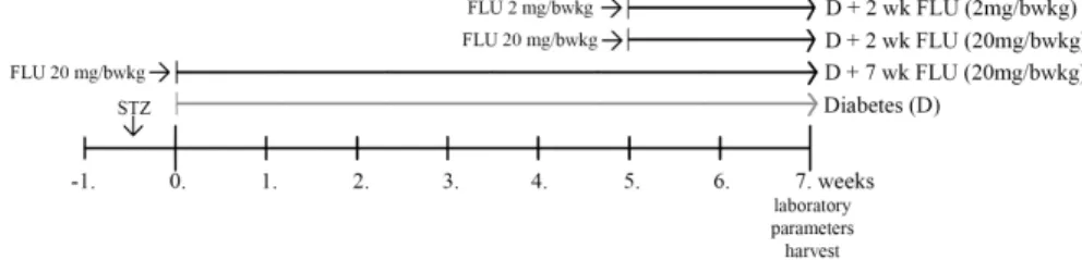
Rat model of type 1 diabetes mellitus (DM1) and experimental groups
DM1 was induced by a single intraperitoneal injection of 65 mg/bwkg streptozotocin (STZ) in freshly prepared 0.1 M citrate buffer. DM1 rats were randomly divided into four groups and were treated by oral gavage daily at 10:00 AM as follows:
(i) 20 mg/bwkg FLU for 7 weeks
(ii) 20 mg/bwkgFLU for 2 weeks after 5 weeks of DM1 (iii) 2 mg/bwkgFLU for 2 weeks after 5 weeks of DM1 Age-matched, non-diabetic control rats were treated per os with saline daily at the same time as the diabetic animals.
Figure 3. Experimental design of type 1 diabetes mellitus In vitro model
All in vitro experiments were performed on human proximal tubular epithelial cell line (HK2). Oxidative stress was induced by 400 µM hydrogen-peroxide treatment for 30 min. Cells were treated 30 min prior to harvest as follows:
(i) FLU (10 µM)
(ii) FLU (10 µM) + NE100 (3 µM)
(iii) FLU (10 µM) + AktVIII inhibitor (10 µM)
Experiments
Renal functional and metabolic parameters
The following renal functional and metabolic parameters were measured: serum creatinine (SCr), BUN, serum AST, serum glucose, fructosamine, electrolytes (sodium, potassium, chloride), albumin, total protein, triglycerides, total cholesterol and glutamate-pyruvate transaminase.
Histology
Tubular injury was evaluated on hematoxylin-eosin and periodic acid-Schiff stained kidney sections.
Masson’s trichrome staining was used to evaluate tubulointerstitial fibrosis and Sirius red staining to evaluate collagen accumulation.
S1R localization was determined with S1R-specific DAB staining and fluorescent immunohistochemistry.
In vivo measurement of renal perfusion and structure
Two-photon microscopy was used to assess renal injury and to measure peritubular capillary diameters in living rats.
Protein abundance measurements
Western blot was used to measure the followin proteins:
S1R, phospho-Akt (Ser473), phospho-eNOS (Ser1177) and nNOS.
Bands of interest were factored for Ponceau red staining to correct for any variations in total protein loading.
Measurement of tubular injury markers mRNA
Kidney injury markers Hif-1α, Ngal, Kim-1 and Mcp-1 were measured using quantitative real-time PCR and normalized against Gapdh as a housekeeping gene.
NO measurement
The total stable oxidation products of NO metabolism (NO2/NO3) of serum and HK2 cell homogenates were assessed using Griess reagent.
S1R gene silencing
HK2 cells were transfected with S1R specific siRNA or negative control siRNA using Lipofectamine 2000. The efficacy of knockdown was determined by Western blot.
Statistical analysis
Parametrical data are expressed as means ± SEM, while non- parametrical data as median ± range. Statistical analyzes were performed using Prism software (version 5.00; GraphPad Prism Software). Survival studies were assessed by Logrank test. Multiple comparisons and possible interactions were evaluated by one-way ANOVA followed by Bonferroni post-hoc test. For non-parametrical data the Kruskal–Wallis ANOVA on ranks followed by Fischer exacts test was used. P values of <0.05 were considered significant.
Results
Sigma-1 receptor is expressed in the kidney
Our group is the first to describe the region-specific distribution of Sigma-1 receptor (S1R) expression in the kidney.
S1R-specific DAB staining and Western blot revealed that S1R expression was most prominent in the renal cortex, but was also present in the medulla and papilla. Co-staining with proximal tubular brush border-specific PAS and anti-S1R DAB revealed that S1R is definitely expressed in proximal tubules but not in glomeruli. S1R fluorescent immunohistochemistry double staining with S1R and antibodies specific to different nephron segments supported the finding that S1R is mainly present in proximal tubules: S1R was co- localized with proximal tubule-specific gamma-glutamyltransferase, but not with Na+/K+-ATP-ase in distal tubules, eNOS in glomeruli or neuronal NOS (nNOS) in the macula densa.
DHEA improves post-ischemic kidney function and ameliorates structural injury
After confirming the presence of S1R in the kidney our goal was to investigate the effect of S1R agonist treatment on renal IRI.
DHEA is a highly abundant steroid hormone, which is also an endogenous agonist of S1R.
Post-ischemic survival was followed and compared to sham- operated controls for seven days. DHEA-pretreated rats survived longer than vehicle-treated ones (median survival: 72 versus 36 hours, P<0.001).
Another series of animals were sacrificed after 24 hours of reperfusion to investigate the acute effects of IRI. Intravital two- photon microscopy allowed us to measure changes of peritubular capillary diameters in live rats. Post-ischemic vasoconstriction was apparent suggesting that the subsequent decline in renal blood flow
could be a causative factor of renal functional and structural damage.
DHEA pretreatment improved kidney function and prevented peritubular vasoconstriction.
Histologic changes were consistent with functional decline:
brush borders disappeared, the majority of tubules showed cell necrosis and extensive cast formation. DHEA pretreatment considerably reduced tubular necrosis and partly preserved brush borders.
High affinity S1R agonist FLU is renoprotective following kidney IRI
To substantiate the crucial role of S1R in IRI, in a second set of experiments rats were pretreated with FLU that has much higher affinity to S1R than DHEA.
FLU-treated rats survived significantly longer than vehicle or FLU+NE100 (selective S1R antagonist)-treated ones (median survival: 67 versus 36 and 49 hours respectively, P<0.001).
SCr, BUN and serum AST were less elevated in FLU-treated rats after 24 hours of reperfusion and the improvement was neutralized by NE100 suggesting that S1R agonism successfully diminishes acute renal injury. Ngal and Kim-1 are specific, early indicators of tubular injury. Increased mRNA expression of these markers was ameliorated by FLU suggesting milder proximal tubular injury.
FLU ameliorates renal structural damage
IRI-induced histologic injury was assessed on PAS-stained kidney sections as well as in vivo using two-photon microscopy.
necrosis were only moderate, and brush borders were intact in several regions of FLU-treated kidneys after 24 hours of reperfusion.
The role of S1R in proximal tubular cells
To our best knowledge our group is the first to show that S1R is expressed in human proximal tubular cells. S1R abundance remained unchanged after FLU or H2O2 treatment; however localization was different under normal conditions and oxidative stress. The receptor showed perinuclear localization in control cells, but was detected everywhere in the cytosol and also in nuclei after H2O2 or FLU treatment.
FLU induces S1R-mediated NO production in HK2 cells
A possible signaling pathway by which vasodilatative NO could be produced is the Akt-eNOS pathway. FLU increased phospho-eNOS (peNOS, Ser1177; the active form of the eNOS enzyme) protein levels in HK2 cells both under normal conditions and oxidative stress. Parallel with successful S1R gene silencing peNOS protein was lower compared to scrambled siRNA-treated negative control cells. Decreased peNOS production in FLU-treated S1R gene silenced cells verified the regulatory role of S1R in peNOS expression. To confirm the role of Akt in FLU-induced NOS activation and NO production, both upstream (Akt IV) and downstream (Akt VIII) Akt inhibitors were used. Inhibition of Akt suppressed peNOS and subsequently NO production in FLU-treated cells.
S1R-mediated renal vasoregulation in SHAM-operated rats Once the role of S1R in NO production was characterized under in vitro circumstances the next step was to investigate the vasoregulatory effect of FLU in the rat kidney. Intravital two-photon
microscopy was used to measure peritubular capillary diameters in SHAM-operated rats after 30 minutes of FLU treatment. Parallel with increased peNOS and NO production FLU pretreatment caused peritubular capillary dilatation.
Peritubular capillaries showed significant vasoconstriction after 24 hours of reperfusion. Parallel with increased nitrite production capillary dilatation was significant in FLU-treated rats after 24 hours of reperfusion as well, which was diminished by NE100 or various NOS inhibitors supporting that the vasodilative effect of FLU is S1R-mediated and NOS-dependent.
The S1R - Akt - NOS signaling pathway in the kidney
FLU increased pAkt (Ser473) and peNOS (Ser1177) protein abundance as soon as 30 minutes after treatment, whereas S1R and nNOS remained unchanged. On the other hand after 24 hours of reperfusion all measured proteins were increased especially in FLU- treated rats. These results indicate that S1R signaling is instantly activated by FLU and is rapidly followed by peNOS production, but nNOS generation only increases later during reperfusion. As a result of increased NOS abundance NO production was also considerably increased contributing to vasodilatation in the post-ischemic kidney.
The effect of FLU-treatment on the transplanted kidney
Kidney function was substantially improved in FLU-treated rats as shown by decreased SCr and AST levels, however BUN was not decreased at this early time point yet. Early markers of kidney injury Ngal, Kim-1 and Mcp-1 were also significantly less elevated in FLU-treated, transplanted kidneys.
were perfused with FLU, but harvested after 2 hours of cold ischemia.
Chronic FLU-treatment is protective in DNP
DM1 induced severe renal impairment with increased SCr and BUN values. Fractional sodium excretion (FeNa) (reflecting tubular function) was increased and significant albuminuria (representing mainly glomerular function) were present indicating the development of DNP. Both long- and short-term treatment with FLU remarkably improved renal functional parameters.
FLU decreased DM1-induced mesangial matrix expansion in long-term, as well as short-term treatment in both dosages.
Fibrotic areas were extensive in DM1 kidneys. Both long- and short-term treatment with FLU successfully reduced tubulointerstitial fibrosis.
The amount of extracellular matrix was doubled in DM1 kidneys compared to controls, but was significantly reduced after long-term FLU treatment. Short-term FLU treatment did not reduce collagen production.
Changes in the S1R-Akt-eNOS signaling pathway were evaluated after long- and short-term FLU treatment. There was an increasing tendency in S1R and pAkt protein levels, however did not reach the level of significance (p=0.06). On the other hand depressed peNOS production in DM was rescued by both long- and short-term FLU treatment.
Conclusions
1. We were the first to clarify that S1R is expressed in different nephron segments, predominantly in the cortex, but also in the medulla and papilla.
2. Our in vivo and in vitro experiments revealed that upon stimulation S1R translocates from the mitochondria- associated ER to the cytoplasm and nucleus.
3. We showed that treatment with endogenous S1R agonist DHEA is renoprotective in a rat model of IRI.
4. We demonstrated that exogenous, high-affinity S1R agonist FLU treatment is protective in renal IRI. It substantially improves post-ischemic survival, ameliorates functional and structural kidney damage, as well as inflammation.
5. We described the role of S1R in improving perfusion in the post-ischemic kidney by inducing Akt-NOS signaling and NO production.
6. We demonstrated the renoprotective effect of FLU treatment in KTx.
7. We showed that chronic FLU treatment ameliorates functional and structural kidney damage in diabetic nephropathy, presumably via hindering fibrosis and rescuing eNOS production.
Bibliography of the candidate’s publications
1. Hosszu, A, Antal, Z, Lenart, L, Hodrea, J, Koszegi, S, Balogh, DB, Banki, NF, Wagner, L, Denes, A, Hamar, P, Degrell, P, Vannay, A, Szabo, AJ, Fekete, A. (2016) Sigma1-Receptor Agonism Protects against Renal Ischemia-Reperfusion Injury. J Am Soc Nephrol In press. IF=8.491
2. Lenart, L, Hodrea, J, Hosszu, A, Koszegi, S, Zelena, D, Balogh, D, Szkibinszkij, E, Veres-Szekely, A, Wagner, L, Vannay, A, Szabo, AJ, Fekete, A. (2016) The role of sigma-1 receptor and brain-derived neurotrophic factor in the development of diabetes and comorbid depression in streptozotocin-induced diabetic rats.
Psychopharmacol, 233: 1269-1278. IF=3.540
3. Gellai, R, Hodrea, J, Lenart, L, Hosszu, A, Koszegi, S, Balogh, D, Ver, A, Banki, NF, Fulop, N, Molnar, A, Wagner, LJ, Vannay, A, Szabo, AJ, Fekete, A. (2016) The role of O-linked N- Acetylglucosamine modification in diabetic nephropathy. Am J Physiol Ren Physiol: ajprenal.00545.02015. IF=3.390
4. Denes, J, Katona, M, Hosszu, A, Czuczy, N, Takats, Z.
(2009) Analysis of biological fluids by direct combination of solid phase extraction and desorption electrospray ionization mass spectrometry. Anal Chem, 81: 1669-1675. IF=5.214
Acknowledgment
First and foremost I am profoundly thankful to my supervisor Andrea Fekete for being a role model and showing me the value of hard work. Her tireless dedication and guidance is indispensable to everyone in our lab.
I wish to sincerely thank Professors Attila Szabo and Tivadar Tulassay for the proud privilege to work as a PhD student in their Research Laboratory of the 1st Department of Pediatrics, Semmelweis University.
I wish to express my gratitude to my PhD supervisors at Georgetown University, Prof. Christopher Wilcox and Dr. William Welch for their constant support and guidance. I also thank the Rostoczy Foundation for the financial support that made my stay possible.
I am also very grateful to the „seniors” of our lab, Adam Vannay, Laszlo Wagner and Judit Hodrea for their valuable experience and suggestions that helped me throughout the experiments. I learned a lot from their critical advice when I was writing my manuscripts.
I would like to thank all my colleagues at the lab for creating a motivating and friendly atmosphere. Special thanks to Sandor Koszegi for preparing the histological evalution, to Zsuzsanna Antal for her invaluable help in animal surgeries, to Agnes Prokai for teaching me two-photon microscopy. I could not be more thankful to Maria Bernath for her help with cell cultures and laboratory work, not to mention the delicious birthday cakes. I am priviliged of having the Lendulet colleagues as well: Fanni Banki, Dora Balogh, Edgar Szkibinszkij, Renata Gellai, thanks to you all for helping me survive these years.