Investigating the pathomechanisms of renal fibrosis in animal models –
Diabetic and toxic nephropathy
PhD thesis
Csaba Imre Szalay MD
Basic Medicine Doctoral School Semmelweis University
Supervisor:
Péter Hamar, MD, D.Sc
Official reviewers:
Csaba Kálmán Ambrus, MD, Ph.D Attila Marcell Szász, MD, Ph.D
Head of the Final Examination Committee:
Tamás Ivanics MD, Ph.D
Members of the Final Examination Committee:
László Wagner MD, Ph.D Ákos Thuma, DVM, Ph.D Andrea Fekete MD, Ph.D
Budapest 2016
1
1. Introduction
Chronic kidney disease (CKD) is a progressive and irreversible loss of kidney function over a longer period of time. CKD slowly progresses to end-stage renal disease (ESRD), independently from the primary insult. ESRD is a life threatening condition, which requires renal replacement therapy in the forms of dialysis or kidney transplantation. The most common cause of CKD is diabetes mellitus (DM). The severity of symptoms and the rate of CKD progression are influenced by age, gender and numerous pieces of evidence support a role for genetic background in progression. Over the last decade significant progress has been made in the understanding of the molecular mechanisms behind CKD, however no specific treatment is available that would be able to arrest or reverse the progression.
Glomerular injury is a key step in the development of many forms of CKD. The role of podocytes in glomerular injury is under intense investigation. Targeting toxins to the podocyte appears to be sufficient for inducing focal segmental glomerular sclerosis (FSGS) like lesions with albuminuria in rodents. For these reasons our workgroup performed several experiments to uncover new mechanisms behind the glomerular changes in CKD focusing on the role of podocytes.
In the present thesis we focused on two aspects of CKD: 1. Glomerular injury as a result of diabetes mellitus, 2. The role of genetic background in the progression of doxorubicin toxicity induced CKD.
1.1. Diabetic nephropathy:
Diabetic nephropathy (DN) is a serious complication of diabetes mellitus occurring in approximately 20–30% of patients with diabetes mellitus, and it accounts for almost 50%
of all ESRD. The earliest symptom of DN is microalbuminuria that progresses into nephrotic syndrome with high blood pressure and progressively impaired kidney function, which manifests as a decrease in glomerular filtration rate (GFR).
Recent studies suggest that insulin signaling in podocytes is essential to maintain normal glomerular ultrafiltration. Deletion of the insulin receptor specifically from podocytes leads to albuminuria and histological changes resembling DN even under normoglycemic conditions. Treatment of cultured podocytes with insulin induces rapid remodeling of the
2
actin cytoskeleton. Proteins involved in actin organization can be therapeutic targets in the treatment of glomerular diseases, including DN.
Ezrin is an important linker of membrane proteins to the underlying actin cytoskeleton with its interaction partner sodium-hydrogen exchange regulatory cofactor (NHERF2).
Ezrin and NHERF2 are expressed in podocytes, where they connect the cell surface sialoprotein podocalyxin to actin. Also the N-terminal fragment of ezrin has been shown to bind to advanced glycation end products (AGEs) in the kidneys of diabetic rats. For these reasons ezrin may play an important role in podocyte injury in DN.
1.2. The role of genetic background in CKD:
The rate of progression of CKD varies between individuals with the same initial disease.
Familial clustering and genome-wide association studies (GWAS) identified several gene mutations responsible for the development of CKD. These identified genes include: the Nephrosis 1 (NPHS1) gene which encodes nephrin, the alpha-actinin-4 encoding gene (ACTN4) and the myosin heavy chain 9 encoding gene (MYH9). The products of these genes are important to the structural and functional integrity of the podocytes and the slit diaphragm.
3
2 Objectives
We investigated the pathomechanisms involved in the development of CKD and renal fibrosis using two different animal models of renal injury.
In the diabetic nephropathy study we investigated early molecular changes in DN. The objectives were:
1. To explore the changes in the protein expression profile of glomeruli at an early stage of the STZ-induced diabetes in rats.
2. To identify glomerular proteins with altered expression compared to healthy controls.
3. To identify the role of these proteins in the development of diabetic nephropathy.
The toxic nephropathy study was based on an earlier study of our group. Rowett, black hooded (BH) rats were resistant to renal fibrosis induced by subtotal nephrectomy plus salt and protein loading. We conducted experiments to identify the possible mechanisms behind this resistance. Our previous study suggested that less oxidative stress may be responsible for the observed resistance of the Rowett rats to renal fibrosis. We used the Doxorubicin nephropathy model for our investigations. The objectives were:
1. To investigate whether Rowett rats develop less severe renal injury than control CD rats in a model of chronic renal fibrosis initiated by toxic podocyte injury.
2. To identify structural changes in the kidney of Rowett rats after direct toxic damage to podocytes.
3. To analyze the possible pathogenic role of oxidative and nitrative stress in the kidney of Rowett rats after renal injury.
4
3 Methods
3.1. Animals and experimental design
We used male Sprague-Dawley (SD) rats weighing 230 +/- 10 grams, 12 weeks old obese (fa/fa) and lean (fa/+) Zucker rats in the diabetic nephropathy study. In the toxic nephropathy study, BH rats were compared to Charles Dawley (CD) rats at eight weeks of age.
3.2. The diabetic nephropathy study 3.2.1. Induction of diabetes
Male SD rats were injected intraperitoneally with 60 mg/kg streptozotocin (STZ) dissolved in citrate buffer, pH 4.5. The control group received citrate buffer only (n=5/group). Development of diabetes was confirmed one week after streptozotocin injection by measuring post-prandial blood glucose. Oral glucose tolerance test was performed at sacrifice, four weeks after STZ injection. Urinary albumin excretion was measured before and 4 weeks after STZ injection when the experiment was terminated.
12 weeks old obese (fa/fa) and lean (fa/+) Zucker rats were used to analyze the ezrin expression level in a different animal model of DN. The obese Zucker rats develop severe hyperphagia, obesity and type II DM-like characteristics spontaneously over time.
3.2.2. Urinary albumin determination
Urine was collected for 24 hours in diuresis cages before STZ injection for self-control purposes and 4 weeks later, before terminating the experiment. Urinary albumin was measured in the diabetic nephropathy study by a rat albumin-specific ELISA.
3.2.3. Sacrifice and sample collection
The STZ- or citrate-injected SD rats were anesthetized with ketamine + xylazine. Rats were bled from the aorto-iliac bifurcation, and were perfused through the aorta with 60 ml cold physiological saline. Both kidneys were removed for further analysis. One kidney from each animal was used for isolation of glomeruli by graded sieving.
3.2.4. Two-dimensional fluorescence difference gel electrophoresis
The isolated glomeruli from the diabetic or the control SD rats were lysed in protein isolation buffer. Protein concentrations were measured with 2D Quant Kit. 50 µg
5
glomerular lysate from rats with STZ-induced diabetes or controls were labeled individually with Cy3 and Cy5, respectively, using CyDye DIGE Fluor minimal labeling kit. An internal standard (a pool of all samples) was labeled with Cy2. Isoelectric focusing was performed using linear pH 3-10, 24 cm Immobiline™ DryStrips. Proteins were resolved in 12% polyacrylamide gels and imaged with Typhoon 9400. A comparison of the images was performed using DeCyder 2D 7.0 software. Reference gel was randomly selected from the control gels, and spots from the other gels were matched to those in the reference gel. The intensities of the spots were normalized by dividing each Cy3 or Cy5 spot volume with the corresponding Cy2 (internal standard) spot volume. Normalized intensities of matched spots were compared between the groups, and spots with intensity changes >1.5-fold with a confidence interval (CI) above 95% (student’s t-test ANOVA analyses; p < 0.05) was considered differentially expressed and significant.
3.2.5. Identification of proteins by LC-MS/MS
Spots of interest were excised from a silver-stained 2D SDS-PAGE gel, in-gel digested with trypsin, and the resulting peptides were analyzed by liquid chromatography tandem mass spectrometry (LC-MS/MS). The LC-MS/MS data were searched with in-house Mascot through ProteinPilot 2.0 interface against the SwissProt database using the following criteria: rodent-specific taxonomy, trypsin digestion with one missed cleavage allowed, carbamidomethyl modification of cysteine as a fixed modification, and oxidation of methionine as a variable modification. All of the reported protein identifications were statistically significant (P < 0.05).
3.2.6. Immunoblotting
Glomerular lysates of three individual STZ-injected and three individual control rats were used for confirming the 2D-DIGE results. Glomerular lysates of 12 weeks old six individual obese (fa/fa) and six individual lean (fa/+) Zucker rats were used for analyzing the expression level of ezrin in type 2 diabetes.
3.2.7. Immunohistochemistry of rat kidney samples
Rat kidney cryosections from the STZ-, or citrate-injected SD rats were fixed with acetone and stained with mouse anti-ezrin, rabbit anti-phospho-ezrin rabbit anti-NHERF2 and rabbit anti-podocin.
6
3.2.8. Immunohistochemistry of human kidney samples
Kidney samples of renal cancer patients with or without type 2 diabetes were obtained from surgical nephrectomies performed for diagnostic purposes at Helsinki and Uusimaa Hospital district, and were from the nonmalignant part of the kidney. Albuminuria, the clinical sign of diabetic nephropathy, was determined from the medical records. Tissue samples were stained for ezrin. Glomeruli (six/sample) were analyzed from 13 patients with type 2 diabetes and 14 controls. The staining intensity of ezrin was visually graded by two researchers independently and blinded from the diabetes status. For histopathological analysis the kidney samples were stained with PAS.
3.3. The toxic nephropathy study
3.3.1. Induction of toxic glomerular damage
In the toxic nephropathy experimental series we performed the following two experiments:
1. Renal functional and morphological experiment in DXR-induced renal failure;
2. Long term survival study with low dose DXR;
In the functional and morphological experiment (exp. 1) rats from both strains (n = 8/group) were intravenously injected with 5 mg/kg body weight DXR dissolved in saline.
The BH and CD rats in the negative control groups were injected with equal volume of saline only. Urinary protein and NGAL excretion was followed for 8 weeks when the experiment was terminated and renal morphology was investigated.
Survival (exp. 2) was evaluated in age matched BH and CD rats (n = 8/group) (5 mg/kg DXR, iv). In the survival experiment animals were euthanized upon signs of uremia.
In order to investigate whether the difference in the degree of tubulointerstitial fibrosis between the two rat strains was the consequence of different tubular protein load, or BH rats were resistant to tubulointerstitial fibrosis per se, we formed two sub-groups. In this analysis CD and BH rats injected with DXR (CD/DXRp, n = 4 and BH/DXRp, n = 5) were matched for urinary protein excretion and sensitive tubular, inflammatory and fibrosis parameters were compared.
7 3.3.2. Urinary protein and NGAL determinations
In the renal functional and morphological experiment urine was collected for 24 hours in diuresis cages before and biweekly after DXR injection until the 8th week, when the experiment was terminated. In the toxic nephropathy study 24 hours total urine protein excretion was measured with a pyrogallol red colorimetric assay. Urine NGAL levels were measured with rat Lipocalin-2/NGAL DuoSet ELISA Development kit according to the manufacturer’s instructions.
3.3.3. Sacrifice and sample collection
When the experiments were terminated the rats were anesthetized, bled from the aorto- iliac bifurcation and were perfused through the aorta. Both kidneys and the heart were removed. The heart and a third of the left kidney were embedded in paraffin for basic histological and immunohistochemical analysis. The remaining two thirds of the left kidney cortex and medulla were separated, frozen in liquid nitrogen and stored at -80°C for molecular studies.
3.3.4. Renal morphology
Kidney paraffin sections form the DXR-, or saline-injected rats were stained with hematoxylin-eosin (HE), periodic acid–Schiff (PAS), or Picro-Sirius Red.
Glomerulosclerosis was assessed on PAS stained sections according to a modified scoring system (scores 0–4) of El Nahas et al. (magnification ×400). The glomerular score of each animal was derived as the arithmetic mean of 100 glomeruli.
Tubulointerstitial damage was assessed on PAS stained sections with a semiquantitative scale (magnification ×100) of percent area affected by tubulointerstitial changes (scores 0–4). The overall score was the mean of 15 fvs.
Inflammatory infiltration was assessed on HE stained sections with a semiquantitative scale (magnification ×400) of percent area infiltrated by inflammatory cells (scores 0–4).
The overall score was the mean of 120 fvs.
Collagen deposition in the renal interstitium was demonstrated by Picro-Sirius Red staining, fibrotic areas were quantified using Image J software.
8 3.3.5. Immunoblotting
Renal cortex samples from the DXR-, or saline-injected CD and BH rats were lysed in RIPA Buffer, protein concentration was determined by the bicinchoninic acid (BCA) protein assay. 20-20 μg protein were used form each rat to detect 4-hydroxy-2-nonenal (HNE) or to detect nitrotyrosine (NT).
3.3.6. Immunohistochemistry of rat kidney samples
Fibronectin immunohistochemistry was performed with anti-fibronectin antibody , using the avidin–biotin method. HNE and NT immunohistochemistry was performed with mouse monoclonal antibody. Color development was induced by incubation with diaminobenzidine (DAB) kit.
3.3.7. Heart fibrosis markers
In a separate group of DXR-injected BH and CD rats, the hearts were removed and embedded similarly to the renal samples 8 weeks after 5 mg/kg DXR administration.
Consecutive sections were stained with Masson’s trichrome to detect collagen deposition as a sign of chronic fibrosis, and direct immunofluorescence was performed for connexin- 43 (Cx43), an early marker of cardiomyocyte damage.
3.3.8. Monitoring mRNA levels with Real-Time quantitative Polymerase Chain Reaction (RT-qPCR)
Total RNA for RT-qPCR was extracted by homogenizing 50-80 mg pieces of renal cortex in TRI. Reverse transcription of 1 μg total renal RNA into cDNA was carried out using random hexamer primers and the High-Capacity cDNA Archive Kit. Messenger RNA levels of NADPH oxidase-2 (NOX-2, p91phox, cytochrome b-245 beta polypeptide), neutrophil cytosolic factor 1 (Ncf1, p47phox), collagen type I, alpha 1 (COL1A1), transforming growth factor β1 (TGF-β1), connective tissue growth factor (CTGF) and macrophage chemotactic protein 1, (MCP-1, chemokine (C-C motif) ligand 2, Ccl2) were measured by RT-qPCR and target mRNA levels were normalized to actin mRNA levels.
Nephrin mRNA levels were measured by double-stranded DNA (dsDNA) dye based RT- qPCR with Maxima SYBR Green RT-qPCR Master Mix, and the mRNA values were normalized to glyceraldehyde-3-phosphate dehydrogenase. Mean values are expressed as fold mRNA levels relative to the control.
9 3.4. Statistics
Unpaired t-test was used to evaluate the differences between the STZ-injected and the citrate-injected control SD rats, also to compare the variables of the two sub-groups within the BH/DXR and the CD/DXR groups. Two-way ANOVA with or without repeated measures were used for multiple comparisons. Post hoc analyses were done with Holm- Sidak‘s test. Logarithmic transformation of data was used if Bartlett’s test indicated a significant inhomogeneity of variances. Survival was analyzed according to the Kaplan- Meier method. The null hypothesis was rejected if p < 0.05.
10
4. Results
4.1. Results of the diabetic nephropathy study
4.1.1. Streptozotocin-induced diabetes leads to differential expression of glomerular proteins
We compared total soluble protein fractions of glomeruli isolated from rats with streptozotocin-induced diabetes and controls using 2D-DIGE coupled with mass spectrometry 4 weeks after induction of diabetes. Analysis with DeCyder software revealed 2274 spots that were present in all five gels. Of the 2274 spots, 29 exhibited a statistically significant (Student’s t-test value ≤0.05) difference >1.5-fold between the diabetic and control rat glomerular samples. Fifteen spots were up-regulated (maximum, 3.16-fold) and 14 were down-regulated (maximum, 3.11-fold) in the diabetic kidney glomeruli. Of the 29 differentially expressed spots, mass spectrometry identified multiple proteins in 17 spots, and a single protein in 12 spots, including actin binding and actin cytoskeleton organizing proteins, apoptosis-associated proteins, regulators of oxidative tolerance and DNA binding, and repair proteins.
4.1.2. Ezrin and NHERF2 were down-regulated in the glomeruli of diabetic rats
Ezrin and NHERF2 were both single identifications in the mass spectrometry, and showed 2.1- and 1.94-fold decreases in diabetic glomeruli. Semiquantitative Western blot analysis confirmed that treatment with streptozotocin decreased the amount of ezrin by 49% and NHERF2 by 42% compared with citrate-injected rat glomeruli. Phosphorylation of ezrin at threonine 567 was reduced by 49%, indicating that both the total amount and the active ezrin are decreased in the glomeruli of rats with experimental diabetes. Also, podocalyxin, which is connected to actin through ezrin/NHERF2 complex, was down-regulated by 35%
in the glomeruli of streptozotocin injected rats. The expression of podocin remained unchanged. Immunostaining revealed decreased expression of ezrin, NHERF2, and phosphorylated ezrin (p-ezrin) in the glomeruli of diabetic rats whereas podocin expression remained unchanged (Figure 1).
11
Figure 1: Immunofluorescence images of glomeruli of control (A-D) and streptozotocin-injected (E-H) rats stained for ezrin, NHERF2, phosphorylated ezrin (p-ezrin), and podocin.
Ctrl: saline-injected, stz: Streptozotocin-injected rats. Scale bar = 30 µm.
4.1.3. Expression of ezrin was reduced in glomeruli of obese Zucker rats and in glomeruli of patients with type 2 diabetes
Semiquantitative Western blot analysis revealed that the expression of ezrin was decreased by 20% in glomeruli of obese Zucker rats when compared with that in lean Zucker rats. Immunohistochemistry (IHC) in human kidney samples obtained from nephrectomies showed significantly lower expression of ezrin in glomeruli of patients with diabetes. The patients did not have clinical nephropathy, and histopathological analysis revealed no diagnostic signs of diabetic nephropathy.
4.2. Result of the toxic nephropathy study
4.2.1. Heart toxicity was absent 8 weeks after injection with DXR at 5 mg/kg
Histology of the heart did not show necrosis or other morphological alterations of cardiomyocytes. Massons’s trichrom staining was devoid of collagen deposition, and connexin-43 immunostaining did not demonstrate any sign of cardiomyocyte damage.
4.2.2. CD rats became moribund earlier than BH rats
BH rats became moribund significantly later following the 5 mg/kg DXR dose, compared to CD rats The mean survival after DXR was 85 days for the CD rats, while it was 108 days for the BH rats (p<0.05).
12
4.2.3. DXR inhibited bodyweight gain more in CD than in BH rats
Compared to strain identical controls, body weight gain was significantly inhibited in DXR-injected CD rats (CD/DXR) already starting at week 4. On the contrary, significant weight gain reduction was observed in BH rats (BH/DXR) only at week 8.
4.2.4. Proteinuria was milder in BH than in CD rats after DXR-injection
Treatment with 5 mg/kg DXR induced progressive proteinuria commencing 2 weeks after DXR in CD rats. Proteinuria started later and progressed slower and proteinuria was significantly milder at each time point in BH/DXR rats. Urinary NGAL a marker of distal tubular epithelial damage, increased in both DXR-injected groups after 4 weeks. Similarly to proteinuria, NGAL excretion was significantly milder in the BH/DXR group at all times.
4.2.5. Renal histological damage and inflammation were more severe in CD than in BH rats
The DXR-injected BH and CD rats developed FSGS-like lesions. Significantly more intact glomeruli (Score: 0) were found in DXR-injected BH rats (Table 1). Mild (Score:
0.5–1.5) (CD: 50.7 vs. BH: 28.4%) and severe (Score ≤ 2) (CD: 13.3 vs. BH: 3.3%) glomerular damage was significantly more common in CD/DXR rats. In parallel with less proteinuria and morphological damage, inflammation was significantly milder in BH/DXR rats.
13
Table1: Renal morphology: score values
CD: Charles Dawley rats, BH: Rowett, black hooded rats, /c: Saline-injected control rats, /DXR: Doxorubicin- injected rats. (dose: 5 mg/kg). n=8/group
Groups Intact
glomeruli (%)
Glomerulosclerosis score
Tubular score Inflammation score CD/DXR 36.3 ± 13.4 0.79 ± 0.22 2.01 ± 0.64 1.61 ± 0.32 BH/DXR 68.3 ± 8.4 0.32 ± 0.11 0.86 ± 0.44 1.06 ± 0.20 Control 93.3 ± 4.4 0.06 ± 0.04 0.00 ± 0.00 0.18 ± 0.06
P value
(CD/DXR vs.
BH/DXR)
<0.001 <0.001 <0.001 <0.01
4.2.6. Milder fibrosis was associated with less oxidative stress and inflammation in BH rats
Fibrosis was strikingly more intense in CD/DXR than in BH/DXR rats as demonstrated by Sirius red staining. Fibronectin immunostaining was detected only in 5.2±0.6% of the scanned areas in the saline-injected control groups, but increased significantly in the DXR-injected CD group. Significantly less fibronectin staining was detected in BH/DXR.
TGF-β1 and CTGF mRNA levels in the kidney cortex were not significantly different in the control groups compared to the DXR-injected BH rats, but were significantly elevated in the CD/DXR group. COL1A1 mRNA levels were elevated in the DXR-injected rats, but the elevation was significantly higher in the CD/DXR group.
Nephrin mRNA levels decreased in the kidney cortex of the CD/DXR group, but it was not reduced in the BH/DXR group, supporting a milder glomerular damage.
The mRNA levels of pro-inflammatory monocyte chemotactic protein 1 (MCP-1) and pro-oxidant markers: p91phox and p47phox increased in both DXR-injected groups;
however the elevation was milder in the BH/DXR group.
In the background of more severe kidney function deterioration demonstrated by proteinuria and fibrosis markers, severe lipid peroxidation and nitrative stress were detected in the kidneys of DXR-injected CD rats, while very mild changes were seen in the HNE or NT stained paraffin sections from the BH/DXR rats. Less staining was corroborated by Western blot demonstrating significantly more renal HNE and NT in CD rats 8 weeks after DXR administration.
14
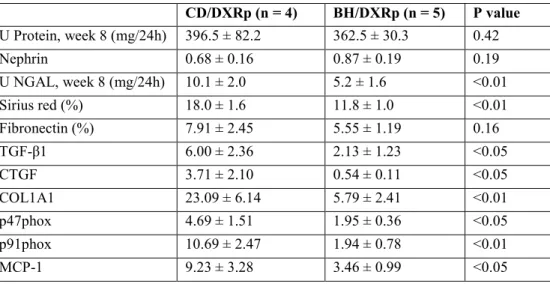
4.2.7. Tubulointerstitial fibrosis and inflammation were milder in DXR-injected BH vs.
CD rats despite similar proteinuria
Urinary protein excretion and renal nephrin mRNA levels were similar in (BH/DXRp, and CD/DXRp rats with similar proteinuria. Markers of fibrosis were significantly lower in BH/DXRp rats. Paralleling less fibrosis and tubular damage, markers of oxidative damage were significantly lower in BH/DXRp rats (Table 2).
Table 2: Comparison of doxorubicin-injected (DXR) Rowett, black hooded (BH) and Charles Dawley (CD) rats with similar proteinuria (BH/DXRp and CD/DXRp subgroups).
CD/DXRp (n = 4) BH/DXRp (n = 5) P value U Protein, week 8 (mg/24h) 396.5 ± 82.2 362.5 ± 30.3 0.42
Nephrin 0.68 ± 0.16 0.87 ± 0.19 0.19
U NGAL, week 8 (mg/24h) 10.1 ± 2.0 5.2 ± 1.6 <0.01
Sirius red (%) 18.0 ± 1.6 11.8 ± 1.0 <0.01
Fibronectin (%) 7.91 ± 2.45 5.55 ± 1.19 0.16
TGF-β1 6.00 ± 2.36 2.13 ± 1.23 <0.05
CTGF 3.71 ± 2.10 0.54 ± 0.11 <0.05
COL1A1 23.09 ± 6.14 5.79 ± 2.41 <0.01
p47phox 4.69 ± 1.51 1.95 ± 0.36 <0.05
p91phox 10.69 ± 2.47 1.94 ± 0.78 <0.01
MCP-1 9.23 ± 3.28 3.46 ± 0.99 <0.05
15
5. Conclusions
Diabetic nephropathy (DN) is a serious complication of DM, and despite the extensive investigations of this disease, there are very few options for specific treatment.
Fluorescence-based two-dimensional difference gel electrophoresis, coupled with mass spectrometry, identified 29 differentially expressed spots, including the actin-binding protein ezrin, and its interaction partner, NHERF2, which were down-regulated in streptozotocin-injected rats. Not just the total amount, but the active, phosphorylated ezrin was also reduced in diabetic rats. Podocalyxin is connected to actin through ezrin/NHERF2 complex, and it was down-regulated in STZ-induced diabetic rats. Ezrin expression was also lower in obese, diabetic Zucker rats and in human patients with diabetes. In conclusion, our data suggest that down-regulation of ezrin may associate with the development of the kidney complication in diabetes.
In our toxic nephropathy study, BH rats were resistant to renal fibrosis compared to CD rats. Fibrosis sensitive CD rats developed progressive proteinuria after DXR injection, with significant loss of nephrin, and FSGS-like histological lesions. DXR-injected BH rats had similar nephrin mRNA levels to that in the control rats of the same strain, with milder proteinuria and less severe glomerular changes. Consequent proteinuria contributed to the development of interstitial fibrosis and tubular atrophy. BH rats had reduced urinary NGAL excretion, accompanied by milder signs of tubulointerstitial inflammation and fibrosis, with less severe lipid peroxidation and protein nitrosylation. Interestingly, markers of tubular atrophy, interstitial fibrosis, oxidative- and nitrative stress were milder in BH rats even with similar levels of proteinuria. BH rats also had prolonged survival. In conclusion, resistance of BH rats against renal fibrosis highlighted the role of inflammation induced oxidative/nitrative stress in chronic podocyte injury leading to glomerulosclerosis and consequent proteinuria in DXR nephropathy.
16
6. Bibliography of the candidate's publications
6.1. Publications related to the PhD thesis
Wasik AA, Koskelainen S, Hyvonen ME, Musante L, Lehtonen E, Koskenniemi K, Tienari J, Vaheri A, Kerjaschki D, Szalay C, Revesz C, Varmanen P, Nyman TA, Hamar P, Holthofer H, Lehtonen S (2014) Ezrin is down-regulated in diabetic kidney glomeruli and regulates actin reorganization and glucose uptake via GLUT1 in cultured podocytes. Am J Pathol 184: 1727-1739.
IF: 4,591
Szalay CI, Erdelyi K, Kokeny G, Lajtar E, Godo M, Revesz C, Kaucsar T, Kiss N, Sarkozy M, Csont T, Krenacs T, Szenasi G, Pacher P, Hamar P (2015) Oxidative/Nitrative Stress and Inflammation Drive Progression of Doxorubicin- Induced Renal Fibrosis in Rats as Revealed by Comparing a Normal and a Fibrosis- Resistant Rat Strain. PLoS One 10: e0127090.
IF: 3,234
6.2. Publications unrelated to the PhD thesis
Kaucsar T, Revesz C, Godo M, Krenacs T, Albert M, Szalay CI, Rosivall L, Benyo Z, Batkai S, Thum T, Szenasi G, Hamar P (2013) Activation of the miR-17 family and miR-21 during murine kidney ischemia-reperfusion injury. Nucleic Acid Ther 23: 344- 354.
IF: 2,888
Kaucsar T, Bodor C, Godo M, Szalay C, Revesz C, Nemeth Z, Mozes M, Szenasi G, Rosivall L, Soti C, Hamar P (2014) LPS-induced delayed preconditioning is mediated by Hsp90 and involves the heat shock response in mouse kidney. PLoS One 9:
e92004.
IF: 3,234
17
7. Acknowledgements:
First and foremost I would like to thank my supervisor, Dr. Péter Hamar for his efforts in teaching and supporting me during my PhD studies.
I am indebted to all my colleagues from the Institute of Pathophysiology: Prof. Dr.
László Rosivall, Head of the Basic Medicine Doctoral School for the opportunity to carry out my PhD studies; Dr. Gábor Szénási for his continuous guidance; Mária Godó, Dr.
Tamás Kaucsár, Csaba Révész, Ágnes Cser, Dr. Anna Buday, Dr. Gábor Kökény, Dr. Nóra É. Bukosza and Dr. Norbert Kiss for their kind help in the experiments and for teaching me numerous methods. I want to express my gratitude to Dr. Miklós Mózes, whose comments were especially helpful to complete this thesis.
I would also like to thank Sanna Lehtonen, Anita A. Wasik and Niina Rouho and all the members of the Department of Pathology of the Haartman Institute (University of Helsinki, Finland) for the possibility to be involved in their research. Without their help, this thesis would not have been possible.
I would like to offer my special thanks to Dr. Pál Pacher and Katalin Erdélyi and all the members of the Laboratory of Cardiovascular Physiology and Tissue Injury (National Institute of Health, Bethesda, Maryland, USA) for all their technical and financial support.
Deepest gratitude to Dr. Tibor Krenács and Dr. Nóra Meggyesházi from the 1st Department of Pathology and Experimental Cancer Research (Semmelweis University, Hungary), who co-operated especially with the immunohistological examinations.
I would like to show my appreciation to Dr. Márta Sárközy and Dr. Tamás Csont from the Department of Biochemistry (University of Szeged, Hungary); for all their contribution to this thesis.
I also wish to thank Dr. Róbert Glávits, for his tutoring and introducing me to the interesting world of histopathology.
Furthermore, I am very grateful to my parents and my siblings for trusting me and for the unconditional support and encouragement during my professional trainings.
Research in this thesis was supported by the Hungarian Scientific Research Fund (OTKA- ANN(FWF) 110810 and OTKA-SNN 114619), and by the Intramural Research Program of NIAAA/NIH.