Subgroups of Paediatric Acute Lymphoblastic Leukaemia Might Differ Significantly in
Genetic Predisposition to Asparaginase Hypersensitivity
Nóra Kutszegi1, Ágnes F. Semsei1, András Gézsi1, Judit C. Sági1, Viktória Nagy2, Katalin Csordás2, Zsuzsanna Jakab2, Orsolya Lautner-Csorba1, Krisztina Míta Gábor3, Gábor T. Kovács2, Dániel J. Erdélyi2, Csaba Szalai2,4*
1Department of Genetics, Cell- and Immunobiology, Semmelweis University, Budapest, Hungary,22nd Department of Paediatrics, Semmelweis University, Budapest, Hungary,3Department of Pediatrics and Pediatric Health Care Center, Faculty of Medicine, University of Szeged, Szeged, Hungary,4Central Laboratory, Heim Pal Children Hospital, Budapest, Hungary
*szalaics@gmail.com
Abstract
L-asparaginase (ASP) is a key element in the treatment of paediatric acute lymphoblastic leukaemia (ALL). However, hypersensitivity reactions (HSRs) to ASP are major challenges in paediatric patients. Our aim was to investigate genetic variants that may influence the risk toEscherichia coli-derived ASP hypersensitivity. Sample and clinical data collection was carried out from 576 paediatric ALL patients who were treated according to protocols from the Berlin—Frankfurt—Münster Study Group. A total of 20 single nucleotide polymor- phisms (SNPs) inGRIA1andGALNT10genes were genotyped. Patients withGRIA1 rs4958351 AA/AG genotype showed significantly reduced risk to ASP hypersensitivity com- pared to patients with GG genotype in the T-cell ALL subgroup (OR = 0.05 (0.01–0.26); p = 4.70E-04), while no such association was found in pre-B-cell ALL. In the medium risk group two SNPs ofGRIA1(rs2055083 and rs707176) were associated significantly with the occur- rence of ASP hypersensitivity (OR = 0.21 (0.09–0.53); p = 8.48E-04 and OR = 3.02 (1.36– 6.73); p = 6.76E-03, respectively). Evaluating the genders separately, however, the associ- ation of rs707176 with ASP HSRs was confined only to females. Our results suggest that genetic variants ofGRIA1might influence the risk to ASP hypersensitivity, but subgroups of patients can differ significantly in this respect.
Introduction
Acute lymphoblastic leukaemia (ALL) is the most common paediatric malignancy, comprising approximately 25–30% of the annually registered cases of cancer among children [1,2]. Survival rates for paediatric and adolescent patients treated according to protocols by the Berlin—
a11111
OPEN ACCESS
Citation:Kutszegi N, Semsei ÁF, Gézsi A, Sági JC, Nagy V, Csordás K, et al. (2015) Subgroups of Paediatric Acute Lymphoblastic Leukaemia Might Differ Significantly in Genetic Predisposition to Asparaginase Hypersensitivity. PLoS ONE 10(10):
e0140136. doi:10.1371/journal.pone.0140136
Editor:Obul Reddy Bandapalli, University of Heidelberg, GERMANY
Received:June 22, 2015 Accepted:September 22, 2015 Published:October 12, 2015
Copyright:© 2015 Kutszegi et al. This is an open access article distributed under the terms of the Creative Commons Attribution License, which permits unrestricted use, distribution, and reproduction in any medium, provided the original author and source are credited.
Data Availability Statement:All relevant data are within the paper and its Supporting Information files.
Funding:This study was supported by OTKA (Hungarian Scientific Research Fund): K81941, K112872 (CS); K115861 (DJE) and the Economic Competitiveness Operational Program, Hungary GVOP 3.1.1-2004-05-0022/3.0 (DJE); and NKTH (National Research and Technology) TECH_08-A1/2- 2008-0120: (CS). The funders had no role in study design, data collection and analysis, decision to publish, or preparation of the manuscript.
Frankfurt—Münster (BFM) Study Group have remarkably improved over the last decades with higher than 80% of children are cured [1,3,4]. The extensive use of L-asparaginase (ASP), which has been a crucial component of paediatric therapies since 1970s, contributes to this achievement [5–9].
ASP enzymes mainly derived from bacteria among which three different preparations from two bacteria are currently used: native and pegylated form ofEscherichia coliASP (E.coli-ASP and PEG-ASP, respectively) and anErwinia chrysanthemi-derived ASP (Erwinia-ASP) with more or less different pharmacokinetic, pharmacodynamic and immunogenic properties [5].
Hypersensitivity reactions (HSRs) and silent inactivation via neutralizing antibody production against the enzyme are major challenges in paediatric patients, because both can lead to subop- timal treatment response. In addition, an anaphylactic reaction can be a potential threat to life requiring urgent interventions [7].
In BFM protocolsE.coli-ASP is considered as first-line treatment. Clinical HSRs occur in up to 45% of paediatric ALL patients, which necessitate the discontinuation ofE.coli-ASP administration and subsequent switch to PEG-ASP orErwinia-ASP [10–12]. The most com- mon manifestation of HSRs is urticaria [13]. However, the signs and symptoms can range from local reactions of erythema, swelling or pain at the injection site to severe symptoms, including laryngeal edema, bronchospasm, hypotension and occasionally systemic anaphylaxis [14]. Sev- eral risk factors of ASP hypersensitivity have been described including different preparations, dosage, route of administration, readministration after a hiatus and concomitant chemother- apy. However, this type of adverse reaction is unpredictable and exhibits large interindividual differences [11].
Recently, a genome wide association study has been carried out in an ethnically diverse pop- ulation to identify germline genetic variations contributing to the risk of ASP allergy in chil- dren with ALL. In this study variants of theGRIA1(Glutamate Receptor, Ionotropic, AMPA 1) gene located at 5q33 have been found associated with ASP allergy [15]. This result has later been replicated in a small Slovenian population [16].
In gene association studies, due to the high number of inconclusive results, it is generally accepted that the role of a gene or a genetic variation can only be acknowledged, if it is con- firmed by independent studies. In the present study our aim was to investigate the possible roles of genetic variations in theGRIA1gene in the susceptibility toE.coli-ASP hypersensitivity in a large Hungarian paediatric ALL population. Additionally, we also involved SNPs in GALNT10(Polypeptide N-Acetylgalactosaminyltransferase 10) gene, located also at 5q33, which in the original study were also found to be associated with ASP allergy in the discovery cohort, but could not be confirmed in a relatively small validation cohort.
Patients and Methods Study population
Samples and clinical data collection were carried out from 576 paediatric acute lymphoblastic leukaemia (ALL) patients who were treated between 1990 and 2012 in 9 Hungarian paediatric haematology centres according to four consecutive chemotherapy protocols from the Berlin— Frankfurt—Münster Study Group (ALL-BFM 90, 95, ALL IC-BFM 2002 and 2009). These pro- tocols in detail have been described elsewhere [3,4,17,18]. In this period of time twoE.coli- ASP, Kidrolase (Jazz Pharmaceuticals, Inc.) or Asparaginase medac (Kyowa-Hakko) were available as first-line ASP preparations.
The treatment according to the different risk arms focusing onE.coli-ASP dosing schedules with the number of patients included are shown inS1 Table. Comparison of the treatments on different risk arms can also be found inS1 Text.
Competing Interests:The authors have declared that no competing interests exist.
Data collection was carried out retrospectively from the files of the patients. We excluded 42 patients for the following reasons: lack of clinical information (n = 15), discontinuation of ASP administration not due to hypersensitivity (n = 1), switch to a different type of ASP not due to hypersensitivity (n = 1), genotyping call rate of the sample<50% (n = 28), significant deviation from the protocol (n = 6) and use of other treatment protocols (10). The inclusion criterion for controls was the completion of at least two blocks ofE.coli-ASP containing treatment without any HSR. Therefore, we excluded further ten patients. Finally, a total of 505 patients were included in the analysis (Table 1).
The National Cancer Institute Common Toxicity Criteria (CTC) system v3.0 was used to assess the grade of hypersensitivity. For analyses, we regarded a case as asparaginase hypersen- sitivity when signs of allergic reactions or anaphylactic reactions CTC grade 1 and above were noted which occurred during the infusion or within 4 hours, plus the clinical team decided to discontinue, not readminister the given aparaginase preparation in the later treatments. The descriptions of the HSRs were included in the medical records of the patients.
Written informed consent was obtained from the study participants or from the next of kin, caretakers, or guardians on the behalf of the minors/children participants involved in the study. All the associated documents have been stored. The study was conducted according to the principles expressed in the Declaration of Helsinki and the whole study including the informed consent procedure were approved by the Hungarian Scientific and Research Ethics Committee of the Medical Research Council (ETT TUKEB; Case No.:8-374/2009-1018EKU 914/PI/08).
Genes and polymorphisms
SNPs ofGRIA1andGALNT10genes were searched for in online databases. The criteria of the SNP selection were the minor allele frequency10% in Caucasian population, their estimated function based on dbSNP database of National Center of Biotechnology Information (NCBI;
www.ncbi.nlm.nih.gov) or any relevant results published previously [19,20]. In case of GALNT10the whole 3’-UTR region were covered (S2 Table).
Genotyping
In most cases, peripheral blood was obtained retrospectively from patients in remission phase.
In some cases, only diagnostic peripheral blood or bone marrow cell suspension samples were
Table 1. Patient characteristics.
Gender (%)
Male 280 (55.4)
Female 225 (44.6)
Years of age at diagnosis
Mean (±SD) 6.2 (±4.1)
Median (range) 4.9 (1–18)
Risk category (%)
Low Risk, LR 147 (29.1)
Medium Risk, MR 305 (60.4)
High Risk, HR 53 (10.5)
Immunophenotype (%)
pre-B ALL 402 (79.6)
T-ALL 69 (13.7)
doi:10.1371/journal.pone.0140136.t001
available (n = 69). The genomic DNA was extracted by using the QIAamp DNA Blood Midi Kit (Qiagen, Valencia, CA, USA) according to the manufacturer’s instructions. In case of low amount or low concentration of DNA (a total of 87 samples) whole genome amplification was performed by using the REPLI-g Mini Kit (Qiagen, Valencia, CA, USA) according to the man- ufacturer’s instructions.
A total of 20 SNPs (10 in each gene) were genotyped by using KASPar-on-Demand prevali- dated assays (LGC Genomics, Berlin, Germany) on 7900HT Fast Real-Time PCR System (Applied Biosystems, Foster City, CA, USA). The genotyping call rate for all SNPs was higher than 80%.
Statistical analysis
Multi-adjusted logistic regression was performed by using IBM SPSS Statistic software, version 20.0 to test for associations. Gender, ALL immunophenotype, age at diagnosis, risk group,E.
coli-ASP dosage during induction phase, BFM protocol and the polymorphisms in additive (11 vs. 12 vs. 22), dominant (11 vs. 12/22) and recessive (11/12 vs. 22) models were included in the analysis as categorical covariates (1, major allele; 2, minor allele). Odds ratios (ORs) and 95%
confidence intervals (CIs) were obtained to estimate risks for each SNP toE.coli-ASP hyper- sensitivity. The analyses were performed not only for the overall ALL cohort, but also for sub- groups created by gender, ALL immunophenotype (pre-B vs. T-ALL), age at diagnosis (10 years vs.<10 years) and risk category (standard vs. medium vs. high risk).
In order to deal with multiple comparisons the Benjamini-Hochberg false discovery rate (FDR) method with type I error rate of 5% (p6.76E-03) was applied as correction [21,22].
Power analysis was conducted by bootstrapping using R statistical software (R Foundation for Statistical Computing, Vienna, Austria; version 3.0.3) in the following way. First, we simu- lated 1 000 data replicates by sampling with replacement from the original dataset. Next, we applied logistic regression to each generated data set, and estimated power as the percentage of cases in which the null hypothesis was rejected.
Discrete-time survival analysis was used to assess the impact of SNPs on the minimum number of doses at whichE.coli-ASP hypersensitivity developed. The hazard rate of thei-th patient was modeled by logistic regression:
logit hi¼ aDiþbXi
whereDrepresents the minimum number of doses at whichE.coli-ASP hypersensitivity devel- oped by each study sample (more formally, for thei-th patient:Dij= 1, if the patient developed allergyexactlyat thej-th dose of ASP, andDik= 0 for all dose countskwherek6¼j);Xicontains the value of the covariate (SNP) that might predict hazard function differences, andαandβare the appropriate regression coefficients. Analyses were performed using R statistical software.
The deviation from Hardy-Weinberg Equilibrium (HWE) was analysed byχ2goodness-of- fit test using an online application (http://ihg.gsf.de/cgi-bin/hw/hwa1.pl).
Results
We investigated the impact ofGRIA1andGALNT10polymorphisms on the risk toE.coli-ASP hypersensitivity in paediatric ALL patients.
Out of the 20 SNPs, 18 were statistically evaluated, since two of them did not meet the requirements of HWE. The genotype and minor allele frequencies (MAF) with the results of the HWE are shown inS2 Table.
To examine the differences in genotype frequencies in patients withE.coli-ASP hypersensi- tivity and patients without HSRs toE.coli-ASP, multi-adjusted logistic regression analyses were applied on the overall cohort and on various subgroups.
Due to multiple testing, statistical corrections were applied. Applying FDR = 5% (p6.76E- 03) significance threshold three SNPs inGRIA1gene showed statistically significant associa- tions on certain subgroups.
The association of rs4958351 withE.coli-ASP hypersensitivity was not replicated in our total study cohort. The allele and genotype distributions for each SNP can be found inS3 Table. When we evaluated the associations between SNPs andE.coli-ASP hypersensitivity in T-cell and pre-B-cell ALL separately, in case of rs4958351 a significant difference between the subgroups was detected. In the dominant model patients with rs4958351 AA/AG genotype showed significantly reduced risk to ASP hypersensitivity compared to patients with GG geno- type in the T-ALL subgroup (OR = 0.05 (0.01–0.26); p = 4.70E-04; power = 0.78);Table 2). To examine a more homogeneous group we also analysed T-ALL patients on MR arm. In this way, the association still could be detected (N = 50; OR = 0.05 (0.01–0.29); p = 8.43E-04).
The overall rate ofE.coli-ASP hypersensitivity was 37% (37%, 30% and 77% in standard (SR), medium (MR) and high risk (HR) group, respectively). We found association between two polymorphisms (rs2055083 and rs707176) and the occurrence of ASP hypersensitivity in the MR group. On the one hand, in the dominant model, patients with rs2055083 AA/AG genotype had significantly lower risk to develop ASP hypersensitivity compared to patients with GG genotype (OR = 0.21 (0.09–0.53); p = 8.48E-04; power = 0.97;Table 3).
On the other hand, in recessive model, patients with CC genotype of rs707176 had signifi- cantly higher risk to have ASP hypersensitivity until the end of the reinduction compared to patients harbouring at least one T allele (OR = 3.02 (1.36–6.73); p = 6.76E-03; power = 0.73;
Table 4). The risk toE.coli-ASP hypersensitivity was approximately four times higher for female patients with CC genotype compared to patients with at least one T allele (OR = 4.03 (1.48–10.94), p = 6.28E-03; power = 0.78), while no such association was found in male patients.
When we evaluated the genders separately in each risk category, it turned out that in the MR group the association of rs707176 with ASP HSR was confined only to females with a very high OR (OR = 11.56 (2.56–52.27); p = 1.48E-03;Table 5).
The highest incidence of HSRs toE.coli-ASP in the MR group was observed during the first dose of ASP in the reinduction (approximately 70% of the overall numbers of cases) after a three-month-long break in ASP therapy. By the end of theE.coli-ASP treatment the cumula- tive incidence ofE.coli-ASP hypersensitivity was 34% (84 out of 244) and 11% (6 out of 55) for patients with rs2055083 GG and AA/AG genotypes (p = 1.3E-3), respectively (Fig 1A). In case of rs707176 this value was 48% (14 out of 29) and 27% (71 out of 264) for patients with CC and CT/TT genotypes (p = 3.1E-2), respectively (Fig 1B). In order to be comparable we analysed T-ALL subgroup restricted to MR patients: the cumulative incidence ofE.coli-ASP hypersensi- tivity was 60% (12 out of 20) and 10% (3 out of 30) for patients with GG and AA/AG genotypes (p = 5.0E-4), respectively (Fig 1C). After completing the reinduction therapy on MR arm (a total of 12 doses ofE.coli-ASP) without any HSR toE.coli-ASP none of the patients developed E.coli-ASP hypersensitivity in further blocks.
Furthermore, focusing on MR patients we used discrete-time survival analysis to assess the effect of the SNPs on the minimum number of doses at whichE.coli-ASP hypersensitivity developed. A strong effect of rs2055083 could be detected on the minimum number of doses.
The hazard ratio of the minor A allele in dominant model was 0.25 (N = 299; 95% CI: (0.11– 0.60); p = 1.8E-03). This means that the hazard rate of patients who have at least one A allele is approximately 75% less than that of the patients who are homozygous to the G allele. In
contrast, the minor C allele of rs707176 in recessive model was proved to be a strong risk factor (N = 293; hazard ratio: 2.38; 95% CI: (1.23–4.62); p = 0.0105). We also found a strong effect of rs4958351 on the minimum number of doses in the T-ALL subgroup on MR arm. The hazard rate of patients harbouring at least one A allele was about 91% less than patients with GG geno- type (N = 50; hazard ratio: 0.09; 95% CI: (0.02–0.36); p = 6.1E-04). Hence, the A allele of rs4958351 appeared to be a preventive factor in T-cell ALL.
Univariate and multivariate logistic regression was applied to evaluate the differences in the rate ofE.coli-ASP hypersensitivity in various subgroups. Patients on HR treatment arm had significantly increased odds of developingE.coli-ASP hypersensitivity compared to those on SR and MR arm (OR = 5.88 (2.85–12.15); p = 2.00E-06 and OR = 10.50 (2.82–39.02);
p = 4.49E-04, respectively). We found no differences inE.coli-ASP hypersensitivity rate between further subgroups (Table 6).
Discussion
The aim of our study was to investigate the impact of genetic variants ofGRIA1andGALNT10 genes on ASP allergy in a large Hungarian population of 576 ALL patients. We identified one synonymous and two intronicGRIA1SNPs associated withE.coli-ASP hypersensitivity on cer- tain ALL subgroups. We could find no association regarding theGALNT10polymorphisms.
Previously, Chenet al. analysed more than 500,000 SNPs in a genome-wide association study of 485 children with ALL. They found that the rs4958351 and four additional intronic SNPs of theGRIA1gene on chromosome 5q33 region were associated with ASP
Table 2. Associations of rs4958351 with the occurrence ofE.coli-ASP hypersensitivity in the total cohort and in the immunophenotypic subgroups.
rs4958351 GG vs. AG/AA
Subgroup N p value OR (95% CI)
Total 464 0.500 0.87 (0.58–1.31)
Immunophenotype pre-B ALL 364 0.522 1.16 (0.73–1.85)
T-ALL 66 4.70E-04 0.05 (0.01–0.26)
Results that reached the significance threshold (FDR(α) = 5%; p6.76E-03) are in bold.
doi:10.1371/journal.pone.0140136.t002
Table 3. Associations of rs2055083 with the occurrence ofE.coli-ASP hypersensitivity in the total cohort and in different risk groups.
rs2055083 GG vs. AG/AA
Subgroup N p value OR (95% CI)
Total 490 0.104 0.66 (0.39–1.09)
Risk category SR 141 0.090 2.08 (0.89–4.84)
MR 298 8.48E-04 0.21 (0.09–0.53)
HR 51 0.245 4.73 (0.34–64.90)
Results that reached the significance threshold (FDR(α) = 5%; p6.76E-03) are in bold.
doi:10.1371/journal.pone.0140136.t003
hypersensitivity [15]. Recently, these results were replicated in an independent, relatively small Slovenian population of 146 paediatric ALL patients [16]. Chenet al. also identified genetic variants ofGALNT10(located in the same region) in the initial cohort of 322 paediatric patients, but these associations could not be detected in the smaller validation cohort of 163 children [15].
GRIA1encodes a subunit (GluR1) of the ionotropic alpha-amino-3-hydroxy-5-methyl- 4-isoxazole propionate (AMPA) receptor. These receptors are homomeric or heteromeric pro- tein complexes consisting of GluR1-4 subunits, which are arranged to form a ligand-gated ion channel transmitting glutamatergic signals in the central nervous system (http://www.
genecards.org/cgi-bin/carddisp.pl?gene=GRIA1).
Recent studies have shown that glutamate acts also as an immunomodulator in addition to being a neurotransmitter [15,16,23]. For the first time, in 2001 Lombardiet al. described the presence of ionotropic glutamate receptors (iGluRs) on the surface of human T-lymphocytes.
The same study has revealed the functionality of AMPA receptors by potentiation of Ca2+
influx upon T-cell activation [24]. Later, other studies have demonstrated that glutamate induced both a pro-adhesive and a pro-migratory effect on naïve/resting T-cells by acting via its AMPA receptors [25–27]. Moreover, the expression of several glutamate receptors and transporters has been described in various types of immune cells including dendritic cells (DCs). DCs release glutamate upon interacting with T-lymphocytes in lymph nodes. Pacheco et al. demonstrated that during this interaction glutamate acts as a highly effective regulator in the initiation of T-cell mediated immune responses [27]. Besides, in addition to theab ovoele- vated level of plasma glutamate due to malignancy, the glutaminase activity ofE.coli-ASP results in a significant rise of the cleavage product glutamate during treatment with a high interindividual variability [28]. Taken together, these data suggest that genetic polymorphisms of glutamate signalling pathways in immune system may influence the risk of developing ASP hypersensitivity.
The rs4958351 SNP showed no association withE.coli-ASP hypersensitivity in our total study cohort. However, we found that the effect of the A allele drastically differs on the suscep- tibility to ASP hypersensitivity in the different subtypes of ALL. While it is associated with sig- nificantly reduced risk to HSR in T-cell ALL (OR = 0.05), it is associated with a slightly although not significantly increased risk to HSR in pre-B-cell ALL. In contrast, Chenet al. and
Table 4. Associations of rs707176 with the occurrence of E. coli-ASP hypersensitivity in the total cohort and in different subgroups.
rs707176a CT/TT vs. CC
Subgroup N p value OR (95% CI)
Total 477 0.041 1.90 (1.02–3.48)
Risk category SR 136 0.689 1.25 (0.42–3.68)
MR 292 6.76E-03 3.02 (1.36–6.73)
HR 49 0.385 0.36 (0.03–3.66)
Gender Male 263 0.770 1.14 (0.48–2.66)
Female 214 6.28E-03 4.03 (1.48–10.94)
Results that reached the significance threshold (FDR(α) = 5%; p6.76E-03) are in bold.
athe cofactor of ASP dosage during induction was not included in the analysis due to numerical problems of the logistic regression model created by the presence of cell values equal to zero.
doi:10.1371/journal.pone.0140136.t004
Rajićet al. have identified the A allele as a risk allele in the whole ALL cohort. However, per- haps because of the low number of T-ALL patients in their study cohorts, the different immu- nophenotypic subgroups have not been evaluated separately. To our knowledge, ours is the first study on the relation of rs4958351 to the risk ofE.coli-ASP hypersensitivity in T-ALL as a separate subgroup; hence this opposite association in this subgroup of ALL patients could have been hidden up to this point.
In our study the intronic rs2055083 polymorphism in theGRIA1gene appeared to be a strong preventive factor ofE.coli-ASP hypersensitivity in a homogeneous cohort of MR ALL patients. No result has been published so far related to rs2055083. According to the results of the 1000 Genomes Project the rs2055083 is in linkage disequilibrium (LD) with another intro- nic SNP rs10515693 (http://www.ensembl.org/Homo_sapiens/Variation/HighLD?db = core;r=
5:153487464-153667465;v = rs2055083;vdb = variation;vf=1720088#13083_tablePanel; r2= 0.953; D’= 1.000; Ensembl release 81—July 2015) in the CEPH population. However, in the European (EUR) super population the regarding values were lower (r2= 0.7; D’= 0.85). Using
Table 5. Associations of rs707176 with the occurrence of E. coli-ASP hypersensitivity in subgroups created by risk category and gender.
rs707176a CT/TT vs. CC
Subgroup N p value OR (95% CI)
Risk category SR Male 65 0.806 1.24 (0.23–6.77)
Female 71 0.726 1.30 (0.30–5.74)
MR Male 165 0.767 1.19 (0.38–3.77)
Female 127 1.48E-03 11.56 (2.56–52.27)
HR Male n.a.b n.a.b n.a.b
Female n.a.b n.a.b n.a.b
Results that reached the significance threshold (FDR(α) = 5%; p6.76E-03) are in bold.
athe cofactor of ASP dosage during induction was not included in the analysis due to numerical problems of the logistic regression model created by the presence of cell values equal to zero.
bnot analysed due to the presence of cell values equal to zero.
doi:10.1371/journal.pone.0140136.t005
Fig 1. The cumulative incidence ofE.coli-ASP hypersensitivity with the number of ASP doses.(A) In case of rs2055083 the cumulative incidence ofE. coli-ASP by the end of the of the second block on MR arm was 34% (84 out of 244) and 11% (6 out of 55) for patients with GG and AA/AG genotypes, respectively. (B) In case of rs707176 the same value was 48% (14 out of 29) and 27% (71 out of 264) for patients with CC and CT/TT genotypes,
respectively. (C) Analysing T-ALL patients on MR arm the cumulative incidence ofE.coli-ASP hypersensitivity by the end of the of the second block was 60%
(12 out of 20) and 10% (3 out of 30) for patients with GG and AA/AG genotypes, respectively.
doi:10.1371/journal.pone.0140136.g001
the online Variant Effect Predictor (VEP) tool of Ensembl it has been revealed that the latter polymorphism is located in a regulatory region (ENSR00001294367), which is active in a human B-lymphocyte cell line GM12878 and acts as an enhancer (http://www.ensembl.org/
Homo_sapiens/Regulation/Summary?db = core;fdb = funcgen;r=5:153554305-153557304;rf=
ENSR00001294367;tl = gEE3ksgDRibSzsCu-737322). Based on this, one possible reason of association between rs2055083 and ASP allergy that rs2055083 is in LD with a regulatory vari- ant in B-lymphocytes.
It has been shown in an Italian case-control association study that the allele distributions of rs707176 were different in DSM-IV-TR (Diagnostic and Statistical Manual of Mental Disor- ders, Fourth Edition, Text Revision) schizophrenia controls and in cases [20]. In our study population this SNP, which is a synonymous C/T transition in exon 3 of theGRIA1gene, was associated withE.coli-ASP hypersensitivity in MR subgroup (p = 6.76E-03). The risk of CC homozygotes to HSR was very high in females (OR = 4.03 (1.48–10.94)), while no such effect could be observed in males which suggests a sex difference in the genetic background of ASP hypersensitivity.
All the genetic variations we genotyped inGRIA1gene were intronic or synonymous variants.
The functionality of these so-called silent polymorphisms is not yet clear. Intronic polymor- phisms in the glutamate receptor subunitGluR2have roles in directing RNA editing of the GluR2coding sequence [15,16,29]. Furthermore, it is known thatGRIA1has alternatively spliced transcript variants encoding different isoforms (http://www.genecards.org/cgi-bin/carddisp.pl?
gene=GRIA1). Emerging data show that the fine balance of alternative splicing isoforms may also be altered by variants of exonic or intronic splicing regulatory elements [30]. These findings suggest mechanisms by which these silent polymorphisms could influence gene function.
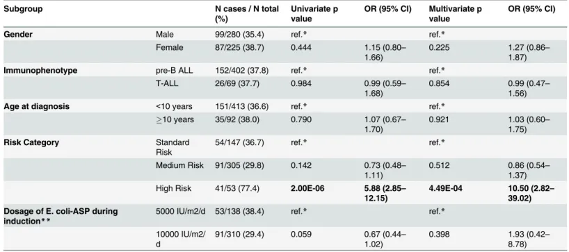
Table 6. Differences between subgroups inE.coli-ASP hypersensitivity.
Subgroup N cases / N total
(%)
Univariate p value
OR (95% CI) Multivariate p value
OR (95% CI)
Gender Male 99/280 (35.4) ref.* ref.*
Female 87/225 (38.7) 0.444 1.15 (0.80–
1.66)
0.225 1.27 (0.86–
1.87)
Immunophenotype pre-B ALL 152/402 (37.8) ref.* ref.*
T-ALL 26/69 (37.7) 0.984 0.99 (0.59–
1.68)
0.854 0.99 (0.47–
1.56)
Age at diagnosis <10 years 151/413 (36.6) ref.* ref.*
10 years 35/92 (38.0) 0.790 1.07 (0.67–
1.70)
0.921 1.03 (0.60–
1.75)
Risk Category Standard
Risk
54/147 (36.7) ref.* ref.*
Medium Risk 91/305 (29.8) 0.142 0.73 (0.48– 1.11)
0.512 0.86 (0.54–
1.37) High Risk 41/53 (77.4) 2.00E-06 5.88 (2.85–
12.15)
4.49E-04 10.50 (2.82– 39.02) Dosage of E. coli-ASP during
induction** 5000 IU/m2/d 53/138 (38.4) ref.* ref.*
10000 IU/m2/
d
91/310 (29.4) 0.059 0.67 (0.44–
1.02)
0.398 1.93 (0.42–
8.78) Results that reached the significance threshold (p0.05 in univariate and FDR(α) = 5%; p6.76E-03 in multivariate analysis) are in bold.
*ref. refers to the reference group to which the others groups are to be compared
**on standard and medium risk arm doi:10.1371/journal.pone.0140136.t006
We tested the differences in the occurrence ofE.coli-ASP hypersensitivity by logistic regres- sion between subgroups created by gender, age at diagnosis, risk category, immunophenotype of leukaemia and the dose ofE.coli-ASP during induction on SR and MR arms. HR patients were at approximately six times higher odds of developingE.coli-ASP hypersensitivity com- pared to SR patients (p = 2.00E-06). The occurrence ofE.coli-ASP hypersensitivity did not dif- fer significantly between the SR and the MR groups. These findings can be explained by the considerably different treatment with more and greater doses of ASP as well as with many breaks in ASP therapy on HR arm compared to SR and MR arms.
The highest occurrence ofE.coli-ASP hypersensitivity reactions was after a three-month- long break (consolidation phase) in ASP treatment during the first dose of reinduction on MR arm. It is in accordance with previously observed phenomena that reexposure of ASP after a hiatus increases the risk to HSRs [10,11]. The proliferation and antibody production of compe- tent B-cells during consolidation can be one of the possible explanations [16,31].
This study has several strengths and limitations. First, although the rate of relapsed patients of our cohort is similar to the relapse rate of the whole ALL population, the rate of died patients, however, is lower in our study population. Patients who died during the chemother- apy due to therapy resistant progressive disease or due to infections or toxicities of therapy are underrepresented in our ALL cohort. Furthermore, HSR data toE.coli-ASP was collected retro- spectively from the files of the patients. This manner does not allow meticulous documentation and fine grading of hypersensitivity reactions.
Our findings pertain to reactions toE.coli-ASP. Fernandez et al. recently reported that the association between the rs4958351 variant and hypersensitivity was strongest among patients receiving nativeE.coliasparaginase compared to PEG-asparaginase [32].
A small proportion of our DNA samples had been originated from the diagnostic clone of lymphoblasts. We hypothesized that the probability of the genetic alteration of the investigated polymorphisms was extremely low during leukaemogenesis. To verify this, we also performed the analysis excluding these samples and calculated the allele frequencies for the remission and for the diagnostic samples. Even in this way, the results remained statistically significant and we could not find substantial differences in the allele frequencies either (data not shown).
In conclusion, in our relatively large population we confirmed that genetic variations in the GRIA1gene could significantly influence the risk of ASP hypersensitivity. Furthermore, we found that the direction of the effect could be significantly different in the different subgroups of patients. In addition to previously published associations, we identified novel polymor- phisms, which may serve to enlighten new details in the genetic background ofE.coli-ASP hypersensitivity reactions. Replication in other cohorts is warranted to confirm our findings.
Functional analysis of variants is needed, especially in case of the rs4958351 in T-ALL to eluci- date its role in ASP allergy development.
Supporting Information
S1 Table. The treatment focusing on ASP dosing schedules according to different risk arms.
(PDF)
S2 Table. Genes and SNPs included in the analysis.
(PDF)
S3 Table. Allele and genotype distributions of the total cohort of ALL patients.
(PDF)
S1 Text. Comparison of the different chemotherapy protocols.
(PDF)
Author Contributions
Conceived and designed the experiments: NK ÁFS DJE CS GTK. Performed the experiments:
NK JCS VN. Analyzed the data: NK AG ÁFS. Contributed reagents/materials/analysis tools:
OLC KC KMG ZJ. Wrote the paper: NK CS ÁFS DJE AG.
References
1. Garami M, Schuler D, Jakab Z. (2014) [Importance of the National Childhood Cancer Registry in the field of paediatric oncology care in Hungary]. Orv Hetil 155: 732–739. doi:10.1556/OH.2014.29918 PMID:24796778
2. Steliarova-Foucher E, Stiller C, Kaatsch P, Berrino F, Coebergh JW, Lacour B, et al. (2004) Geographi- cal patterns and time trends of cancer incidence and survival among children and adolescents in Europe since the 1970s (the ACCIS project): an epidemiological study. Lancet 364: 2097–2105. PMID:
15589307
3. Stary J, Zimmermann M, Campbell M, Castillo L, Dibar E, Donska S, et al. (2014) Intensive chemother- apy for childhood acute lymphoblastic leukemia: results of the randomized intercontinental trial ALL IC- BFM 2002. J Clin Oncol 32: 174–184. doi:10.1200/JCO.2013.48.6522PMID:24344215
4. Moricke A, Reiter A, Zimmermann M, Gadner H, Stanulla M, Dordelmann M, et al. (2009) Risk-adjusted therapy of acute lymphoblastic leukemia can decrease treatment burden and improve survival: treat- ment results of 2169 unselected pediatric and adolescent patients enrolled in the trial ALL-BFM 95 (vol 111, pg 4477, 2008). Blood 113: 4478–4478.
5. Andrade AF, Borges KS, Silveira VS. (2014) Update on the Use of l-Asparaginase in Infants and Ado- lescent Patients with Acute Lymphoblastic Leukemia. Clin Med Insights Oncol 8: 95–100. doi:10.
4137/CMO.S10242PMID:25210485
6. Avramis VI, Tiwari PN. (2006) Asparaginase (native ASNase or pegylated ASNase) in the treatment of acute lymphoblastic leukemia. Int J Nanomedicine 1: 241–254. PMID:17717965
7. Kawedia JD, Rytting ME. (2014) Asparaginase in acute lymphoblastic leukemia. Clin Lymphoma Mye- loma Leuk 14 Suppl: S14–17. doi:10.1016/j.clml.2014.06.017PMID:25486949
8. Pieters R, Hunger SP, Boos J, Rizzari C, Silverman L, Baruchel A, et al. (2011) L-asparaginase treat- ment in acute lymphoblastic leukemia: a focus on Erwinia asparaginase. Cancer 117: 238–249. doi:
10.1002/cncr.25489PMID:20824725
9. Pession A, Valsecchi MG, Masera G, Kamps WA, Magyarosy E, Rizzari C, et al. (2005) Long-term results of a randomized trial on extended use of high dose L-asparaginase for standard risk childhood acute lymphoblastic leukemia. J Clin Oncol 23: 7161–7167. PMID:16192600
10. Rizzari C, Conter V, Stary J, Colombini A, Moericke A, Schrappe M. (2013) Optimizing asparaginase therapy for acute lymphoblastic leukemia. Curr Opin Oncol 25 Suppl 1: S1–9. doi:10.1097/CCO.
0b013e32835d7d85PMID:23380829
11. Woo MH, Hak LJ, Storm MC, Sandlund JT, Ribeiro RC, Rivera GK, et al. (2000) Hypersensitivity or development of antibodies to asparaginase does not impact treatment outcome of childhood acute lym- phoblastic leukemia. Journal of Clinical Oncology 18: 1525–1532. PMID:10735901
12. Muller HJ, Boos J. (1998) Use of L-asparaginase in childhood ALL. Crit Rev Oncol Hematol 28: 97– 113. PMID:9768345
13. Narta UK, Kanwar SS, Azmi W (2007) Pharmacological and clinical evaluation of L-asparaginase in the treatment of leukemia. Crit Rev Oncol Hematol 61: 208–221. PMID:17011787
14. Shinnick SE, Browning ML, Koontz SE. (2013) Managing hypersensitivity to asparaginase in pediatrics, adolescents, and young adults. J Pediatr Oncol Nurs 30: 63–77. doi:10.1177/1043454212471728 PMID:23380527
15. Chen SH, Pei D, Yang W, Cheng C, Jeha S, Cox NJ, et al. (2010) Genetic variations in GRIA1 on chro- mosome 5q33 related to asparaginase hypersensitivity. Clin Pharmacol Ther 88: 191–196. doi:10.
1038/clpt.2010.94PMID:20592726
16. Rajic V, Debeljak M, Goricar K, Jazbec J. (2015) Polymorphisms in GRIA1 gene are a risk factor for asparaginase hypersensitivity during the treatment of childhood acute lymphoblastic leukemia. Leuk Lymphoma: 1–6.
17. Erdelyi DJ, Kamory E, Csokay B, Andrikovics H, Tordai A, Kiss C, et al. (2008) Synergistic interaction of ABCB1 and ABCG2 polymorphisms predicts the prevalence of toxic encephalopathy during antican- cer chemotherapy. Pharmacogenomics J 8: 321–327. PMID:17938643
18. Gezsi A, Lautner-Csorba O, Erdelyi DJ, Hullam G, Antal P, Semsei AF, et al. (2014) In interaction with gender a common CYP3A4 polymorphism may influence the survival rate of chemotherapy for child- hood acute lymphoblastic leukemia. Pharmacogenomics J.
19. Bishop JR, Chae SS, Patel S, Moline J, Ellingrod VL. (2012) Pharmacogenetics of glutamate system genes and SSRI-associated sexual dysfunction. Psychiatry Res 199: 74–76. doi:10.1016/j.psychres.
2012.03.048PMID:22534499
20. Magri C, Gardella R, Barlati SD, Podavini D, Iatropoulos P, Bonomi S, et al. (2006) Glutamate AMPA receptor subunit 1 gene (GRIA1) and DSM-IV-TR schizophrenia: a pilot case-control association study in an Italian sample. Am J Med Genet B Neuropsychiatr Genet 141B: 287–293. PMID:16526023 21. Benjamini Y, Hochberg Y. (1995) Controlling the False Discovery Rate—a Practical and Powerful Approach to Multiple Testing. Journal of the Royal Statistical Society Series B-Methodological 57:
289–300.
22. Storey JD. (2002) A direct approach to false discovery rates. Journal of the Royal Statistical Society Series B-Statistical Methodology 64: 479–498.
23. Ma Q, Lu AY. (2011) Pharmacogenetics, pharmacogenomics, and individualized medicine. Pharmacol Rev 63: 437–459. doi:10.1124/pr.110.003533PMID:21436344
24. Lombardi G, Dianzani C, Miglio G, Canonico PL, Fantozzi R. (2001) Characterization of ionotropic glu- tamate receptors in human lymphocytes. Br J Pharmacol 133: 936–944. PMID:11454668
25. Ganor Y, Besser M, Ben-Zakay N, Unger T, Levite M. (2003) Human T cells express a functional iono- tropic glutamate receptor GluR3, and glutamate by itself triggers integrin-mediated adhesion to laminin and fibronectin and chemotactic migration. J Immunol 170: 4362–4372. PMID:12682273
26. Ganor Y, Levite M. (2014) The neurotransmitter glutamate and human T cells: glutamate receptors and glutamate-induced direct and potent effects on normal human T cells, cancerous human leukemia and lymphoma T cells, and autoimmune human T cells. J Neural Transm 121: 983–1006. doi:10.1007/
s00702-014-1167-5PMID:24584970
27. Pacheco R, Oliva H, Martinez-Navio JM, Climent N, Ciruela F, Gatell JM, et al. (2006) Glutamate released by dendritic cells as a novel modulator of T cell activation. J Immunol 177: 6695–6704. PMID:
17082582
28. Pieters R, Appel I, Kuehnel HJ, Tetzlaff-Fohr I, Pichlmeier U, van der Vaart I, et al. (2008) Pharmacoki- netics, pharmacodynamics, efficacy, and safety of a new recombinant asparaginase preparation in chil- dren with previously untreated acute lymphoblastic leukemia: a randomized phase 2 clinical trial. Blood 112: 4832–4838. doi:10.1182/blood-2008-04-149443PMID:18805963
29. Egebjerg J, Kukekov V, Heinemann SF. (1994) Intron sequence directs RNA editing of the glutamate receptor subunit GluR2 coding sequence. Proc Natl Acad Sci U S A 91: 10270–10274. PMID:
7937939
30. Pagani F, Baralle FE. (2004) Genomic variants in exons and introns: identifying the splicing spoilers.
Nat Rev Genet 5: 389–396. PMID:15168696
31. Pagani M. (2010) The complex clinical picture of presumably allergic side effects to cytostatic drugs:
symptoms, pathomechanism, reexposure, and desensitization. Med Clin North Am 94: 835–852, xiii.
doi:10.1016/j.mcna.2010.03.002PMID:20609866
32. Fernandez CA, Smith C, Yang W, Mullighan CG, Qu C, Larsen E, et al. (2015) Genome-wide analysis links NFATC2 with asparaginase hypersensitivity. Blood 126: 69–75. doi:10.1182/blood-2015-02- 628800PMID:25987655