Circulating microRNAs as minimal residual disease biomarkers in childhood acute lymphoblastic leukaemia
Ph.D thesis
Andrea Rzepiel
Semmelweis University Clinical medicine PhD School
Supervisior: Dr. Dániel János Erdélyi, MD, Ph.D.
Official reviewers: Dr. Zsuzsanna Gaál, MD, Ph.D., Infant and paediatric specialist
Dr. Zsolt Rónai, Ph.D., Associate professor
Head of the Final Examination Committee: Prof. Dr. László Rosivall, D.Sc.
Members of the Final Examination Committee:
Prof. Dr. Barna Vásárhelyi, D.Sc.
Dr. László Szabó, Ph.D.
Dr. András Fogarasi, D.Sc.
Budapest
2020
2 Introduction
Paediatric acute lymphoblastic leukaemia (ALL) is a clonal disease of hematopoietic stem cells. ALL is the most common paediatric malignancy. In Hungary there are 60-70 new ALL cases per year. The most common subgroup is the precursor B cell subgroup, it takes approximately 85% of all ALL cases. The remaining 15% belongs to T cell subtype subgroup. Lymphoblasts are typical cells in ALL. Lymphoblasts can be found in bone marrow and most of the cases in peripheral blood. Treatment protocols vary in different countries. In Hungary ALL IC-BFM 2009 protocol is currently used. The protocol includes 6 phases: induction, early intensification, consolidation, reinduction, late reinduction and maintenance therapy. Induction therapy is the treatment of the first 33 days of therapy. It aims to destroy the initial malignant cell mass. The whole ALL therapy lasts 2 years, although there are breaks during the treatment letting the regeneration of the healthy bone marrow. Survivor rates are improving during the last decades and the 5 year survivor rate is 83% in our country.
Stratification based on early therapy response and minimal residual disease (MRD) monitoring are essential parts of the successful therapy.
Minimal residual disease is the name given to those malignant cells remaining after the treatment at low level (under 5%) detected with a sensitive diagnostic tool. Traditionally microscopic analysis of blood and bone marrow was used for this purpose. Although this was not sensitive enough. Nowadays flow cytometry (FC) and PCR techniques are used for monitoring the minimal residual disease. Sensitivity of flow cytometry is approximately 10-4, while sensitivity of PCR is 10-5.
MicroRNAs are short ~22 nucleotide long, non-coding RNAs. Most of them are transcribed from DNA sequences of primary microRNA or precursor microRNA, and ultimately mature to microRNA. Typically they bind to the 3' untranslated region (UTR) of the target mRNA and suppress their expression. Sometimes they bind to 5' UTR, coding sequences or to a gene promoter.
, In the human genome there are about 24 000 non coding genes, while the number of mature microRNAs is approximately 2300, which regulate 30-60% of the protein coding genes.
3
MicroRNAs affect metabolic and signalling pathways through cell proliferation, differentiation, and regulation of survival. Altered microRNA expression profile is common in many malignancies. MicroRNAs are cell and tissue specific thus their assessment whether a microRNA is oncogenic or tumour suppressor is difficult. In addition, it is not known yet that altered microRNA expression is a direct cause of malignancy or an indirect consequence. It makes even more difficult that a microRNA has multiply targets. Thus, one particular microRNA in a given environment is oncogenic, while somewhere else it has a tumour suppressor function.
MicroRNAs can be found in extracellular environment. Scientists have isolated microRNAs from blood plasma and serum, cerebrospinal fluid, urine, saliva, milk, tears and other body fluids.
Extracellular microRNAs are very stable. They can be stored at room temperature for days even in extreme condition without any degradation. MicroRNAs can also be found in extracellular vesicles or in complexes with proteins such as AGO2 in body fluids.
Studying healthy and aberrant haematopoiesis, scientists discovered that microRNAs have important role in regulation of maturation during haematopoiesis.
Extracellular vesicles (EV) are surrounded by double-layer phospholipid membrane.
There are three main groups based on their size and origin: exosomes, microvesicles and apoptotic bodies.
Extracellular vesicles are involved in communication between cells, angiogenesis, removal of intracellular components, immunoreactions and blood coagulation. Due to them, protein, RNA and DNA exchange is possible between cells. Healthy and malignant cells produce vesicles and scientists successfully isolated them from many body fluids.
The most common resources of exosomes are immune cells and malignant cells.
Exosomes are able to pack certain microRNAs and keep their integrity even in blood circulation (protected against RNase) thus their origin is identifiable even in circulation.
4 Aims
I had the following aims during my work:
Based on previous studies 46 microRNAs were selected and their extracellular expressions were examined in peripheral blood, bone marrow and cerebrospinal fluid samples of paediatric patients with acute lymphoblastic leukaemia.
The evaluation of expression changes of examined microRNAs in peripheral blood plasma whether they are suitable as minimal residual disease biomarkers.
5 Methods
Patients with acute lymphoblastic leukaemia were diagnosed and treated at 2nd Department of Paediatrics Semmelweis University and Heim Pál Children's Hospital Onco-haematology Department. Peripheral blood (3.5 ml) and bone marrow (1 ml) samples were collected into citrate blood collection tubes and native tubes were used for cerebrospinal fluid (1 ml) sample collection. In case of peripheral blood and bone marrow, samples were centrifuged within 2 hours of collection in order to isolate platelet-free plasma (PFP). Samples were stored at -80°C. Patients were treated according to ALL-IC BFM 2009 protocol. Specimens were collected during the first 33 days of therapy.
Peripheral blood samples were collected at diagnosis, day 8, 9, 15, 29, 30 and day 33.
Bone marrow and cerebrospinal fluid samples were collected at diagnosis, day 15 and day 33.
Altogether samples of 28 de novo, 5 relapsed and 2 central nervous system involved precursor B-cell ALL patients were examined. Samples of relapsed patients were collected during the first relapse. Patients were 1-18 years old. In case of de novo patients 3 cytogenetic subgroups were made: hyperdiploid (number of chromosomes 47 or more), patients with t(12;21)(p13;q22) translocation and normal karyotype subgroup (normal findings on a set FISH panel targeting frequent ALL rearrangements, found to be diploid by flow cytometry DNA index, either normal karyotype or unsuccessful karyotyping).
Additional peripheral blood samples of 10 control patients were included.
RNA was isolated from platelet-free plasma samples then cDNA was synthetized.
Expression of 46 preselected microRNAs were measured on custom TaqMan Low Density Array (TLDA) cards. Selection criteria was their confirmed high intracellular expression in ALL samples at least in two previous studies. QPCR methodology was used for validation on expanded population. Exosome enriched fraction was isolated by ultracentrifugation (Beckman Coulter Optima XP) from platelet-free plasma of peripheral blood then RNA was isolated and cDNA synthetized and examined by qPCR.
Statistical analysis was carried out using R software by Dr. András Gézsi.
6 Results
On discovery population setup, platelet-free plasma of peripheral blood, bone marrow and cerebrospinal fluid samples were examined on custom TLDA cards (Figure 1).
Significant correlation between peripheral blood and bone marrow microRNA expression was not found.
7
Figure. 1 Normalized log2 transformed expression levels measured on TaqMan Low Density array cards in peripheral blood and bone marrow PFP samples of ALL subgroups and controls. Yellow indicates microRNA down-expression, red indicates overexpression compared to the normalizing miR. Black boxes on left side indicates microRNAs were selected for further measurement. Value: transformed log2 expression level Count: height of histogram
Out of forty-six, 21 microRNAs were expressed differently in platelet-free plasma of peripheral blood samples of patients with de novo and relapsed ALL compared to controls. When relapsed patients were excluded from analysis (due to low specimen number, short follow up), 19 microRNAs were expressed differently. 18 microRNAs were overexpressed, while one microRNA was underexpressed.
Samples of 3 karyotype subgroups were studied separately and compared to healthy controls. Five microRNAs showed overexpression in each subgroups: miR-128-3p, miR- 146a-5p, miR-181b-5p, miR-222-3p and miR-532-5p. We did not find unique microRNA pattern in any subgroup.
In case of platelet-free peripheral blood plasma of patients with de novo ALL 4 microRNAs were overexpressed at diagnosis as well as at day 33 compared to control samples: miR-128-3p, miR-181a-5p, miR-181b-5p and miR-222-3p. Thus these microRNAs were selected for further qPCR analysis.
8
Using qPCR microRNA expression detected in diagnostic de novo ALL samples were compared to healthy control in peripheral blood. (Table 1)
Table 1. MicroRNA expression in diagnostic de novo ALL peripheral blood PFP compared to healthy controls. NS: not significant
Subgroups
miR-128-3p log2 FC (adj. p value)
miR-181a-5p log2 FC (adj. p value)
miR-181b-5p log2 FC (adj. p value)
miR-222-3p log2 FC (adj. p value) Hyperdiploid
subgroup
3.39
(7.05*10-4) NS NS 2.09
(4.06*10-2) t(12;21)
subgroup
5.93 (9.59*10-10)
2.56 (1.38*10-2)
3.84 (2.26*10-4)
3.34 (2.21*10-4) Normal
subgroup
4.23 (1.78*10-5)
3.45 (5.35*10-4)
4.31 (4.73*10-5)
2.63 (4.76*10-3) Total ALL
group
4.52 (3.48*10-9)
2.46 (2.11*10-3)
3.46 (4.73*10-5)
2.68 (2.15*10-4)
9
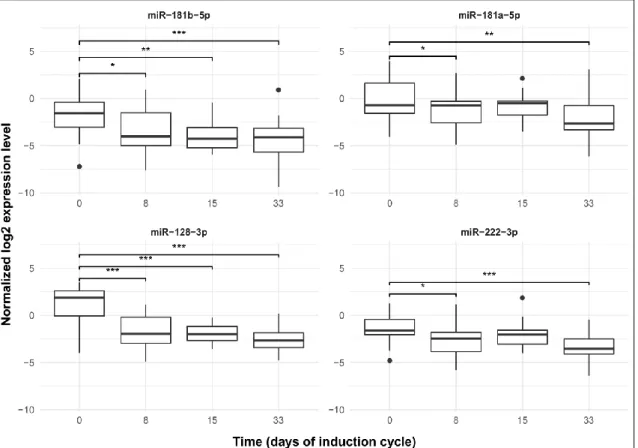
On an expanded population, platelet-free peripheral blood plasma samples were tested by qPCR during the induction therapy at 4 time points. As the treatment had started expression of every microRNA was reduced significantly by day 8 and by day 33. (Figure 2). Although continuous decrease was not detected.
Figure 2. MiR expressions in PB PFP during the first month of therapy. Box: the 2nd and 3rd quartiles; thick line in the box: median; whiskers: minimum and maximum values if there are no outrange values, or Q1−1.5*IQR; dots: outliers, lines above the boxes:
significant correlation (p < 0.05); ***p value: 0≤p < 0.001, **p value: 0.001≤p < 0.01,
*p value: 0.01≤p < 0.05
We analysed microRNA expressions and expression changes in all platelet-free peripheral blood plasma samples. Results were further analysed with day 15 flow cytometry MRD results and other prognostic factors. MiR-128-3p and miR-222-3p expression changes by day 8 was correlated with day 15 flow cytometry MRD result (adj.
p: 2.71*10-4 and 3.00*10-3, respectively). The other two tested microRNAs or their expression changes by day 15 did not show statistically significant association.
10
The predictive accuracy of microRNAs to differentiate patients with high MRD (day 15 flow cytometry MRD > 1%) versus low MRD (< 1%) was assessed by ROC analysis.
MiR-128-3p (AUC=0.91) and miR-222-3p (AUC=0.79) had high predictive accuracy.
MiR-128-3p and miR-222-3p expression was examined in exosome enriched fraction at 4 time point during the induction therapy. Expression of miR-128-3p significantly decreased by day 15 and day 33, while significant expression decrease was not detected by day 8. In case of miR-222-3p monotonous, one-way expression changes was not found. Results were correlated with day 15 bone marrow flow cytometry MRD and other prognostic factors. We found that miR-128-3p expression by day 8, day 15 and by day 33 correlated with day 15 flow cytometry MRD result. In case of miR-222-3p statistically significant association was not found.
11 Conclusions
Several differentially expressed microRNAs were successfully identified in platelet-free peripheral blood and bone marrow plasma samples of patients with acute lymphoblastic leukaemia on custom TaqMan Low Density Array cards compared to healthy control platelet-free peripheral blood plasma samples.
Real-time PCR methodology was used for validation and for examination of peripheral blood samples on expanded population. Statistical analysis was carried out using microRNA expression and expression changes during the induction therapy. As a result, expressions of 4 microRNAs were decreased by day 8 compared to their levels of diagnosis, although further reduction (by day 15 and day 33) was not detected.
It can be concluded that change in the normalised expression level is a better putative MRD biomarker than the expression level itself.
Correlation was found between day 0 to day 8 expression changes of miR-128-3p and miR-222-3p and the day 15 flow cytometry MRD detected in platelet-free peripheral blood plasma samples.
Based on the examination of exosome enriched fraction, we found that expression of miR- 128-3p decreased by day 8 compared to the day of the diagnosis, and by day 15 in addition by day 33. Expression changes correlated significantly with the day 15 flow cytometry MRD.
To summarize our results, the selected circulating miRs can indeed be used as biomarkers of residual leukemia burden but are less sensitive than the day 15 bone marrow flow cytometry MRD measurement, the standard response measurement.
12 Bibliography
Publications used in doctoral thesis
Rzepiel Andrea, Nóra Kutszegi, András Gézsi, Judit C. Sági, Bálint Egyed, György Péter, Henriett Butz, Gábor Nyírő, Judit Müller, Gábor T. Kovács, Csaba Szalai, Ágnes F. Semsei, Dániel J. Erdélyi: Circulating microRNAs as minimal residual disease biomarkers in childhood acute lymphoblastic leukemia. Journal of translational medicine 17:372. 2019.
IF: 4,124
Rzepiel Andrea, Nóra Kutszegi, Judit C. Sági, Andrea Kelemen, Krisztina Palóczi, Ágnes F. Semsei, Edit Buzás, Dániel J. Erdélyi: Extracellular vesicles and their role in haematological malignancies. Orvosi Hetilap 157 (35): 1379-1384. 2016.
IF: 0,349
Bálint Egyed, Nóra Kutszegi, Judit C. Sági, András Gézsi, Andrea Rzepiel, Tamás Visnovitz, Péter Lőrincz, Judit Müller, Marianna Zombori, Csaba Szalai, Dániel J.
Erdélyi, Gábor T. Kovács, Ágnes F. Semsei: MicroRNA-181a as novel liquid biopsy marker of central nervous system involvement in pediatric acute lymphoblastic leukemia.
Journal of translational medicine 18:250. 2020.
IF: 4,124
13 Publications relevant to the thesis
Egyed B., G. T. Kovács, N. B. Kutszegi, A. Rzepiel, J. Cs. Sági, D. J. Erdélyi, J. Müller, A. F. Semsei: New and traditional directions in the biology and management of childhood acute lymphoblastic leukemia . Orvosi Hetilap 159 (20): 786-797. 2018.
IF: 0,564
Sági J. C., B. Egyed, A. Kelemen, N. Kutszegi, M. Hegyi, A. Gézsi, M. A. Herlitschke, A. Rzepiel, L. E. Fodor, G. Ottóffy, G. T. Kovács, D. J. Erdélyi, Cs. Szalai, A. F. Semsei:
Variations in ABCC2, NQO1 and CYP3A5 genes might influence the risk of cardiotoxicity in pediatric acute lymphoblastic leukemia and osteosarcoma. BMC Cancer 18(1):704. 2018.
IF: 2,933
14 Publications not related to the thesis
Laczikó D., E. Répási, A. Rzepiel, E. Kerekes, K. Shenker-Horváth, A. Koller, G. Elbert, Zs. B. Nagy: Role of genetic factors in hypertension. Hypertonia és Nephrologia 19(6):
252-6. 2015.
Kerekes É., K. Shenker-Horváth, E. Répási, V. Szokolai, A. Rzepiel, A. Koller, Zs. B.
Nagy: Potential roles of the gene variants of angiotensin-converting enzyme in hypertension. Új Diéta 5: 7-9. 2015.
Dinnyés A., A. Rzepiel, V. Vass: Orvosi Nobel-díj 2012. Reprogrammed cells. Temészet világa 144(2): 50-53. 2013.
Rzepiel A., Zs. Bíró, P. Lehotzky, L. Ózsvári, M. Horvai-Szabó, Z. B. Nagy: The establishment and usefulness of domestic cat genom bank through the model of Maine coon breed. Magyar Állatorvosok Lapja 135: 543-548. 2013.
IF: 0,185
Nagy Zs. B., A. Rzepiel, Á. Szabára, M. Heltai, S. Csányi, P. Lehotzky, L. Ózsvári: The occurrence of golden jackal in Hungary and importance of gene mapping and use of its gene bank. Magyar Állatorvosok Lapja 135: 149-158. 2013.
IF: 0,185
Rzepiel A., M. Horvai-Szabó, Sz. Monoki, L. Ózsvári, P. Lehotzky, Zs. B. Nagy: The establishment and fields of application of the komondor biobank in Hungary. Magyar Állatorvosok Lapja 134: 620–627. 2012.
IF: 0,146