Cancer Medicine. 2021;00:1–10. wileyonlinelibrary.com/journal/cam4
|
1O R I G I N A L R E S E A R C H
The genomic landscape of teenage and young adult T- cell acute lymphoblastic leukemia
Marcela B. Mansur
1,2,3| Caroline L. Furness
1| Sirintra Nakjang
4,5| Amir Enshaei
4|
Donat Alpar
1,6| Sue M. Colman
1| Lynne Minto
4| Julie Irving
4| Beth V. Poole
4|
Elda P. Noronha
2| Suvi Savola
7| Sameena Iqbal
8| John Gribben
8|
Maria S. Pombo- de- Oliveira
2| Tony M. Ford
1| Mel F. Greaves
1| Frederik W. van Delft
1,41Centre for Evolution and Cancer, The Institute of Cancer Research, London, UK
2Paediatric Haematology- Oncology Program, Research Centre, Instituto Nacional de Câncer, Rio de Janeiro, Brazil
3Division of Clinical Research, Research Centre, Instituto Nacional de Câncer, Rio de Janeiro, Brazil
4Wolfson Childhood Cancer Research Centre, Newcastle University Centre for Cancer, Newcastle upon Tyne, UK
5Bioinformatics Support Unit, Faculty of Medical Sciences, Newcastle University, Newcastle upon Tyne, UK
6HCEMM- SE Molecular Oncohematology Research Group, 1st Department of Pathology and Experimental Cancer Research, Semmelweis University, Budapest, Hungary
7Oncogenetics, MRC- Holland, Amsterdam, The Netherlands
8Centre for Haemato- Oncology, Barts Cancer Institute, London, UK
This is an open access article under the terms of the Creative Commons Attribution License, which permits use, distribution and reproduction in any medium, provided the original work is properly cited.
© 2021 The Authors. Cancer Medicine published by John Wiley & Sons Ltd.
Marcela B. Mansur and Caroline L. Furness contributed equally to this work and are joint first authors.
Correspondence
Frederik W. van Delft, Wolfson Childhood Cancer Research Centre, Newcastle University Centre for Cancer, Hershel Building, 6th floor, Newcastle upon Tyne NE1 7RU, UK.
Email: frederik.van-delft@newcastle.
ac.uk
Funding information
Kay Kendall Leukaemia Fund, Grant/
Award Number: KKLF417; Leukaemia
& Lymphoma Research; The European Hematology Association— EHA Partner Fellowship, Grant/Award Number:
2011/01; Lady Tata Memorial Trust—
LTMT International Award for Research in Leukaemia and the Ministry of Health, INCA- Brazil; Bloodwise; INCA, CNPq, Grant/Award Number: 301594/2015- 5 and 310877/2019- 5; FAPERJ, Grant/
Award Number: E- 26/110.712/2012 and E- 26/202.577/2019; Fundação do Cancer, SwissBridge Fund; The Hungarian National Research, Development and
Abstract
Background: Treatment on risk adapted intensive pediatric protocols has improved outcome for teenagers and young adults (TYA) with T- cell acute lymphoblastic leuke- mia (T- ALL). Understanding the biology of disease in this age group and the genetic basis of relapse is a key goal as patients with relapsed/refractory disease have poor outcomes with conventional chemotherapy and novel molecular targets are required.
This study examines the question of whether TYA T- ALL has a specific biological- molecular profile distinct from pediatric or adult T- ALL.
Methods: Genomic characterization was undertaken of a retrospective discovery cohort of 80 patients aged 15– 26 years with primary or relapsed T- ALL, using a combination of Genome- Wide Human SNP Array 6.0, targeted gene mutation and promoter methylation analyses. Findings were confirmed by MLPA, real- time quan- titative PCR, and FISH. Whole Exome Sequencing was performed in 4 patients with matched presentation and relapse to model clonal evolution. A prevalence analysis was performed on a final data set of 1,792 individual cases to identify genetic lesions with age specific frequency patterns, including 972 pediatric (1– 14 years), 439 TYA
1 | INTRODUCTION
T- cell acute lymphoblastic leukemia (T- ALL) is a highly aggressive hematological malignancy affecting 15% of children and 25% of adults with ALL. Current treat- ment protocols result in long- term cure for 70%– 80%
of children and 50%– 60% of adults with T- ALL.1,2 The outcome for teenage and young adult T- ALL (TYA T- ALL) has improved substantially using pediatric- style regimens and is no longer significantly different from childhood T- ALL.3 With this approach, the majority of relapses occur on treatment and are associated with abysmal outcomes.4
The genomic landscape of T- ALL has been extensively explored in infants, children and adults, but no specific effort has been undertaken to explore the incidence of molecular aberrations in TYA T- ALL. In the present study we set out to establish the existence of TYA T- ALL specific biology through genomic characterization of an in- house discov- ery cohort of 80 patients with TYA T- ALL and to explore the origin of relapse in 6 cases with subsequent recurrence.
Moreover, we performed a genetic analysis of 1792 cases of T- ALL (including in- house and published cases) to describe in detail age- related incidence of genomic abnormalities.
This approach will ascertain whether T- ALL biology in TYA patients is distinct from younger and older patients and guide selection of appropriate patient populations for targeted therapies.
2 | MATERIAL AND METHODS 2.1 | Patients and eligibility criteria
Samples from 80 patients aged 15– 26 years with primary or relapsed T- ALL were obtained from The Royal Marsden Hospital/Institute of Cancer Research, London, UK, Haemato- Oncology Centre, Barts Cancer Institute, London, UK, and Paediatric Haematology- Oncology Program, Instituto Nacional de Câncer, Rio de Janeiro, Brazil (ascer- tainment period 2001– 2012) (Table S1). The ethics commit- tee or institutional review board of each participating center approved the study (South West Wales REC reference 12/
WA/0324, CEP_INCA/CONEP#888.277). Informed con- sent was obtained from all subjects in accordance with the Declaration of Helsinki. In total, 104 archival bone marrow or peripheral blood samples were collected (presentation, n = 79; relapse, n = 8; matched remission, n = 17).
2.2 | Identification of age specific genomic aberrations
A PubMed search was undertaken for research papers re- porting DNA copy number (microarray based) and gene se- quence (Sanger and/or NGS) abnormalities in T- ALL. The authors were approached to provide age at diagnosis for in- dividual patients. The analysis presented in this manuscript
Innovation Office— NKFIH, Grant/
Award Number: 119950 and 134253;
New National Excellence Program of the Ministry for Innovation and Technology, Grant/Award Number: ÚNKP- 20- 5- SE- 22; Hungarian Academy of Sciences, Grant/Award Number: BO/00320/18/5;
EU’s Horizon 2020 Research and Innovation Program, Grant/Award Number: 739593; The Institute of Cancer Research; Wellcome Trust, Grant/Award Number: 105104/Z/14/Z
(15– 24 years) and 381 adult (≥25 years) cases. These cases were extracted from 19 publications with comparable genomic data identified through a PubMed search.
Results: Genomic characterization of this large cohort of TYA T- ALL patients iden- tified recurrent isochromosome 7q i(7q) in our discovery cohort (n = 3). Prevalence analysis did not identify any age specific genetic abnormalities. Genomic analysis of 6 pairs of matched presentation – relapsed T- ALL established that all relapses were clonally related to the initial leukemia. Whole exome sequencing analysis revealed re- current, targetable, mutations disrupting NOTCH, PI3K/AKT/mTOR, FLT3, NRAS as well as drug metabolism pathways.
Conclusions: All genetic aberrations in TYA T- ALL occurred with an incidence similar or intermediate to that reported in the pediatric and adult literature, demon- strating that overall TYA T- ALL exhibits a transitional genomic profile. Analysis of matched presentation – relapse supported the hypothesis that relapse is driven by the Darwinian evolution of sub- clones associated with drug resistance (NT5C2 and TP53 mutations) and re- iterative mutation of known key T- ALL drivers, including NOTCH1.
K E Y W O R D S
clonal selection, genomics, relapse, T- ALL, teenagers and young adults
is based on 19 publications for which this information was made available (see relevant result section). Patients were separated into three age groups, namely 1– 14 (pediatric), 15–
24 (TYA), and over 25 (adult) years of age at diagnosis, in line with currently used UK- based age criteria.5 A prevalence analysis was performed on a data set of 1792 individual cases (including in- house cohort), including 439 TYA patients, to identify genetic lesions with age specific frequency patterns using the Fisher's exact test.
Additional methods can be found in Supporting Information.
3 | RESULTS
3.1 | Comprehensive genetic characterization of TYA T- ALL
The initial genetic characterization of the in- house discovery cohort consisted of high- resolution copy number alteration (CNA) analysis in 69 of 80 samples. Three hundred fifty- six CNAs were identified at diagnosis with an average of 8.1 CNA per sample (Table S2). The regions of recurrent losses and gains identified in at least 2 samples at diagnosis are shown in Table 1 and Table S3.
Relevant to the pathogenesis of T- ALL, this analysis identified recurrent deletion of CDKN2A, CDKN2B, STIL, LEF1, PTEN, 9q34.11– 13, FBXW7, 1p33/STIL and gain of MYB.6– 13 The presence of the STIL- TAL1 fusion gene was confirmed by breakpoint- specific PCR followed by Sanger sequencing in 8 cases, as was the presence of the SET- NUP214 fusion in case UPN6.14– 16 (Figures S1– S4;
Table S4).
The target gene within frequent deletions on the long arm of chromosome 6 in lymphoid malignancies remains unknown.17 Two common regions of deletion were identi- fied in 9 patient samples, encompassing many putative tar- get genes, namely SYNCRIP, SNHG5, CASP8AP2, BACH2, EPHA7, CCNC and GRIK2.18– 23 (Figure S5) Absence of bi- allelic T- cell receptor γ locus deletion (ABD) was observed
in 12 out of 51 patient samples.24 Chromosome 9p was the most frequent region of copy number neutral LOH af- fected in this cohort (n = 12), with all cases harboring a CDKN2A/B deletion.
3.2 | Identification of recurrent isochromosome 7q i(7q) in TYA T- ALL
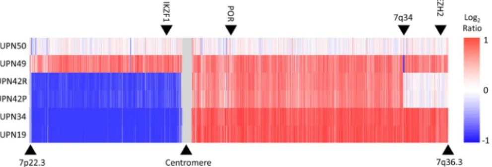
Isochromosome (7q), with resultant IKZF1 deletion, was de- tected in 3 cases at diagnosis (UPN19, 34 and 42). We set out to confirm the SNP array discovery of i(7q) using MLPA analyses to determine the copy number of exons in EZH2 (7q36.1), IKZF1 (7p12.2) and POR (7q11.23) (Figure 1;
Figure S6). Three copies of EZH2 were observed in UNP49 (trisomy 7) as expected. UPN42 carried 2 copies of EZH2 ex- plained by the diploid status of the distal part of the long arm of chromosome 7. Analysis of sample UPN19 and UPN34 showed 1 copy of IKZF1 and 3 copies of POR and EZH2 confirming the presence of i(7q), initially identified by SNP array. Interestingly, as UPN42P and UPN42R are matched diagnostic and relapse samples from the same patient, our analyses suggest that this lesion is preserved throughout dis- ease progression.
3.3 | Validation of identified CNAs
MLPA was performed in 48 samples confirming all previ- ously identified DNA losses and gains. The MLPA analysis was suggestive of LEF1 intragenic deletions in 2 samples (UPN 2, 32), which had not been detected by the initial SNP array screening (Figures S7 and S8). Exon specific copy number qPCR (exon 4, 7, 13) confirmed deletion of exon 4 in both cases (Figure S9). MLPA analysis of 4 samples (UPN 11, 26, 32, 50) was further suggestive of intragenic deletions within the PTEN gene also not detected by SNP array. Sanger sequencing of PTEN exon 7 confirmed indel mutations in 2 samples (UPN 11, 50), providing a potential explanation for these findings.25
Gene/region No Gene/region No Gene/
region No Gene/region No
CDKN2A 36 PAX5 6 PTEN 4 5q34 2
CDKN2B 29 LEF1 5 ETV6 4 9q34.11– 34.13 2
MLLT3 16 IKZF1 5 SUZ12 4 WT1 2
STIL 10 BNC2/ORF93 5 NR3C1 3 CREBBP 2
CASP8AP2 8 RB1 5 i7(q) 3 16q22.1 2
PTPRD 7 CTCF 5 DLEU7 3
CDKN1B 7 6q21 4 NF1 3 MYB gain 7
6q16.2– 16.3 6 CREB5 4 EBF1 2
TABLE 1 Recurrent DNA copy number alterations in teenagers and young adults T- cell acute lymphoblastic leukemia at diagnosis
3.4 | Target gene mutation screening
Gene mutation screening was undertaken in se- lected genes implicated in the pathogenesis of T- ALL (Table S5). Our study confirmed the NOTCH signaling pathway as the main mutated target in T- ALL. At diagno- sis, NOTCH1 mutations were identified in 46/79 (58.2%) and FBXW7 mutations in 17/79 (21.5%). NOTCH1 muta- tions were found in the HD, PEST/TAD domains or both in 58.7%, 21.7% and 19.6%, respectively. Eight cases car- ried both NOTCH1 and FBXW7 mutations, and overall 55/79 (78.5%) diagnostic samples carried mutations lead- ing to aberrant NOTCH pathway activation. PTEN and IL7R mutations were found in 12/79 (15.2%) and 5/79 (6.3%) of diagnostic cases, respectively (Figure S10).
Screening of Janus kinases identified no mutations in JAK2 exon 14. One potential new mutation (p.V651L) was observed in JAK1. The latter occurred in the pseudo- kinase domain that is essential for normal kinase activ- ity. This variation is absent from both the dbSNP and the
1000 genome project databases. Two known mutations in TP53 (V218A, R248Q) were identified. Mutation screen- ing of the RAS pathway identified 2 mutations in NRAS (G12S, Q61P), 1 mutation in CBL, 1 mutation in KRAS (G13D), and no mutations in PTPN11 (SHP2, exons 3 and 13), or FLT3 (exons 14 and 20).26,27
3.5 | Inactivation of the CDKN2A locus in T- ALL
DNA copy number analysis identified CDKN2A deletion in 70.6% of presentation samples. Retention of wild- type CDKN2A was confirmed by FISH in 4 cases (UPN 6, 11, 23, 27) (Figure S11). The CDKN2A locus could also be in- activated through gene mutation or promoter methylation.28 Promoter methylation analysis in patient samples that re- tained one or both gene copies of CDKN2A demonstrated clear methylation in only 2 out of 23 tested samples (UPN 6, 27) (Figure S12).
FIGURE 1 Recurrent isochromosome 7q in TYA T- ALL. Nexus copy number analysis of chromosome 7 in 6 TYA T- ALL samples.
Analysis of UNP49 (trisomy 7) and UPN50 (disomy 7) are shown for comparative reasons. The location of MLPA probes for IKZF1, POR and EZH2 is indicated by the black arrows at the top. The color scale represents change in DNA copy number— red and blue represent gain and loss respectively. T- ALL, T- cell acute lymphoblastic leukemia; TYA, teenagers and young adults
FIGURE 2 Incidence of gene abnormalities with significant enrichment in T- ALL age groups. Bar diagrams depict the relative incidence of gene deletions and mutations (mGene) with significant enrichment in Paediatric, TYA and/or Adult patients with T- ALL. T- ALL, T- cell acute lymphoblastic leukemia; TYA, teenagers and young adults
3.6 | Identification of age specific genomic aberrations
The genomic landscape of pediatric and young adult T- ALL was recently characterized.29 We used this extensive data set in combination with our in- house and other pub- lished data to perform a comprehensive incidence analysis aiming to identify any potential TYA specific genomic ab- errations. Nineteen studies were identified with available age at diagnosis, DNA copy number and/or gene sequence data.6,7,9– 12,24,29– 41 The total patient cohort included 1792 individual patients, who were then separated into three age groups: 1– 14 (n = 972); 15– 24 (n = 439) and ≥25 (n = 381) years of age at diagnosis. Statistical analyses were performed on 148 genes and/or gene regions to identify genetic lesions with age- specific incidence patterns (Table S6). The inci- dence of NOTCH1 mutations in the 3 age groups was 61.5%, 58.8% and 64% respectively, and was not significantly en- riched in any age group. Similarly, FBXW7 mutations were equally distributed (Table S6). The incidence of 17 abnor- malities was significantly enriched or depleted in one or two of the three age groups (Figure 2).
The pediatric group was enriched for deletion of CDKN2A/B and LEF1 as well as mutation of PTEN. The adult cohort was enriched for deletion of CREBBP, ABD, as well as mutation of DNMT3A, JAK3, PHF6 and RUNX1.
Deletion of NOTCH1 and RUNX1, as well as mutation of CDKN2A and PTPN2, were significantly enriched in adult T- ALL. However, the latter four abnormalities were detected in
<10 cases overall, hence these results should be interpreted with caution and were removed from Figure 2.
Pediatric and TYA T- ALL were significantly enriched for 1p33/STIL abnormalities, whilst TYA/adult T- ALL were en- riched for gene mutations in ETV6 and GATA3. No aberration was solely enriched in our TYA cohort.
The therapeutic consequences of these different muta- tion patterns were explored by performing pathway analyses.
Gene alterations were functionally mapped into four path- ways. Using the initial 148 gene panel, two of these were significantly enriched in the pediatric group providing ac- tionable targets, such as PI3K- AKT and RAS. We observed a trend for increased incidence of JAK- STAT and NOTCH pathway abnormal activation, particularly in the pediatric group (Table S7).
3.7 | The genetics of relapsed TYA T- ALL
The DNA copy number analysis of 8 in- house relapse samples showed alterations of known drivers, such as loss of CDKN2A/B, MLLT3, PTEN, 6q16.2– 16.3, 6q21, IKZF1, NR3C1, CASP8AP2, TOX, PAX5, 9q34.11– 34.13, RAG1/2, WT1, RB1, DLEU7, CTCF, NT5C3 and gain of 10p (Table S3). When compared with matched presenta- tion, additional loss of 6q14.3– 21 (including CASP8AP2) and MLLT3/CDKN2A/B was observed in one patient each at relapse.
Whole exome sequencing (WES) was undertaken in 4 in- house cases of TYA T- ALL with available matched presen- tation, remission and relapse samples. Overall, 739 somatic Single Nucleotide Variation (SNVs) and 213 insertion and/
or deletions (indels) were identified. Three hundred and eight (41.7%) of the somatic mutations were predicted to be protein altering, with the majority being missense mutations (34.2%);
the rest were frameshift indels (2.5%), in- frame indels (2.3%) and nonsense (2.3%) mutations (Table S8). Protein- altering abnormalities occurred across 308 genes (Table S9). On av- erage, each sample contained 32.4 somatic protein- altering SNVs and indels. In our case series we observed non- significant increase in average number of protein- altering
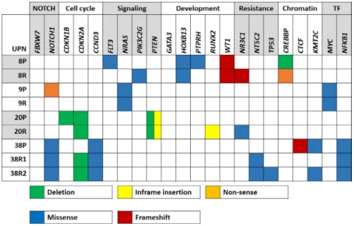
FIGURE 3 Heatmap of recurrently targeted pathways in matched presentation and relapsed TYA T- ALL. Heatmap of selected genes affecting cellular pathways in T- ALL. Validated and presumed protein- altering mutations are shown.
Green boxes indicate deletions; blue boxes missense mutations; yellow boxes in- frame insertions; red boxes frameshift mutations;
orange boxes nonsense mutations. T- ALL, T- cell acute lymphoblastic leukemia; TF, transcription factor; TYA, teenagers and young adults
mutations with 28.5 (range 24– 34) at presentation and 31.3 (19– 38) at relapse.
Whole exome sequencing analyses revealed recurrent mu- tations affecting the NOTCH and PI3K/AKT/mTOR path- ways (Figure 3). Furthermore, mutations in FLT3, NRAS, MYC, NR3C1, NT5C2 and CREBBP suggest disruption of relevant oncogenic pathways and drug metabolism. One case carried an inactivating mutation of Protein Tyrosine Phosphatase, Receptor Type, H (PTPRH) which is predicted to provide additional growth and survival signals through activation of LAT and ZAP70, two kinases involved in pre T- cell receptor signaling.42,43
3.8 | Origin of relapse in TYA T- ALL
Previous studies have demonstrated that a clonal relation- ship is evident in most cases of relapsed T- ALL.8,30 We used SciClone to analyze variant allele frequencies of somatic mutations, in combination with CNA data, to infer clonal architecture and pattern of tumor evolution under treatment pressure. In all 6 in- house matched cases, the relapse clone shared at least a single genetic aberration with the presen- tation leukemia. The molecular composition of the relapse clone either fully resembled the diagnostic clone (UPN9, 42)
or acquired additional mutations (UPN20, 21, 38). In case UPN8, the presentation and relapse leukemia shared a somat- ically acquired WT1 mutation. Strikingly, copy number gains and losses changed completely between the 2 time points (Figure S13).
Clinically, these 6 patients were treated with a variety of historic treatment protocols. All achieved complete remission after initial induction therapy. The relapses in our cohort oc- curred after a median remission duration of 1.0 year (0.3–
2.4 years). Four patients relapsed very early (<18 months from diagnosis). Case UPN8, in which relapse was likely driven by an ancestral clone, exhibited the longest time to re- lapse (2.4 years). Despite 3 patients receiving a bone marrow transplantation, none of the 6 relapsed patients is currently alive.
UNP38 represents an interesting case with 2 subsequent relapses (Figure 4). Presentation and both relapse samples were characterized by gain of 10p and an acquired missense mutation in CCND3. The CCND3 mutation (K268T) was found in a mutational hotspot in exon 5.44 CCND3 muta- tions were shown to increase cell proliferation rate, further enhanced by CDKN2A deletions at relapse.44 The diagnostic sample further harbored 2 distinct NOTCH1 mutations with only one of these mutations present at both relapses, indi- cating subclonal evolution underlying relapse. Inactivating
FIGURE 4 Comparative genetic composition of UPN38 at presentation and subsequent relapses. Top, variant allele frequency (VAF) of missense mutations in presentation, relapse 1 and 2. Bottom, Inferred clonal evolution from a single hematopoietic stem cell to presentation (P), relapse 1 (R1) and relapse 2 (R2). Phylogenetic tree depicting shared and acquired gene mutations
mutations in KMT2C (at presentation and second relapse) have previously been described in refractory diffuse large B- cell lymphoma and suggest a role for chromatin remod- eling and histone modulation.45 Both relapses carried an NT5C2 missense mutation suggesting escape of a drug re- sistant clone, which ultimately also acquired or selected for a TP53 mutation.
4 | DISCUSSION
Genome- wide DNA copy number and target gene mutation analyses in our in- house TYA cohort confirmed the plethora of genetic markers known to play a role in T- ALL.46,47 The average number of CNAs identified was similar as reported for pediatric (3.3– 7.1) and adult (6– 6.8) T- ALL.7,10,11,34 Remarkably, 3 out of 51 (5.8%) presentation cases were char- acterized by i(7q). Although this abnormality has previously been described in T- ALL, it mostly occurs in hyperdiploid B- lineage ALL.48 Moreover, i(7q) is a typical feature of Hepato Splenic T- cell Lymphoma.49 With an estimated incidence of i(7q) in ALL around 0.9%, our analysis showed an increased incidence in TYA T- ALL.48 Isochromosome 7q was not re- ported or observed in other TYA T- ALL cases (n = 388) in our extended analysis. The incidence reported in our discov- ery cohort might thus be an underestimate and can only be fully appreciated if genome wide copy number DNA analysis are performed.
We have previously established that the rare subtype of in- fant T- ALL has a distinctive genomic profile with MLF1 and KMT2A gene abnormalities and a relatively low incidence of CDKN2A deletions.50 Our current statistical analysis of nearly 1800 cases (in- house and published) revealed uneven distribution of only 17 out of 148 genes/gene regions tested, underlining broad biological similarities in T- ALL biology across the age groups evaluated (pediatric, TYA and adults) (Table S10). No single DNA abnormality was specific for the TYA age group.
Our dataset confirmed a trend for the decreasing occurrence of LEF1, PTEN, RPL10, STIL, TLX3 and increase in CNOT3, CREBBP, DNMT3A, JAK1, PHF6, PICALM- MLLT10, TLX1 abnormalities with older age.12,43,51,52 Phenotyping data were unfortunately not available for our TYA cohort. We pro- pose however that the increased incidence of the early T- cell precursor- ALL phenotype in adults explains the observed en- richment in ABD and mutations in ETV6, GATA3, RUNX1 and JAK3 in adult T- ALL.11,24,29,37,41,53,54
The genetic basis of relapsed T- ALL was explored using SNP arrays and/or WES. Previous work has highlighted the lack of acquisition of additional CNAs in relapsed childhood T- ALL, in contrast with relapsed BCP- ALL.30 This was confirmed in our study of 6 matched cases with an average number of CNAs of 7.6 at diagnosis and relapse. Previously,
exome sequencing of T- ALL had identified an average of 8.2 and 21.0 protein- altering mutations in pediatric and adult pa- tients, respectively.43 The higher incidence of somatic mu- tations in our data set could reflect the true mutation rate in TYA T- ALL. However, it is conceivable that the difference in mutation rate may be due to the small cohort analyzed and the difference in bioinformatic workflows used. WES analy- ses of matched pediatric T- ALL at diagnosis and relapse has previously shown an increase in SNVs and small indels at relapse, which we could not corroborate.8
The TYA genomic landscape presents ample scope for introduction of targeted therapies at relapse, as exemplified by aberrations in NOTCH, PI3K/AKT/mTOR, IL7R/JAK/
STAT and RAS pathways. Gene mutations associated with drug resistance (NR3C1, NT5C2, CREBBP and TP53) were gained at relapse, suggesting evolutionary selection of sub- clones endowed with survival advantage.55,56 Encouragingly, synergy between small molecule inhibitors and dexametha- sone has been shown to overcome glucocorticoid resistance in T- ALL.57– 60
Notably, several mutations (NOTCH1, FLT3, PTEN) were
‘lost’ at relapse, indicating they might represent non- founder or subclonal mutations.15,30,61 This underlined our previous data, which highlighted at single cell level that NOTCH1 and PTEN mutations were subclonal and demonstrated conver- gent evolution.15
Our study was limited to retrospective analysis of historic samples with incomplete information on immunopheno- typing, protocol and outcomes. This impeded identification of prognostic biomarkers. This further precluded complete genetic annotation with RNA sequencing to identify onco- gene expression and co- occurrence of mutations in individ- ual samples. Such an approach would have allowed further comparison with pediatric T- ALL, whilst such data in adult T- ALL are mostly lacking.
In conclusion, our study has described for the first time the genomic landscape of TYA T- ALL and identified a rela- tively high incidence of isochromosome 7q. Our observations support the existence of age- related biological differences between infants, children, TYA and adult patients. WES anal- yses suggested the major route to relapse is selection of drug resistant clones under therapy pressure and identified bona fide drug targets. Although these observations will inform the design of future salvage therapies, they should not be seen in isolation and require integration with functional profiling of relapse cells incorporating the protective role of the bone marrow niche.
ACKNOWLEDGMENTS
We would like to thank Dr David Gonzalez and Ms Sue Lillis for the TP53 gene mutation screening, Dr Brian Walker for help with the exome library preparation (The Institute of Cancer Research, London, UK), Dr Emily
Winterman for additional MLPA analyses, and Ms Janet Matthews for data collection for patients treated at The Barts Cancer Institute, London, UK. We are grateful to all authors who kindly provided age at diagnosis. We thank patients, parents, and hospital staff for their valuable col- laboration. This work was supported by an intermedi- ate fellowship from The Kay Kendall Leukaemia Fund (FWvD); Leukaemia & Lymphoma Research (MG, AMF, CF); The European Hematology Association— EHA Partner Fellowship #2011/01, the Lady Tata Memorial Trust—
LTMT International Award for Research in Leukaemia and the Ministry of Health, INCA- Brazil (MBM); Bloodwise (formerly Leukaemia & Lymphoma Research)— Clinical Research Training Fellowship (CLF); Bloodwise Integrative Biology Fellowship (AE); INCA, CNPq #301594/2015- 5; #310877/2019- 5; FAPERJ #E- 26/110.712/2012; #E- 26/202.577/2019; Fundação do Cancer, SwissBridge Fund; The Hungarian National Research, Development and Innovation Office— NKFIH (IDs: K_16 #119950 and FK_20 #134253), the New National Excellence Program of the Ministry for Innovation and Technology (ID: ÚNKP- 20- 5- SE- 22), the János Bolyai Scholarship Program (BO/00320/18/5) of the Hungarian Academy of Sciences, as well as by the EU’s Horizon 2020 Research and Innovation Program under grant agreement No. 739593 (DA); The Institute of Cancer Research (AMF and SMC) and by a Wellcome Trust Strategic Award (105104/Z/14/Z) (MG).
CONFLICT OF INTEREST The authors declare no conflict of interest.
ETHICAL APPROVAL
The ethics committee or institutional review board of each participating center approved the study (South West Wales REC reference 12/WA/0324, CEP_INCA/CONEP#888.277).
Informed consent was obtained from all subjects in accord- ance with the Declaration of Helsinki.
DATA AVAILABILITY STATEMENT
The raw data CEL and fastq files are available upon request and will be accessible through the Newcastle University data repository (data.NCL).
ORCID
Maria S. Pombo- de- Oliveira https://orcid.
org/0000-0002-1507-004X
Frederik W. van Delft https://orcid.org/0000-0002-3704-3720 REFERENCES
1. Beldjord K, Chevret S, Asnafi V, et al. Oncogenetics and min- imal residual disease are independent outcome predictors in adult patients with acute lymphoblastic leukemia. Blood.
2014;123(24):3739- 3749.
2. Patrick K, Wade R, Goulden N, et al. Outcome for children and young people with early T- cell precursor acute lymphoblastic leu- kaemia treated on a contemporary protocol, UKALL 2003. Br J Haematol. 2014;166(3):421- 424.
3. Patrick K, Wade R, Goulden N, et al. Improved outcome for chil- dren and young people with T- acute lymphoblastic leukaemia: re- sults of the UKALL 2003 trial. Blood. 2014;124(21):3702.
4. Sellar RS, Rowntree C, Vora AJ, et al. Relapse in teenage and young adult patients treated on a paediatric minimal residual dis- ease stratified ALL treatment protocol is associated with a poor outcome: results from UKALL2003. Br J Haematol. 2018;181(4):
515- 522.
5. Hough R, Sandhu S, Khan M, et al. Are survival and mortality rates associated with recruitment to clinical trials in teenage and young adult patients with acute lymphoblastic leukaemia? A retrospective observational analysis in England. BMJ Open. 2017;7:e017052.
6. Gutierrez A, Sanda T, Ma W, et al. Inactivation of LEF1 in T- cell acute lymphoblastic leukemia. Blood. 2010;115(14):2845- 2851.
7. Karrman K, Castor A, Behrendtz M, et al. Deep sequencing and SNP array analyses of pediatric T- cell acute lymphoblastic leuke- mia reveal NOTCH1 mutations in minor subclones and a high in- cidence of uniparental isodisomies affecting CDKN2A. J Hematol Oncol. 2015;8:42.
8. Kunz JB, Rausch T, Bandapalli OR, et al. Pediatric T- cell lympho- blastic leukemia evolves into relapse by clonal selection, acquisi- tion of mutations and promoter hypomethylation. Haematologica.
2015;100(11):1442- 1450.
9. La Starza R, Lettieri A, Pierini V, et al. Linking genomic lesions with minimal residual disease improves prognostic stratification in children with T- cell acute lymphoblastic leukaemia. Leuk Res.
2013;37(8):928- 935.
10. Mullighan CG, Goorha S, Radtke I, et al. Genome- wide analysis of genetic alterations in acute lymphoblastic leukaemia. Nature.
2007;446(7137):758- 764.
11. Van Vlierberghe P, Ambesi- Impiombato A, De Keersmaecker K, et al. Prognostic relevance of integrated genetic profiling in adult T- cell acute lymphoblastic leukemia. Blood. 2013;122(1):74- 82.
12. Vicente C, Schwab C, Broux M, et al. Targeted sequencing identifies associations between IL7R- JAK mutations and epi- genetic modulators in T- cell acute lymphoblastic leukemia.
Haematologica. 2015;100(10):1301- 1310.
13. Yu L, Slovak ML, Mannoor K, et al. Microarray detection of multiple recurring submicroscopic chromosomal aberrations in pediatric T- cell acute lymphoblastic leukemia. Leukemia.
2011;25(6):1042- 1046.
14. Mansur MB, Ford AM, van Delft FW, et al. Occurrence of iden- tical NOTCH1 mutation in non- twinned sisters with T- cell acute lymphoblastic leukemia. Leukemia. 2011;25(8):1368- 1370.
15. Furness CL, Mansur MB, Weston VJ, et al. The subclonal com- plexity of STIL- TAL1+ T- cell acute lymphoblastic leukaemia.
Leukemia. 2018;32(9):1984- 1993.
16. Van Vlierberghe P, van Grotel M, Tchinda J, et al. The recur- rent SET- NUP214 fusion as a new HOXA activation mecha- nism in pediatric T- cell acute lymphoblastic leukemia. Blood.
2008;111(9):4668- 4680.
17. Hayashi Y, Raimondi SC, Look AT, et al. Abnormalities of the long arm of chromosome 6 in childhood acute lymphoblastic leu- kemia. Blood. 1990;76(8):1626- 1630.
18. Flotho C, Coustan- Smith E, Pei D, et al. Genes contributing to minimal residual disease in childhood acute lymphoblastic
leukemia: prognostic significance of CASP8AP2. Blood.
2006;108(3):1050- 1057.
19. Li N, Fassl A, Chick J, et al. Cyclin C is a haploinsufficient tumour suppressor. Nat Cell Biol. 2014;16(11):1080- 1091.
20. Lopez- Nieva P, Vaquero C, Fernandez- Navarro P, et al. EPHA7, a new target gene for 6q deletion in T- cell lymphoblastic lympho- mas. Carcinogenesis. 2012;33(2):452- 458.
21. Sinclair PB, Sorour A, Martineau M, et al. A fluorescence in situ hybridization map of 6q deletions in acute lymphocytic leukemia:
identification and analysis of a candidate tumor suppressor gene.
Can Res. 2004;64(12):4089- 4098.
22. Swaminathan S, Huang C, Geng H, et al. BACH2 mediates neg- ative selection and p53- dependent tumor suppression at the pre- B cell receptor checkpoint. Nat Med. 2013;19(8):1014- 1022.
23. Chaar T, Avran D, Genesca L, et al. Combined Haploinsufficiency of Syncrip/Hnrnp- Q and Snhg5/Snornas in Deletion 6Q Promotes T- Cell Acute Leukemogenesis. Pavia, Italy: Ferrata Storti Foundation; 2013.
24. Gutierrez A, Dahlberg SE, Neuberg DS, et al. Absence of bial- lelic TCRgamma deletion predicts early treatment failure in pediatric T- cell acute lymphoblastic leukemia. J Clin Oncol.
2010;28(24):3816- 3823.
25. Sandell S, Schuit RJ, Bunyan DJ. An intronic polymorphic dele- tion in the PTEN gene: implications for molecular diagnostic test- ing. Br J Cancer. 2013;108(2):438- 441.
26. Ward AF, Braun BS, Shannon KM. Targeting oncogenic Ras signal- ing in hematologic malignancies. Blood. 2012;120(17):3397- 3406.
27. Case M, Matheson E, Minto L, et al. Mutation of genes affecting the RAS pathway is common in childhood acute lymphoblastic leukemia. Cancer Res. 2008;68(16):6803- 6809.
28. Sulong S, Moorman AV, Irving JA, et al. A comprehensive analysis of the CDKN2A gene in childhood acute lymphoblastic leukemia reveals genomic deletion, copy number neutral loss of heterozy- gosity, and association with specific cytogenetic subgroups. Blood.
2009;113(1):100- 107.
29. Liu Y, Easton J, Shao Y, et al. The genomic landscape of pediat- ric and young adult T- lineage acute lymphoblastic leukemia. Nat Genet. 2017;49(8):1211- 1218.
30. Mullighan CG, Phillips LA, Su X, et al. Genomic analysis of the clonal origins of relapsed acute lymphoblastic leukemia. Science.
2008;322(5906):1377- 1380.
31. Gutierrez A, Sanda T, Grebliunaite R, et al. High frequency of PTEN, PI3K, and AKT abnormalities in T- cell acute lymphoblas- tic leukemia. Blood. 2009;114(3):647- 650.
32. Kunz JB, Rausch T, Bandapalli OR, et al. Pediatric T- lymphoblastic leukemia evolves into relapse by clonal selection, acquisition of mutations and promoter hypomethylation. Haematologica.
2015;100:1442.
33. Marks DI, Paietta EM, Moorman AV, et al. T- cell acute lympho- blastic leukemia in adults: clinical features, immunophenotype, cytogenetics, and outcome from the large randomized prospective trial (UKALL XII/ECOG 2993). Blood. 2009;114(25):5136- 5145.
34. Safavi S, Hansson M, Karlsson K, Biloglav A, Johansson B, Paulsson K. Novel gene targets detected by genomic profiling in a consecutive series of 126 adults with acute lymphoblastic leuke- mia. Haematologica. 2015;100(1):55- 61.
35. Yuan L, Lu L, Yang Y, et al. Genetic mutational profiling analy- sis of T cell acute lymphoblastic leukemia reveal mutant FBXW7 as a prognostic indicator for inferior survival. Ann Hematol.
2015;94(11):1817- 1828.
36. Grossmann V, Haferlach C, Weissmann S, et al. The molecular profile of adult T- cell acute lymphoblastic leukemia: mutations in RUNX1 and DNMT3A are associated with poor prognosis in T- ALL. Genes Chromosom Cancer. 2013;52(4):410- 422.
37. Yang YL, Hsiao CC, Chen HY, et al. Absence of biallelic TCRgamma deletion predicts induction failure and poorer out- comes in childhood T- cell acute lymphoblastic leukemia. Pediatr Blood Cancer. 2012;58(6):846- 851.
38. Jenkinson S, Kirkwood AA, Goulden N, Vora A, Linch DC, Gale RE. Impact of PTEN abnormalities on outcome in pediatric pa- tients with T- cell acute lymphoblastic leukemia treated on the MRC UKALL2003 trial. Leukemia. 2016;30(1):39- 47.
39. Jenkinson S, Koo K, Mansour MR, et al. Impact of NOTCH1/
FBXW7 mutations on outcome in pediatric T- cell acute lympho- blastic leukemia patients treated on the MRC UKALL 2003 trial.
Leukemia. 2013;27(1):41- 47.
40. Bandapalli OR, Zimmermann M, Kox C, et al. NOTCH1 activation clinically antagonizes the unfavorable effect of PTEN inactivation in BFM- treated children with precursor T- cell acute lymphoblastic leukemia. Haematologica. 2013;98(6):928- 936.
41. Zhang J, Ding L, Holmfeldt L, et al. The genetic basis of early T- cell precursor acute lymphoblastic leukaemia. Nature.
2012;481(7380):157- 163.
42. Van Vlierberghe P, Pieters R, Beverloo HB, Meijerink JP.
Molecular- genetic insights in paediatric T- cell acute lymphoblastic leukaemia. Br J Haematol. 2008;143(2):153- 168.
43. De Keersmaecker K, Atak ZK, Li N, et al. Exome sequenc- ing identifies mutation in CNOT3 and ribosomal genes RPL5 and RPL10 in T- cell acute lymphoblastic leukemia. Nat Genet.
2013;45(2):186- 190.
44. Schmitz R, Young RM, Ceribelli M, et al. Burkitt lymphoma pathogenesis and therapeutic targets from structural and functional genomics. Nature. 2012;490(7418):116- 120.
45. Morin RD, Assouline S, Alcaide M, et al. Genetic landscapes of relapsed and refractory diffuse large B- cell lymphomas. Clin Cancer Res 2015;22:2290- 2300.
46. Meijerink JP. Genetic rearrangements in relation to immunophe- notype and outcome in T- cell acute lymphoblastic leukaemia. Best Pract Res Clin Haematol. 2010;23(3):307- 318.
47. Van Vlierberghe P, Ferrando A. The molecular basis of T cell acute lymphoblastic leukemia. J Clin Investig. 2012;122(10):3398- 3406.
48. Martineau M, Clark R, Farrell DM, Hawkins JM, Moorman AV, Secker- Walker LM. Isochromosomes in acute lymphoblastic leu- kaemia: i(21q) is a significant finding. Genes Chromosom Cancer.
1996;17(1):21- 30.
49. Jonveaux P, Daniel MT, Martel V, Maarek O, Berger R.
Isochromosome 7q and trisomy 8 are consistent primary, non- random chromosomal abnormalities associated with hepatosplenic T gamma/delta lymphoma. Leukemia. 1996;10(9):1453- 1455.
50. Mansur MB, van Delft FW, Colman SM, et al. Distinctive geno- types in infants with T- cell acute lymphoblastic leukaemia. Br J Haematol. 2015;171(4):574- 584.
51. Asnafi V, Beldjord K, Libura M, et al. Age- related phenotypic and oncogenic differences in T- cell acute lymphoblastic leukemias may reflect thymic atrophy. Blood. 2004;104(13):4173- 4180.
52. Flex E, Petrangeli V, Stella L, et al. Somatically acquired JAK1 mutations in adult acute lymphoblastic leukemia. J Exp Med.
2008;205(4):751- 758.
53. Haydu JE, Ferrando AA. Early T- cell precursor acute lymphoblas- tic leukaemia. Curr Opin Hematol. 2013;20(4):369- 373.
54. Van Vlierberghe P, Ambesi- Impiombato A, Perez- Garcia A, et al.
ETV6 mutations in early immature human T cell leukemias. J Exp Med. 2011;208(13):2571- 2579.
55. Mullighan CG, Zhang J, Kasper LH, et al. CREBBP mu- tations in relapsed acute lymphoblastic leukaemia. Nature.
2011;471(7337):235- 239.
56. Tzoneva G, Perez- Garcia A, Carpenter Z, et al. Activating muta- tions in the NT5C2 nucleotidase gene drive chemotherapy resis- tance in relapsed ALL. Nat Med. 2013;19(3):368- 371.
57. Piovan E, Yu J, Tosello V, et al. Direct reversal of glucocorticoid resistance by AKT inhibition in acute lymphoblastic leukemia.
Cancer Cell. 2013;24(6):766- 776.
58. Li Y, Buijs- Gladdines JG, Cante- Barrett K, et al. IL- 7 receptor mutations and steroid resistance in pediatric T cell acute lympho- blastic leukemia: a genome sequencing study. PLoS Medicine.
2016;13(12):e1002200.
59. Serafin V, Capuzzo G, Milani G, et al. Glucocorticoid resistance is reverted by LCK inhibition in pediatric T- cell acute lymphoblastic leukemia. Blood. 2017;130(25):2750- 2761.
60. Shi Y, Beckett MC, Blair HJ, et al. Phase II- like murine trial identi- fies synergy between dexamethasone and dasatinib in T- cell acute lymphoblastic leukemia. Haematologica. 2020;106:1056.
61. Greaves M, Maley CC. Clonal evolution in cancer. Nature.
2012;481(7381):306- 313.
SUPPORTING INFORMATION
Additional supporting information may be found online in the Supporting Information section.
How to cite this article: Mansur MB, Furness CL, Nakjang S, et al. The genomic landscape of teenage and young adult T- cell acute lymphoblastic leukemia.
Cancer Med. 2021;00:1– 10. https://doi.org/10.1002/
cam4.4024